|
|
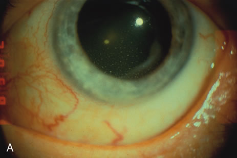
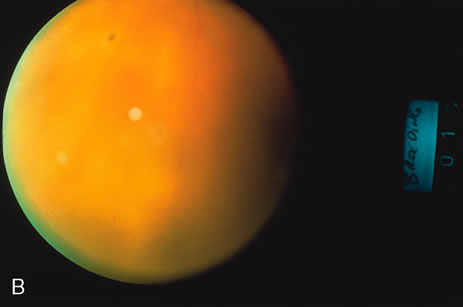
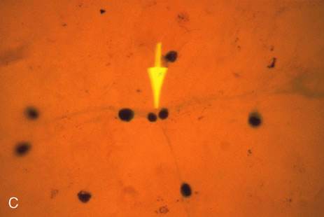
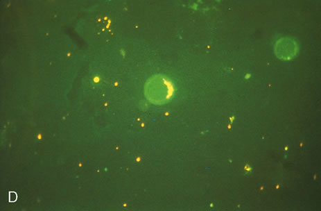
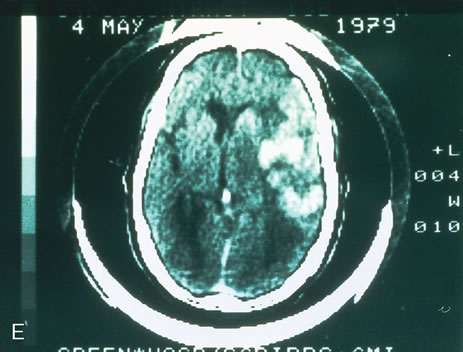
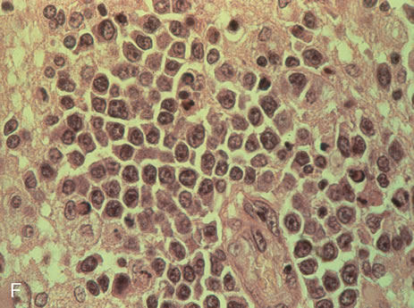
| Fig. 5. A. External view of the eye of a 76-year-old man in whom reticulum cell sarcoma (large cell lymphoma) is suspected. Note large mutton-fat keratic precipitates. B. Fundus view of the same patient demonstrates 3\+ vitreous cells and white subretinal infiltration. C. Vitreous aspirate demonstrates large cell lymphoma infiltrate. Note two smaller cells in mitosis (arrow). D. Vitreous cells stained for their surface immunoglobulin light chains. Fluorescein discloses IgM light chains only, as a corroboration of the monoclonal nature of this malignant infiltration. E. CT scan of same patient (different time) shows intracerebral microgliomatosis of large cell lymphoma infiltration. F. Histologic appearance of this brain tumor of “reticulum cell sarcoma” or large cell lymphoma. |