Chapter 54e
Glaucoma Associated with Retinal Disorders and Retinal Surgery
GEORGE R. REISS and JACK O. SIPPERLEY
Main Menu   Table Of Contents

Search

OPEN-ANGLE GLAUCOMAS
ANGLE-CLOSURE GLAUCOMAS
OTHER RETINAL DISORDERS ASSOCIATED WITH ANGLE-CLOSURE
CONCLUSION
REFERENCES

1Patients with retinal disorders may develop glaucoma of both a primary and secondary type. When several glaucoma mechanisms are operative, the primary diagnosis may be obscured and result in delayed treatment and greater morbidity. We present a pathogenic approach to the diagnosis of glaucoma associated with retinal disorders to further clarify the interpretation and treatment of these complicated cases. Our purpose is to highlight the most frequent associations seen in practice and, where possible, explain them according to pathogenesis. This chapter should not be considered a complete source of information for each entity or for every possible association.

For simplicity, this chapter has been divided anatomically into glaucomas having open- and closed-angles (Table 1). Some retinal disorders are associated with glaucomas having either or both of these features, and this will be mentioned where possible.


TABLE 1. Classification for Glaucoma Associated with Retinal Disorders and Retinal Surgery


Open-Angle GlaucomasAngle ClosureOther
MultifactorialStructuralexpanding gas
 Retinal detachments PHPV 
 Retinitis pigmentosa Retinal dysplasia 
 Stickler's Syndrome Retinopathy of prematurity 
  Iris retraction of Campbell 
  Post vitrectomy pupillary block 
Obstructive-particulate matter  
 UveitisCyclocongestive/ciliary body rotation 
 Red blood cells Postoperative scleral buckling 
 Tumors Postoperative pan retinal laser 
 Schwartz Syndrome Metastatic choroidal tumor 
 Silicone oil/gas Nanophthalmos 
 Foreign body siderosis Acute choroidal/retinal hemorrhage 
 Intra-Vitreal Steroids Central vein occlusion 
  Other retinal entities 
Raised episcleral venous pressure  
 Superior Vena Cava SyndromeNeovascular 
 Carotid cavernous fistula Central retinal vein occlusion 
 Sturge-Weber Syndrome Diabetes mellitus 
  Carotid artery disease 
  Tumors of the choroid 
  Central retinal artery occlusion 
  Other retinal entities 


The authors and editors wish to acknowledge the contributions of the late Randy V. Campo, M.D. who authored the original chapter. Some of this material has been used in this revision.
Back to Top
OPEN-ANGLE GLAUCOMAS

MULTIFACTORIAL LINKAGE

Glaucoma occurs at a higher frequency in patients with certain retinal disorders than might be expected in an age-matched control group. The retinal disorder itself does not appear to have a direct role in the glaucoma, but the frequency of the association and the clinical relevance bear consideration.

Retinal Detachments

In the general population the incidence of primary open-angle glaucoma approaches 1%1; by comparison Becker reported 5.8% of patients with retinal detachments (31 of 530 patients) had unrecognized primary open-angle glaucoma in the contralateral eye, with the diagnosis based on disc analysis, visual fields, tonometry, and/or tonography.2 In a review of 817 patients undergoing primary retinal detachment repair, Phelps and Burton found glaucoma in 9.5% (78 of 817 patients); the glaucoma preceded the retinal detachment historically or by clinical evidence in 7.3% (60 of 817 patients) and followed the retinal detachment in 2.2% (18 of 817 patients).3 The reason for the association between retinal detachment and glaucoma was unclear and, surprisingly, the authors could find no definite risk factors in myopia, trauma, or the use of miotics. In aphakic eyes, however, the risk of glaucoma was three to four times more likely compared with the phakic group.

Shammas and colleagues reviewed glaucoma risk factors in 30 patients with primary, atraumatic retinal detachments. Following the chronic administration of topical dexamethasone drops to the contralateral, nondetached eye, they found that 20% (6 of 30 patients) had an intraocular pressure rise of 16 mm Hg or higher.4 This was a significantly higher proportion than the expected 5% to 6% high-pressure corticosteroid responders reported previously in a general population group supporting a strong predisposition in patients with retinal detachment.5,6

A possible association between the use of miotics and acute retinal detachments has been suggested in numerous reports.7–9 These agents were of more clinical significance in the past, when they were a mainstay of glaucoma treatment, and the authors now limit their use to pseudophakic patients in which other agents have failed. Regardless, the association with retinal detachment should be kept in mind, particularly in susceptible patients (ex: high myopia).

One study of patients with pigment dispersion syndrome (PDS) showed an increased prevalence of lattice changes and retinal detachment as compared to non-PDS patients having a similar distribution of myopic eyes.10 For this reason, a good peripheral examination in such patients is warranted, and recently symptomatic patients should be treated. This association may be genetically determined, either by a single gene or by two genes at closely linked loci. Unique anatomical variations at both the peripheral iris and vitreous base may explain this increased risk.10

Retinitis Pigmentosa

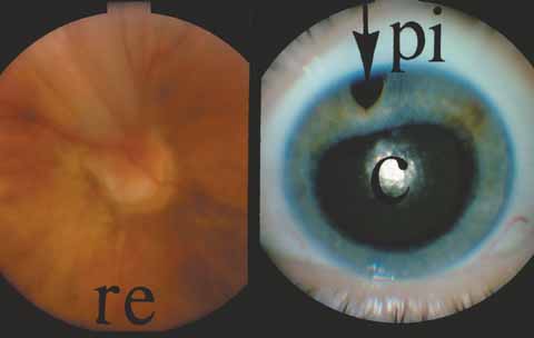
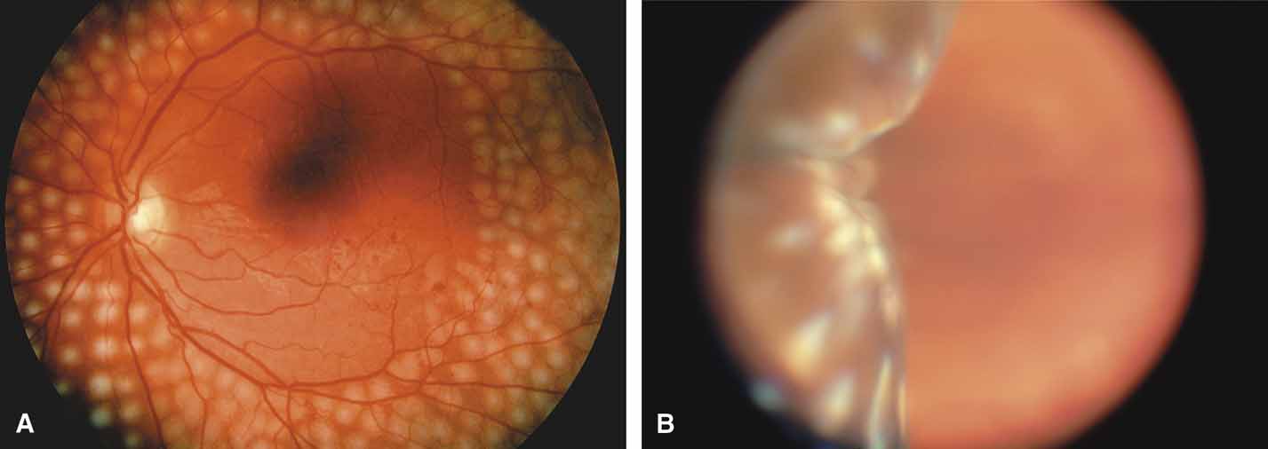
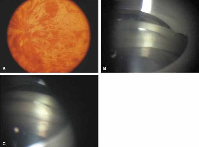
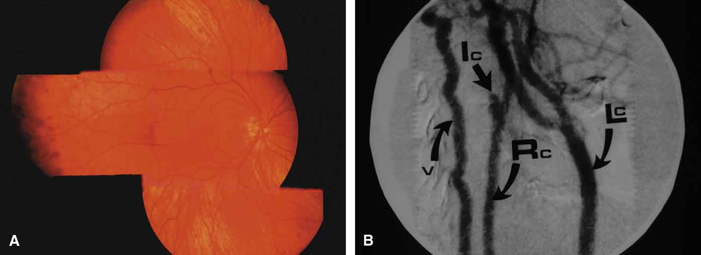
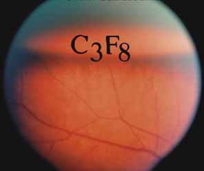
The possible association between retinitis pigmentosa and glaucoma dates back to 1862.11 Over the past century the reported glaucoma incidence has varied between 2.98% to 10% of all cases of retinitis pigmentosa.12,13 Of concern in all previous reports is the accuracy of the diagnosis of retinitis pigmentosa itself, a hereditary disease with diverse manifestations. Retinitis pigmentosa-like syndromes may mimic glaucomatous field loss, and the pattern of advancement of visual field loss in true retinitis pigmentosa may parallel that of typical open-angle glaucoma. Patients with undiagnosed retinitis pigmentosa may have subtle fundus findings, especially those with the sine pigmento form (Fig. 1). A clue to diagnosis is the disproportionate peripheral field loss in comparison to the degree of optic nerve cupping and rim pallor. Patients with atypical field loss and minimal optic nerve cupping should have an electroretinogram to exclude unsuspected retinitis pigmentosa.

Fig. 1 Subtle fundus findings in sine pigmento form of retinitis pigmentosa. This patient presented with small central islands on visual field testing that mimicked end-stage glaucoma A. Relatively normal posterior segment. B. Occasional bone spicule pigmentation only seen in the far periphery.

Stickler's Syndrome

Glaucoma has been reported in Stickler's vitreoretinopathy, an autosomal dominant disorder that demonstrates multiple systemic abnormalities in addition to the ocular abnormalities emphasized in seminal reports by Wagner14 and Janssen.15 Ophthalmic manifestations include an optically clear vitreous with prominent peripheral vitreoretinal membranes that have been associated with retinal detachment. High myopia, nuclear sclerotic cataracts, and glaucoma complete the eye findings. Systemic findings include flattened facies, high-arched palate, hearing loss, scapular abnormalities, and knee pain due to spondyloepiphyseal dysplasia. The glaucoma has been described as an open-angle type with both congenital and later-onset varieties. Phelps16 has reported normal angles on gonioscopy and a good response to medication. He recommended avoiding miotics in light of the peripheral retinal abnormalities, high myopia, and potentially higher risk of retinal detachment. Neovascular glaucoma has been reported, but occurs most often following multiple surgeries.17

OBSTRUCTIVE/PARTICULATE MATTER

Particulate matter of varying sizes can cause a mechanical obstruction to outflow or in other ways damage the trabecular meshwork of an otherwise normal open-angle. The degree and duration of the pressure elevation, the underlying health of the optic nerve, and systemic factors determine the outcome and the ultimate effect on vision.

Uveitis

Disorders in this category affect the eye and adnexa in three general locations that frequently overlap: (1) anterior segment, (2) posterior segment, and (3) orbit (Table 2).18 Involvement of the anterior segment may produce a high, low, or normal intraocular pressure, depending on the balance between inflow and outflow.


TABLE 2. Mechanisms of Glaucoma in Uveitis


Increased viscosity of aqueous humor
Obstruction of the trabecular meshwork
Swelling and dysfunction of the trabecular meshwork
Liberation of active substances such as prostaglandins and substance P
Scarring of the outflow channels
Development of a cuticular membrane over the angle
Neovascularization
Elevation of episcleral venous pressure
Forward movement of the iris lens diaphragm
Pupillary block
Formation of peripheral anterior synechiae

(Modified from Hoskins HD Jr, Kass M: Becker-Schaffer's diagnosis and therapy of the glaucomas. 6th ed. St Louis: CV Mosby, 1989)


Specific posterior segment entities known to cause glaucoma include sarcoidosis, toxoplasmosis, and syphilis. In a study of 202 patients with sarcoid affecting the eyes, 10.9% (22 of 202) were found to have glaucoma at some time during the clinical course of the illness.19 Lichter20 reported that syphilis was associated with glaucoma of three distinct types: (1) iritic, which demonstrated peripheral anterior synechiae, a residua of inflammation; (2) narrow angle; and (3) irregular angle-closure, in which intraepithelial cysts of the ciliary body and iris are seen on gonioscopy and account for the variable narrowing of the angle approach.

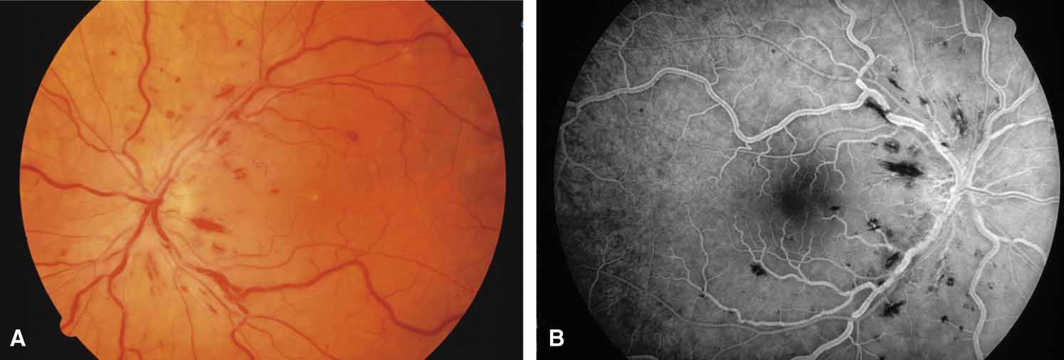
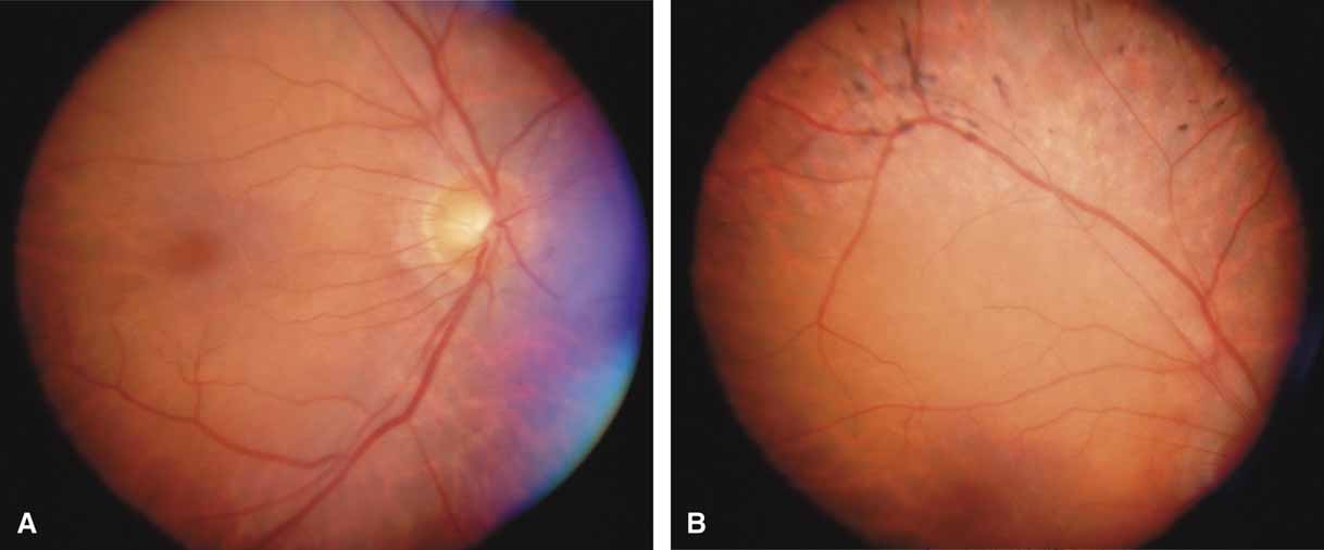
Active retinal involvement in acute toxoplasmosis may be associated with an acute elevation of intraocular pressure, sometimes producing corneal edema. When the cornea is cleared with topical glycerin, the angle is found to be open and a minimal anterior chamber reaction is present. The mechanism of the glaucoma is believed to be immune complex (antigen–antibody) deposition in the trabecular meshwork, rather than obstruction by cellular debris.21 The elevated pressure responds well to timolol or acetazolamide and does not require topical corticosteroids. Systemic treatment of the active retinitis should follow only if the optic nerve (positive Marcus Gunn), macular center, or peripapillary retina is involved. This unique entity will be missed if a fundus examination is not performed in all cases of hypertensive uveitis (Fig. 2).

Fig. 2 Recurrent, active toxoplasmic retinochoroiditis in right eye. Patient presented with acute glaucoma, intraocular pressure of 45 mm Hg, and a cloudy edematous cornea. Note active, fluffy white retinitis nasal to optic nerve and adjacent to old pigmented scar.

Active anterior or posterior uveitis, although minimally active, may upset the inflow/outflow balance severely and produce pressure elevations out of proportion to the anterior chamber reaction. Careful gonioscopy may reveal a localized trabeculitis manifesting as an occasional keratic precipitate on the trabecular meshwork or more anteriorly on the corneal endothelium. This easily overlooked syndrome mimics a medically unresponsive open-angle glaucoma and is important to correctly diagnose because the pressure can be lowered with the application of topical corticosteroids.22 In corticosteroid-sensitive persons, topical and periocular corticosteroid-related pressure elevations may become intractable. Long-acting depot preparations may require surgical removal of the depot corticosteroid to ameliorate the pressure elevation.23

Treatment with corticosteroids may hasten the recovery from hypotony in dramatic fashion, producing an acute pressure elevation as the blood aqueous barrier recovers and inflow overwhelms outflow. For this reason, careful titration of the corticosteroid dosage and simultaneous observation of the effect on intraocular pressure is advised.

Red Blood Cells

Long-standing vitreous hemorrhage can produce an acute glaucoma of an open-angle type. Originally, the term hemolytic glaucoma was employed to describe these events because pathologic studies showed macrophages containing hemoglobin in the anterior chamber.24 The situation was thought to be analogous to phacolytic glaucoma (hypermature liquefied lens protein engulfed by macrophages). Later, Campbell and co-workers25,26 demonstrated that the major mechanism was trabecular meshwork obstruction by ghost cells, rather than red blood cells, hemoglobin, or macrophages. Red blood cells in the vitreous release their hemoglobin within a few days and become the less pliable membranous container called a ghost cell. For simplicity, we consider the conditions hemolytic glaucoma and ghost cell glaucoma to be interchangeable. Two other disorders, hemosiderotic and siderotic glaucoma, add confusion to the terminology. These entities are separate glaucomatous disorders due to iron-induced damage of an open trabecular meshwork. The source of the iron may be blood (hemosiderotic) or an occult iron-containing foreign body (siderotic).

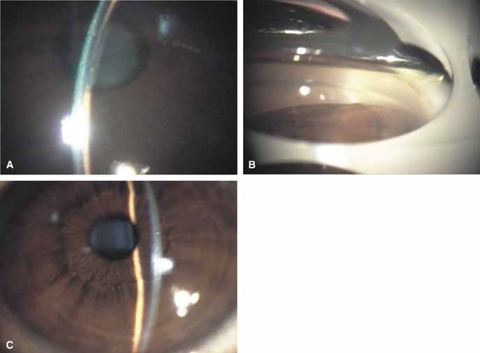
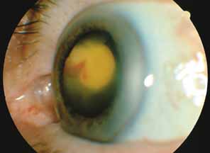
The majority of cases of ghost cell glaucoma occur following vitrectomy for diabetic retinopathy, vitreous hemorrhage due to trauma, or vitreous hemorrhage following anterior segment surgery (i.e., cataract extraction or corneal transplant). Although studies suggest that a disruption of the anterior hyaloid face may be a necessary precedent, ghost cell glaucoma probably can occur with an intact hyaloid if a sufficient quantity of vitreous blood is present.27 The clinical presentation includes acute pressure elevation, corneal edema, and lack of keratic precipitates. Minute tan cells (ghost cells) may layer out, producing a pseudohypopyon or, if admixed with blood, a “candy-striped” hypopyon. (Fig. 3) Intraocular pressure elevations after diabetic vitrectomy raise concerns regarding concomitant neovascular glaucoma. However, neovascular glaucoma rarely develops in the immediate postvitrectomy period, unless some anterior segment neovascularization was present preoperatively. Late-onset pressure elevations (> 6 to 8 weeks out) in a postvitrectomized diabetic eye usually signal neovascular glaucoma.

Fig. 3 67-year-old diabetic patient with rubeosis, vitreous hemorrhage, and neovascular glaucoma presents postoperatively with a red-and-white layered pseudohypopyon, the “candy-striped” hypopyon.

Blunt trauma that produces hyphema and vitreous hemorrhage may result in an early pressure elevation (hyphema-induced), which abates in 1 to 2 weeks. If the pressure becomes elevated in the absence of hyphema, the diagnosis of ghost cell glaucoma secondary to residual vitreous hemorrhage should be considered. One should look for fine cells that are smaller than those seen in uveitis and are more uniform and larger than pigment particles. Blunt trauma may also cause angle recession glaucoma, which is usually asymptomatic and can occur weeks, months, or even years later.

Intraocular hemorrhage after anterior segment surgery such as cataract extraction or corneal transplant may be confused with infectious endophthalmitis, which may also result in acute pressure elevations. In clinically equivocal situations, diagnostic vitreous and aqueous smears and cultures and antibiotic therapy may be necessary.

In the treatment of red blood cell–related glaucoma, maximally tolerated medical treatment (Beta Blockers, Alpha 2 Agonists, Acetazolamide) and patience will usually result in a spontaneous abatement of the intraocular pressure elevation. Some patients with severe, unremitting intraocular pressure elevation or those developing corneal bloodstaining will require an anterior chamber washout or vitrectomy. If debris (blood or ghost cells) is present in the anterior chamber, visualization will require anterior chamber lavage prior to vitrectomy. The decision for surgery should be guided by a prior knowledge of the health of the optic nerve, the clarity of the cornea, and the comfort of the patient. A history of sickle cell disorders requires a more aggressive approach to preserve optic nerve perfusion which is more easily compromised by elevated intraocular pressure and systemic use of acetazolamide (produces metabolic acidosis).28 Hemoglobin electrophoresis or a sickle cell preparation is recommended to diagnose the disorder in populations at risk for this hemoglobinopathy.

Tumors

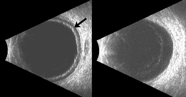
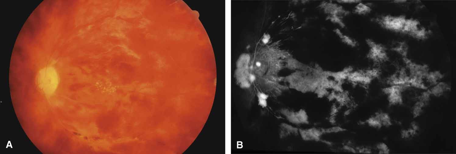
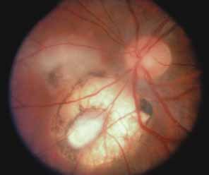
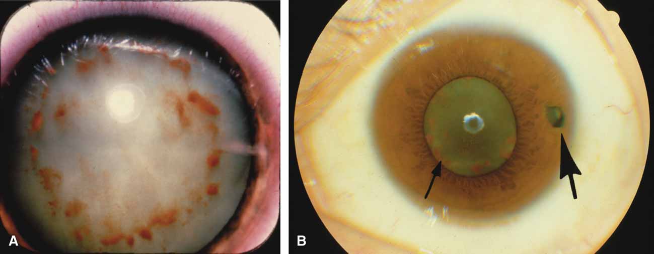
Both benign and malignant tumors can produce a secondary glaucoma. Shields and associates29 found 5% (126) of 2,704 eyes diagnosed with intraocular tumors suffered glaucoma. The glaucoma rate was only 2% in eyes with choroidal melanomas but rose to 17% in eyes with melanomas of the ciliary body.28 The mechanism of glaucoma in posterior segment melanomas was usually iris neovascularization (Fig. 4; Table 3).30 The incidence of glaucoma in tumor-containing eyes was found to rise when retinal detachment was present. In eyes presenting with reduced vision, glaucoma, opaque media, and an uncertain history of visual loss, it is prudent to exclude an unsuspected neoplasm by performing diagnostic B-scan ultrasonography.

Fig. 4 Patients with glaucoma secondary to choroidal melanoma. A. Large inferior melanoma (mm) of ciliary body occupies one-third of the volume of the globe. B. Large diffuse, peripapillary melanoma with partial extension into nerve interiorly. Both eyes were enucleated.


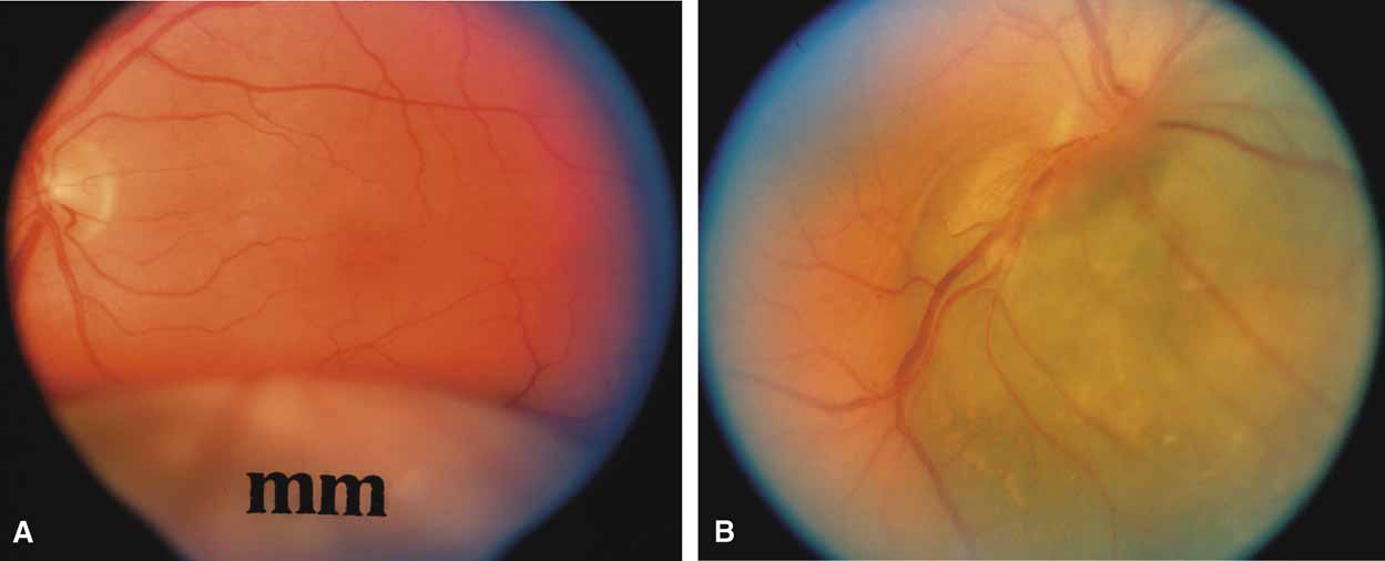
TABLE 3. Glaucoma Mechanisms in Eyes Containing Uveal Malignant Melanoma


MechanismUnderlying Cause
Peripheral anterior synechiae and angle closurePosterior synechiae, iris bombe and peripheral anterior synechiae
 Rubeosis irides and peripheral anterior synechiae
 Diffuse iris nevus or melanoma and peripheral anterior synechiae
  
Cellular obstruction of aqueous drainage area of an open angleSeeding of neoplasm into anterior chamber angle
 Ring melanoma with invasion of anterior chamber angle structures
 Melanin phagocytosis by macrophages with obstruction of anterior chamber angle (melanomalytic glaucoma)

(Yanoff M: Glaucoma mechanisms in eyes containing uveal malignant melanoma. Am J Ophthalmol 70:898, 1970)


Melanomalytic glaucoma is the term applied to glaucoma due to macrophages laden with pigment released from necrosis of a ciliary body melanoma.31 However, because a similar picture may occur with a nonmalignant tumor (melanocytoma), the presence of glaucoma associated with a pigmented mass does not itself confirm the diagnosis of malignancy. Posterior segment metastases caused a pressure elevation due to angle closure–related choroidal mass effect or retinal detachment in only 1% of eyes.29

Leukemic involvement of the eye generally affects the choroid and retina, but iris infiltration, hypopyon, and glaucoma have been reported; irradiation is often successful in these cases.32

Schwartz Syndrome (Rod Outer Segment Obstruction)

In 1973, Schwartz described 11 patients with a unilateral elevation of intraocular pressure, ranging from 29 to 55 mm Hg, who were found to have a rhegmatogenous retinal detachment that in some cases had been overlooked previously.33 Schwartz found the angles were open and normal except for one patient with an angle recession (5 of 11 patients had a history of trauma). The elevated pressures responded poorly to glaucoma medications given prior to the correct diagnosis, and successful retinal repair resulted in a normal intraocular pressure and outflow facility. More recent work34,35 suggests that trabecular obstruction by photoreceptor outer segments may play a role, although reduced aqueous outflow due to pigment dispersion and trabecular damage from previous trauma are all possible mechanisms of the glaucoma in this syndrome. This unique entity should be differentiated from coexistent primary open-angle glaucoma, which is usually bilateral, and from uveitic glaucoma, which generally responds to corticosteroids. It is important to exclude an occult retinal detachment in any medically unresponsive unilateral glaucoma especially prior to instituting miotics. Chronic miotic instillation may impair pupil size and compromise future retinal evaluation.

Silicone Oil/Gas

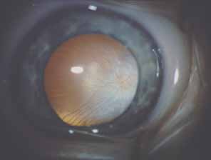
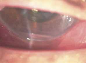
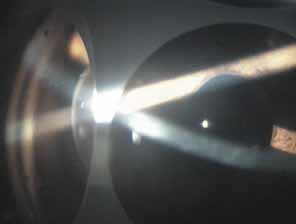
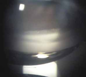
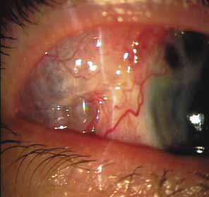
Over the past two decades silicone oil (dimethylpolysiloxane) has enjoyed a resurgence in the repair of complicated retinal detachments. In 1967, Watzke36 found no pressure elevation attributable to silicone but described droplets in the superior angle that result from silicone's lower specific gravity (buoyancy) compared with water. Silicone-induced glaucoma does occur, however, and one possible mechanism is an acute pupillary block due to the buoyancy of the silicone sometimes combined with overfilling the aphakic eye (Fig. 5). This can be averted by performing an inferior iridectomy and by paying careful attention to the silicone volume injected. A second mechanism involves chronic obstruction of an open angle by emulsified silicone microbubbles (more likely with low viscosity silicone, 1,000 centistokes, than high viscosity silicone, 12,500 centistokes) (Fig. 6), pigmented cells, and silicone-laden macrophages or silicone-induced fibrosis of the trabecular meshwork due to possible inherent fibrogenicity of this compound.37,38 Treatment of this open-angle glaucoma is generally medical, although silicone removal may be required. Although some studies have confirmed the presence of emulsified oil in the anterior chamber as a strong predictor of pressure elevation, others do not, suggesting that multiple factors are at play.39,40,41,42 Often this can only be seen on gonioscopy, and removal of the oil, if possible, is beneficial in some cases. Sometimes an expected pressure elevation that would otherwise occur may be muted by concomitant cyclitic membrane formation or hypotony associated with recurrent retinal detachment. The use of shunt procedures in this difficult glaucoma has led to the unusual complication of silicone oil escape into the subconjunctival space43,44 (Fig. 7). Whether placed superiorly or inferiorly, shunts should probably be avoided in these eyes unless the silicone oil can first be safely removed. Transscleral diode laser cyclophotocoagulation has been proven to be an excellent noninvasive option in treating these eyes for recalcitrant pressure elevation.45,46

Fig. 5 Gonioscopic view of pupillary block by silicone oil. Patent iridectomy below allows aqueous to enter the anterior chamber, thereby preventing pupillary block angle closure.

Fig. 6 Fine glistening silicone droplets seen congregating only in the superior angle. Silicone oil has a viscosity lower than aqueous, which causes this superior migration of oil bubbles.

Fig. 7 Subconjunctival mass of silicone oil escaping from the globe through an Ahmed shunt. Similar cases have been reported with inferiorly placed shunts.

Intravitreal gas has become a standard part of complex and routine retinal repairs. The appropriate use of intraocular long-acting gas tamponades requires a thorough knowledge of the time-expansion characteristics of the chosen gas (sulfur hexafluoride; perfluoropropane) as well as the clinical situation (partial or total fill of the vitreous cavity).47 Sulfur hexafluoride has a peak expansion 6 to 12 hours after injection. A 20% mixture usually represents the concentration that will not expand in volume when a total fill of the vitreous cavity is performed. Perfluoropropane expands acutely over a similar time frame. A 14% mixture with room air represents the “safe” nonexpansile percentage when the total volume is replaced (Fig. 8).

Fig. 8 Recently vitrectomized eye with 20% perfluoropropane (C3F8) gas bubble.

OTHER TRABECULAR MESHWORK INSULTS

Foreign Body Siderosis

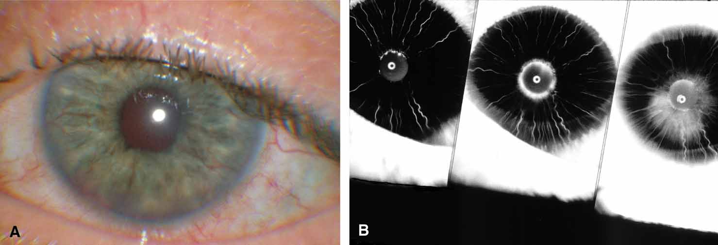
Long periods of exposure of intraocular toxic metals such as iron can cause damage to the cells of the trabecular meshwork. Thus, an open-angle glaucoma can ensue that may be recalcitrant to treatment. The unilaterality may be confusing, particularly when the previous ocular history is unknown and the foreign body is occult (Fig. 9).48

Fig. 9 Ocular siderosis. A. Pigment clumping on the anterior lens capsule in eye penetrated by iron foreign body with nonrecordable electroretinogram. B. Small, occult foreign body (large arrow) had penetrated iris many years previously. Pigment clumps on lens capsule with dense cataract. Elevated pressures were controlled, and the electroretinogram was only mildly affected, allowing 20/20 (metric equivalent 6/6) visual recovery after cataract removal. Metallic foreign body was localized to anterior segment and easily removed by magnet extraction at the time of cataract surgery.

Intravitreal Steroids

Intravitreal steroids,49 both depot and the newer delayed release implants,50 have been proposed for the treatment of recalcitrant macular edema or exudation from diabetes, uveitis, macular degeneration, and other conditions.51

As might be expected, these have been found to cause clinically significant pressure elevations in some individuals.52,53,54 As such, intravitreal steroid treatment mandates careful monitoring of intraocular pressure in these patients.

RAISED EPISCLERAL VENOUS PRESSURE

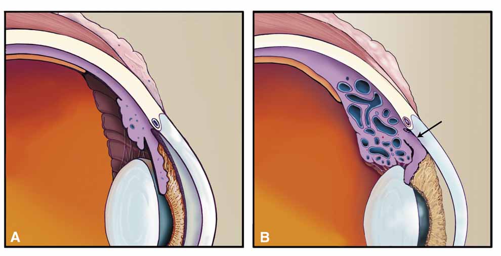
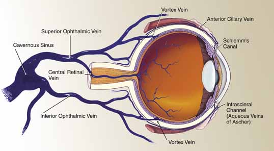
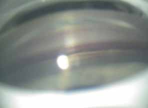
A review of the aqueous blood microcirculation of the eye (Fig. 10) explains how raised venous pressure can affect both retinal vessels and intraocular pressure. From Schlemm's canal, aqueous traverses the intrascleral emissary channels (aqueous veins of Ascher) to the episcleral plexus and then the long ciliary venous vessels. The long ciliary veins, as well as the vortex veins, empty into the ophthalmic vein before entering the cavernous sinus. Increases in episcleral venous backpressure theoretically contribute to a 1:1 mm Hg rise of measured intraocular pressure. The clinical result of an acute rise in external venous pressure may include choroidal effusion, elevated intraocular pressure with blood in Schlemm's canal (Fig. 11), and retinal vein obstruction with intraretinal hemorrhages in a central vein obstruction pattern with or without prominent swelling of the optic nerve. Chronic elevations of venous pressure may permanently damage trabecular meshwork, impairing outflow facility, and result in a chronic open-angle glaucoma.

Fig. 10 Venous drainage system of the eye. Aqueous enters Schlemm's canal and then penetrates through the intrascleral channels (aqueous veins of Ascher). Portions of the aqueous veins are visible at the limbus. The episcleral vessels drain to the anterior ciliary vessels, which along with the vortex veins drain to the ophthalmic vein and then the cavernous sinus.

Fig. 11 Gonioscopy photograph showing blood in Schlemm's canal noted below Schwalbe's line, which may result from elevated venous pressure, uveitis, trauma, or hypotony. This finding may be more subtle in pigmented angles but recognized as the often brick red coloration of the trabecular meshwork band as compared to the other eye.

Superior Vena Cava Syndrome

Obstruction of the venous inflow to the heart may be due to mediastinal tumors, aortic aneurysms, goiters, and enlarged hilar lymph nodes among other causes. A central retinal vein obstruction picture results with engorgement of the retinal veins, peripapillary retinal edema, and elevated intraocular pressure.55 The pressure becomes elevated in the supine position. Correction of the underlying obstruction is ideal, but topical pressure medications may be somewhat effective.

Carotid–Cavernous Fistula

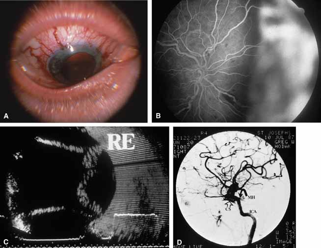
A true shunt between the carotid and cavernous sinusoidal venous system (high-flow type) occurs in 25% of cases and usually results from trauma.56 The more common situation (75% of cases) is a vascular shunt between a dural branch of the external or internal carotid (usually meningohypophyseal branch) and the cavernous sinus (low-flow type).56 There is some evidence that vestigial dural shunts may be congenital and expand when an obstruction occurs in the transverse and sagittal venous drainage systems of the brain.56 The net effect is a direct transmittal of arterial pressure to the retinal and choroidal venous system, which may produce several syndromes. Recent work involving more precise radiologic studies suggests that the specific clinical presentation is determined by the exact location of the vascular anomaly.57 Ocular hypoxia produced by reduced flow may lead to iris neovascularization and glaucoma.58 The more common cause of elevated intraocular pressure is chronic venous pressure elevation that results in permanent damage to the trabecular meshwork. Rarely, vortex venous backpressure may produce choroidal edema, effusion, rotation of the ciliary body, and a congestive glaucoma (Fig. 12).59,60 High- and low-flow arteriovenous shunts pose no threat to human life but have been the object of prodigious surgical efforts. The indications for the surgery should remain intractable eye pain or threatened loss of the eye or other neurological deficits. Shunt flow may be reduced by arterial closure, direct shunt closure, or venous ligation. All these methods, except direct shunt closure, may exacerbate hypoxia and induce iris neovascularization.61

Fig. 12 Dural-type carotid cavernous fistula with acute glaucoma. A. Arterialized episcleral, limbal vessels with shallow chamber and tension of 35 mm Hg. B. Fluorescein angiogram shows nasal choroidal detachment with multiple areas of blocked fluorescence due to concomitant venous obstruction. C. B-scan ultrasonogram demonstrating serous choroidal detachments. D. Selective internal carotid arteriogram showing filling of the cavernous sinus (CS) from the meningohypophyseal branch (MH) of the internal carotid artery (1CA).

Sturge-Weber Syndrome

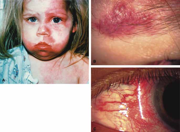
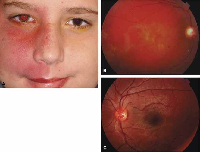
The Sturge-Weber Syndrome is a sporadically occurring phakomatosis that consists of a diffuse choroidal hemangioma, facial nevus flammeus (dermal angioma) (Fig. 13), meningeal angioma with “tram-track” calcification of the brain, and an ipsilateral glaucoma often seen when the dermal angioma involves the upper lid (Fig. 14). The glaucoma may present in infancy or the late teens. In infancy, the glaucoma mechanism may be similar to congenital glaucoma (abnormal trabecular bands) and differ from the later-onset form, which may result from elevated episcleral venous pressure.62 Phelps63 demonstrated elevated episcleral venous pressure in this syndrome and related the extent of the episcleral angioma (often more obvious at surgery when conjunctiva was reflected) to the severity of the glaucoma. Recent work demonstrated thickening of the trabecular beams with abnormalities of the outflow structures, possibly induced by the abnormal vasculature present during embryogenesis.64 Regardless of the exact mechanism, most authorities agree that standard filtration methods carry a high risk of expulsive choroidal hemorrhage. If filtra-tion surgery is contemplated, Bellows and co-workers65 recommend prophylactic sclerostomies prior to decompression of the eye to avoid intraoperative choroidal detachments that may occur in these patients, even when choroidal hemangiomas are not present. Meticulous flap closure that avoids hypotony is the mainstay to preventing choroidal detachments.66 Preoperative scatter laser photocoagulation over the hemangioma, even in the absence of serous retinal detachment, may also reduce the risk of choroidal exudation prior to filtration surgery.66

Fig. 13 (A) Ten-year-old boy with facial nevus flammeus in the distribution of the 2nd division of the right trigeminal nerve and increased intraocular pressure in the right eye. (B) A diffuse choroidal hemangioma of the macula was present along with an overlying serous detachment of the retina and cupping of the right optic disc. (C) Normal left fundus. (Photos courtesy of Carol L. Shields, MD, Philadelphia, PA)

Fig. 14 Sturge Weber Syndrome Findings. A. Facial Hemangioma is usually unilateral but can vary in both size and distribution. B. Localized dermal hemangioma in different patient. C. Episcleral hemangioma can be quite prominent (from patient shown in B).

Back to Top
ANGLE-CLOSURE GLAUCOMAS

STRUCTURAL

The designation “Structural” implies a mechanical crowding of anterior segment structures (lens, ciliary processes, and pars plana) that predisposes the eye to angle closure. Pupillary block may develop primarily from anatomical crowding, although angle closure secondary to ciliary body rotation may result from choroidal congestion. Whereas peripheral iridotomy/iridectomy is successful in pupillary block cases, proper treatment of the congestive component includes laser iridoplasty or angle deepening by cycloplegia or viscoelastic injection.

Postvitrectomy Angle Closure

One of us has recently treated two cases of pupillary block following pars plana vitrectomy with gas instillation for macular hole repair. In both cases, the gas fill was less than 30%, and the angle closure was successfully broken with laser iridotomies. In light of these findings, we presume that postoperative inflammation caused adhesions between the pupil and the crystalline lens with a subsequent angle closure. It is doubtful that the gas bubble contributed to the closure, although a pars plana tap is certainly warranted in cases of overfill. Post–pars plana vitrectomized eyes are at a higher risk for developing several different types of glaucomas, which can be identified by careful anterior segment and gonioscopic examinations.67

Persistent Fetal Vasculature (PFV)

A congenital, unilateral (90%) malformation, persistent fetal vasculature (PFV) is recognizable at birth and is unrelated to prematurity.68 Originally called persistent hyperplastic primary vitreous (PHPV), Goldberg has proposed the newer terminology to reflect our better understanding of this clinical condition.69 In the classic description of this disorder (anterior form), one finds a microphthalmic eye with a shallow anterior chamber (Table 4); the posterior form is less frequently recognized, and glaucoma may occur in the involved eye in latter life.70 The anterior form, if untreated, may develop pupillary block glaucoma by vascular ingrowth of the tunica vasculosis lentis (Fig. 15) through the posterior capsule, leading to a sudden hydrostatic expansion of the lens. Milder forms of the condition have been recognized, and surgical removal of the lens is not always indicted.71 In most cases, pars plana lensectomy employing an anterior postlimbal entry has been advocated to avoid glaucoma and offer a chance for visual rehabilitation.71,72 Most late-onset cases of angle closure due to lenticular swelling occur in severely amblyopic eyes, and the intractable glaucoma in this setting is best treated by atropine, corticosteroids, Timoptic, and even enucleation, if required. In the anterior form of the disorder, the contralateral eye appears to be at risk for open-angle glaucoma later in adult life and should be observed closely.73


TABLE 4. Ocular Findings in Anterior and Posterior Forms of Persistent Fetal Vasculature (PFV)


Anterior FormPosterior Form
LeukocoriaNo leukocoria
MicrophthalmiaMicrocornea (1.5 mm
Initial clear lens; difference)
 later cataractUsually no cataract
Shallow anterior chamberVitreous membranes, stalks,
Elongated ciliary retinal fold
 processesPeripapillary tractional retinal
Engorged iris vessels detachment
Acute secondary-angle closure glaucomaInvolved eye at risk for primary open-angle
Contralateral eye at risk for primary open-angle glaucoma glaucoma

(Adapted from Pruett RC, Schepens CC: Posterior hyperplastic primary vitreous. Am J Ophthalmol 69:535, 1970)


Fig. 15 Persistent fetal vasculature (PFV)/persistent hyperplastic primary vitreous (PHPV). Intraoperative photograph showing the persistent tunica vasculosis lentis present on the posterior capsule of a clear lens in this 6-week-old infant. The eye was microphthalmic with a horizontal corneal diameter of 9 mm.

Retinal Dysplasia

The term retinal dysplasia was originally used to describe a syndrome including abnormalities of the brain, heart, extremities, mouth, and eye. The current, preferred use of the term designates a pathologic retinal lesion involving dysplastic retina in a malformed eye and not a specific clinical syndrome.74 This term may describe the dysplastic retina seen in persistent hyperplastic primary vitreous, chromosomal trisomy syndrome 13–15, and the fetal effects of maternal ingestion of D-lysergic diethylamide (LSD). Secondary angle-closure glaucoma of the pupillary block variety may develop as a result of anterior chamber angle malformation.

Retinopathy of Prematurity

In cases involving a blind eye with an organized total retinal detachment judged inoperable, treatment options should include cycloplegics, corticosteroids, and possibly cyclodestructive therapy.75 Lens aspiration in infants to relieve an acute, secondary angle-closure glaucoma should be considered as a prelude to definitive vitrectomy for complicated retinal detachment (stage 4 or 5). Vitrectomy results in these patients are under evaluation and remain controversial. In later life, angle-closure glaucoma can occur in a seeing eye despite high myopia (stage 1).76 In those eyes with the cicatricial phase of the disease, crowding of the anterior ocular structures predisposes to a pupillary block glaucoma (Fig. 16; Table 5).77–80 Treatment of the acute angle closure would include laser (argon or neodynium: yttrium-aluminum-garnet [Nd: YAG]) or surgical iridectomy. The glaucoma mechanisms in these severely afflicted eyes may be multifactorial and include iris neovascularization.

Fig. 16 Cicatricial stage of retinopathy of prematurity in an adult. A. Right eye with dragging of the optic disc (stage 2), high myopia, and 20/50 (6/15) vision. B. Left eye with cataract (c), amblyopia, and 20/400 (6/120) vision. Previous acute angle closure glaucoma treated by surgical peripheral iridectomy (pi).


TABLE 5. Acute and Chronic Stages of Retinopathy of Prematurity


Acute Stage (Age 3 to 5 Weeks)Cicatricial Stage (Age 3 to 5 Months)
White peripheral line of demarcationHigh myopia, > 6 diopters
White peripheral ridge of tissueDragging of the optic disc
Elevated, reddish neovascularizationFalciform retinal fold
Subtotal retinal detachmentPartial retinal detachment
 ExtrafovealTotal retinal detachment
 Retinal detachment including fovea 
Total retinal detachment: funnel anterior or posterior; open or closed 
Plus disease: enlarged retinal veins and tortuous arteries 

(Data from International Committee for the Classification of the Late Stages of Retinopathy of Prematurity: An international classification of retinopathy of prematurity: II. The classification of retinal detachment. Arch Ophthalmol 105:906, 1987, and Tasman WS: Retrolental fibroplasia. In Tasman WS (ed): Retinal diseases in children. New York: Harper & Row, 1971:59–69)


The goal of panretinal photocoagulation in advanced, acute stage 3 retinopathy of prematurity with plus disease is to induce the regression of neovascularization without traction.78 As more eyes are salvaged by this treatment method, it remains to be seen if these newly converted cicatricial stage eyes will assume a similar risk of acute pupillary block glaucoma.

Iris Retraction Syndrome of Campbell

The iris retraction syndrome of Campbell was described in eyes suffering from severe uveitis secondary to unattended retinal detachment.81 The uveitis produced posterior synechiae, an irregular pupil, and pupillary seclusion. Theoretically there is posterior diversion of aqueous from the ciliary body through the retinal hole and into the choroid, producing low pressure with a deep anterior chamber (iris retraction), owing to a reduced vitreous volume. These eyes may spontaneously improve their ciliary body fluid output or may respond to corticosteroid treatment, leading to a sudden, severe angle-closure glaucoma by a pupillary block mechanism with iris bombé. Peripheral iridotomy by laser (argon or Nd: YAG) or surgery may relieve the acute glaucoma, but the severe retinal detachment complicated by proliferative vitreoretinopathy and uveitis often results in a poor surgical outcome despite modern vitreoretinal techniques.

CYCLOCONGESTIVE/CILIOCHOROIDAL EFFUSION SYNDROME

All disorders in this group have some degree of choroidal congestion (Table 6), which may be noted by the usual clinical signs of clinical moundlike or annular choroidal detachment or be completely undetected, particularly if anteriorly placed and hidden behind the iris.82 Choroidal congestion can produce forward rotation of the ciliary body, resulting in closure of the peripheral angle and a shallow but preserved central anterior chamber (Fig. 17).83 Although this may appear a contradiction of terms because choroidal detachments are usually associated with hypotony, experimental detachments of the ciliary body do not themselves result in aqueous shutdown,84 suggesting that low pressure with choroidal effusion is due to hyposecretion secondary to concurrent iridocyclitis. Continued aqueous production in the presence of a closed peripheral angle results in an acute pressure elevation. Some clinical diseases sharing this mechanism have been mislabeled “malignant glaucoma,” an unfortunate name that adds mystique and confusion to the mechanism underlying these diseases. Though we prefer the term cyclocongestive to describe those disorders sharing this mechanism, other terms have been suggested including ciliochoroidal effusion syndrome.85 Either is descriptive of the pathophysiologic mechanisms that cause this clinical presentation. Miotics and peripheral iridectomy may worsen these conditions, whereas they are a first-line choice in the treatment of primary angle-closure glaucomas. Although cyclocongestive glaucoma may initially begin by ciliary body rotation with peripheral iris blocking the trabecular meshwork, if it is left untreated or diagnosed incorrectly, permanent, chronic angle closure may result, requiring goniosynechialysis or filtering surgery for adequate pressure control. Filtration surgery may actually precipitate the disorder in some cases.86


TABLE 6. Cyclocongestive Angle-Closure Glaucomas Secondary to Retinal Disease


InflammatoryVascularPostoperativeNeoplasticIdiopathic
Scleritis116Post CRVO123,124Post-cataract extraction121,86Malignant melanoma30Uveal effusion100
Pars planitis117Nanophthalmos99Trauma87,89Pseudotumor95 syndrome
Harada's disease118Arteriovenous malformationScleral buckling87,89Ocular metastases95 
Acquired immunodeficiency syndrome (AIDS)110,111 or fistulae60Post-panretinal photocoagulation92,93,94  


Fig. 17 A. Normal anatomy. B. Cyclocongestive glaucoma. Scleral buckling and other conditions may obstruct vortex venous outflow, resulting in choroidal congestion and detachment. Swelling of the ciliary body rotates this structure about the scleral spur, closing the peripheral angle. Note the loss of visible angle structures. Laser iris retraction is the treatment of choice in cases resistant to conservative therapy (cycloplegics, topical corticosteroids, timolol maleate, and acetazolamide).

Postoperative Scleral Buckling

Although postoperative pressure elevation following a routine scleral buckling is common, the incidence of clinically significant pressure elevations manifesting as pain and corneal edema is low.87 Choroidal congestion, which may be obvious or detectable only by ultrasonography, causes forward rotation of the ciliary body and closure of the peripheral angle. This situation is more likely to occur as a result of closure or compression of the vortex veins by broad scleral exoplants, which encourages postoperative choroidal edema.88 Exacerbating factors include excessive cryoretinopexy (choroidal congestion), drainage of subretinal fluid, and paracentesis during surgery.89 Treatment should include a trial of acetazolamide, atropine, corticosteroids, and aqueous suppressants. If no improvement is noted in 5 to 7 days, then peripheral laser iris retraction (laser iridoplasty = laser gonioplasty = iridoretraction) may thermally contract the iris, pull open the angle, and relieve the pressure elevation.90 If this fails, choroidal drainage combined with release of the scleral buckle will eliminate the mechanical obstruction of the vortex system.91

Postoperative Panretinal Photocoagulation

Most of the mild, asymptomatic, and transient pressure elevations noted immediately after panretinal photocoagulation occur by unknown mechanisms in the presence of an open angle.92 Occasionally, heat-induced choroidal congestion, choroidal vascular permeability, and choroidal vascular obstruction may, as discussed previously, result in ciliary body rotation and closure of the peripheral angle (Fig. 18). Usually the narrow or closed angles resolve within several days, and thus far no severe visual complications have been reported. Choroidal congestion/effusion occurs frequently but is usually not clinically significant or recognized. Limiting the number of laser burns and the amount of retinal area treated can prevent this problem from occurring.93 Dividing panretinal photocoagulation treatments into two or more sessions several weeks apart is advisable when possible. Photocoagulative pressure elevations usually respond to cycloplegia, aqueous suppressants (acetazolamide and timolol), and osmotics.94

Fig. 18 Panretinal photocoagulation with elevated intraocular pressure. A. Elevated intraocular pressure was noted in the first few hours following scatter laser photocoagulation of the proliferative diabetic retinopathy. B. Four days later the same patient noted blurred vision and was found to have peripheral choroidal detachments, a closed peripheral angle, and intraocular pressure of 35 mm Hg. Treatment with cycloplegics, topical corticosteroids, timolol maleate, and acetazolamide produced a rapid drop in pressure. The choroidal detachments resolved completely in 10 days.

Metastatic Tumor to the Choroid

Widespread metastases to the choroid usually present as multifocal, 2 to 4 disc diameter, grayish mounds that may have associated subretinal fluid. Massive choroidal tumor invasion may produce forward rotation of the ciliary body due to mass effect and vascular choroidal congestion, leading to an acute glaucoma with peripheral angle closure and a shallow but preserved anterior chamber.95 Although medical treatment (cycloplegics, corticosteroids, acetazolamide, and timolol) may produce respite from pain, enucleation of a nonseeing painful eye seems advised.

Nanophthalmos

Eyes with a cycloplegic refraction of greater than +7.50 diopters or axial diameter less than 20 mm by quantitative biometry are deemed nanophthalmic.96 These eyes may develop angle-closure glaucoma in the fourth to sixth decades of life (Fig. 19). A congenital thickening of the sclera results in obstruction of the vortex veins and secondary choroidal congestion, forward rotation of the ciliary body, and closure of the peripheral angle.97 Uveal effusion and exudative retinal detachment have been found to precede98 or follow ocular surgery and may be a factor leading to forward rotation of the ciliary body and iris lens diaphragm producing a cyclocongestive glaucoma. This severe glaucoma may respond to treatment with mannitol, atropine, and Neo-Synephrine. The failure of iridectomy to reliably relieve the condition suggests that relative pupillary block is not the primary event. Nonetheless, peripheral iridotomy should be tried, and if it is ineffective, then laser iridoplasty (peripheral laser iris retraction) may be attempted to physically “pull” the peripheral iris away from the angle.96,99 As in the other choroidal congestive glaucomas, pilocarpine is ineffective and should be avoided. The contralateral eye is also at risk, and management may also include laser iridoplasty for angles with severe narrowing.70 Vortex vein decompression, the Gass procedure (equatorial sclerectomy with sclerotomy),100 and/or unsutured operative sclerostomies should be considered for both acute ipsilateral glaucoma and if any ocular surgical procedures such as cataract extraction are performed.101 Prophylactic therapy in the other eye would include all these modalities, especially if choroidal effusion is present.96 A recent report describes a nanophthalmic patient who developed bilateral angle closure as a complication of systemic anticoagulation. This patient developed massive subretinal hemorrhage, which precipitated angle closure unresponsive to laser iridotomy and ultimately required vitreoretinal surgery.102 This report provides a good example of why nanophthalmic eyes, owing to their unique intraocular anatomy, must be closely monitored for unexpected responses to common clinical interventions.

Fig. 19 Nanophthalmos. A. Complete loss of anterior chamber with pressure of 58 mm Hg. This eye was unresponsive to laser iridotomy. B. Gonioscopic View. No angle structures are apparent. C. Contralateral eye treated with prophylactic iridotomy.

Acute Choroidal/Retinal Hemorrhage

Approximately 5% of patients with age-related macular disciform scars may develop breakthrough hemorrhage into the vitreous cavity and further loss of peripheral vision. Patients taking aspirin or warfarin, those having a blood dyscrasia and those with systemic hypertension may be at a higher risk for such an occurrence. Acute angle closure glaucoma can occur with a hemorrhagic detachment of the retina, retinal pigment epithelium, and choroid that are so severe, sudden, and forceful as to cause the ciliary body to rotate forward. This occludes the peripheral drainage angle and produces an acutely painful eye with corneal edema and elevated pressure.103,104,105 Medical treatment with acetazolamide, corticosteroids, atropine, beta-blockers, and alpha agonists are sometimes useful in the short term to control the pressure elevation and relieve discomfort. When clot liquefication occurs, a secondary ghost cell glaucoma may ensue. Usually these eyes suffer a profound and permanent loss of vision despite the use of vitrectomy to remove the hemorrhage. Treatments of blind painful eyes include retrobulbar alcohol or Thorazine106,107 transscleral diode laser cyclophotocoagulation,105 or enucleation.105

Expulsive or nonexpulsive (limited choroidal) hemorrhage may develop during or immediately following cataract surgery or more frequently at or following glaucoma filtering surgery.108 Acute management should include immediate wound closure, glaucoma medication, and if the pressure cannot be controlled, vitrectomy with drainage of the suprachoroidal hemorrhage.109 The risk of hemorrhagic choroidal detachment is greater with increasing age, myopia, previous glaucoma, and general anesthetics.108 These eyes are at higher risk for retinal detachment complicated by proliferative vitreoretinopathy.

Other Retinal Entities Associated with Congestive Angle-Closure Glaucoma

A most unusual acute angle-closure glaucoma may develop in young men who are later found to have positive human immunodeficiency virus (HIV) titers.110 The glaucoma appears to be of the choroidal cyclocongestive type, with a myopic shift, angle shallowing, and pressure elevation all related to choroidal effusions. These cases respond to cycloplegia and aqueous suppressants (acetazolamide and timolol). Of significance is the fact that the acute glaucoma was reported as the presenting sign of this ominous disease.111 A rash of case reports has linked Topiramate (Topamax), a newer seizure medication, with acute angle closure attacks.112–115 (Fig. 20). Of particular concern is the use of this medica-tion in mentally challenged individuals who may not be able to alert their caregivers to the symptoms of elevated intraocular pressure. Examples of other retinal disorders with which cyclocongestive angle-closure glaucoma may be associated are listed in Table 6.30,60,87,89,92–95,99,100,110–124

Fig. 20 Topiramate. A. B-scan ultrasonography shows diffuse choroidal thickening (Arrow) B. Following resolution of the angle closure, a subsequent scan shows a normal choroidal appearance with posterior vitreous collapse as the only obvious sequelae. (Reprinted from Banta JT, Hoffman K, Budenz DL, et al: Presumed Topiramate-induced bilateral acute angle-closure glaucoma. Am J Ophthalmol 132(1):112, 2001)

NEOVASCULAR

All diseases in the neovascular classification produce neovascularization of the iris (Table 7). The underlying cause is ocular hypoxia, which results in the production of an angiogenic factor directed primarily to the anterior segment.125 Three stages have been described: (1) rubeosis irides—marginal iris vessels with no increased ocular tension; (2) open-angle phase—radial iris vessels join circumferential angle vessels and cross over the scleral spur, now producing increased ocular ten-sion and possibly hyphema; and (3) angle-closure phase—uveal ectropion, dilated pupil, and peripheral angle synechiae that produce intractable total angle closure.126


TABLE 7. Neovascular Angle-Closure Glaucomas Secondary to Retinal Diseases


Ocular Vascular DiseaseOther Ocular DisordersOcular NeoplasmsExtraocular Vascular Disease
Retinal venous obstructionRhegmatogenous retinalRetinoblastomasCarotid artery obstruction
Central/hemispheric/branch detachmentChoroidal melanomaCarotid-cavernous fistula
Diabetic retinopathyUveitisMetastatic carcinomaCarotid artery ligation
Central retinal artery obstructionEndophthalmitisReticulum cell sarcomaGiant cell arteritis
Combined retinal artery/vein obstructionVitreous wick syndrome (intraocular lymphoma)Takayasu's disease
Coats' disease   (pulseless disease)
Eales' disease   
Sickle cell retinopathy   
Sturge-Weber, choroidal hemangioma   
Angiomatosis retinae (Von Hippel's disease)   
Radiation retinopathy   
Syphilitic vasculitis   
Familial exudative vitreoretinopathy of Criswick and Schepens   
Pars planitis   


Central Retinal Vein Occlusion (CRVO)

The vascular thrombosis in central retinal vein occlusion is believed to lie within the optic nervehead at the level of the lamina cribrosa or behind where the vein and the artery share a common adventitial sheath.127 Rarely, an initial period of hypotony may develop after an acute central retinal vein occlusion.128 If major retinal capillary compromise is present,129 iris neovascularization may develop within 1 to 3 months and produce peripheral angle synechiae and angle closure. Ischemic central retinal vein occlusions represent less than one-third of all central venous obstructions and are characterized by multiple cotton-wool spots and a poor visual acuity, typically less than 20/200 (6/60) (Fig. 21).

Fig. 21 Ischemic central retinal vein obstruction. A. Heavy intraretinal hemorrhages, cotton-wool spots, and vision of 20/400 (6/120). B. Fluorescein angiogram clearly demonstrates severe loss of capillary bed perfusion.

Rarely, an acute central retinal vein occlusion may produce choroidal congestion, ciliary body congestion, and rotation with transient peripheral iris angle narrowing and closure (Fig. 22) (see Table 6).123,124 This acute cyclocongestive glaucoma usually responds well to acetazolamide and cycloplegia.

Fig. 22 58-year-old female with acute CRVO. Several days after loss of vision, intraocular pressure elevated to mid 40s. A. Fundus appearance. B. Gonioscopic appearance with elevated pressure. Note loss of angle structures. C. Gonioscopic appearance after cycloplegic treatment. Angle structures are now visible. Intraocular pressure dropped to mid teens, where it has remained.

Although cotton-wool spots represent infarcts of the nerve fiber layer and may be a clinical sign of ischemia, any eye with a central retinal vein occlusion should be monitored with monthly slit lamp and gonioscopic exams. The onset of neovascularization of iris or angle warrants pan retinal photocoagulation to try to prevent neovascular glaucoma.130,131 Between 10% and 20% of eyes with central retinal vein occlusion may convert from an initial nonischemic angiographic picture to ischemia, especially if the initial vision is poor. The greatest risk of conversion to ischemia is found 1 to 3 months following the occlusion but can occur even later. Nonischemic occlusions (Fig. 23) should be followed closely during this time period.

Fig. 23 Nonischemic central retinal vein obstruction. A. Dilated, tortuous veins, minimal intraretinal hemorrhages, and 20/40 (6/12) vision. B. Fluorescein angiogram showing minimal damage to capillary bed. Despite the lack of ischemic capillary damage on the present study, up to 20% of patients may convert to the ischemic type, necessitating careful follow-up examinations.

Although most practices now follow the clinical exam to evaluate of CRVOs instead of fluorescein angiography, significant nonperfusion on angiography still guides some clinicians toward prophylactic pan retinal photocoagulation. In the past, some utilized the electroretinogram (ERG) to define those eyes at greatest risk for neovascularization.132 The ERG was of particular value in those eyes, which confounded conventional angiographic evaluation due to a small pupil, cataract, or those that might have undetectable peripheral ischemia.

Treatment by photocoagulation (possible with a large pupil and a clear lens) or peripheral retinal pancryoablation (in cases with a small pupil and/or cataract) may abort the neovascular stimulus and may induce regression in established iris neovascularization, producing a temporary or long-lasting drop in intraocular pressure. For established neovascular glaucoma, more vigorous methods of glaucoma control are required,133 including filtration surgery with antimetabolites134 or aqueous shunts.135,136 These methods should be considered in eyes with ambulatory vision. In eyes with hand motions or light perception vision, noninvasive transscleral diode laser cyclophotocoagulation or cyclocryotherapy may be considered, although this latter treatment may result in phthisis and rarely in sympathetic ophthalmia.137

Retrobulbar alcohol and Thorazine106 for pain control or enucleation107 are other options. In 20% to 30% of cases of central retinal vein occlusion, signs of primary open-angle glaucoma are present in the contralateral, uninvolved eye,138 which must be diligently examined.

Diabetes Mellitus

Many cases of iris neovascularization develop following ocular surgery, particularly following pars plana vitrectomy for posterior segment complications of proliferative diabetic retinopathy, especially if lensectomy is required (risk increased by twofold) or if preexisting iris neovascularization is present.139 Preoperative iris neovascularization in diabetic patients may regress with successful retinal reattachment by vitrectomy, whereas persistent, postoperative retinal detachment invariably results in neovascular glaucoma and blindness.140 Cataract surgery alone has been implicated as a risk factor for the development of rubeosis and neovascular glaucoma in diabetic patients with proliferative retinopathy (Fig. 24).141 Spontaneous iris neovascularization developing in the absence of posterior segment disease signs of diabetic retinopathy or venous occlusion should prompt an investigation for other entities that produce ischemia such as carotid artery stenosis, especially in patients older than age 50.142

Fig. 24 Rapid progression of diabetic retinopathy following cataract extraction. A. Five weeks following uneventful extracapsular cataract extraction, this 44-year-old patient with insulin-dependent diabetes of 30 years' duration developed a severe drop in vision to 20/400 (6/120) with multiple cotton wool spots and iris neovascularization. B. Iris angiogram shows early neovascularization in early, mid, and late phases of the angiogram. Prompt panretinal photocoagulation prevented frank neovascular glaucoma.

An entity known as anterior hyaloidal fibrovascular proliferation describes the postvitrectomy proliferation of new vessels on the peripheral retina, pars plana, ciliary body, and sometimes the iris.143 This disorder has replaced neovascular glaucoma as the most frequent postsurgical complication of diabetic vitrectomy (Fig. 25).144 There is increasing recognition that the frequency of anterior hyaloidal fibrovascular proliferation may be related to vitreous traction at the sclerotomy sites or the severity of the diabetic retinopathy.145 Although iris neovascularization and glaucoma may result, the more severe clinical outcome is retinal detachment and hypotony secondary to ciliary body detachment by a cyclitic membrane. Anterior hyaloidal fibrovascular proliferation with retinal detachment or vitreous hemorrhage is treated by repeat vitrectomy combined with lens removal, vitreous base dissection, and endophotocoagulation.

Fig. 25 Anterior hyaloidal fibrovascular proliferation. Clinical photograph showing nasal perilenticular neovascularization in diabetic eye 6 weeks following an uneventful vitrectomy for traction retinal detachment.

In diabetic eyes requiring complicated vitrectomies, usually with lensectomy, a unique syndrome of fibrin pupillary block glaucoma has been described.146 The fibrin is a result of surgical trauma and is especially frequent in eyes with preexisting iris neovascularization. The fibrin clot occludes the pupil, resulting in an acute aphakic pupillary block glaucoma. The treatment of this unique syndrome includes argon or Nd: YAG laser disruption of the pupillary membrane or fibrin clot dissolution by intracameral injection of tissue plasminogen activator.147

Carotid Artery Disease

Features of ocular ischemia including neovascular glaucoma may develop when severe carotid occlusive disease results in hypoperfusion. A dot-blot peripheral retinopathy with microaneurysms (venous stasis retinopathy of Kearns) may be present in 5% of patients with established carotid artery stenosis of greater than 70% flow reduction (Fig. 26). However, iris neovascularization and glaucoma may develop in the absence of posterior segment manifestations148 and make the underlying carotid pathology difficult to diagnose. Since the ciliary body is subject to both hypoxia and hypoperfusion, neovascular glaucoma may be normotensive or even hypotensive.149 Endarterectomy may improve flow and may transiently lead to regression of iris neovascularization and lowering of pressure elevation. When neovascularization of the angle results in increased intraocular pressure, then panretinal photocoagulation is indicated. Other cycloablative methods, such as transscleral diode laser cyclophotocoagulation therapy or continuous wave transscleral Nd: YAG ablation, may be considered when panretinal photocoagulation fails or is not possible.

Fig. 26 Venous stasis retinopathy of carotid artery disease. A. Mid-peripheral dot-blot hemorrhages and microaneurysms in asymptomatic patient with right carotid obstruction. B. Digital subtraction angiogram, venous method, showing complete right internal carotid obstruction (Ic) (v, vertebral; Ic, internal carotid; Rc, right common carotid; Lc, left common carotid).

Tumors of the Choroid

Widespread choroidal metastatic tumors (breast or lung), large posterior or equatorial melanomas, and large exophytic retinoblastomas may grow rapidly and cause choroidal congestion and acute glaucoma. Large tumors may result in anterior segment hypoxia with iris neovascularization and angle-closure glaucoma. Shields and co-workers29 found pressure elevations in 17% of 303 eyes with retinoblastomas, and unlike other tumors, neovascular angle closure occurred in 70% of this group. If tumor growth can be controlled by radiation and if vision can be salvaged, then enucleation may be avoided.150 Nonetheless, modern methods to control tumors (primarily melanomas), either by external beam irradiation (helium or proton beam)151 or episcleral plaque radiation therapy,152 carry a small but significant risk of iris neovascularization and glaucoma, which may result in loss of vision and loss of the eye.

Central Retinal Artery Occlusion

Iris neovascularization following central retinal artery obstruction is a rare event, occurring in 1% to 5% of these patients. It usually develops 1 to 2 months following the acute loss of vision.153 It is important to consider previously undiagnosed carotid obstructive disease as the cause of iris neovascularization, either by release of microembolic debris from a diseased carotid bifurcation or by severe flow reduction. Duker and Brown154 described several cases of neovascular retinopathy and glaucoma developing in eyes following central retinal artery occlusion. Panretinal photocoagulation resulted in neovascular regression, but vision remained poor because of the retinal infarction.

Other Retinal Entities Associated with Neovascular Angle-Closure Glaucoma

Severe ischemic retinopathies due to sickle cell disease are more frequent in the SC and SS variants and result from vascular sludging of nondeformable (sickled) red blood cells.155 These changes are induced by hypoxia and acidosis. Peripheral ischemic retinopathy with neovascularization (sea-fan) is more common, but ischemia may involve the anterior segment and iris to produce neovascular glaucoma. Anterior segment necrosis syndrome (corneal edema, cataract, dilated or neovascular iris vessels) may follow retinal detachment repairs requiring scleral buckling with or without vitrectomy. Previously, exchange transfusions were recommended preoperatively, however, the risk of anterior segment necrosis is now considered minimal and the need for these transfusions is infrequent and the risk unnecessary.156

Patients with Coats' disease (congenital retinal telangiectasis) with four-quadrant retinal involvement are at risk for the development of neovascular glaucoma, especially in eyes that escape treatment with retinal cryopexy or laser photocoagulation.157

End-stage uveitis (e.g., advanced pars planitis117) with retinal neovascularization and occlusive vasculitis syndromes (e.g., Behçet's disease or systemic lupus erythematosus) carry a risk of neovascular glaucoma. Usually iris neovascularization and glaucoma occur as an end-stage event associated with severe visual loss.

Examples of other retinal entities associated with neovascular angle-closure glaucoma are listed in Table 7.

Back to Top
OTHER RETINAL DISORDERS ASSOCIATED WITH ANGLE-CLOSURE

GLAUCOMA

Expanding Gases/Air Travel

Air and expanding gas bubbles are frequently used in retina and vitreous surgery. Major intraocular pressure problems may arise if the patients have to drive to a high altitude or fly while the bubble is still in the eye.158,159,160 Boyle's law dictates that the bubble will expand as the altitude increases. The intraocular pressure rise may result from (1) increasing intraocular volume (open-angle mechanism) or (2) pupillary block (closed-angle mechanism). An air bubble placed in an aphakic postvitrectomized eye may produce an angle-closure attack by a pupillary block mechanism if the pupil opening is occluded. The important risk factors are the size of the bubble and the pupillary diameter. This occurrence can be avoided by proper head positioning, which keeps the air bubble away from the pupil. If the angle closure is diagnosed early, an anterior chamber deepening procedure may be done at the slit lamp by injecting a viscoelastic agent through a limbal paracentesis in aphakic individuals or through a pars plana tap in phakic or pseudophakic patients to reduce the gas volume.161 If performed early, this procedure may avert permanent peripheral angle closure, which might require filtration surgery.

An expanding gas bubble (sulfur hexafluoride or perfluoropropane) may present similar difficulties in pseudophakic or phakic eyes, but the initial mechanism for pressure elevation is an open-angle glaucoma due to inability of the trabecular meshwork to clear fluid faster than the expansion of the gas. Later, the glaucoma mechanism involves simple mechanical crowding of anterior ocular structures because the physical presence of the bubble induces a secondary angle closure. Treatment in this instance would include needle aspiration of a small quantity of gas (0.1 ml) possibly combined with viscoelastic unsealing of the angle, if needed, to relieve iris to corneal adhesion.

Changes in altitude as a result of surface travel or air flight are thought to be safe if the total estimated air bubble is kept at less than 20% of the total vitreous volume (Fig. 27) and the rate of ascent is slow enough to allow the IOP to equilibrate.159 The safest advice for postoperative air travelers with intraocular gas is for them to wait until the bubble has resorbed.158,160 In our experience, pretreatment with acetazolamide or topical agents has been ineffective in reducing this pressure rise during air travel.

Fig. 27 Regardless of the type of gas employed (room air, sulfur hexafluoride [SF6], perfluoropropane [C3F8]), the main consideration for safe air flight is the percent volume of the vitreous cavity occupied by the air bubble at the time of air flight. During air flight takeoffs, a rapid climb in altitude produces expansion of the air bubble. Even a 20% air bubble may expand sufficiently to cause elevation of intraocular pressure. (Reprinted from Diekert J, O'Connor P, Schaklett D, et al: Air travel and intraocular gas. Ophthalmology 93:642, 1986)

Back to Top
CONCLUSION
Retinal disease can be complicated by a variety of glaucomas, usually of the secondary variety. The glaucoma might be easily overlooked while attention is focused on the acute visually threatening retinal affliction. Conversely, glaucoma patients are not immune to retinal disease, which may complicate management of an otherwise simple primary open-angle glaucoma. Understanding a mechanistic approach to the vast array of interrelated disorders will promote diagnostic accuracy and an effective therapeutic plan.
Back to Top
REFERENCES

1. Wright JE: The Bedford glaucoma survey. In Hunt LB (ed): Glaucoma: Epidemiology, early diagnosis, and some aspects of treatment. Edinburgh: E&S Livingstone, 1966:12–17

2. Kolker A, Hetherton J: Becker and Schaffer's diagnosis and therapy of the glaucomas. 3rd ed. St Louis: CV Mosby, 1970:243–244

3. Phelps CD, Burton TC: Glaucoma and retinal detachment. Arch Ophthalmol 95:418, 1977

4. Shammas HF, Halasa AH, Paris BM: Intraocular pressure, cup-disc ratio, and steroid responsiveness in retinal detachment. Arch Ophthalmol 94:1108, 1976

5. Armaly MF: Inheritance of dexamethasone hypertension and glaucoma. Arch Ophthalmol 77:747, 1967

6. Becker B: Intraocular pressure response to corticosteroids. Invest Ophthalmol 4:198, 1965

7. Lemcke HH, Pischel DK: Retinal detachments after the use of phospholine iodide. Transactions of the Pacific Coast Otoophthalmological Society 47:157, 1966

8. Pape LG, Forbes M: Retinal detachment and miotics. Am J Ophthalmol 85:558, 1978

9. Alpar JJ: Miotics and retinal detachments: A survey and case report. Ann Ophthalmol 11:395, 1979

10. Weseley P, Liebmann J, Walsh J, Ritch R: Lattice degeneration of the retina and the pigment dispersion syndrome. AJO 114:539, 1992

11. Galezowski X: Glaucome aigu dans un cas d'atrophie progressive de la retine (retinite pigmentaire des auteurs). Ann Ocul 48:269, 1862

12. Gartner S, Schlossman A: Retinitis pigmentosa associated with glaucoma. Am J Ophthalmol 32:1337, 1971

13. Kogbe OI, Follmann P: Investigations into aqueous humor dynamics in primary pigmentary degeneration of the retina. Ophthalmologic 171:165, 1975

14. Wagner H: Ein bisher unbekanntes Erbleiden des Auges (Degeneratio hyaloideo-retinalis hereditaria), beobachtet im Kanton Zurich. Klin Monatsbl Augenheilkd 100:840, 1938

15. Jansen LMAA: Degeneratio Hyaloideo-Retinalis hereditaria. Ophthalmologica 144:458, 1962

16. Phelps C: Glaucoma associated with retinal disorders. In Ritch R, Shields MB (eds): The secondary glaucomas. St Louis: CV Mosby, 1982:159

17. Young NJA, Hitchings RA, Sehmi K, Bird AC: Stickler's syndrome and neovascular glaucoma. Br J Ophthalmol 63:826, 1979

18. Hoskins HD Jr, Kass M: Becker-Schaffer's diagnosis and therapy of the glaucomas. 6th ed. St Louis: CV Mosby, 1989:332

19. Obenauf CD, Shaw H, Syndor CF, Klintworth GK: Sar-coidosis and its ophthalmic manifestations. Am J Ophthalmol 86:648, 1978

20. Lichter P, Schaffer RN: Interstitial keratitis and glaucoma. Am J Ophthalmol 68:241, 1968

21. O'Connor GR: Manifestations and management of ocular toxoplasmosis. Bull NY Acad Med 50:192, 1974

22. Roth M, Simmons RJ: Glaucoma associated with precipitates on the trabecular meshwork. Ophthalmology 86:1613, 1979

23. Herschler J: Intractable intraocular hypertension induced by repository triamcinolone acetonide. Am J Ophthalmol 74:501, 1972

24. Kiess JD, Burnes KC, Cohen MM, Wolter JR: Hemolytic glaucoma: A difficult clinical diagnosis. Eye, Ear, Nose Throat Month 48:49, 1969

25. Campbell DG, Simmons RJ, Grant WM: Ghost cells as a cause of glaucoma. Am J Ophthalmol 81:441, 1976

26. Campbell DG, Simmons RJ, Tolentino FI, McMeel JW: Glaucoma occurring after closed vitrectomy. Am J Ophthalmol 83:63, 1977

27. Campbell DG: Ghost cell glaucoma. In Ritch R, Shields MB (eds): The secondary glaucomas. St Louis: CV Mosby, 1982:320

28. Deutch TA, Weinreb RN, Goldberg MF: Indications for surgical management of hyphema in patients with sickle cell trait. Arch Ophthalmol 102:566, 1984

29. Shields CL, Shields JA, Shields MB, Augsburger JJ: Prevalence and mechanisms of secondary intraocular pressure elevation in eyes with intraocular tumors. Ophthalmology 94:839, 1987

30. Yanoff M: Glaucoma mechanisms in eyes containing uveal malignant melanoma. Am J Ophthalmol 70:898, 1970

31. Yanoff M, Scheie HG: Melanomalytic glaucoma: Report of a case. Arch Ophthalmol 84:471, 1970

32. Zakka KA, Yee RD, Shorr N, et al: Leukemic iris infiltration. Am J Ophthalmol 89:204, 1980

33. Schwartz A: Chronic open-angle glaucoma secondary to rhegmatogenous retinal detachment. Am J Ophthalmol 75:205, 1973

34. Matsuo N, Takabatke M, Ueno H, et al: Photoreceptor outer segments in the aqueous humor in rhegmatogenous retinal detachment. Am J Ophthalmol 101:673, 1986

35. Lambrou F, Vela A, Woods W: Obstruction of the trabecular meshwork by retinal rod outer segments. Arch Ophthal 107:742, 1989

36. Watzke RC: Silicone retinopiesis for retinal detachment. Arch Ophthalmol 77:185, 1967

37. Cvenkel B, Zupan M, Hvala A: Transmission electron microscopic analysis of trabecular meshwork in secondary glaucoma after intravitreal silicone oil injection. Int Ophthalmol 20:43, 1997

38. Lambrou FH, Burke JM, Aaberg TM: Silicone oil enhances proliferation in experimental proliferative vitreoretinopathy, ARVO abstract. Invest Ophthalmol Vis Sci 28(suppl):290, 1987

39. Honavar S, Goyal M, Majii A, Sen P, Naduvilath T, Dandona L: Glaucoma after pars plana vitrectomy and silicone oil injection for complicated retinal detachments. Ophthalmol 106(1) :169, 1999

40. Valone J, McCarthy M: Emulsified anterior chamber silicone oil and glaucoma. Ophthalmol 101(12) :1908, 1994

41. Nguyen QH, Lloyd MA, Heuer DK, Baerveldt G, Minckler DS, Lean JS, Liggett PE: Incidence and management of glaucoma and intravitreal silicone oil injection for complicated retinal detachments. Ophthalmol 99(10) :1520, 1992

42. Budenz D, Taba KE, Feuer WJ, Eliezer R, Cousins S, Henderer J, Flynn HW: Surgical management of secondary glaucoma after par plana vitrectomy and silicone oil injection for complex retinal detachment. Ophthalmol 108(9) :1628, 2000

43. Nazemi P, Chong L, Varma R, Burnstine M: Migration of intraocular silicone oil into the subconjunctival space and orbit through an ahmed glaucoma valve. Am J Ophthalmol 132(6) :129, 2001

44. Morales J, Shami M, Craenen , Wentlandt T: Silicone oil egressing through an inferiorally implanted ahmed valve. Arch Ophthal 120:831, 2002

45. Kumar A, Dada T, Singh RP, Kedar S: Diode laser trans-scleral cyclophotocoagulation for glaucoma following silicone oil removal. Clin Experiment Ophthalmol 29(4) :220, 2001

46. Han SK, Park KH, Kim DM, Chang BL: Effect of diode laser trans-scleral cyclophotocoagulation in the management of glaucoma after intravitreal silicone oil injection for complicated retinal detachments. Br J Ophthalmol 83(6) :713, 1999

47. Crittenden JJ, deJuan E Jr, Tiedeman J: Expansion of long-acting gas bubbles for intraocular use: Principles and practice. Arch Ophthalmol 103:831, 1985

48. Sneed SR: Ocular siderosis. Arch Ophthalmol 106:997, 1988

49. McCuen BW II, Bessler M, Tano Y, et al: The lack of toxicity of intravitreally administered triamcinolone acetonide. Am J. Ophthal: 91:785, 1981

50. Cheng CK, Berger AS, Pearson PA, et al: Intravitreal sustained release dexamethasone device in the treatment of experiental uveitis. Invest Ophthal Vis Sci 36:442, 1995

51. Tano Y, Chandler D, Machemer R: Treatment of intraocular proliferation with intravitreal injection of triamcinalone acetonide. Am J Ophthalmol 90:810, 1980

52. Jonas JB, Kreissig I, Sofker A, Degenring R: Intravitreal Injection of triamcinolone for diffuse diabetic macular edema. Arch Opthalmol 121:57, 2003

53. Jonas JB, Kreissig I, Degenring R: Intraocular pressure after intravitreal injection of triamcinolone acetonide. Br J Ophthalmol 87:24–27, 2003

54. Antcliff RJ, Spalton DJ, Stanford MR, Graham EM, Fftyche TJ, Marshall J: Intravitreal triamcinalone for uveitic cystoid macular edema: An optical coherence study. Ophthalmol 108(4) :765, 2001

55. Alfano JE, Alfano PA: Glaucoma and superior vena caval syndrome. Am J Ophthalmol 42:685, 1956

56. Grove AS Jr: The dural shunt syndrome: Pathophysiology and clinical course. Ophthalmology 91:31, 1984

57. Steibel-Kalish H, Setton A, Nimii Y, Kalish Y, Hartman J, Huna Bar-on R, Berenstein A, Kupersmith M: Cavernous sinus dural arteriovenous malformations, Patterns of venous drainage are related to clinical signs and symptoms. Ophthalmol 109(9) :1685, 2002

58. Sugar HL: Neovascular glaucoma after carotid-cavernous fistula formation. Ann Ophthalmol 11:1667, 1979

59. Harbison JW, Guerry D, Wiesinger H: Dural arteriovenous fistula and spontaneous choroidal detachment: New cause of an old disease. Br J Ophthalmol 62:483, 1978

60. Buus DR, Tse DT, Parrish RK: Spontaneous carotid cavernous fistual presenting with acute angle closure glaucoma. Arch Ophthalmol 107:596, 1989

61. Weiss DI, Shaffer RN, Nehrenberg TR: Neovascular glaucoma complicating carotid-cavernous fistula. Arch Ophthalmol 69:60, 1963

62. Weiss DI: Dual origin of glaucoma in encephalotrigeminal haemangiomatosis. Trans Ophthalmol Soc UK 93:477, 1973

63. Phelps CD: The pathogenesis of glaucoma in Sturge-Weber syndrome. Ophthalmology 85:276, 1978

64. Cibis GW, Tripathi RC, Tripathi BJ: Glaucoma in Sturge-Weber syndrome. Ophthalmology 91:1061, 1984

65. Bellows AR, Chylack LT Jr, Epstein DL, et al: Choroidal effusion during glaucoma surgery in patients with prominent episcleral vessels. Arch Ophthalmol 97:493, 1979

66. Ritch R, Simmons RJ, Krupin T, Hitchings R: Cases in controversy: Serous retinal detachment after glaucoma filtration surgery in Sturge-Weber Syndrome. Journal of Glaucoma 1(1) :58, 1992

67. Han DP, Lewis H, Lambrou FH, Mieler WF, Hartz A: Mechanisms of intraocular pressure elevation after pars planar vitrectomy. Ophthalmol 96(9) :1357, 1989

68. Reese AB: Persistent hyperplastic primary vitreous. Am J Ophthalmol 40:317, 1955

69. Goldberg MF: Persistent fetal vasculature (PFV); an integrated interpretation of signs and symptoms associated with persistent hyperplastic primary vitreous. The Jackson Memorial Lecture. Am J Ophthalmol 124:587, 1997

70. Pruett RC, Schepens CC: Posterior hyperplastic primary vitreous. Am J Ophthalmol 69:535, 1970

71. Stark WJ, Lindsey PS, Fagadau WR, et al: Persistent hyperplastic primary vitreous: Surgical treatment. Ophthalmology 90:452, 1983

72. Alexandrakis G, Scott I, Flynn HW, Murray TG, Feuer WJ: Visual acuity outcomes with and without surgery in patients with persistent fetal vasculature. Opthalmol 107(6) :1068, 2000

73. Awan KJ, Humayun M: Changes in the contralateral eye in uncomplicated persistent hyperplastic primary vitreous in adults. Am J Ophthalmol 99:122, 1985

74. Green WR: Retina. In Spencer WH (ed): Ophthalmic pathology: An atlas and textbook. Philadelphia: WB Saunders, 1985:615–617

75. Kushner BJ, Sondheimer S: Medical management of glaucoma associated with cicatricial retinopathy of prematurity. Am J Ophthalmol 94:313, 1982

76. Smith J, Shivitz I: Angle closure glaucoma in adults with cicatricial retinopathy of prematurity. Arch Ophthalmol 102:371, 1984

77. Hittner HM, Rhodes LM, McPherson AR: Anterior segment abnormalities in cicatricial retinopathy of prematurity. Ophthalmology 86:803, 1979

78. Multicenter trial of cryotherapy for retinopathy of prematurity: Preliminary results: Cryotherapy for retinopathy of prematurity cooperative group. Arch Ophthalmol 106:471, 1988

79. International committee for the classification of the late stages of retinopathy of prematurity: An international classification of retinopathy of prematurity: II. The classification of retinal detachment. Arch Ophthalmol 105:906, 1987

80. Tasman WS: Retrolental fibroplasia. In Tasman WS (ed): Retinal diseases in children. New York:. Harper & Row, 1971:615–617

81. Campbell DG: Iris retraction associated with rhegmatogenous retinal detachment syndrome and hypotony: A new explanation. Arch Ophthalmol 102:1457, 1984

82. Aaberg TM: Experimental serous hemorrhagic uveal edema associated with retina detachment surgery. Invest Ophthalmol 14:243, 1975

83. Fourman S: Angle-closure glaucoma complicating ciliochoroidal detachment. Ophthalmology 96:646, 1989

84. Pederson JE, Gaasterland DE, MacLellan HM: Experimental ciliochoroidal detachment: Effect on intraocular pressure and aqueous humor flow. Arch Ophthalmol 97:536, 1979

85. Ikeda N, Ikeda T, Nagata M, Mimura O: Ciliochoroidal effusion syndrome induced by sulfa derivatives (letter). Arch Ophthalmol 120:1775, 2002

86. Dugel PU, Heuer DK, Thach AB, Baerveldt G, Lee PP, Lloyd MA, Minckler DS, Green RL: Annular peripheral choroidal detachment simulating aqueous misdirection after glaucoma surgery. Arch Ophthal 104(3):439–444, 1997

87. Smith TR: Acute glaucoma developing after scleral buckling procedures. Am J Ophthalmol 63:1807, 1967

88. Hayreh SS, Baines JA: Occlusion of the vortex veins: An experimental study. Br J Ophthal 57:217, 1973

89. Burton TC, Folk JC: Laser iris retraction for angle-closure glaucoma after retinal detachment surgery. Ophthalmology 95:742, 1988

90. Phelps CD: Glaucoma associated with retinal disorders. In Ritch R, Shields MD (eds): The secondary glaucomas. St Louis: CV Mosby, 1982:615–617

91. Simmons RJ: Angle-closure after scleral buckling operations for separated retina. In Chandler PA, Grant WM (eds): Glaucoma. 2nd ed. Philadelphia: Lea & Febiger, 1979:183

92. Blondea P, Pavan PR, Phelps CD: Acute pressure elevation following panretinal photocoagulation. Arch Ophthalmol 99:1239, 1981

93. Gentile RC, Stegman Z, Liebmann JM, Dayan AR, Tello C, Walsh JB, Ritch R: Risk factors for effusion after pan retinal photocoagulation. Ophthalmol 103(5) :827, 1996

94. Mensher JH: Anterior chamber depth alteration after retinal photocoagulation. Arch Ophthalmol 95:113, 1977

95. Ferry PA, Font RL: Carcinoma metastatic to the eye and orbit: II. A clinicopathological study of 26 patients with carcinoma metastatic to the anterior segment of the eye. Arch Ophthalmol 93:472, 1975

96. Singh OS, Simmons RJ, Brockhurst RJ, et al: Nanophthalmos: A perspective on identification and therapy. Ophthalmology 89:1006, 1982

97. Brockhurst RJ: Vortex vein decompression for nanophthalmic uveal effusion. Arch Ophthalmol 98: 1987

98. Ryan EA, Zwaan J, Chylack LT: Nanophthalmos with uveal effusion: Clinical and embryologic considerations. Ophthalmology 89:1013, 1982

99. Kimbrough RL, Trempe CS, Brockhurst RJ, et al: Angle-closure glaucoma in nanophthalmos. Am J Ophthalmol 88:572, 1979

100. Gass JDM: Uveal effusion syndrome: A new hypothesis concerning pathogenesis and technique of surgical treatment. Retina 3:159, 1983

101. Jin JC, Anderson DR: Laser and unsutured sclerotomy in nanophthalmos. Am Journ Ophthal 109:575, 1990

102. Caronia RM Sturm RT, Fastenberg DM, Berke SJ, Weintraub J: Bilateral secondary angle closure as a complication of anticoagulation in a nanophthalmic patient. Am J Ophthalmol 126(2) :307, 1998

103. Steinemann T, Goins K, Smith T, et al: Acute closed-angle glaucoma complicating hemorrhagic choroidal detachment associated with parenteral thrombolytic agents. Am J Ophthalmol 106:752, 1988

104. Wood WJ, Smith TR: Senile disciform macular degeneration complicated by massive hemorrhagic retinal detachment and angle closure glaucoma. Retina 3:296, 1983

105. Pepsin S, Katz L, Augsburger J, Chien A, Eagle R: Acute angle-closure glaucoma from massive hemorrhagic retinal or choroidal detachment. Ophthalmol 97(1) :76, 1990

106. Chen T, Yuen S, Sangalang M, Fernando R, Leuenberger E: Retrobulbar Chloropromazine injections for the management of blind and seeing painful eyes. Journal Glaucoma 11:209, 2002

107. Cioffi G: If by chance your eye offend you: The blind painful eye. J Glaucoma 11:171, 2002

108. Gressel MG, Parrish RK, Heuer DK: Delayed nonexpulsive suprachoroidal hemorrhage. Arch Ophthalmol 102:1757, 1984

109. Lakhanpal V, Schocket SS, Elman MJ, et al: A new modified vitreoretinal surgical approach in the management of massive suprachoroidal hemorrhage. Ophthalmology 96:793, 1989

110. Ullman S, Wilson RD, Schwartz LS: Bilateral angle-closure glaucoma in association with acquired immune deficiency syndrome. Am J Ophthalmol 101:419, 1986

111. Williams AS, Williams FC, O'Donnell JJ: Letter: AIDS presenting as acute glaucoma. Arch Ophthalmol 106:311, 1988

112. Thambi L, Kapcala LP, Chambers W, et al: Topiramate-associated secondary angle-closure glaucoma: a case series. Arch Ophthalmol 120(8):1108, 2002

113. Sankar PS, Pasquale LR, Grosskreutz CL: Uveal effusion and secondary angle-closure glaucoma associated with topiramate use. Arch Ophthalmol 119(8) :1210, 2001

114. Rhee DJ, Goldberg MJ, Parrish RK: Bilateral angle-closure glaucoma and ciliary body swelling from topiramate. Arch Ophthalmol 119(11) :1721, 2001

115. Banta JT, Hoffman K, Budenz DL, Ceballos E, Greenfield D: Presumed topiramate-induced bilateral acute angle-closure glaucoma. Am J Ophthalmol 132(1) :112, 2001

116. Quinlan MP, Hitchings RA: Angle-closure glaucoma secondary to posterior scleritis. Br J Ophthalmol 62:330, 1978

117. Brockhurst RJ, Schepens CL, Okamura ID: Uveitis II. Peripheral uveitis: Clinical description, complications and differential diagnosis. Am J Ophthalmol 49:1257, 1960

118. Kimura R, Suzuki A, Maekawa N: Four cases of secondary angle-closure glaucoma caused by Harada's disease. Rinsho Ganka Jpn J Clin Ophthalmol 26:827, 1973

119. Dotan S, Oliver M: Shallow anterior chamber and uveal effusion after nonperforating trauma to the eye. Am J Ophthalmol 94:782, 1982

120. Kutner BN: Acute angle closure glaucoma in nonperforating blunt trauma. Arch Ophthalmol 106:19, 1988

121. McClure HL: Massive bilateral choroidal detachment occurring in an aphakic patient six years and nine months postoperative. Am J Ophthalmol 63:295, 1967

122. Gass JDM: Retinal detachment and narrow-angle glaucoma secondary to inflammatory pseudotumor of the uveal tract. Am J Ophthalmol 64:612, 1967

123. Hyman SW, Neumann : Transient angle-closure glaucoma after retinal vein occlusion. Brit J Ophthal 56:353, 1972

124. Grant WM: Shallowing of the anterior chamber following occlusion of the central retinal vein. Am J Ophthal 75(3) :384, 1973

125. Glaser BM, D'Amore PA, Michels RG, et al: The demonstration of angiogenic activity from ocular tissues. Ophthalmology 87:440, 1980

126. Gartner S, Henkind P: Neovascularization of the iris (rubeosis irides): Review. Surv Ophthalmol 22:440, 1978

127. Green WR, Chan CC, Hutchins GM, et al: Central retinal vein occlusion: A prospective histopathologic study of 29 eyes in 28 cases. Retina 1:27, 1981

128. Hayreh S, March W, Phelps CD: Ocular hypotony following retinal vein occlusion. Arch Ophthalmol 96:827, 1978

129. Margargal LE, Donoso LA, Sanborn GE: Retinal ischemia and risk of neovascularization following central retinal vein obstruction. Ophthalmology 89:1241, 1982

130. Margargal LE, Brown GC, Augsburger JJ, Parrish RK: Neovascular glaucoma following central retinal vein obstruction. Ophthalmology 88:1095, 1981

131. The Central Retinal Vein Study Group: Natural History and clinical management of central retinal vein occlusion. Arch Ophthalmol 115:486, 1997

132. Bresnick GH: Following up patients with central retinal vein occlusion. Arch Ophthalmol 106:324, 1988

133. Sivak-Calcott JA, O'Day D, Gass JD, Tsai JC: Evidence-based recommendations for the diagnosis and treatment of neovascular glaucoma. Ophthalmology 108(1) :1767, 2001

134. Rockwood EJ, Parris RK, Heuer DK, et al: Glaucoma filtering surgery with 5-fluorouracil. Ophthalmology 94:1071, 1987

135. Minckler DS, Heuer DK, Hasty B, et al: Clinical experience with the single-plate Molteno implant in complicated glaucomas. Ophthalmology 95:1181, 1988

136. Schocket SS, Nirankari VS, Lakhanpal V, et al: Anterior chamber tube shunt to an encircling band in the treatment of neovascular and other refractory glaucomas: A long-term study. Ophthalmology 92:553, 1985

137. Green WR: Inflammatory diseases and conditions of the eye. In Spencer WH (ed): Ophthalmic pathology: An atlas and textbook. Philadelphia: WB Saunders, 1985:1915

138. Vannas S, Tarkkanen A: Retinal vein occlusion and glaucoma: Tonographic study of the incidence of glaucoma and of its prognostic significance. Br J Ophthalmol 44:583, 1960

139. Rice TA, Michels RG, Maguire M, et al: The effect of lensectomy on the incidence of iris neovascularization and neovascular glaucoma after vitrectomy for diabetic retinopathy. Am J Ophthalmol 95:1, 1983

140. Scuderi JJ, Blumenkranz MS, Blankenship G: Regression of diabetic rubeosis irides following successful surgical reattachment of the retina by vitrectomy. Retina 2:193, 1982

141. Aiello LM, Wand M, Liang G: Neovascular glaucoma and vitreous hemorrhage following cataract surgery in patients with diabetes mellitus. Ophthalmology 90:814, 1983

142. Valone JA Jr, McMeel JW, Franks EP: Unilateral proliferative diabetic retinopathy: I. Initial findings. Arch Ophthalmol 99:1357, 1981

143. Lewis H, Abrams GW, Williams GW: Anterior hyaloidal fibrovascular proliferation after diabetic vitrectomy. Am J Ophthalmol 104:607, 1987

144. Lewis H, Aaberg TM, Abrams GW, et al: Results and current causes of failure after diabetic vitrectomy. Invest Ophthalmol Vis Sci 29(suppl):220, 1988

145. Campo RV, Sipperley JO: Anterior hyaloidal fibrovascular proliferation after diabetic vitrectomy. Am J Ophthalmol 105:432, 1988

146. Lewis H, Han D, Williams GA: Management of fibrin pupillary-block glaucoma after pars plana vitrectomy with intravitreal gas injection. Am J Ophthalmol 103:180, 1987

147. Jaffe GJ, Lewis H, Han DP, et al: Treatment of postvitrectomy fibrin pupillary block with tissue plasminogen activator. Am J Ophthalmol 108:170, 1989

148. Campo RV: Ocular manifestations of carotid artery disease. Ocular Ther 2:2, 1985

149. Coppeto JR, Wand M, Bear L, et al: Neovascular glaucoma and carotid artery obstructive disease. Am J Ophthalmol 99:567, 1985

150. Shields JA, Giblin ME, Shields CL, et al: Episcleral plaque radiotherapy for retinoblastoma. Ophthalmology 96:530, 1989

151. Kirn MK, Char DH, Castro JL, et al: Neovascular glaucoma after helium ion irradiation for uveal melanoma. Ophthalmology 93:189, 1986

152. Shields CL, Shields JA, Karlson U, et al: Reasons for enucleation after plaque radiotherapy for posterior uveal melanoma. Ophthalmology 96:919, 1989

153. Duker JS, Brown GC: Neovascularization of the optic disc associated with obstruction of the central retinal artery. Ophthalmology 96:87, 1989

154. Duker JS, Brown GC: The efficacy of panretinal photocoagulation for neovascularization of the iris after central retinal artery obstruction. Ophthalmology 96:92, 1989

155. Welch RB, Goldberg MF: Sickle-cell hemoglobin and its relation to fundus abnormality. Arch Opthalmol 75:353, 1966

156. Morgan CM, D'Amico DJ: Vitrectomy surgery in proliferative sickle retinopathy. Am J Opthalmol 104:133, 1987

157. Ridley ME, Shields JA, Brown GC, et al: Coats' disease: Evaluation of management. Ophthalmology 89:1381, 1982

158. Diekert J, O'Connor P, Schaklett D, Tredici T, Lambert H, Fanton D, Sipperly J, Rasahid E: Air travel and intraocular gas. Ophthalmology 93:642, 1986

159. Lincoff H, Weinberger D, Stergiu P: Air travel with intraocular gas: II. Clinical considerations. Arch Ophthalmol 107:907, 1989

160. Mills MD, Devenyi RG, Lam W, Berger AR, Beijer D, Lam S: An assessment of intraocular pressure rise in patients with gas-filled eyes during simulated air flight. Ophthalmol 108:40, 2001

161. Han DP, Lewis H, Williams GA: Management of complete iridocorneal apposition after vitrectomy. Am J Ophthalmol 103:108, 1987

Back to Top