|
|
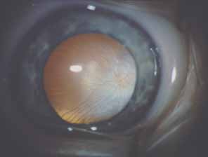
| Fig. 15 Persistent fetal vasculature (PFV)/persistent hyperplastic primary vitreous (PHPV). Intraoperative photograph showing the persistent tunica vasculosis lentis present on the posterior capsule of a clear lens in this 6-week-old infant. The eye was microphthalmic with a horizontal corneal diameter of 9 mm. |