|
|
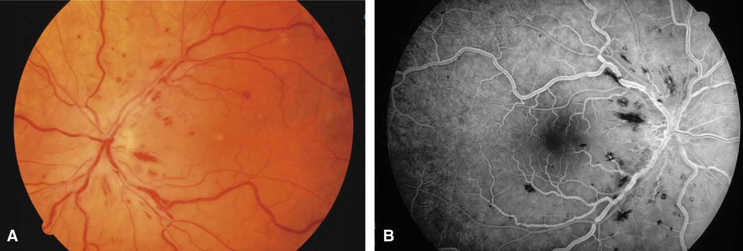
| Fig. 23 Nonischemic central retinal vein obstruction. A. Dilated, tortuous veins, minimal intraretinal hemorrhages, and 20/40 (6/12) vision. B. Fluorescein angiogram showing minimal damage to capillary bed. Despite the lack of ischemic capillary damage on the present study, up to 20% of patients may convert to the ischemic type, necessitating careful follow-up examinations. |