Chapter 29
Ophthalmic Manifestations of Defects in Metabolism
SHIRLEY H. WRAY and ROSEMARY N. BOUSTANY
Main Menu   Table Of Contents

Search

THE GANGLIOSIDOSES
NIEMANN-PICK DISEASE
GAUCHER'S DISEASE
FARBER'S DISEASE (LIPOGRANULOMATOSIS)
THE SIALIDOSES
METACHROMATIC LEUKODYSTROPHY
KRABBE'S DISEASE (GLOBOID CELL LEUKODYSTROPHY)
FABRY'S DISEASE
REFERENCES

The lysosomal storage diseases are a rare class of neurometabolic disorders resulting from genetic enzyme deficiencies. As a result, metabolites of complex lipid accumulate within neurons. Most of the diseases are inherited in an autosomal recessive fashion, and biochemical diagnosis is essential for genetic counseling and for monitoring future pregnancies.

Neurologically, the sphingolipidoses are characterized by a progressive degenerative disease of the nervous system, with blindness, dementia, epilepsy, ataxia, paralysis, and hyperreflexia being prominent features. Ophthalmologically, a cherry-red spot at the macula and optic atrophy are the most common signs, but all parts of the eye may be involved and in many cases the ophthalmologic examination can provide an important clue to the diagnosis.

The lysosomal storage diseases are ultimately fatal because there is no effective treatment. Nevertheless, a more complete understanding of these diseases and their chemical defects has resulted from advances in molecular biology, biochemistry, and enzymology. Today a biochemically based terminology is used instead of eponyms, and almost all the disorders can now be diagnosed by enzyme analysis. Easily accessible cells such as leukocytes and cultured skin fibroblasts or serum can be assayed for enzyme activity, thus circumventing the need for biopsy of the liver, brain, or other tissues. Prenatal diagnosis is also possible for all those persons with known enzyme defects.

Back to Top
THE GANGLIOSIDOSES
The gangliosidoses are neuronal lipid storage disorders due to deficiencies of certain lysosomal hydrolases. In general they are characterized by autosomal recessive inheritance and progressive mental and motor deterioration due to the storage of GM1- or GM2-ganglioside in neurons.

Gangliosides are important constituents of gray matter. They are glycosphingolipids that contain sialic acid in the oligosaccharide chain. At least ten different gangliosides have been identified, four of which constitute over 90% of the total: GM1, GDla, GD1b, and GT. Gangliosides GM2 and GM3 together constitute less than 2% of the total brain ganglioside content. The metabolism of ganglioside involves the removal of the terminal galactose to convert GM1-ganglioside to GM2-ganglioside. GM2-ganglioside is then hydrolyzed to GM3-ganglioside by the removal of N-acetylgalactosamine (Fig. 1).

Fig. 1. Biochemical and nosologic divisions of the sphingolipidoses.

Ten ganglioside storage diseases are now recognized (Table 1). Seven of the disorders involve storage of GM2-ganglioside, and three involve the accumulation of GM1-ganglioside.


TABLE 1. The Gangliosidoses


PresentationDiseaseAge at OnsetSigns and SymptomsEye FindingsDefectStored Ganglioside
InfantileInfantile Tay- Sachs2–3 moIncreased startle, hypotonia, developmental and motor regression, megalencephaly, seizuresCherry-red spotsDeficient hexosaminidase A, α-locus mutation(chromosome 15)GM2- ganglioside
 G m2-gangliosidosis AB variant and B1variant2–3 moIncreased startle, hypotonia, developmental and motor regression, megalencephaly, seizuresCherry-red spotsNormal hexosaminidase A and B with either activator deficiency(chromosom e 5) or in B1variant mutation in the active site for the α-locus(chromosome 15)GM2- ganglioside
 Sandhoff's disease2–3 moHypotonia, developmental and motor regression, megalencephaly, occasional organomegaly, seizuresCherry-red spotsAbsent hexosaminidase A and B, ß-locus mutation(chromosome 5)Gm2- ganglioside GA2- ganglioside Globoside
 G M1-gangliosidosis type IBirth-3 moIncreased startle, hypotonia, developmental and motor regression, megalencephaly, organomegaly, coarse facial features, seizuresCherry-red spots (50% of cases), mild corneal cloudingDeficient ß-galsctosidase (chromosome 3)GM1- ganglioside
JuvenileJuvenile GM2-gangliosidosis2–6 yrDysarthria spasticity, ataxia, progressive dementia, seizuresAtypical cherry red spot, optic atrophy, pigmentary retinopathyDeficient hexosaminidase A, α-locus mutation(chromosome 15)GM2- ganglioside
 Juvenile G M1-gangliosidosis2–4 yrCerebellar ataxia, dystonia, myoclonus, dementia, seizuresCherry-red spotsß-Galactosidase(chromosome 3)GM1- ganglioside
 Juvenile Sandhoff's disease2–6 yrProgressive cerebellar ataxia, psychomotor retardation, spasticity Deficient hexosaminidase A and B, ß-locus mutation(chromosome 5)GM2- ganglioside GA2- ganglioside Globoside
 Chronic Gm2- gangliosidosisFirst and second decadeSpinocerebellar degeneration, disabling dysarthria, tremor, late-appearing proximal muscle weakness and atrophy 10% to 15% residual hexosaminidase A activity, α-locus mutation(ch romosome 15)GM2- ganglioside
AdultAdult G M1-gangliosidosisSecond decadeAtaxia, slurred speech, intellectual decline, mild vertebral changes, angiokeratoma Deficient ß-galactosidase (chromosome 3)GM1- ganglioside
 Adult GM2gangliosidosisSecond to fourth decadeMinimal spinocerebellar signs, prominent anterior horn cell disease with proximal muscle weakness and fasciculations, presence of manic-depressive illness in proband or family history of manic-depression in 40% of cases 5% to 10% residual hexosaminidase A activity, ß-locus mutation(chromosome 15)GM2- ganglioside


THE GM2-GANGLIOSIDOSES

The GM2-gangliosidoses are a class of disorders resulting from the deficiency of ß-hexosaminidase A (Hex A) or ß-hexosaminidase B (Hex B) and Hex A. Hex A is an essential enzyme that normally cleaves the terminal sugar from the Tay-Sachs ganglioside and is identified by its action on artificial substrates.

Expression of Hex A activity requires three separate gene products: an α-subunit, a ß-subunit, and an activator protein, all encoded by genes located on two different chromosomes. Various forms of GM2-gangliosidosis have been described (Tay-Sachs disease, Sandhoff's disease, and variants) that can be traced to mutations in one or another of these three loci. The effect is usually a deficiency of Hex A activity or a concomitant Hex B and Hex A deficiency, permitting a pathologic accumulation of GM2-ganglioside in neurons.

Hex A is made up of two ß-subunits and two α-subunits, the latter coded for on the long arm of chromosome 15, The activator protein locus is located on chromosome 5. Hex B is a tetramer made up of four identical ß-subunits also coded for on chromosome 5 (Fig. 2).

Fig. 2. Molecular genetics of the GM2-gangliosidoses.

The extent of the deficiency of hexosaminidase determines the rate of ganglioside accumulation and hence the time of onset and the clinical severity of the disease. The most severe form is classic infantile Tay-Sachs disease characterized by an almost complete absence of Hex A activity with preservation of Hex B; hence it is called the B variant. Less severe forms have been described that are characterized by the presence of residual but insufficient levels of Hex A activity. These milder variants present as juvenile, chronic, or adult forms of GM2-gangliosidosis (see Table 1).

Tay-Sachs Disease (Infantile GM2-Gangliosidosis Type I)

Infantile GM2-gangliosidosis type I is the most common example of a sphingolipid storage disease. It is inherited as an autosomal recessive disorder because of a mutation in the α-locus on chromosome 15, resulting in a deficiency of Hex A. The carrier frequency for this gene is very high among those of Ashkenazi Jewish or French Canadian descent (1/27 and 1/25 individuals, respectively). Tay-Sachs screening programs primarily target these two populations.

CLINICAL MANIFESTATIONS. Children with Tay-Sachs disease show neurologic signs at birth. Observant parents notice an increased startle reaction to sound and hypotonia at 2 to 3 months of age. The startle response is a reflex myoclonic jerk consisting of tonic extension, adduction and elevation of the arms, clenching of the hands, flexion or extension of the legs, a startled facial expression, and a sharp cry. A brief metallic bang elicits it more reliably than a hand clap, and although it has been called hyperacusis, the sign does not denote increased auditory sensitivity. Occasionally the response is produced by light flashes and tactile stimuli. The increased startle reflex is characteristic of the early stages of Tay-Sachs disease and is without overt electroencephalogram (EEG) correlate. At 12 to 18 months of age it is superseded by segmental and diffuse myoclonus and by prolonged tonic seizures, both spontaneous and stimulus driven. The spikes and sharp waves of the EEG in the second year are replaced by a flattened, featureless, slow EEG by the third year.

The exaggerated startle response is not restricted to Tay-Sachs and Sandhoff's diseases. It occurs in other conditions, such as GM1-gangliosidosis, although not as early or as persistently. Apart from this sign and mild hypotonia the child appears to develop normally until the fourth to sixth month of life when signs of mental retardation and loss of motor skills become manifest. The infant fails to sit up or walk, marked axial hypotonia in combination with pyramidal signs develop, and there is variable spasticity of the limbs. After 18 months of age, progressive deafness, blindness, and megalencephaly occur as a result of storage of GM2-ganglioside and cerebral gliosis. By the end of the third year the child is demented, decerebrate, and blind. Progressive cachexia and aspiration pneumonia usually lead to death before age 10.

OCULAR MANIFESTATIONS. Tay, a British ophthalmologist, was the first to recognize the macular cherry-red spot in 1881 .1 In 1896, Sachs, an American neurologist, emphasized the association of this ocular manifestation with signs of progressive involvement of the central nervous system (CNS) characterized by dementia, blindness, convulsions, and early death.2

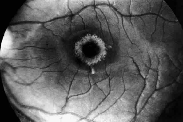
Recognition of the cherry-red spot at the macula is a major diagnostic criterion of Tay-Sachs disease (Fig. 3). It is caused by the accumulation of intracytoplasmic membranous bodies in retinal ganglion cells.

Fig. 3. Fundus of an 18-month-old boy with Tay-Sachs disease. Note cherry-red spot and small white parafoveal halo.

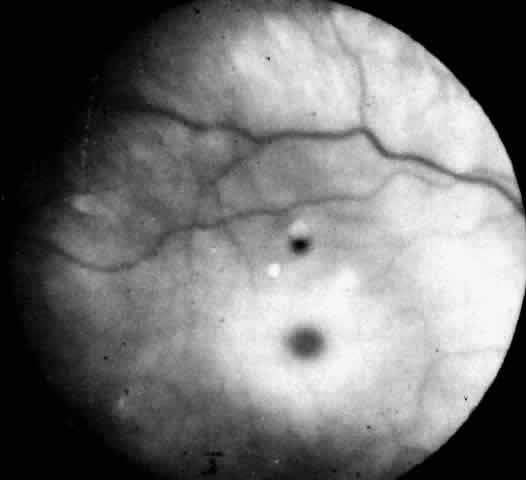
The circular appearance of the fundoscopic lesion reflects the anatomy of the macula. No ganglion cells are present at the very center of the macular region, the foveola, and the central red spot simply represents the normal choroidal background color. The ganglion cell layer surrounding the foveola is several cells thick, and loading of these neurons by storage products results in loss of retinal transparency and a white parafoveal halo. Peripheral to the macular region the ganglion cell layer is only one cell thick, and lipid accumulation in these cells is, therefore, less conspicuous.

Tay-Sachs disease is also the most common storage disease causing macular cherry-red spots (Table 2).3 The variation in the shade of the red spot reflects racial fundus pigmentation. The halo is opaque, slightly elevated, and 1.5 disc diameters in width. The outer border is less sharp than the inner border.


TABLE 2. Storage Diseases with Macular Cherry-Red Spots


DiseaseEnzymeDiagnostic Sample(s)
GM2-gangliosidosis  
Tay-Sachs diseaseHex A deficientLeukocyte serum
Sandhoff's diseaseHex A and B deficientLeukocyte serum
AB variantHex A, B normalGM2loading studies in fibroblasts
B1variantHex A deficient with sulfated substrateLeukocyte fibroblasts
Juvenile GM2-gangliosidosisPartial Hex A deficiencyLeukocyte serum
Infantile GM1-gangliosidosisß-Galactosidase deficientLeukocyte fibroblasts
Niemann-Pick disease  
Type A, infantileSphingomyelinase deficientLeukocyte fibroblasts
Type BSphingomyelinase deficientLeukocyte fibroblasts
Farber's lipogranulomatosisCeramidaseLeukocyte fibroblasts
Sialidoses  
Type 1Sialidase deficientFresh fibroblasts
Type 2Sialidase and ß-galactosidase deficientFresh fibroblasts


Hex, hexosaminidase.

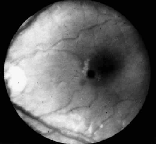
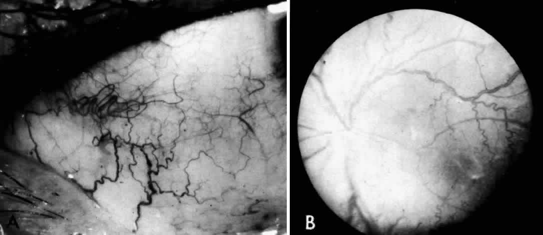
A more widespread opacification of the retina can occur due to involvement of the ganglion cells in the posterior pole. This was observed by Wray4 in a 3½-year-old child with Tay-Sachs disease. The child had black hair and brown eyes, and the cherry-red spot at the macula was brown. The patient was blind, with marked optic atrophy (Fig. 4).

Fig. 4. Fundus of 3 1/2-year-old girl with Tay-Sachs disease. Note cherry-red spot, an extensive white parafoveal halo, and optic atrophy.

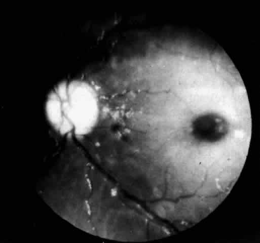
A dynamic process of development of the macular cherry-red spot occurs paralleling the infant's progressive neurologic disorder. The cherry-red spot can be observed as early as 2 months of age and is conspicuous at age 4 to 6 months. Loss of visual acuity may occur without noticeable change in the circular halo. But, in time, the ganglion cells atrophy and optic atrophy and loss of the nerve fiber layer occurs. At this stage blindness coincides.

The frequency of the disappearance of the cherry-red spot is unknown,3 and Cogan has actually never seen it disappear. Nevertheless, the physician should include biochemical testing in the evaluation of a neurologically impaired child presenting with only optic atrophy.

A number of patients reported to have classic Tay-Sachs disease have presented without the expected cherry-red spots. Most of these cases were documented between 1908 and 1964 before biochemical confirmations were available. This suggests that the patients had another storage disease or, alternatively, the diagnosis of Tay-Sachs disease may have been correct but the eye examination was performed after the cherry-red spots had disappeared.

In the final prognosis, infants with Tay-Sachs disease become blind, usually by 18 months of age. The pupils are areflexic to light. In the rare patient noted to have brisk pupillary responses despite blindness, the visual loss may be cortical in origin. At this stage, the visual evoked response (VER) is no longer elicited although the flash electroretinogram (ERG) remains intact.

Infants with Tay-Sachs disease also show a degradation of eye movements relative to the stage of the illness.5 In the early stage there is loss of exploratory and voluntary eye movements, followed by loss of pursuit and optically elicited and vestibular movements. In the late stage, conjugate vertical eye movements elicited by the doll's head maneuver are lost. Terminally, the eyes may become fixed in a downward deviated position.

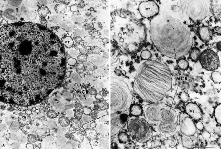
NEUROPATHOLOGY. The pathology in Tay-Sachs disease is diffuse. Intraneuronal storage of GM2-ganglioside occurs in neurons throughout the cortex, brain stem, cerebellum, spinal cord, autonomic ganglia, rectal submucosa, and retina. The affected cells are distended by intralysosomal inclusion bodies composed of closely packed, eccentrically arranged, electron-dense lamellae—the membranous cytoplasmic bodies. The membranous cytoplasmic bodies are characteristic of all the gangliosidoses and represent aggregates of gangliosides and their derivatives with cholesterol and phospholipid, which have oriented to form membranes.

The first histologic study of the eye in Tay-Sachs disease was performed by Collins in 1892.6 Emphasis has since been placed on the staining properties of the storage material, and loss of ganglion cells and extravasation of storage material occurs.7 In Tay-Sachs disease, unlike most other retinal storage diseases, the ganglion cells die early and all of them are involved.

The retinal ganglion cells in the inner portion of the bipolar cell layer and in the inner reticular layer store GM2-ganglioside. As they become affected, the nucleus in the swollen cell locates eccentrically and membranous cytoplasmic bodies fill the cytoplasm; when the cells burst the material is deposited extracellularly without significant phagocytosis or gliosis.

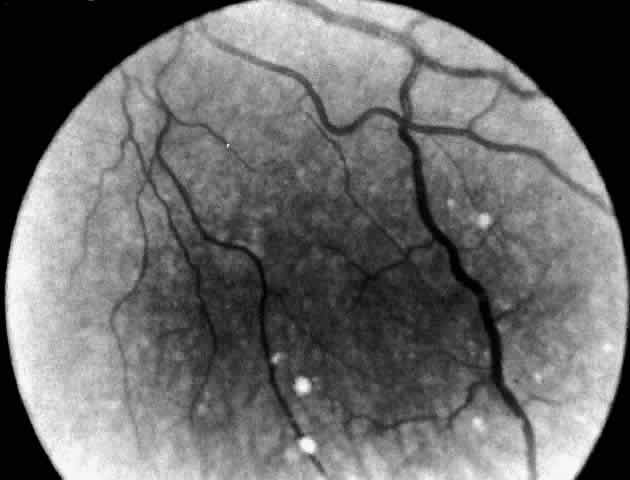
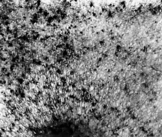
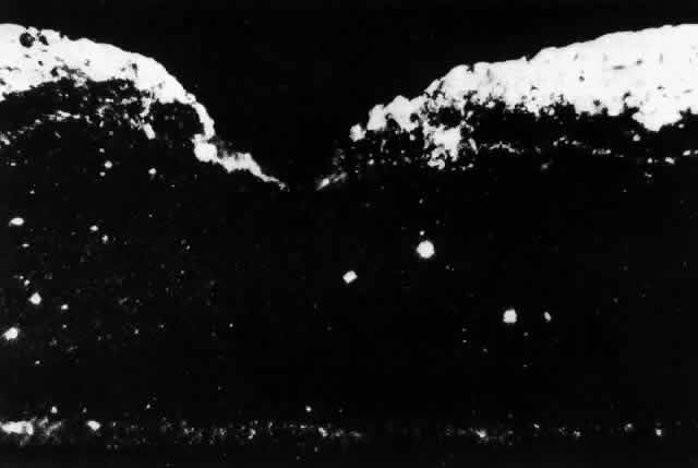
These deposits are strongly birefringent, and striking macular birefringence may be readily visualized in sections of the retina (Fig. 5) even in the unstained state as well as when lightly stained with cresyl violet or sudanophilic dyes.8 Loss of the ganglion cells and atrophy of the optic nerve, which is also evident, are accompanied by a thinning of the nerve fiber layer.

Fig. 5. Birefringence of ganglion cell layer of macula in Tay-Sachs disease. Frozen section, cresyl violet stain, photographed between crossed polaroids (x 80). (Cogan DG, Kuwabara T: The sphingolipidoses and the eye. Arch Ophthalmol 79:437, 1968. Copyright © 1968, American Medical Association)

Histochemical stains have characterized the retinal lipid as a glycolipid,7 and lipid chromatography of retinal cells shows a prominent spot of GM2-ganglioside.9

Although ultrastructural studies of the eye have focused primarily on the ganglion cell,8–10 the amacrine cells in the inner nuclear layer of the retina are equally loaded with GM2-ganglioside yet the horizontal cells, bipolar cells, and photoreceptor cells are unaffected.9

DIAGNOSTIC TESTS. The diagnostic test in Tay-Sachs disease is the assay for Hex A in serum and/or leukocytes, cultured skin fibroblasts, or cultured amniotic fluid cells obtained by amniocentesis at 16 weeks' gestation. In the homozygous Tay-Sachs infant or fetus, Hex A is markedly deficient and the test confirms the diagnosis.

Early prenatal diagnosis, between the 8th and 11th weeks of gestation, is now possible by aspiration of tissue from the fetal chorionic villus through a transcervical catheter. The enzyme assay is performed on fresh and cultured villus cells. This technique reduces the waiting period for anxious parents, and earlier termination of pregnancy can be carried out if the fetus is affected.

Heterozygous carriers of the gene for Tay-Sachs disease can also be detected by serum and leukocyte assay. Heterozygotes have reduced levels of Hex A. Genetic counseling of carriers permits reduction in the incidence of the disease through the use of early prenatal monitoring of a pregnancy.

Infantile GM2-Gangliosidosis: The AB and B1 Variants

The AB and B1 variants of infantile GM2-gangliosidosis are rare. These variants are distinguished biochemically from classic Tay-Sachs disease and Sandhoff's disease by the presence of normal Hex A and Hex B levels in the serum. The defect in the AB variant is a deficiency in activator protein due to a mutation on chromosome 5.11 The defect in the B1 variant is secondary to a mutation involving the active site for the a-locus on chromosome 15 (see Fig. 2). The end result is that GM2-ganglioside accumulates in neurons.12

CLINICAL AND OCULAR MANIFESTATIONS. The story of the index case of the AB variant of Tay-Sachs disease is not widely known. The patient was a male infant of American Indian and English descent. He was said to have developed normally during the first few months of life but failed to sit at the appropriate period and mental retardation was questioned. Cogan consulted on the case in 1965 at the Osteopathic Hospital in Portland. Examination at the age of 21 months showed a typical cherry-red spot, but the nerve-heads were pink and the patient responded to appropriate visual stimuli. The pupils reacted vigorously to light. Neurologically the child had moderate spastic quadriparesis and extensor plantar responses. The case was reported in the literature by Harrell as Tay-Sachs disease in a non-Jewish male.13 The patient died at age 32 months.

NEUROPATHOLOGY. Cogan considered the case reported by Harrell so atypical for Tay-Sachs disease that he asked to be notified when the patient died. Kuwabara removed the eyes. Histopathologic study of the eyes showed the deposition of a substance that was weakly sudanophilic, strongly periodic acid-Schiff (PAS) positive, cresyl violet positive, and strongly birefringent in intact ganglion cells of the retina. The staining characteristics were similar to those of classic Tay-Sachs disease but did not show customary destruction of the ganglion cells nor the usual optic atrophy. By electron microscopy the laminated bodies were abundant in the ganglion cells, presenting both as intact membrane-bound inclusions and free unbound bodies in the cytoplasm.

Pathologic examination of the brain showed a similar storage substance in the neurons as was present in the eyes and similarly a relatively good preservation of white matter. This later was especially evident in the cerebellum, which provided a striking contrast to the usual loss of white matter in Tay-Sachs disease.14

A sample of frozen liver tissue was sent to Sandhoff, who made the first demonstration of normal hexosaminidase levels, thus establishing the AB variant as a biochemical entity.

DIAGNOSTIC TESTS. TO establish the diagnosis it is necessary to evaluate GM2-ganglioside metabolism in cultured skin fibroblasts or amniotic fluid cells over a period of time. In both the AB and B1 variants there are normal levels of Hex A and Hex B but cultured cells from an affected infant or fetus are unable to metabolize 88% to 95% of radioactively labeled GM2-ganglioside.15 In the B1 variant Hex A is shown to be deficient if the enzyme assay is performed using a sulfated synthetic substrate.

Sandhoff's Disease (GM2-Gangliosidosis Type 2)

In 1968, Sandhoff and colleagues16 described a phenotypic variant of Tay-Sachs disease. The disorder is panethnic. It is characterized by the neuronal and visceral deposition of GM2-ganglioside, its asialo derivative, and GA2-globoside. Storage of GA2-globoside is particularly prominent in the kidney. The hexosaminidase deficiency is marked by the absence of Hex A and Hex B; hence Sandhoff called this the O variant. Others have called it Tay-Sachs type 2. The hexosaminidase deficiency is due to a defect in the ß-locus on chromosome 5 that codes for the ß-subunit, an essential gene product for both enzymes (see Fig. 2).

CLINICAL MANIFESTATIONS. Clinical features of Sandhoff's disease are almost indistinguishable from classic Tay-Sachs disease except for, in some cases, the presence of moderate hepatosplenomegaly and mild skeletal dysostosis. The infants have no signs of renal disease. Death occurs before 10 years of age, generally from pneumonia.

OCULAR MANIFESTATIONS. Cherry-red spots in the macula were present in four infants with Sandhoff's disease known to O'Brien.17 Additional cases have now been observed. The halo is opaque white and funduscopically identical to Tay-Sachs cherry-red spots. Blindness and optic atrophy occur.

The cornea in infants with Sandhoff's disease is usually clear clinically, but Tremblay and Szots18 reported a unique patient with slight corneal clouding. The combination of macular cherry-red spots and corneal clouding found in Sandhoff's disease also occurs in GM1-gangliosidosis type I, Niemann-Pick disease, and sialodosis type 1 with ß-galactosidase deficiency.

NEUROPATHOLOGY. In Sandhoff's disease there is an accumulation of GM2-ganglioside as in Tay-Sachs disease, as well as of asialo-GM2 and GA2-globoside. Ultrastructurally, the metabolic products are stored in almost every tissue as cytoplasmic bodies or cytosomes. These vary in their concentration and configuration, resembling typical membranous cytoplasmic bodies or pleomorphic cytoplasmic inclusions, zebra bodies, vesicles, or granules. Histologic evidence of lipid storage in the viscera is present. Lipid-laden histiocytes appear in the liver and spleen, and there are droplets of fat in the cytoplasm of renal tubular epithelial cells. The accumulation of asialo-GM2-ganglioside is much more marked in CNS tissue and in the liver. Characteristically there is storage also of another glycolipid (a kidney globoside) in the kidney, liver, and spleen as well as in the brain.

There are few reports in the literature describing the ocular pathology of Sandhoff's disease,18–20 but the findings hardly differ from that of Tay-Sachs disease. The major distinctions are the character of the membranous cytoplasmic bodies within the ganglion cells of the retina and the involvement of the cornea. In an electron microscopic study, Brownstein and co-workers2° found abundant pleomorphic storage cytosomes in all neurons of the retina, including the inner segments of the photoreceptor cells, and in astrocytes in the optic nerve.

The ultrastructure of the cornea was also studied even when the corneas were normal on clinical examination. Storage cytosomes in keratocytes were found resembling those noted in the mucopolysaccharidoses (MPS) and mucolipidoses, in which the storage material has been identified as acid mucopolysaccharide. An excess accumulation of acid mucopolysaccharide was not evident histochemically however, implying that the storage material in the cornea was a different substance although one that leaves an ultrastructural residue similar to that observed in the MPS. Brownstein and co-workers explained the normal clinical appearance of the cornea as due to the relatively low concentration of storage material in the keratocytes.

DIAGNOSTIC TESTS. Enzyme assay for Hex A and Hex B levels in the serum or leukocytes confirms the diagnosis of Sandhoff's disease. This test shows homozygous patients with Sandhoff's disease to have absent Hex A and Hex B and clearly distinguishes infants with Sandhoff's disease from Tay-Sachs cases. Prenatal diagnosis of Sandhoff's disease has been successfully performed.21 An additional test in Sandhoff's disease is thin layer chromatography of the urine for oligosaccharides. This procedure detects a pattern of complex carbohydrate excretion typical for Sandhoff's disease.

Juvenile Sandhoff's Disease

Juvenile Sandhoff's disease is rare. The absence of Hex A and Hex B in this disorder is attributed to a mutation in the ß-locus on chromosome 5 allelic to the mutation in infantile Sandhoff's disease.22

CLINICAL MANIFESTATIONS. Onset of neurologic symptoms occurs at age 2 to 6 years. A 10-year-old boy with progressive cerebellar ataxia and psychomotor retardation was described by Wood and McDougall.23 Cherry-red spots have not been reported.

DIAGNOSTIC TESTS. Assay for hexosaminidase activity shows an absence of Hex A and Hex B.

Juvenile GM2-Gangliosidosis

Juvenile GM2-gangliosidosis differs from classic Tay-Sachs disease by its later onset and more protracted course.

CLINICAL MANIFESTATIONS. In juvenile GM2-gangliosidoses ataxia is the dominant feature. Four families of Puerto Rican descent and a family of Lebanese Canadian origin have been described by Andermann.24 The children present between 2 and 6 years of age with progressive dysarthria, spasticity, and cerebellar ataxia. Seizures occur later. Death occurs in the teen years.

OCULAR MANIFESTATIONS. By 1984, 24 patients with late-onset GM2-gangliosidosis had been reported but only 4 had macular changes in the form of atypical cherry-red spots, unlike those seen in cases of Tay-Sachs disease. In two of the three cases (cases 4 and 5),25 symptoms began in the late-infantile period (the second year of life). The cherry-red spots were of an unusual nature and described by Brett and colleagues25 as an irregular, poorly defined area of pallor surrounding the fovea. The fovea itself was not clearly demonstrated and was not circular. A photograph of this maculopathy in a blind boy aged 5 years, 8 months (case 4) with onset of the disease at age 14 months is available for comparison with the typical cherry-red spot in classic Tay-Sachs disease.25 The child died at 6 years, 10 months of age. No autopsy examination was made.

The onset of symptoms in four additional cases of late-onset GM2-gangliosidosis was in the juvenile period (the fourth year or later). An atypical cherry-red spot was seen in only one child (case 6) with mild bilateral optic atrophy.25 Cherry-red spots at the macula are not mentioned in previously reported cases of juvenile GM2-gangliosidosis reviewed by Brett and colleagues25 or in the single case studies by Menkes and associates26 but one child showed the beginnings of retinitis pigmentosa 4 years from onset.

Johnson and associates27 found a boy with mild hand tremor since age 21/2 years to have macular cherry-red spots at age 4 years and juvenile cerebellar ataxia. Visual acuity was 20/100 (6/30)* with optic atrophy. The electroretinogram (ERG) was normal. The child had full eye movements and no nystagmus, but both eyes showed slowing of voluntary and optokinetic saccadic movements.


* Metric equivalent given in parentheses following Snellen notation.

DIAGNOSTIC TESTS. Assay of hexosaminidase shows a marked deficiency of Hex A. This is attributed to a mutation in the a-locus on chromosome 15 at a site allelic to the classic Tay-Sachs mutation (see Fig. 2).

Chronic GM2-Gangliosidosis

CLINICAL MANIFESTATIONS. Chronic GM2-gangliosidosis usually presents in the first or second decade with signs of spinocerebellar degeneration, dysarthria, and disabling tremor. Later in the course signs of anterior horn cell disease develop with muscle wasting and fasciculations. The affected teenager shows a deterioration in school performance as intellectual function declines.28

OCULAR MANIFESTATIONS. The fundi are normal. Macular cherry-red spots have not been observed. Episodic uncontrollable vertical eye movements, facial grimacing, and dystonic postures were observed in affected members of a family with atypical spinocerebellar degeneration.29 Oculogyric crisis may occur.

DIAGNOSTIC TESTS. Biochemical tests show a 10% to 15% residual Hex A activity.

Adult GM2-Gangliosidosis

All cases of adult GM2-gangliosidosis described have been in persons of Ashkenazi Jewish descent but unreported non-Jewish cases are known to us.

CLINICAL MANIFESTATIONS. Intellect is usually preserved, but probands may have a manic-depressive illness. Proximal muscle weakness manifests itself in the second to fourth decade. Fasciculations are prominent, and muscle biopsies confirm the presence of group atrophy. Computed tomography of the brain shows cerebellar atrophy.

DIAGNOSTIC TESTS. In adult GM2-gangliosidosis a 5% to 10% residual Hex A activity is present.

THE GM1-GANGLIOSIDOSES

Infantile GM1-Gangliosidosis (GM1-Gangliosidosis Type I)

The earliest well-documented report of GM1-gangliosidoses type I appeared in 1959,30,31 followed by seven cases in 1964.32 The disease is inherited as an autosomal recessive disorder caused by a deficiency of ß-galactosidase.

The absence of ß-galactosidase isoenzymes A, B, and C leads to the storage of a variety of macromolecules with a terminal ß-galactosyl residue. These are primarily GM1-ganglioside and complex carbohydrates. The accumulation of the latter compound explains the visceral and skeletal manifestations of this disease. The storage of GM1-ganglioside in the brain is responsible for the neurologic manifestations.33

CLINICAL MANIFESTATIONS. Dysmorphic facial features, macular cherry-red spots, and an enlarged liver delineate GM1-gangliosidosis. Unlike the GM2-gangliosidoses, symptoms of infantile GM1-gangliosidosis may be present at birth with edema of the extremities, hypotonia, and failure to thrive. Psychomotor development halts during the third to sixth months of life. Spasticity, tonic spasm, and hyperreflexia then develop. Head size may be normal, or megalencephaly and seizures occur. The infant becomes decerebrate and blind. Frontal bossing, depressed nasal bridge, hypertelorism, epicanthal folds, large low-set ears, and macroglossia are variable features. Progressive hepatomegaly is noted early with or without splenomegaly. Mild changes of dysostosis multiplex are often present. The peripheral blood smear contains vacuolated lymphocytes, and the bone marrow contains foamy histiocytes. Death from pneumonia usually occurs by 2 years of age.

OCULAR MANIFESTATIONS. A systematic ocular examination is most important in infantile GM1-gangliosidosis. Blindness develops early, and cherry-red spots have been found in half the patients. The macular cherry-red spot resembles that seen in Tay-Sachs disease. Nystagmus and optic atrophy have been reported34 as well as somewhat tortuous retinal vessels and flame-shaped retinal hemorrhages.35 Mild clouding of the cornea may also occur, but slit lamp examination of the cornea has not been done routinely in these infants to determine the frequency of small corneal opacities. Strabismus, lid edema, and ptosis are also described.

NEUROPATHOLOGY. Membranous cytoplasmic bodies are present in central and autonomic nervous system neurons. Vacuoles in cells of the liver, spleen, and cerebral blood vessels are indicative of widespread accumulation of intracellular complex carbohydrates. Renal glomerular epithelial cells also store complex carbohydrates.

The first pathologic report of the eye in infantile GM1-gangliosidosis was made by Emery and associates.35 The infant had cherry-red spots of the macula and mild diffuse corneal clouding. Light and electron microscopy showed lipid storage in the ganglion cells of the retina. The cytoplasm of these cells, particularly of those in the macular region, appeared foamy and filled with membranous cytoplasmic bodies with a morphologic appearance resembling the intralysosomal inclusions present in cerebral neurons.34

DIAGNOSTIC TESTS. In early infantile GM1-gangliosidosis type I, ß-galactosidase activity is deficient. The assay can be performed on leukocytes, cultured skin fibroblasts, amniotic fluid cells, and chorionic villus samples.

Urine thin-layer chromatography for oligosaccharides is abnormal, showing a pattern of complex carbohydrate excretion typical for this disease.

Juvenile GM1-Gangliosidosis (GM1-Gangliosidosis Type II)

Infantile GM1-gangliosidosis type I and juvenile GM1-gangliosidosis type II are recognized to be allelic mutations at the structural gene locus for acid-ß-galactosidase on chromosome 3.

CLINICAL MANIFESTATIONS. The disease begins in the first or second year of life with motor and mental regression, quadriplegia, dystonia, pseudobulbar palsy, cerebellar ataxia, dementia, and occasionally seizures. Foamy histiocytes are present in the bone marrow. There is no hepatosplenomegaly or significant facial or skeletal deformity. Because the clinical features are quite uncharacteristic, biochemical studies are easily overlooked. Survival until 10 years of age is possible.36

OCULAR MANIFESTATIONS. The eye does not provide a clue to the diagnosis. Vision is normal, and changes in the retina and cornea are absent. Nonparalytic squints are frequent. Late in the course optic atrophy may occur.

DIAGNOSTIC TESTS. Marked deficiency of ß-galactosidase activity in leukocytes and cultured fibroblasts confirms the diagnosis.

Adult GM1-Gangliosidosis (GM1-Gangliosidosis Type III)

CLINICAL MANIFESTATIONS. Adult GM1-gangliosidosis type III presents in the second decade with progressive cerebellar ataxia, dysarthria, and spasticity. The patient maintains normal intellect until quite late in the course. Seizures are rare. Vertebral changes, if present, are mild, and organomegaly is not usually a feature. Occasionally skin angiokeratomas develop.37

OCULAR MANIFESTATIONS. The eye may not be affected although some patients have had mild optic atrophy. Occasional mild corneal clouding occurs.38

DIAGNOSTIC TESTS. Markedly deficient ß-galactosidase activity in leukocytes or cultured fibroblasts confirms the diagnosis.

Back to Top
NIEMANN-PICK DISEASE
Niemann-Pick disease is an autosomal recessive lipidosis due to impaired sphingomyelin metabolism.

Sphingomyelin is a constituent of many cell membranes and a major lipid of the myelin sheath and of the stroma of red blood cells. Sphingomyelin is normally metabolized by the cleavage of phosphorylcholine from ceramide by the action of the enzyme sphingomyelinase. This enzyme is deficient in Niemann-Pick disease type A and type B. The two types are believed to be allelic.39

In Niemann-Pick disease type C, sphingomyelinase activity is usually normal; the metabolic block is said to be in the esterification of cholesterol within the cells.40 The biochemical defects in Niemann-Pick disease type D and in type E remain to be discovered.

The broad classification of Niemann-Pick disease into five phenotypic variants (A through E) is based on the age at onset, the severity and type of neurologic involvement, and the evolution of the disease. In all variants hepatosplenomegaly and foam cells in the bone marrow are constant features. Pulmonary infiltration and CNS involvement are variable findings (Table 3).


TABLE 3. Classification: Niemann-Pick Disease


  Involvement   
TypeEthnic BackgroundVisceralCNSOcular SignsEnzyme Deficiency
AInfantile neuronopathic++++Cherry-red spotSphingomyelinase
 (Ashkenazi Jewish)(50%)   
BChildhood non-neuronopathic (panethnic)++--Sphingomyelinase
 Late on-neuronopathic++-Macular haloSphingomyelinasen
CChronic neuronopathic (panethnic)+(early jaundice)++Vertical supranuclear palsy, oculomotor apraxiaCholesterol esterification defect
DNova Scotia variant(Acadian)+(jaundice)+-?
EAdult non-neuronopathic+-Cherry-red spot ±?

+ = involvement present, - = involvement absent.


Niemann-Pick disease type A was described by Niemann in 191441 in an 18-month-old Jewish child with hepatosplenomegaly and mental retardation. In 1927, Pick42 described the histopathology characterized by large numbers of vacuolated or “foam” cells in many organs and tissues in the body. Subsequently, Klenk, in 1934,43,44 identified the lipid stored as sphingomyelin.

CLINICAL MANIFESTATIONS. Infantile Niemann-Pick disease type A has its onset in early infancy at 6 months of age. The disease is characterized by failure to thrive, a protuberant abdomen due to an enlarging spleen, and severe involvement of the CNS with progressive loss of motor function. Death occurs by the third year of life. Approximately half of these patients are the offspring of Ashkenazi Jews.

In Niemann-Pick disease type B, only visceral involvement is noted. Onset is in late childhood between 3 and 11 years of age or in adult life with splenomegaly and pulmonary infiltrates. Intellect is normal. Infants affected with type B are of mixed ethnic background.

Niemann-Pick disease type C, the subacute form, presents in infancy with neonatal hepatitis or later in childhood with moderate splenomegaly and gradual neurologic deterioration. Many patients have seizures and limitation of vertical gaze. The presence of down-gaze paresis is the most typical defect. Sea-blue histiocytes in the bone marrow are common. These patients usually die before the end of their second decade.

Niemann-Pick disease type D is clinically similar to type C. This designation is limited to an inbred group of cases traced to a couple who lived in Yarmouth County in Nova Scotia in the late 1600s.45 These patients store sphingomyelin in the tissues in variable amounts. They present with hepatosplenomegaly at 4 to 6 years of age and follow a protracted course. Involvement of the CNS occurs late. Many patients become ataxic and develop seizures.

The type E phenotype is characterized by adult onset, mild splenomegaly, foam cells in the bone marrow, and the absence of neurologic signs.

OCULAR MANIFESTATIONS. Niemann-Pick Disease Type A. A cherry-red spot of the macula is present in 50% of cases.46 Cogan47 noted no distinction between the appearance of the cherry-red spot in infantile Niemann-Pick disease and that seen in Tay-Sachs disease. Others have reported subtle differences in the color of the fovea and/or the halo and in the degree of opacification of the peripheral retina.48–51

All infants with Niemann-Pick type A disease become blind late in the course of the illness, and optic atrophy develops. Walton and associates51 also noted subtle lens opacities and peculiar corneal clouding, suggesting widespread ocular involvement in this disorder of sphingolipid metabolism.

Niemann-Pick Disease Type B. A unique retinal abnormality, the macular halo syndrome has been reported in seven cases of Niemann-Pick disease type B, three enzymatically proven52–54 and four prior to enzyme verification described between 1950 and 1970.55–58 Symmetrical punctate crystalloid ring-form opacities about the foveas were observed in each case with no visual impairment. Cogan and Federman56 in one of the early cases were the first to publish a fundus photograph of the macular halo (Fig. 6). The patient was a 24-year-old woman with hepatomegaly, no neurologic signs, and a reticuloendotheliosis of unclassified type. Both fundi showed doughnut-shaped opacities about the foveas. The opacities were described as yellowish white scintillating granules forming a relatively sharp border on the inner edge of the ring and a ragged border on the outer edge of the ring. Despite this condition the patient had normal visual acuity (20/15 [6/5]) and no scotoma.

Fig. 6. Fundus of patient with Niemann-Pick disease type B showing the macular halo syndrome. (Cogan DG, Chu FC, Barranger JA, Gregg RE: Macular halo syndrome; variant of Niemann-Pick disease. Arch Ophthalmol 101:1698, 1983. Copyright © 1983, American Medical Association)

The first case verified enzymatically was reported by Harzer and associates in 1973.52 The assay showed a sphingomyelinase level in leukocytes that was less than 10% of normal.

Although these scattered reports were in the literature, it was Cogan who clearly identified the lesion and made the association with Niemann-Pick disease type B. Cogan and co-workers53 reexamined their initial patient and added a 21-year-old man with a history of splenomegaly and hyperlipidemia. The diagnosis of Niemann-Pick type B disease in each of these patients was confirmed by finding significantly lower sphingomyelinase levels in cultured skin fibroblasts. They named the condition macular halo syndrome. In these cases, the opacities in the retina formed a halo approximately one-half disc diameter at their outer edge. The halo had a crystalloid appearance. By stereo-ophthalmoscopy, slit lamp biomicroscopy, and fluoroangiography the opacities appeared to occupy various depths of the retina but were most numerous in Henle's fiber layer, causing only minor obscuration of the overlying vessels.

The foveal lesion in Matthews and associates' patient54 is similar in size and appearance to the fundus photograph published by Cogan and coworkers. On the basis of stereo biomicroscopy and contact lens examination, Matthews and associates located the lesion in the ganglion cell layer of the retina. The masking effect that the ring lesion had on the perifoveal vasculature in the early fluorescence angiogram was taken as confirmation that the accumulated material is in this superficial layer of the retina. As a result, Matthews and associates proposed that the macular halo represents the smallest or mildest form of a cherry-red spot—findings in conflict with those of Cogan. The precise location of the opacities in the retina remains uncertain, however, because of the lack of histopathology.

The available clinical data suggest that such opacities are permanent. For example, the appearance of the macular halo remained unchanged for 15 months in one case,57 4 years in another,52 and more than 20 years in one of Cogan's cases.53 Cogan concluded that the remarkable preservation of normal visual function in all of the cases either was due to hiatuses in the opacities or, less likely, to a localization of the opacities behind the photoreceptors.

Niemann-Pick Disease Type C, In Niemann-Pick disease type C neither the macular cherry-red spot nor the macular halo syndrome has been observed. However, differentiating Neimann-Pick disease type C from the other types of Niemann-Pick disease is a clinical syndrome that has been referred to in the literature as ophthalmologic neurovisceral lipidosis,59 vertical supranuclear ophthalmoplegia lipidosis,60–63 and Neville's disease.64 Cogan and co-workers65 have added nine cases to the 30 patients already reported Under these various headings and have recommended an acronym, the DAF syndrome, to denote the three essential features: downgaze paralysis, ataxia/ athetosis, and foam cells in the spleen, liver, and bone marrow.

Characteristic of the Niemann-Pick disease type C DAF syndrome is an onset in the first 2 decades of life. There is a characteristic progression of the down gaze paralysis to involve upgaze and eventually horizontal gaze and a total supranuclear ophthalmoplegia. Athetosis and spasticity develop in addition to ataxia. There is extensive infiltration of bone marrow, spleen, liver, and other tissues with foam cells and a variable degree of intellectual impairment. Sphingomyelinase activity in leukocytes and cultured fibroblasts is decreased or normal. The underlying metabolic block is reported to be in the esterification of cholesterol within the cells.

Niemann-Pick Disease Types D and E. Niemann-Pick type D disease has no ocular manifestations. Patients with the type E variant may have macular cherry-red spots.

NEUROPATHOLOGY. In Niemann-Pick disease sphingomyelin accumulates in excess in the brain and autonomic ganglia. The neurons appear swollen with pale cytoplasm. Ultrastructurally the cells contain concentric lamellated bodies representing storage cytosomes. Inclusion profiles in the viscera, lymph nodes, and foam cells also correlate with an increase in sphingomyelin content.

In the eye in Niemann-Pick disease type A with macular cherry-red spots, stored lipid is localized to the ganglion and amacrine cells of the retina most obviously in the parafoveal region. The ganglion cells of the other nuclear layers are unaffected.66

Larsen and Ehlers67 report, however, that the affected ganglion cells are not ballooned, degenerated, or reduced in number. This may account for the retention of vision in their patient and in similar nonblind infants despite the presence of a cherry-red spot at the macula. Degenerated, necrotic engorged ganglion cells, interspersed with structurally intact lipid-laden ganglion cells, have been described by others.66

It appears that the retinal ganglion cells in Niemann-Pick disease are conspicuously unlike those in Tay-Sachs disease in their ability to tolerate the presence of excess intracellular lipid so well.

In frozen sections of formalin-fixed retina, both ganglion cells and amacrine cells show marked granular cytoplasmic birefringence. In the study by Robb and Kuwabara68 frozen sections of other portions of the eye revealed a remarkably wide distribution of storage material giving the granular birefringence. Corneal stromal cells were markedly positive, as were corneal endothelium and lens epithelium. The vascular endothelium, the nonpigmented epithelium of the ciliary body, and the retinal pigment epithelium were uniformly birefringent.

Electron microscopic examination of the cornea and retina showed the lipid stored was in the form of membranous cytoplasmic bodies (Fig. 7).68 The distribution of these inclusion bodies is similar to that of the birefringent material seen on light microscopy. The bodies are most abundant in retinal ganglion cells and retinal pigment epithelium. They are present in moderate numbers in corneal stromal cells, lens epithelium, corneal endothelium, vascular endothelium, and the sphincter muscle of the iris. They are infrequent in Müller cells, glial cells, and rod and cone inner segments.

Fig. 7. Retinal ganglion cell. (A) Portion of cell in Niemann-Pick disease, showing numerous membranous cytoplasmic bodies. Mitochondria (m) and dilated endoplasmic reticulum (er) are also evident. Area outlined in lower right is shown in greater magnification in B. (× 15,000) (B) Portion of cytoplasm of ganglion cell shown in A. Membranous cytoplasmic bodies cut in several different planes are evident, as are mitochondria (m) and endoplasmic reticulum (er) (×42,000). (Robb RM, Kuwabara T: The ocular pathology of type A Niemann-Pick disease: A light and electron microscopic study. Invest Ophthalmol 12:366, 1973)

The morphology of the membranous cytoplasmic bodies found in the eye in Niemann-Pick disease type A corresponds closely to the previously reported ultrastructure of lipid inclusions in the brain and viscera in other patients with Niemann-Pick disease type A. An ocular ultrastructural study of a 23-week-old fetus with Niemann-Pick disease type A also demonstrated rather extensive ocular involvement.69

In the eye in Niemann-Pick disease type C, Palmer and colleagues70 observed extensive lipid storage in ocular tissues. Intracellular pleomorphic cytoplasmic bodies were visualized in the conjunctiva, cornea, lens, retinal ganglion cells, and retinal pigment epithelium and in optic nerve astrocytes. Only the pathology of the optic nerve and retinal ganglion cells correlated with the clinical findings of optic atrophy and a perimacular gray discoloration.

DIAGNOSTIC TESTS. The diagnostic test in Niemann-Pick disease type A and type B is measurement of sphingomyelin activity in leukocytes or cultured skin fibroblasts. The enzyme is deficient in both types. Prenatal diagnosis has been accomplished by assaying cultured amniotic fluid cells in type A and type B.71

In Niemann-Pick disease type C the metabolic block is in intracellular cholesterol esterification. Cultured fibroblasts from affected persons store high levels of unesterified cholesterol. This is the basis for the laboratory diagnosis of Niemann-Pick type C. Carriers are also identified by this test.

The underlying metabolic defect in Niemann-Pick type D and type E disease is still not understood, but these two variants are not diagnosable by a single laboratory test. Increase in organ sphingomyelin content and/or presence of foam cells in the bone marrow help to corroborate clinical suspicions.

Back to Top
GAUCHER'S DISEASE
Gaucher's disease is an autosomal recessive sphingolipidosis. The disease is due to a deficiency of the enzyme ß-glucosidase, leading to the accumulation of glucosylceramide in cells of the reticuloendothelial system.

The disease, first described by Gaucher in 1882,72 is characterized by hepatosplenomegaly and the presence of lipid-laden storage cells called Gaucher's cells in the bone marrow and organs.

The disease is panethnic, although Gaucher's disease type I has a high prevalence among Ashkenazi Jews. A subset of' Gaucher's disease type III has been described with a high incidence in a Swedish isolate.73

CLINICAL MANIFESTATIONS. Three types of Gaucher's disease are distinguished: type II acute infantile neuronopathic, type III subacute juvenile neuronopathic, and type I chronic adult non-neuronopathic.74

Type II acute infantile neuronopathic Gaucher's disease is a unique neurovisceral disorder. Clinically it simulates infantile Niemann-Pick disease with the insidious onset of symptoms in early infancy, including failure to thrive, progressive hepatosplenomegaly, laryngeal stridor, and dysphagia. Anemia, thrombocytopenia, and leukopenia are present. Every infant with type II Gaucher's disease has evidence of severe and progressive CNS dysfunction before death. Signs of neurologic deterioration usually appear before 6 months and frequently before 3 months of age. A rather stereotyped neurologic syndrome has been observed in which hypotonia gives way to spastic weakness associated with a persistent retroflexion of the neck, with internal strabismus, seizures, and signs of pseudobulbar palsy. Motor skills and mental awareness are rapidly lost. The usual cause of death as the bulbar palsy worsens is bronchial aspiration and pneumonia.

Type III subacute juvenile neuronopathic Gaucher's disease describes children and young adults who do not fit into either of the other two groups and whose illness is complicated by neurologic manifestations that differ from, and have a later age at onset and more chronic course than, those of the infantile form. Two clinical pictures are distinguished, depending on the age at onset of neurologic symptoms. The older patients,75–77 including two patients reported by Winkelman and co-workers,78 in whom symptoms began in late childhood, adolescence, or' early adult years, presented with similar neurologic syndromes, consisting of myoclonic epilepsy and supranuclear disorders of gaze. In some,75 including Winkelman's second patient,76 the gaze disorder was the more prominent; in others,76 myoclonic epilepsy was the most evident feature.

Freedom from signs of cerebral involvement is the sine qua non for the diagnosis of type I chronic adult non-neuronopathic Gaucher's disease. Patients with this variant can have onset of the disease in childhood or in adulthood and a prolonged course. They manifest hepatosplenomegaly, anemia, thrombocytopenia, periods of bleeding, and pathologic bone fractures. Radiographic abnormalities of bone are present in 50% to 75% of patients. The most common sign is an Erlenmeyer flask deformity at the lower end of the femur due to expansion of the marrow cavity.

OCULAR MANIFESTATIONS. The validity of two cited cases79,80 of a cherry-red spot in the retina of patients with Gaucher's disease has been questioned by Cogan and colleagues.81 In addition, one reported case of macular cherry-red spots in a 20-year-old woman believed to have Gaucher's disease, along with other similar cases, in retrospect now seems to have been an example of the cherry-red spot-myoclonus syndrome of sialidase deficiency.82,83 All three suspected Gaucher's disease cases antedated the biochemical means for establishing the diagnosis. In fact, it was not until 1965 that the enzyme assay for Gaucher's disease became available.

To determine for certain the incidence of macular cherry-red spots in Gaucher's disease, Cogan and colleagues81 examined 42 enzymatically proven cases of the disease and reviewed the records of 9 others. In none was a cherry-red spot seen.

In three patients with type III subacute juvenile neuronopathic Gaucher's disease however, the retina showed a unique retinopathy. The findings in Cogan's case 1 are described: “Both fundi showed discrete white spots randomly distributed in the posterior fundus, especially along the inferior vascular arcades (Fig. 8). The spots varied in diameter from just visible to approximately 0.1 mm and were situated in the superficial retina or on the surface of the retina. Several covered the retinal vessels. The disc and retinal vessels were normal.” The child, an 11-year-old boy, had normal acuity and a full field of vision by confrontation. He had presented at age 3 years with splenomegaly.

Fig. 8. Fundus of a patient with type III subacute juvenile neuronopathic Gaucher"s disease showing discrete white spots in or on the retina along the inferior vascular arcades. At least one spot overlies a vein. The optic disc and retinal vessels were normal. (Cogan DG, Chu FC, Gittinger J, Tyshsen L: Fundal abnormalities of Gaucher"s disease. Arch Ophthalmol 98:2202, 1980. Copyright © 1980, American Medical Association)

Normal vision and similar retinal abnormalities were observed in a mildly mentally retarded 18-year-old woman (case 2) presenting with splenomegaly at age 1 year and in a 6½-year-old boy (case 3) noted to have hepatosplenomegaly in the first year of life. The patients in cases 1 and 3 had conspicuous supranuclear defects of eye movement.

Japanese observers Yanagida, Matsumoto, Tokudo, and Hirose were the first to report the peculiar retinal spots in type III juvenile Gaucher's disease between 1950 and 1965. Their clinical reports in Japanese journals are referenced in Cogan and colleagues' paper.81

Type III juvenile neuronopathic Gaucher's disease is also associated with a distinctive supranuclear eye movement disorder affecting primarily horizontal gaze and only occasionally vertical gaze.75–78,84,85 The early defect in horizontal gaze involves the saccadic system, and the disorder mimics closely congenital ocular motor apraxia. To distinguish Gaucher's disease cases from benign congenital apraxia, it is essential to obtain the appropriate enzyme studies.

NEUROPATHOLOGY. The liver, spleen, and long bones are the primary organs affected by the storage of glucosylceramide. The dominating precursors to the accumulation of glucosylceramide are derived from the normal turnover of leukocytes and erythrocytes, but other tissue cells also contribute.

The highly cytotoxic substance glucosylsphingosine (the non-acyl derivative of glucosylceramide) is also stored in excess in the viscera in all types of Gaucher's disease and in the brain in the neuronopathic types, type II and III. This accumulation leads to cell death.

Like the clinical features, the neuropathologic changes in patients with type II infantile neuronopathic Gaucher's disease are quite uniform. Nerve cell loss and neuronophagia involving the brain stem and deep cerebellar nuclei predominate, but the thalamus, basal ganglia, and spinal cord are also affected. Such neuronal destruction is diffuse and, except for oculomotor abnormalities and dysphagia, causes no localizing signs.

Perivascular nodular collections of Gaucher's cells are found in the brain, invading the meninges and in Virchow Robin spaces. Gaucher's cells have a unique appearance. The cytoplasm has a wrinkled tissue-paper appearance because of interwoven fibrils contained within it. Under the electron microscope the cells contain large lysosomes filled with tubular profiles. These cells are distinct in their appearance and differ markedly from foam cells seen in other lipidoses.86

A pathologic study of two siblings with neuronopathic Gaucher disease and supranuclear upgaze and horizontal gaze palsies has demonstrated similar widespread involvement of the cortex, cerebellum, and spinal cord. The midbrain, pretectal region, oculomotor and red nuclei, superior colliculus, substantia nigra, and reticular formation were all involved. In the pons, the paramedian reticular formation showed pathologic change, as did the abducens and superior vestibular nuclei.78

An electron microscopic study of Gaucher's cells in the eye in type III juvenile neuronopathic disease is reported from Japan by Ueno and associates87,88 who noted white spots in an arcuate pattern in the fundi of an 8-year-old Japanese boy. At autopsy the retinal spots corresponded to polymorphonuclear giant cells within and on the surface of the retina. These cells were large and stained positively for glycolipid and acid phosphatase.

DIAGNOSTIC TESTS. The disease is confirmed by demonstration of a deficiency of glucocerebroside-ß-glucosidase in leukocytes and fibroblasts. In spite of the range of biologic expression, the same enzyme is deficient in all three types of the disease. Moreover, lack of complementation in cell fusion studies strongly suggests that these forms of Gaucher's disease represent allelic mutations at the same gene locus.89 There is a rare variant of Gaucher's disease clinically similar to adult non-neuronopathic disease type I but in which glucocerebroside-ß-glucosidase activity is normal. In this variant the defect is believed to be due to absence of the A activator protein.90 Prenatal diagnosis is available for all three types. It is seldom requested for type I Gaucher's disease.

Back to Top
FARBER'S DISEASE (LIPOGRANULOMATOSIS)
Farber's disease is a rare autosomal recessive disease. The inborn error is in ceramide metabolism due to lack of the enzyme acid ceramidase. The first case of disseminated lipogranulomatosis was described by Farher in 1949; by 1952 he had observed two additional cases.91 There are now more than 28 reported cases in the literature.92,93

CLINICAL MANIFESTATIONS. The disease begins in the first few weeks of life and presents as irritability, vomiting, hoarseness, failure to thrive, and painful swollen joints. Subcutaneous periarticular nodules develop, especially at the ankle and elbow joints. Hoarseness is severe because of progressive granulomatous infiltration of the epiglottis and larynx. Disturbance in swallowing with episodic bouts of fever and pulmonary consolidation is common. Some infants develop systolic murmurs owing to valvular involvement. Most have a generalized lymphadenopathy with occasionally moderate hepatomegaly and rarely splenomegaly. Characteristically intellect is unaffected and involvement of the nervous system is not a prominent feature.91 However, hypotonia and diminished deep tendon reflexes due to a peripheral neuropathy and anterior horn cell involvement have been reported. In such cases electromyographic studies show signs of denervation and elevated cerebrospinal fluid protein. Most children do not survive the fourth year, although a few cases of intermediate severity with longer survival have been reported.94

OCULAR MANIFESTATIONS. The retinal changes in Farber's disease are subtle and easily overlooked. The fundi of one 8-month-old patient had been examined several times following the onset of the disease at age 2 weeks and considered to be normal. Cogan and colleagues95 subsequently observed and reported a diffuse grayish opacification of the retina about the fovea, producing a mild cherry-red spot (Fig. 9). The parafoveal opacity differed from that seen in Tay-Sachs disease in that the opacity was pale gray instead of white and it was accompanied by little, if any, pallor of the optic disc. Shortly before death at age 11 months there was a suggestion of a peppery pigmentation of the entire retina and some abnormal pigmentation in the macula. The retinal vessels showed no abnormality. Visual function, as judged by the patient's attentiveness, retention of the optokinetic response, and absence of nystagmus, was normal.

Fig. 9. Fundus of patient with Farber"s disease at age 8 months. Macula shows abnormal grayness with suggestion of cherry-red center. Retinal vessels are normal, and color of disc is normal. (Cogan DG, Kuwabara T, Moser H et al: Retinopathy in a case of Farber"s lipogranulomatosis. Arch Ophthalmol 74:752, 1966. Copyright © 1966, American Medical Association)

Zetterstrom's patient with Farber's disease had no retinopathy.96 At age 17 months he developed a xanthoma-like growth approximately the size of a rice grain in the conjunctiva over the left eye. Microscopic examination of the excised conjunctival granuloma showed a similar histologic picture to that of a subcutaneous granuloma, with groups of irregularly formed large foam cells with granular cytoplasm weakly positive in reaction to fat stains.

François97 has reported central macroscopic subepithelial corneal opacities in two children with Farber's disease.

NEUROPATHOLOGY. The characteristic pathologic lesion in Farber's disease is a granuloma containing foam cells filled with PAS positive material extractable with lipid solvents. Ultrastructural examination of these cells has shown cytoplasmic vacuoles limited by a single membrane and containing comma-shaped tubular structures. The vacuoles are acid phosphatase positive and probably represent lysosomes. They are called Farber bodies. Elevated levels of ceramide have been found in subcutaneous nodules in the liver, kidney, brain, and lungs. In the brain, neurons and glial cells are distended owing to the accumulation of ceramide and gangliosides.

Routine sections of the paraffin-embedded eye tissue show no histologic abnormalities in Farber's disease, but frozen, unstained sections show birefringent lipid crystals in the ganglion cells of the retina. The accumulation of lipid is most conspicuous in the parafoveal region of the macula where the ganglion cells are abundant. In spite of their distention, the engorged cells remain intact and there are no extracellular deposits of lipid in the retina. The optic nerve is not atrophic.95Cogan and colleagues95 performed solubility studies in several of the organic solvents and determined that the intracellular deposit in the ganglion cells was likely to be a complex lipid rather than neutral fat or cholesterol.

DIAGNOSTIC TESTS. The isolation of excess ceramide in biopsied tissue is no longer required to confirm the diagnosis of Farber's disease.98 Determination of ceramidase levels in leukocytes and cultured fibroblasts is the diagnostic procedure. The assay shows low to absent enzyme activity. Carriers are identified by demonstration of half normal levels of enzyme activity in cultured fibroblasts. Prenatal diagnosis has been accomplished in four pregnancies at risk by assaying cultured amniocytes .99

Back to Top
THE SIALIDOSES
In 1970 Spranger and Wiedemann100 defined a new category of mucolipidoses to classify patients who exhibit clinical features of the mucopolysaccharidoses without evidence of excess excretion of urinary mucopolysaccharides.

Under this broad category of the mucolipidoses four separate entities were initially described: mucolipidosis type I or sialidosis, mucolipidosis type II or inclusion cell disease, mucolipidosis type III, and mucolipidosis type IV. Only one group of these disorders, mucolipidosis type I, has a primary deficiency of the enzyme sialidase and retinal manifestations. This group is now classified as sialidosis type 1 and sialidosis type 2.86

The gene defect in sialidosis type I is not allelic with the gene defect in sialidosis type 2. Complementation studies between sialidosis type 1 and type 2 fibroblasts result in restoration of both ß-galactosidase and sialidase activities in fused cells.101 Sialidosis type 1 is due to a defect in the structural gene for sialidase, which is located on chromosome 10, and sialidosis type 2 may be a result of a defect in the gene for a protective protein coded for on chromosome 20.102 This may in part explain the marked clinical variations between the various subtypes (Table 4).


TABLE 4. The Sialidoses


TypeAge at OnsetAppearanceMental RetardationEye FindingsMyoclonusSialidaseß-Galactosidase
Sialidosis type 1 Cherry-red spot-myoclonus syndromeAdolescenceNormomorphic Cherry-red spots, corneal clouding, lens opacities+++DeficientNormal
Sialidosis type 2CongenitalBirth(stillborn)Dysmorphic hydrops fetalis, organomegaly Not known DeficientDeficient
InfantileBirthDysmorphic dysostosis multiplex puffy facies++Cherry-red spots, punctate lens opacities++DeficientSome cases deficient
Juvenile8–15 yearsDysmorphic dysostosis, scoliosis+Cherry-red spots, corneal clouding++DeficientDeficient


SIALIDOSIS TYPE 1 (CHERRY-RED SPOT-MYOCLONUS SYNDROME)

CLINICAL MANIFESTATIONS. Sialidosis type 1 is called the cherry-red spot-myoclonus syndrome.103,104 Retinopathy and myoclonus occur simultaneously at the onset of the disease in early adolescence.

The striking neurologic manifestation is stimulus-sensitive myoclonus that limits daily activities. Generalized grand mal seizures may also occur. Intellect is preserved however, and patients with this rare disorder can survive beyond age 30 years.

OCULAR MANIFESTATIONS. Bilateral visual loss is an early sign, and all patients develop macular cherry-red spots similar in appearance to classic Tay-Sachs disease. As the disease advances, acuity is impaired and some patients become blind. The macular lesions fade, the spot becomes brown and less evident, and this important retinopathy can easily be missed at this late stage.

NEUROPATHOLOGY. Vacuolated lymphocytes are occasionally seen in the peripheral blood, and foamy histiocytes have been observed in the bone marrow. The pathology of the brain is diffuse, and neurons in the cerebral cortex store PAS-positive material extractable with alcohol. Electron microscopic studies of the liver and reticuloendothelial system show empty vacuoles in the histiocytes and Kupffer cells. Some of these vacuoles contain fine fibrillar material. No ocular pathology has been reported.

DIAGNOSTIC TESTS. An important diagnostic screening test in this disorder is the examination of the urine for excess excretion of sialic acid-containing compounds. Urine thin-layer chromatography stained with resorcinol gives a blue color, suggesting the presence of sialylated oligosaccharides. The definitive test is the demonstration of deficient sialidase in fresh cultured skin fibroblasts.

SIALIDOSIS TYPE 2 (CONGENITAL, INFANTILE, AND JUVENILE TYPES)

CLINICAL MANIFESTATIONS. Congenital sialidosis type 2 cases have all been stillborn infants with hydrops fetalis, ascites, hepatosplenomegaly, stippling of the epiphyses, and periosteal cloaking of the long bones.105

Infants with late-infantile sialidosis type 2 have coarse puffy facies at birth and a depressed nasal bridge. Development is slow. Skeletal abnormalities are prominent: the trunk is short and thoracic deformities are present. Stippled epiphyses, ovoid vertebral bodies, and a thickened calvarium are evident on roentgenography. Hepatomegaly and gingival hyperplasia are also present. Peripheral blood lymphocytes are vacuolated, and foam cells are present in the bone marrow.

The neurologic signs are mental retardation and a gait ataxia. Myoclonus is present. Grand mal seizures, deafness, and a peripheral neuropathy may occur.

Juvenile sialidosis type 2 can begin anywhere between the ages of 8 to 15 years, and it can also present later in adult life.106 A majority of the reported cases occur in Japanese patients. There is mild coarsening of the facial features with or without scoliosis and mild lumbar vertebral changes. Neurologic signs of cerebellar ataxia, myoclonus, and grand real seizures may occur. Progression of the disease is slow, and mentation is only minimally impaired.

OCULAR MANIFESTATIONS. The eye in congenital sialidosis type 2 has not been examined. In late-infantile sialidosis type 2 macular cherry-red spots and punctate lens opacities occur. Vision is retained. The ocular signs in juvenile sialidosis type 2 are macular cherry-red spots and mild corneal clouding.

NEUROPATHOLOGY. In late-infantile sialidosis type 2 neuronal storage of a glycolipid has been demonstrated in the brain. In those cases with a peripheral neuropathy, the nerve shows myelin and axonal degeneration. In juvenile sialidosis type 2 brain stem nuclei and anterior horn cells are swollen with stored lipid that stains positive with Sudan black B. No ocular pathology is reported in the sialidoses.

DIAGNOSTIC TESTS. In congenital sialidosis type 2 cultured fibroblasts are deficient in sialidase and ß-galactosidase activity. In the infantile form of sialidosis type 2 sialidase is deficient in cultured fibroblasts and ß-galactosidase has only been found to be deficient in a few cases. Juvenile sialidosis type 2 leukocytes are deficient in ß-galactosidase, and cultured fibroblasts are deficient in both ß-galactosidase and sialidase activities. Theoretically, prenatal diagnosis should be possible since cultured amniotic fluid cells behave similarly to cultured fibroblasts; however, there are no published reports.

Back to Top
METACHROMATIC LEUKODYSTROPHY
Metachromatic leukodystrophy (MLD) is an autosomal recessive disorder affecting the metabolism of myelin. Currently MLD is classified into three clinical categories according to age at onset: late-infantile, juvenile, and adult types.107

Although the possible biochemical differences among the different subtypes of MLD have not yet been elucidated, it seems clear that the subgrouping by age at onset is useful for two reasons. First, different forms of the disease generally do not occur in families. Second, the clinical syndrome that evolves is fairly consistent within groups of patients with a similar age at onset of the symptoms.

The biochemical defect is a deficiency of the enzyme arylsulfatase A. This deficiency results in abnormal sulfatide metabolism with demyelination and storage of sulfatides in both the central and peripheral nervous system. Pathologic excretion of sulfatides is found in the urine.

LATE-INFANTILE METACHROMATIC LEUKODYSTROPHY

CLINICAL MANIFESTATIONS. The late-infantile form of MLD is the most common. It begins before the age of 3 years and results in death between 2 and 6 years of age. The earliest sign is a gait disorder secondary to a severe demyelinating peripheral neuropathy characterized by flaccid paraparesis, hypotonia, and absent tendon reflexes. This stage may last several months before pyramidal signs are superimposed. Ataxia may also be present.

As the disease progresses mental retardation develops and speech deteriorates. This stage is short, lasting only 3 to 6 months. By the onset of the third stage the child is bedridden, with quadriplegia, a combined bulbar and pseudobulbar palsy, and optic atrophy. Within the next 3 years, decorticate, decerebrate, or dystonic postures and flexor spasms occur. The fourth and final stage is reached when the patient is mute, blind, unresponsive to stimuli, and without volitional movements. The terminal stage is very protracted, often lasting as long as 7 years. Death usually occurs from bronchopneumonia.

Characteristically, marked slowing of nerve conduction velocities in motor and sensory nerves and a high cerebrospinal fluid protein level are early and constant features. Computed tomography of the brain shows white matter destruction, particularly in the periventricular regions, often associated with a concomitant increase in ventricular size. The hypodense lesions may be focal or diffuse.

OCULAR MANIFESTATIONS. An abnormal grayness about the fovea and optic atrophy are the major ocular manifestations of MLD. Cogan and colleagues108 are convinced that the subtle macular change can be detected before disturbances of vision are apparent. They described the appearance as follows:

At first glance the maculae appeared normal but on further examination it was apparent that the area surrounding the foveolas was abnormally gray and the central area stood darkly in the manner of a central red spot. This gray area, comprising about one disc diameter, had the general appearance of that seen in Tay-Sachs disease but it was faint gray instead of white and therefore much less obvious. It was also unaccompanied by the optic atrophy and blindness of Tay-Sachs disease.

Among the infantile cases of MLD the probability of a child having ocular manifestations increases with advancing age. Three patients diagnosed between 2 and 3 years, including one who died at 21/2 years, had normal fundi. Optic disc pallor developed in another child at the age of 3 years, a few months before death. A fifth patient had macular cherry-red spots in addition to optic atrophy at the age of 47 months.107 Overall, it appears that about 50% of cases of infantile MLD develop optic atrophy and 20% have macular changes.

NEUROPATHOLOGY. The histologic hallmark of MLD is the metachromatic staining of excess sulfatide deposits in the glial cells of the white matter and in macrophages in zones of widespread demyelination in the cerebrum, brain stem, cerebellum, and spinal cord. A small amount may accumulate in neurons in the basal ganglia, brain stem, spinal cord, cerebellum, and spinal and autonomic ganglia. Destruction of medullated nerve fibers and axons is widespread.

In the peripheral nerves, sulfatide accumulates in the Schwann cells and in macrophages. There is segmental demyelination, and the myelin structure in preserved axons is abnormal. Excess sulfatide is also present in the epithelium of the renal tubule and in gallbladder and liver cells.

Frozen tissue from MLD patients shows a striking pink metachromasia when stained with toluidine blue and a brownish color when stained with cresyl violet in acetic acid. This latter phenomenon is specific for MLD and useful for clinical diagnosis.

Histochemical and ultrastructural studies of the eye in MLD have been reported by Cogan and colleagues.109 The retinal ganglion cells were found histologically to contain a metachromatic complex and, on electron microscopy, inclusion bodies with a suggestively laminated structure.

The detailed distribution of the metachromatic material was studied in cross sections of the retina cut in the frozen state, and despite the clinical finding of grayness of the macula, Cogan and colleagues observed that the stored material was most obvious in the large ganglion cells away from the fovea. No perceptible opacification of the peripheral retina was visible in the affected eye. They also observed that most of the ganglion cells remained microscopically intact despite the accumulation of intracellular material and suggested that this might explain the preservation of normal vision until late in the course of the disease.

In the same study, flat preparations of the retina illustrated strikingly the many small, dense metachromatic deposits in glial cells in the nerve fiber layer of the retina especially about the disc. The retina showed only moderate birefringence when viewed with crossed polaroids.

A similar storage substance was present in the oligodendrocytes of the optic nerve and in the Schwann cells of ciliary nerves.

Libert and associates110 performed an electron microscopic study of the eye on patients with different clinical and genetic variants of MLD. The optic nerve showed profound demyelination and axon loss in all cases. A variety of membrane-bound inclusion profiles were present in the cytoplasm of glial cells. Some appeared whorled, homogeneous, and granular; others had a lamellar configuration or were prismatic. The varied ultrastructure of the storage inclusions is explained by the progressive transformation of their content, since the nonmetabolized sulfatide moiety of myelin becomes relatively more concentrated as the molecule undergoes catabolism. The final stage of this process is represented by the prismatic inclusions, which are believed to consist of sulfatide. Demyelination was also found in corneal and conjunctival nerves. The axons appeared intact.

DIAGNOSTIC TESTS. Elevated cerebrospinal fluid protein levels, delayed nerve conduction, and white matter changes on computed tomography or magnetic resonance imaging are highly suggestive of MLD. However, diagnosis relies on the demonstration of deficient arylsulfatase A or sulfatidase in leukocytes or cultured fibroblasts and excess excretion of sulfatide in the urine. The latter test is important because it uncovers cases of MLD with activator protein deficiency and normal arylsulfatase A activity.

Prenatal diagnosis has been successfully achieved using cultured amniotic fluid cells.

JUVENILE METACHROMATIC LEUKODYSTROPHY

CLINICAL MANIFESTATIONS. Juvenile MLD begins before the age of 10 years, and death usually occurs before the age of 20 years. The patient presents with motor dysfunction and decline in school performance. Frequently there is an unexplained progressive dystonia.

OCULAR MANIFESTATIONS. Slight visual disturbances may be the initial or early symptom of juvenile MLD. The fundus changes are similar to those seen in the late-infantile form of the disease; however, optic atrophy rather than a maculopathy is more common.

NEUROPATHOLOGY. In microscopic studies of the eye in juvenile MLD and adult MLD111 no lesions were found in the retina. Inclusions were limited to the optic, ciliary, conjunctival, and corneal nerves. Optic atrophy may or may not be present.

DIAGNOSTIC TESTS. The cerebrospinal fluid protein level is elevated in juvenile MLD, but there may be no delay in nerve conduction velocities. White matter lucencies are evident on computed tomography and magnetic resonance imaging. Absence of arylsulfatase A activity in leukocytes or cultured fibroblasts and increased excretion of urine sulfatide establish the diagnosis.

ADULT METACHROMATIC LEUKODYSTROPHY

CLINICAL MANIFESTATIONS. Adult MLD begins anywhere from the late teens to middle life or, rarely, late life. Psychic disorders, sometimes simulating schizophrenia and progressive dementia, predominate at the onset. As the disease progresses a frontal lobe syndrome develops. The course is variable, with some patients reaching a plateau after a period of decline. Others continue to deteriorate, with the terminal stage resembling the vegetative state of the late-infantile disease.

OCULAR MANIFESTATIONS. Wray112 observed a parafoveal gray halo and optic atrophy as the only fundus abnormality in the eyes of a patient with late adolescent MLD. The symptoms of the disease, a schizophrenic-like illness and impaired vision, had appeared at 18 years of age. The patient was blind and demented when examined at 26 years of age.

In strictly adult cases of MLD with onset after age 21, visual symptoms and blindness are considered to be most exceptional. Austin and associates113 followed two adults with progressive disease for 25 to 30 years without observing retinal changes or optic atrophy.

Only isolated reports are available on visual evoked responses (VER) in MLD.114–118 In the late-infantile or juvenile forms, flash-evoked VERs were normal, poorly formed, or absent.114

Pattern VERs were delayed in a 12-year-old child with juvenile MLD114 and in three adults with adult MLD.115,116 The latency of the major positive component (P100) (normal 93 msec ± 3.8) was 136 msec for the left eye and 140 msec for the right eye in case 2 (a man aged 25 years) and 112 msec in both eyes in case 3 (a man aged 27 years). The ERG in case 3 recorded with skin electrodes was normal. Unfortunately, visual acuity and fundoscopy were not reported.

DIAGNOSTIC TESTS. The cerebrospinal fluid protein level is usually elevated. Delayed nerve conduction velocities are not necessarily present. Computed tomography and magnetic resonance imaging show white matter lesions, and in rare instances the area of demyelination may be so severe that the shrunken brain on computed tomography gives the appearance of cortical atrophy. The diagnosis is confirmed by serum and leukocyte enzyme assay and measurement of urine sulfatide excretion studies. Arylsulfatase A is deficient, and sulfatide excretion is increased.

Back to Top
KRABBE'S DISEASE (GLOBOID CELL LEUKODYSTROPHY)
Krabbe's disease was first described in 1916.119 Collier and Greenfield120 used the term globoid in 1924 to describe the typical phagocytic cells found in the central white matter. Similar changes also occur in the myelin of peripheral nerves.121

The metabolic block is a severe deficiency of the enzyme galactocerebroside-ß-galactosidase, which normally breaks down galactocerebroside to ceramide and galactose. It is postulated that the accumulation of galactocerebroside and its deacylated derivative psychosine results in the destruction of myelin-producing cells, the oligodendroglia, causing dysmyelination and a widespread leukodystrophy. The total brain content of galactocerebroside is not increased.

There are three forms of Krabbe's disease classified according to the age at onset: infantile, juvenile, and adult.

INFANTILE KRABBE'S DISEASE

CLINICAL MANIFESTATIONS. The most common form is late-infantile Krabbe's disease. The age at onset is prior to 6 months of age. Initially, flexed upper extremities, excessive crying, hypotonic lower extremities, an arched back, seizures, and absent reflexes are present. The cerebrospinal fluid protein level is elevated, and nerve conduction velocities are slowed. Computed tomography and magnetic resonance imaging show symmetrical periventricular patches of demyelination, very rarely accompanied by calcification. Optic atrophy is always present. Death occurs by the second to fifth years of life.

NEUROPATHOLOGY. The brain is usually decreased in size. The white matter is gliotic and hypercellular owing to astrocytosis. Globoid cells are present and are identifiable as giant multinucleated cells containing lamellated inclusions, some enclosed by a membrane. The sarcoplasmic reticulum contains dilated sacs and tubular angulated inclusions. Other inclusions are crystalline and octagonal.122 Inclusions are also seen in pericytes and endothelial cells.

The peripheral nerves show segmental demyelination. Globoid cells are present in areas of myelin breakdown. The cytoplasm of Schwann cells frequently contains inclusions similar to those seen in CNS white matter.

DIAGNOSTIC TEST. The diagnostic test is the measurement of galactocerebroside-ß-galactosidase activity in leukocytes or cultured fibroblasts. The result shows low to absent values. Prenatal diagnosis has been achieved using cultured amniotic fluid cells and chorionic villus samples.123

JUVENILE KRABBE'S DISEASE

The age at onset of juvenile Krabbe's disease is between 2 and 8 years. Spasticity, developmental delay, and optic atrophy are always present.

Dystonia and cerebellar ataxia are variably present. The cerebrospinal fluid protein level is usually elevated but may be normal. Nerve conduction velocities are frequently normal. Computed tomographic and neuropathologic findings are identical to those of the infantile form. The enzyme galactocerebroside-ß-galactosidase is deficient. Cell complementation studies have shown the infantile and juvenile forms to be allelic.124

ADULT KRABBE'S DISEASE

Boustany and co-workers125 have reported a 73-year-old woman of Italian extraction with adult Krabbe's disease who began having symptoms of weakness at age 49, beginning proximally and progressing distally to involve the extremities. Examination showed a severe generalized mixed motor and sensory polyneuropathy confirmed by electrophysiological tests. The cerebrospinal fluid protein level was normal. Computed tomography revealed symmetrical periventricular areas of demyelination. In the last year of her illness the patient developed dysphagia, partial vocal cord paralysis, and inability to speak. Intellect was preserved. No optic atrophy was noted. Death was due to pneumonia. Diagnostic enzyme studies showed a deficiency in galactocerebroside-ß-galactosidase in leukocytes and cultured fibroblasts.

Back to Top
FABRY'S DISEASE
Fabry's disease is a sex-linked metabolic disease due to a deficiency of a-galactosidase. This defect results in the storage of two closely related neutral glycosphingolipids: trihexosyl ceramide and dihexosyl ceramide.

The first reports of Fabry's disease appeared in the dermatologic literature in 1898. Anderson126 reported his patient in England as a case of angiokeratoma; Fabry127 reported his patient independently in Germany as a case of purpura nodularis, and, because of the prominence of the cutaneous vascular lesion, Fabry's disease became known as angiokeratoma corporis diffusum. It was not suspected to be a lipid storage disease until 1947 when Ruiter, Pompen, and others128,129 observed vacuoles in the media of abnormal blood vessels throughout the body in two men known to have the disease. Hornbostel and Scriba130 were the first however to establish the diagnosis of Fabry's disease in a living patient by demonstrating a refractile lipid in the blood vessels of a skin biopsy specimen.

The genetic defect in Fabry's disease is the absence of the enzyme a-galactosidase, which catalyzes the cleavage of the terminal molecule of galactose to trihexosyl ceramide. The pattern of accumulation of trihexosyl ceramide in Fabry's disease, particularly the predilection for blood vessels, is uniquely different from that seen in the other sphingolipidoses.

CLINICAL MANIFESTATIONS. Few tissues in the body escape damage. Early signs of Fabry's disease appear in childhood or adolescence, with burning paresthesias or excruciating pain in the extremities. Disturbances of sweating and temperature control and dependent edema develop. Abdominal pain occurs and simulates renal colic. Episodic fever, evanescent proteinuria, and the appearance of telangiectases are among the earliest manifestations and lead to the diagnosis of Fabry's disease in children.131

The typical cutaneous lesions develop slowly as clusters of individual punctate, dark red angiectases in the superficial layers of the skin (Fig. 10). They occur in a “bathing trunk” distribution on hips, buttocks, back, thighs, penis, and scrotum. Involvement of the oral mucosa is also common. The lesions may be flat or raised, and they fail to blanch on pressure. As the child grows they increase in number. Not all males with Fabry's disease have cutaneous lesions.

Fig. 10. Telangiectases of the thigh in Fabry"s disease. (Cogan DG, Kuwabara T: The sphingolipidoses and the eye. Arch Ophthalmol 79:437, 1968. Copyright © 1968, American Medical Association)

In early adulthood the symptoms become more diverse but less bothersome; malaise, generalized weakness, and anemia may be present. Involvement of the heart, CNS, gastrointestinal system, or other organs may occur, but the most frequent and most serious manifestation is renal disease with its attendant complications.

Cardiovascular signs become apparent by early adult life and include hypertension, cardiomegaly, and myocardial ischemia or infarction. Neurologic complications occur early, before the age of 25 years, even when renal disease and hypertension are not prominent. They are the result of premature cerebrovascular disease and include cerebral thromboses, hemorrhage, hemiplegia, hemianesthesia, aphasia, seizures, and early death.

Death most often results from kidney disease. Most patients die of uremia between 40 and 50 years of age.

Obligate female carriers for the disease occasionally have mild clinical symptoms: acral pain, dryness of the skin, and ocular signs. Nevertheless, the clinical course is mild and the prognosis is better than that for affected men.

OCULAR MANIFESTATIONS. Spaeth and Frost133 have emphasized that the ophthalmologist is in an excellent position to diagnose Fabry's disease because the eye findings are conspicuous. The posterior spokelike cataracts seem to be pathognomonic, the corneal opacities highly indicative, and the conjunctival and retinal vessel changes nonspecific but nevertheless most suggestive of the underlying ailment.

We should not, however, expect all these ocular signs to be present in every patient. Corneal opacities occur in 90% of cases, conjunctival vascular changes in 60%, retinal vessel tortuosity in 55%, and cataracts in only 50% of patients.133 Corneal opacities are found in males and in many heterozygous females with the disease when other manifestations are very slight.134 They occur as early as 6 months of age133 and are due to the deposition of glycosphingolipid in the corneal epithelium, which coats the cornea with a diffuse, delicate haze. In more advanced cases, the opacities are seen as fine, curving, creamy white lines radiating from a point below the center of the cornea or as whorled streaks extending to the periphery.134,135 They only vaguely have the appearance of “fingerprint” lines.136The glycosphingolipid deposition is partly a function of time, as is illustrated by events after corneal biopsy. In these cases the regenerating corneal epithelium is initially clear, but then within 3 months, it develops clouding and a golden haze.137

The conjunctival and retinal vascular changes are part of the generalized involvement of the blood vessel epithelium. The conjunctival changes characteristically include aneurysmal dilatation of thin-walled venules and angulation and segmental, sausage-like dilatation of veins (Fig. 11).8,133

Fig. 11. Fabry's disease. (A) Tortuosity of conjunctival vessels of right eye. (B) Dilatation and “corkscrew” tortuosity of retinal vessels. (Cogan DG, Kuwabara T: The sphingolipidoses and the eye. Arch Ophthalmol 79:437, 1968. Copyright © 1968, American Medical Association)

Vascular tortuosity of the fundus vessels was first reported in Fabry's disease by Steiner and Vorner138 in 1909. Dilatation and corkscrew tortuosity of the retinal vessels, primarily of the veins, may be quite prominent (See Fig. 11B).

The lens abnormalities are specific but quite subtle. They are best seen on retroillumination with a fundus camera. Under these circumstances a shadow consisting of wiggly spokes extends from the center of the lens. On direct illumination, the spokes appear as narrow, feathery white branching lines radiating from the posterior pole of the lens. This appearance is pathognomonic for the cataract in Fabry's disease.

NEUROPATHOLOGY. A pathologic feature of the disease is the deposition of glycosphingolipid in neurons of the autonomic nervous system. There is also loss of small myelinated and nonmyelinated fibers and small cells in the spinal ganglia. Lipid deposits are also seen in brain-stem nuclei and cortical cells.139

In the peripheral nerves ischemic necrosis due to vascular involvement is present as well as lipid deposition in the perineurium. These changes account for delayed conduction in the peripheral nerves.

The pathologic changes in the eye correlate well with the clinical ocular manifestations.

Biopsy specimens from the conjunctiva133,140 show granular storage material in the endothelia, perivascular cells, and smooth muscle cells of clinically abnormal tortuous blood vessels. The appearance of the ultrastructure140 of these granules is identical to that of granules observed in cutaneous blood vessels in skin biopsy specimens.

Almost every structure in the eye is involved in this disease. Abnormal granular deposits have been observed in the smooth muscle of the iris and ciliary body,141 in perineural cells, in connective tissue of the lens and cornea, and in the epithelium of the conjunctiva, cornea, and lens.141,142

Weingeist and Blodi135 first reported the pathology responsible for the characteristic corneal opacity. In their patient, a female carrier with Fabry's disease, the whorl-like pattern of the corneal dystrophy was believed to arise from the formation of a series of subepithelial ridges. These ridges consist of two elements, a thin band formed by reduplication of the basement membrane and an amorphous material of unknown origin located between the basement membrane and Bowman's membrane. No deposits were found in the stroma or corneal endothelium.

DIAGNOSTIC TESTS. The diagnosis of Fabry's disease can be made before the onset of clinical manifestations by finding low levels of α-galactosidase activity in leukocytes or cultured fibroblasts.

Identification of female carriers is possible by demonstrating an elevated level of urinary excretion of ceramide trihexoside and occasionally reduced levels of α-galactosidase activity in leukocytes. A frequent clinical finding in heterozygous females is the characteristic whorl-like corneal epithelial dystrophy observed by slit lamp examination.

Prenatal diagnosis can be accomplished by identifying a male karyotype and subsequently demonstrating deficient α-galactosidase activity in cultured amniocytes.143

Back to Top
REFERENCES

1. Tay W: Symmetrical changes in the region of the yellow spot in each eye of an infant. Trans Ophthalmol Soc UK 1:55, 1881

2. Sachs B: A family form of idiocy, generally fatal, associated with early blindness (amaurotic family idiocy). J Nerv Ment Dis 21:475, 1896

3. Kivlin JD, Sanborn GE, Myers GG: The cherry-red spot in Tay-Sachs and other storage diseases. Ann Neurol 17: 356, 1985

4. Wray SH: Patient seen by courtesy of E.H. Kolodny, 1973

5. Jampel RS, Quaglio ND: Eye movements in Tay-Sachs disease. Neurology 14: 1013, 1964

6. Collins T, cited in Kingdon EC: A rare fatal disease of infancy, with symmetrical changes at the macula lutea. Trans Ophthalmol Soc UK 12: 126, 1892

7. Cogan DG, Kuwabara T: Histochemistry of the retina in Tay-Sachs disease. Arch Ophthalmol 61:414, 1959

8. Cogan DG, Kuwabara T: The sphingolipidoses and the eye. Arch Ophthalmol 79:437, 1968

9. Nagashima K, Kikuchi F, Suzuki Y, Abe T: Retinal amacrine cell involvement in Tay-Sachs disease, Acta Neuropathol 53:333, 1981

10. Harcourt RB, Dobbs RH: Ultrastructure of the retina in Tay-Sachs disease. Br J Ophthalmol 52:898, 1968

11. Burg J, Conzelmann E, Sandhoff K et al: Mapping of the gene coding for the human GM2 activator protein to chromosome 5. Ann Hum Genet 49:41, 1985

12. Conzelmann E, Sandhoff K: AB variant of infantile GM2-gangliosidosis: Deficiency of a factor necessary for stimulation of hexosaminidase A-catalyzed degradation of ganglioside GM2 and glycolipid GA2. Proc Natl Acad Sci USA 75:3979, 1978

13. Harrell LS: Tay-Sachs disease (amaurotic family idiocy) in a non-Jewish male: A case report. J Am Osteopath Assoc 66:303, 1966

14. Cogan DG, Kuwabara T, Kolodny EH: A variant of Tay-Sachs disease. Transactions of the XXI Concilium of Ophthalmology, p. 700. Paris, 1974

15. Raghavan S, Krusell A, Lyerla TA et al: GM2-ganglioside metabolism in cultured human skin fibroblasts: Unambiguous diagnosis of GM2-gangliosidosis. Biochim Biophys Acta 834:238, 1985

16. Sandhoff K, Andreae U, Jatzkewitz H: Deficient hexosaminidase activity in an exceptional case of Tay-Sachs disease with additional storage of kidney globoside in visceral organs. Life Sci 7:283, 1968

17. O'Brien ,IS: Ganglioside-storage diseases. In Harris H, Hirschhorn K (eds): Advances in Human Genetics, Vol 3, p. 39. New York, Plenum Press, 1972

18. Tremblay M, Szots F: GM2 type 2-gangliosidosis (Sandhoff's disease): Ocular and pathological manifestations. Can J Ophthalmol 9:338, 1974

19. Garner A: Ocular pathology of GM2 gangliosidosis-type 2 (Sandhoff's disease). Br J Ophthalmol 57:514, 1973

20. Brownstein S, Carpenter S, Polomeno RC, Little JM: Sandhoff's disease (GM2 gangliosidosis type 2): Histopathology and ultrastructure of the eye. Arch Ophthalmol 98: 1089, 1980

21. Kolodny EH, Boustany R-M: Personal communication, 1988

22. Spence MW, Ripley BA, Embil JA, Tilles AP: A new variant of Sandhoff's disease. Pediatr Res 8:628, 1974

23. Wood S, MacDougall BG: Juvenile Sandhoff disease: Some properties of the residual hexosaminidase in cultured fibroblasts. Am J Hum Genet 28:489, 1976

24. Andermann E, Scriver CR, Wolfe LS et al: Genetic variants of Tay-Sachs disease: Tay-Sachs disease and Sandhoff's disease in French Canadians, juvenile Tay-Sachs disease in Lebanese Canadians and a Tay-Sachs screening program in the French Canadian population. In Kaback MM (ed): Tay-Sachs Disease: Screening and Prevention, pp 161–188. New York, Alan R Liss, 1977

25. Brett EM, Ellis RB, Haas L et al: Late onset GM2-gangliosidosis: Clinical, pathological and biochemical studies on 8 patients. Arch Dis Child 48:775, 1973

26. Menkes JH, O'Brien JS, Okada S et al: Juvenile gangliosidosis: Biochemical and ultrastructural studies on a new variant of Tay-Sachs disease. Arch Neurol 25: 14, 1971

27. Johnson W, Chutorian A, Miranda A: A new juvenile hexosaminidase deficiency disease presenting as cerebellar ataxia: Clinical and biochemical studies. Neurology 27:1012, 1977

28. Kolodny EH, Raghavan SS: GM2-gangliosidosis: Hexosaminidase mutations not of the Tay-Sachs type produce unusual clinical variants, Trends Neurosci 6: 16–20, 1983

29. Rapin I, Suzuki K, Suzuki K, Valsamis MP: Adult (chronic) GM2-gangliosidosis: Atypical spinocerebellar degeneration in a Jewish sibship. Arch Neurol 33:120, 1976

30. Craig JM, Clark JT, Banker BQ: Metabolic neuro-visceral disorder with accumulation of an unidentified substance: Variant of Hurler's syndrome? Am J Dis Child 98:577, 1959

31. Norman RM, Urich H, Tingey AH: Tay-Sachs disease with visceral involvement and its relationship to Niemann-Pick disease. J Pathol Bacteriol 78:409, 1959

32. Landing BH, Silverman FN, Crain MM et al: Familial neurovisceral lipidosis. Am J Dis Child 108:503, 1964

33. Kolodny EH, Boustany R-M: Storage diseases of the reticuloendothelial system. In Nathan DG, Oski SA (eds): Hematology of Infancy and Childhood, 3rd ed, pp 1229–1230. Philadelphia, WB Saunders, 1987

34. O'Brien JS: Generalized gangliosidosis. J Pediatr 75: 167, 1969

35. Emery JM, Green WR, Wyllie RG, Howell RR: GM1-gangliosidosis: Ocular and pathological manifestations. Arch Ophthalmol 85: 177,1971

36. Goldmann JE, Katz D, Rapin I et al: Chronic gangliosidosis presenting as dystonia. I. Clinical and pathological features. Ann Neurol 9:465, 1981

37. Wenger DA, Sattler M, Mueller OT et al: Adult GM1-gangliosidosis: Clinical and biochemical studies on two patients and comparison to other patients called variant or adult GM1-gangliosidosis. Clin Genet 17:323, 1980

38. O'Brien JS, Gugler E, Giedion A et al: Spondyloepiphyseal dysplasia, corneal clouding, normal intelligence and beta-galactosidase deficiency. Clin Genet 9:495, 1976

39. Brady RO: Sphingomyelin lipidosis: Niemann-Pick disease. In Stanbury JB, Wyngaarden JB, Fredrickson DS et al (eds): The Metabolic Basis of Inherited Disease, 5th ed, pp 831–841. New York, McGraw-Hill, 1983

40. Pentchev PG, Comly ME, Krath HS et al: A defect in cholesterol esterification in Niemann-Pick (type C) patients. Proc Natl Acad Sci USA 82:8247–8251, 1985

41. Niemann A: Ein unbekanntes Krankheitsbild. Jahrb Kinderheilkd 79:1, 1914

42. Pick L: Uber die lipoidzellige Splenchepatomegalie Typus Niemann-Pick als Staoffwechselerkrankung. Med Klin 23:1483, 1927

43. Klenk E: Über die Natur der Phosphatide der Milz bei der Niemann-Pickschen Krankheit. Z Physiol Chem 229: 151, 1934

44. Klenk E: Über die Natur der Phosphatide und anderer Lipoide des Gehirns und der Leber bei der Niemann-Pickschen Krankheit. Hoppe Seylers Z Physiol Chem 235:24, 1935

45. Winsor EJT, Welch JP: Genetic and demographic aspects of Nova Scotia Niemann-Pick disease (type D). Am J Hum Genet 30:530, 1978

46. Fredrickson RE: Sphingomyelin lipidosis: Niemann-Pick disease. In Startbury JB, Wyngaarden JB, Frederickson DS (eds): The Metabolic Basis of Inherited Disease, 2nd ed, p 568. New York, McGraw-Hill, 1960

47. Cogan DG: Personal communication, 1973

48. Rothstein JL, Welt S: Infantile amaurotic family idiocy. Am J Dis Child 62:801, 1941

49. Wascowitz B: Niemann-Pick's disease (essential lipoid histiocytosis). Am J Dis Child 43:356, 1931

50. Goldstein I, Wexler D: Niemann-Pick's disease with cherry-red spots in the macula. Arch Ophthalmol 5:704, 1931

51. Walton DS, Robb RM, Crocker AC: Ocular manifestations of group A Niemann-Pick Disease. Am J Ophthalmol 85:174, 1978

52. Harzer K, Ruprecht KW, Seuffer-Schulze D, Jans U: Morbus Niemann-Pick Typ B-enzymatisch gesichertmit unerwarteter retinaler Beteiligung. Graefes Arch Clin Exp Ophthalmol 206:79, 1978

53. Cogan DG, Chu FC, Barranger JA, Gregg RE: Macular halo syndrome: Variant of Niemann-Pick disease. Arch Ophthalmol 101: 1698, 1983

54. Mathews JD, Welter JJ, Kolodny EH: Macular halos associated with Niemann-Pick type B disease. Ophthalmology 93:933, 1986

55. Wewalka F: Zur Frage der “blauen Pigmentmakropha-gen” im Sternalpunktat. Wien Klin Wochenschr 62:788, 1950

56. Cogan DG, Federman DD: Retinal involvement with reticuloendotheliosis of unclassified type. Arch Ophthalmol 71:489, 1964

57. Sebestyen J, Galfi I: Retinal functions in Niemann-Picklipidosis: Ophthalmological aspects of the chronic form of sphyngomyelin lipidosis. Ophthalmologica 157:349, 1969

58. Saidi P, Azizi SP, Sarlati R, Sayar N: Rare variant lipid storage disorders. Blood 35:533, 1970

59. Neville BGR, Lake BD, Stephens R, Sanders MD: A neurovisceral storage disease with vertical supranuclear ophthalmoplegia and its relationship to Niemann-Pick disease. Brain 96:97, 1973

60. Brett EM, Berry CL: Value of rectal biopsy in paediatric neurology: Report of 165 biopsies. Br Med J 3:400, 1967

61. Brett EM, Lake BD: Reassessment of rectal approach to neuropathology in childhood. Arch Dis Child 50:753, 1975

62. Sanders MD, Wybar KC: Vertical supranuclear ophthalmoplegia with compensatory head movements. In Transactions of the Concilium Europaeum Strasbismi Studio Denditum Congress, pp 63–69. London, Kimpton, 1969

63. Grover WD, Naiman JL: Progressive paresis of vertical gaze in lipid storage disease. Neurology 21:896, 1971

64. Pons G, Portsot G, Leveque B, Lyon G: Maladie de surcharge neuro-viscérale avec ophthalmoplégie supra-nucléaire de la verticalité et presence dans la mõelle d'histiocytes bleutés ou maladie de Neville. Ann Pediatr 23:503, 1976

65. Cogan DG, Chu FC, Bachman DM, Barranger J: The DAF syndrome. Neuro-ophthalmology 2:7, 1981

66. Goldstein I, Wexler D: Niemann-Pick's disease with cherry-red spots in the macula. Arch Ophthalmol 5:704, 1931

67. Larsen HW, Ehlers N: Ocular manifestations in Tay-Sachs and Niemann-Pick diseases. Acta Ophthalmol 43: 285, 1965

68. Robb RM, Kuwabara T: The ocular pathology of type A Niemann-Pick disease: A light and electron microscopic study. Invest Ophthalmol 12:366, 1973

69. Howes EL Jr, Wood IS, Colbus M et al: Ocular pathology of infantile Niemann-Pick disease: Study of a fetus of 23 weeks' gestation. Arch Ophthalmol 93:494, 1975

70. Palmer M, Green WR, Maumenee IH et al: Niemann-Pick disease Type C: Ocular histopathologic and electron microscopic studies. Arch Ophthalmol 103:817, 1985

71. Wenger DA, Kudoh T, Sattler M et al: Niemann-Pick disease type B: Prenatal diagnosis and enzymatic and chemical studies on fetal brain and liver. Am J Hum Genet 33:337, 1981

72. Gaucher P: De l'epithelioma primitif de la rate, thesis. Paris, 1882

73. Svennerholm L, Dreborg S, Erikson A et al: Gaucher's disease of the Norbottian type (type III): Phenotypic manifestations. In Desnick RE, Gatt S, Grabowski GA (eds): Gaucher's Disease: A century of delineation and research, pp 67–94. New York, Alan R Liss, 1982

74. Fredrickson DS, Sloan HR: Glucosyl ceramide lipidoses: Gaucher's disease. In Stanbury JB, Wyngaarden JB, Fredrickson DS (eds): The Metabolic Basis of Inherited Disease, 3rd ed, pp 730–759. New York, McGraw-Hill, 1972

75. King JO: Progressive myoclonic epilepsy due to Gaucher's disease in an adult. J Neurol Neurosurg Psychiatry 38:849, 1975

76. Tripp, JH, Lake BD, Young E et al: Juvenile Gaucher's disease with horizontal gaze palsy in three siblings. J Neurol Neurosurg Psychiatry 40:470, 1977

77. Nishimura RN, Barranger JA: Neurologic complications of Gaucher's disease, type 3. Arch Neurol 37:92, 1980

78. Winkelman MD, Banker BQ, Victor M, Moser HW: Non-infantile neuronopathic Gaucher's disease: A clinicopathologic study. Neurology 33:994, 1983

79. Eyb C: Augenhintergrund bei der kindlichen Gauchers-chen Ferkrankung. Wien Klin Wochenschr 74:38, 1952

80. Glasgow GL: A case of amaurotic family idiocy with lipid storage disease of bone. Aust Ann Med 6:295, 1957

81. Cogan DG, Chu FC, Gittinger J, Tychsen L: Fundal abnormalities of Gaucher's disease. Arch Ophthalmol 98: 2202, 1980

82. Rapin I, Goldfischer S, Katzman R: The cherry-red spot-myoclonus syndrome. Ann Neurol 3:234, 1978

83. O'Brien JS: Neuraminidase deficiency in the cherry-red spot-myoclonus syndrome. Biochem Biophys Res Commun 79:1136, 1977

84. Cogan DG, Chu FC, Reingold D, Barranger J: Ocular motor signs in some metabolic diseases. Arch Ophthalmol 99:1802, 1981

85. Boltshauser E, Schimert G, Gitzelmann R, Henn V: Ocular apraxia in Gaucher's disease type III. Personal communication, 1984

86. Kolodny EH, Boustany R-M: Storage disease of the reticuloendothelial system. In Nathan DG, Oski SA (eds): Hematology of Infancy and Childhood, 3rd ed, pp 1212–1247. Philadelphia, WB Saunders, 1987

87. Ueno H, Ueno S, Kajitani T: Clinical and histopathological studies of a case with juvenile form of Gaucher's disease. Jpn J Ophthalmol 21:98, 1977

88. Ueno H, Ueno S, Matsuo N et al: Electron microscopic study of Gaucher cells in the eye. Jpn J Ophthalmol 24:75, 1980

89. Gravel RA, Leung A: Complementation analysis in Gaucher disease using single cell microassay techniques: Evidence for a single Gaucher gene. Hum Genet 65:112, 1983

90. Christomanou H, Aignes-Berger A, Linke RP: Immunochemical characterization of two activator proteins stimulating enzymic sphingomyelin degradation in vitro: Absence of one of them in a human Gaucher disease variant. Biol Chem 367:879, 1986

91. Farber S: Lipid metabolic disorder—disseminated “lipogranulomatosis”: A syndrome with similarity to, and important difference from, Niemann-Pick and Hand-Schüller-Christian disease. Am J Dis Child 84:499, 1952

92. Moser HW, Chi WW: Ceramidase deficiency: Farber's lipogranulomatosis. In Stanbury JB, Wyngaarden JB, Fredrickson DS et al (eds): The Metabolic Basis of Inherited Disease, 5th ed, pp 820–827. New York, McGraw-Hill, 1983

93. Cartigny B, Liberr J, Fensom AH et al: Clinical diagnosis of a new case of ceramidase deficiency (Farber's disease). J Inherit Metab Dis 8:8, 1985

94. Antonorakis SE, Valle D, Moser HW et al: Phenotypic variability in siblings with Farber's disease. J Pediatr 104: 406, 1984

95. Cogan DG, Kuwabara T, Moser H, Hazard GW: Retinopathy in a case of Farber's lipogranulomatosis. Arch Ophthalmol 74:752, 1966

96. Zetterstrom R: Disseminated lipogranulomatosis (Farber's disease). Acta Paediatr 47:50, 1958

97. François J: Heredity in Ophthalmology, pp 290–346. St. Louis, CV Mosby, 1951

98. Abul-Haj SK, Martz DG, Douglas WF, Geppert LJ: Farber's disease: Report of a case with observations of its histogenesis and notes on the nature of the stored material. J Pediatr 61:221, 1962

99. Fensom AH, Neville BRG, Moser AE et al: Prenatal diagnosis of Farber's disease. Lancet 2:990, 1979

100. Spranger JW, Wiedemann HR: The genetic mucolipidoses: Diagnosis and differential diagnosis. Hum Genet 9: 113, 1970

101. Mueller OT, Shows TB: Human beta-galactosidase and alpha-neuraminidase deficient mucolipidosis: Genetic complementation analysis of the neuraminidase deficiency. Hum Genet 60:158, 1982

102. Verheijen FW, Palmieri S, Hoogeven AT, Galjaard H: Human placental neuraminidase: Activation, stabilization and association with beta-galactosidase and its “protective” protein. Eur J Biochem 149:315, 1985

103. Thomas PK, Abrams JD, Swallow D, Stewart G: Sialidosis type 1: Cherry-red spot-myoclonus syndrome with sialidase deficiency and altered electrophoretic mobilities of some enzymes known to be glycoproteins. I. Clinical findings. J Neurol Neurosurg Psychiatry 42:873, 1979

104. O'Brien JS, Warner TG: Sialidosis: Delineation of subtypes by neuraminidase assay. Clin Genet 17:35, 1980

105. Gravel RA, Lowden JA, Callahart JW et al: Infantile sialidosis: a Phenocopy of type 1 GM1-gangliosidosis distinguished by genetic complementation and urinary oligosaccharides. Am J Hum Genet 31:669, 1979

106. Matsuo T, Egaua I, Okada S et al: Sialidosis type 2 in Japan. J Neurol Sci 58:45, 1983

107. Kolodny EH, Moser HW: Sulfatide lipidosis: Metachromatic leukodystrophy. In Stanbury JB, Wyngaarden JB, Fredrickson DS et al (eds): The Metabolic Basis of Inherited Disease, 5th ed, pp 881–905. New York, Mc-Graw-Hill, 1983

108. Cogan DG, Kuwabara T, Moser H: Metachromatic leukodystrophy. Ophthalmologica 160:2, 1970

109. Cogan DG, Kuwabara T, Richardson EP, Lyon G: Histochemistry of the eye in metachromatic leukoencephalopathy. Arch Ophthalmol 60:397, 1958

110. Libert J, Van Hoff F, Toussaint D et al: Ocular findings in metachromatic leukodystrophy: An electron microscopic and enzyme study in different clinical and genetic variants. Arch Ophthalmol 97: 1495, 1979

111. Quigley HA, Green WR: Clinical and ultrastructural ocular histopathologic studies of adult-onset metachromatic leukodystrophy. Am J Ophthalmol 82:472, 1975

112. Wray SH: Unpublished data, 1973

113. Austin JH, Armstrong D, Fouch S et al: Metachromatic leukodystrophy (MLD). VIII. MLD in adults: Diagnosis and pathogenesis.Arch Neurol 18:225, 1968

114. Harden A, Pampiglione G: Visual evoked potential, electroretinogram, and electroencephalogram studies in progressive neurometabolic “storage” diseases of childhood. In Desmedt JE (ed): Visual Evoked Potentials in Man: New Developments. Oxford, Clarendon Press, 1977

115. Markand ON, DeMyer WE, Worth RM, Warren C: Multimodality evoked responses in leukodystrophies. In Courjon J, Mauguiere F, Revol M (eds): Clinical Applications of Evoked Potentials in Neurology, pp 409–416. New York, Raven Press, 1981.

116. Carlin L, Road ES, Riela A et al: Juvenile metachromatic leukodystrophy: Evoked potentials and computed tomography. Ann Neurol 13: 105, 1983

117. Pilz H, Duensing I, Heipertz R et al: Adult metachromatic leukodystrophy. I. Clinical manifestation in a female aged 44 years, previously diagnosed in the preclinical state. Eur Neurol 15:301, 1977

118. Wulff CH, Trojaborg W: Adult metachromatic leukodystrophy: Neurophysiologic findings. Neurology 35:1776, 1985

119. Krabbe K: A new familial, infantile form of diffuse brain sclerosis. Brain 39:74, 1916

120. Collier J, Greenfield JG: The encephalitis periaxialis of Schilder: A clinical and pathological study with an account of two cases, one of which was diagnosed during life. Brain 47:489, 1924

121. Jooster EM, Krijgsman JB, Gabreels-Festen AA et al: Infantile globoid cell leukodystrophy (Krabbe's disease): Some remarks on clinical and sural nerve biopsy findings. Dev Med Child Neurol 16:228, 1974

122. Shae C-M, Carlson CB: Crystalline structures in globoid-epitheloid cells. An electron microscopic study of globoid leukodystrophy (Krabbe's disease). J Neuropathol Exp Neurol 29:306, 1970

123. Giles L, Cooper A, Fowler B et al: Krabbe's disease: First trimester diagnosis confirmed on cultured amniotic fluid cells and fetal tissues. Prenatal Diagn 7:329-332, 1987

124. Loonen MCB, Van Diggelen OP, Janse HC et al: Late onset globoid cell leukodystrophy (Krabbe's disease): Clinical and genetic delineation of two forms and their relation to the early infantile form. Neuropediatrics 16: 137–142, 1985

125. Boustany R-M, Riskind P, Raghavan S, Kolodny EH: Adult onset Krabbe's disease: Clinical and biochemical features of an unusual case. Ann Neurol 22: 167–168, 1987

126. Anderson W: A case of angiokeratoma. Br J Dermatol 10: 113, 1898

127. Fabry J: Ein Beltrag zur Kenntnis der Purpura haemorrhagica nodularis (Purpura papulosa hemorrhagica Hebrae). Arch Dermatol Syph 43:187, 1898

128. Pompen AWM, Ruiter M, Wyers JJG: Angiokeratoma corporis diffusum (universale) Fabry, as a sign of an unknown internal disease: Two autopsy reports. Acta Med Scand 128:234, 1947

129. Ruiter M, Pompen AWM, Wyers JJG: Über interne und pathologisch-anatomische Befunde bei Angiokeratoma corporis diffusum (Fabry). Dermatologica (Basel) 94:1, 1947

130. Hornbostel H, Scriba K: Zur Diagnostik des Angiokeratoma Fabry mit cardio-vasorenalem Symptomenkomplex als Phosphatidspeicherungskrankheit durch Probeexcision der Haut. Klin Wochenschr 31:68, 1953

131. Stiles FD, Opitz JM: Diffuse angiokeratosis (Fabry's disease) in children, abstracted. Meeting of Midwest Society for Pediatric Research, Chicago, November 1963

132. Desnick RJ, Sweeley ChC: Fabry's disease: Alpha-galactosidase deficiency. In Stunbury JB, Wyngaarden JB, Fredrickson DS et al (eds): The Metabolic Basis of Inherited disease, 5th ed, p 906. New York, McGraw-Hill, 1983

133. Spaeth GL, Frost P: Fabry's disease: Its ocular manifestations. Arch Ophthalmol 74:760, 1965

134. Wise D, Wallace HJ, Jellinek EH: Angiokeratoma corporis diffusum: A clinical study of eight affected families. Q J Med 31:177, 1962

135. Weingeist TA, Blodi FC: Fabry's disease: Ocular findings in a female carrier. Arch Ophthalmol 85: 169, 1971

136. Guerry D III: Fingerprint-like lines in the cornea. Am J Ophthalmol 33:724, 1950

137. Dilorenze PA, Kleinfeld J, Tellman W, Nay L: Angiokeraroma corporis diffusum (Fabry's disease). Acta Derm Venereol 49:319, 1969

138. Steiner L, Vorner H: Angiomatosis Miliaris “Eine idiopathische Gefasserkrankung.” Dtsch Arch Klin Med 96: 105, 1909

139. Grunnet ML, Spilsbury PR: The central nervous system in Fabry's disease. Arch Neurol 28:231, 1973

140. Frost P, Tanaka Y, Spaeth GL: Fabry's disease—glycolipid lipidosis: Histochemical and electron microscopic studies of two cases. Am J Med 40:618, 1966

141. Witschel H, Mathyl J: Morphological elements of the specific ocular changes in Motbus Fabry. Klin Monatsbl Augenheilkd 154:599, 1969

142. Witschel H, Meyer W: Fabry's disease: Clinical and pathologic studies of a clinical case. Kolin Wochenschr 46:305, 1968

143. Bladon MT, Milunsky A: Use of microtechniques for the detection of lysosomal enzyme disorders: Tay-Sachs disease, GM1-gangliosidosis and Fabry disease. Clin Genet 14:359, 1978

Back to Top