|
|
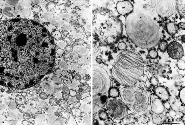
| Fig. 7. Retinal ganglion cell. (A) Portion of cell in Niemann-Pick disease, showing numerous membranous cytoplasmic bodies. Mitochondria (m) and dilated endoplasmic reticulum (er) are also evident. Area outlined in lower right is shown in greater magnification in B. (× 15,000) (B) Portion of cytoplasm of ganglion cell shown in A. Membranous cytoplasmic bodies cut in several different planes are evident, as are mitochondria (m) and endoplasmic reticulum (er) (×42,000). (Robb RM, Kuwabara T: The ocular pathology of type A Niemann-Pick disease: A light and electron microscopic study. Invest Ophthalmol 12:366, 1973) |