Chapter 27
Rhegmatogenous Retinal Detachment
BRIAN P. CONNOLLY and CARL D. REGILLO
Main Menu   Table Of Contents

Search

PATHOGENESIS
RISK FACTORS
CLINICAL FEATURES
DIFFERENTIAL DIAGNOSIS
TREATMENT OF RETINAL BREAKS TREATMENT MODALITIES
MANAGEMENT
REFERENCES

A rhegmatogenous retinal detachment (RRD) occurs when fluid from the vitreous cavity passes through a break in the neurosensory retina into the potential space between the retinal pigment epithelium (RPE) and the neurosensory retina. Throughout this chapter, the terms neurosensory retina and retina are used interchangeably. Although retinal breaks are present in 5% to 10% of the general public, most never lead to an RRD.1–7 In the general population, the estimated incidence of RRD is 1 in 10,000.8 After an RRD in one eye, the risk of RRD is estimated to be approximately 10% in the fellow eye.9–12

Various characteristics of a retinal break and the remaining ocular structures determine whether a retinal break will remain asymptomatic or cause an RRD. This chapter discusses the pathogenesis of RRD and the predisposing factors. The prophylactic management of retinal tears and the treatment of RRD are introduced as well.

Back to Top
PATHOGENESIS

POSTERIOR VITREOUS DETACHMENT

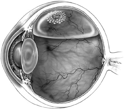
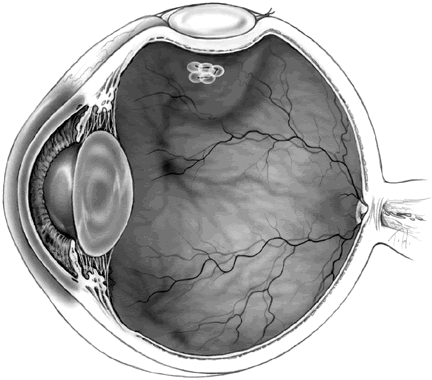
Many retinal breaks that lead to RRDs are precipitated by posterior vitreous detachments (PVDs). As one ages, biochemical changes in the matrix of the vitreous gel cause progressive liquefaction (syneresis) of the central vitreous.13–15 Throughout the vitreous cavity, liquefied cavities develop and ultimately coalesce. In many patients, the posterior cortical vitreous develops breaks that allow liquefied vitreous to pass into the potential space between the posterior hyaloid face and the retina. Once this occurs, a partial PVD can progress to a complete PVD rapidly.13 At the vitreous base, the cortical vitreous adheres strongly to the retinal surface. Vitreous traction at the posterior aspect of the vitreous base predisposes to retinal breaks at this location (Fig. 1).16

Fig. 1. Vitreous traction at the posterior aspect of the vitreous base predisposes to retinal breaks at this location. Both flap tears (horseshoe tears) and operculated tears may occur. (Courtesy of Hilel Lewis, MD, Cleveland, OH, and Allan Kreiger, MD, Los Angeles, CA.)

When patients experience an acute PVD, they often note the onset of floaters. These floaters are often shadows cast by remnants of glial tissue adherent to the posterior hyaloid face.17 Up to half of patients who experience an acute PVD note the onset of photopsias (flashes of light) in addition to the floaters.18–20

RETINAL BREAKS

The incidence of retinal tears after a symptomatic PVD is approximately 15%.18,20–25 Up to 20% of patients note the presence of a large number of small floaters, which usually correspond to a vitreous hemorrhage. Such hemorrhages frequently result from a torn blood vessel on the surface of the retina or optic nerve.20,22,24,26 When a PVD is accompanied by any vitreous hemorrhage, the incidence of retinal tears is approximately 70%. By comparison, only 2% to 4% of eyes have tears when a PVD is not accompanied by vitreous hemorrhage.18,20–25 The presence of pigment clumps in the anterior vitreous or anterior chamber (“tobacco dust”) alerts the clinician to a high likelihood of a retinal tear. This finding indicates the presence of RPE cells that have passed through a retinal break.

When a patient has potential symptoms of a PVD, a complete ocular examination is essential, including indirect ophthalmoscopy with scleral depression or fundus biomicroscopy with a three-mirror contact lens. When a vitreous hemorrhage obscures the peripheral retina, B-scan ultrasonography can be used to rule out an RRD and to evaluate a variety of other pathologic conditions. Although a skilled ultrasonographer can identify some (usually larger) retinal tears, a negative study does not rule out a retinal tear. Careful follow-up is suggested for such patients until the vitreous hemorrhage has cleared enough to allow a complete retinal examination. If an RRD or tear is present in the setting of dense vitreous hemorrhage, pars plana vitrectomy is generally indicated to obtain a view of the retina and to allow treatment of the underlying pathology.

Some authors have estimated that only 1 in 70 eyes with retinal breaks goes on to develop an RRD.6,7 There are several mechanisms in place that normally keep the retina attached. The physical interdigitation of the apical portions of the RPE cells with the photoreceptors of the neurosensory retina is one such mechanism.27 Actin contained in the cell sheaths of the RPE cells is believed to play an active role in this process.28 A second active mechanism is the removal of subretinal fluid by the RPE. A variety of ion channels and cotransporters are involved in this process.29

Several passive mechanisms also contribute to keeping the retina attached. First, the interphotoreceptor matrix between the RPE and the neurosensory retina acts as a biologic glue that secures the two surfaces.30 Further resistance to the development of an RRD is provided by the high oncotic pressure in the protein-rich choroid that promotes the absorption of fluid from the inner ocular structures.28 Because the retina offers hydraulic resistance to the diffusion of fluid from the vitreous to the choroid, the relative pressure of the vitreous helps to hold the retina in place.31–33 Finally, in eyes with little vitreous liquefaction, the formed cortical vitreous impedes the flow of fluid into the subretinal space in the event of a retinal break.34

RETINAL TEARS AND DIALYSES

When a retinal break occurs, several factors can catalyze detachment of the retina. Retinal breaks associated with vitreous traction are referred to as tears, and they tend to cause RRDs.9,35,36 In particular, flap tears are associated with a higher risk of RRD than other retinal breaks because the breaks are held open, facilitating the flow of fluid from the vitreous cavity into the subretinal space (Fig. 2). Also, liquefied vitreous can pass through retinal breaks more easily than more formed vitreous, especially if the flow of the vitreous is turbulent.37 In the case of an operculated retinal tear, the traction on the surrounding retina is relieved, and the risk of subsequent RRD is minimized (Fig. 3). A retinal dialysis is a tear at the junction of the neurosensory retina and the nonpigmented epithelial layer of the pars plana. Although not strictly a retinal tear, a retinal dialysis allows liquefied vitreous to enter the subretinal space, causing an RRD. Retinal dialyses frequently result from blunt trauma to the globe, and they are most likely to occur inferotemporally or superonasally (Fig. 4).38–40

Fig. 2. A. Flap (horseshoe tear) with a rolled posterior edge. Bridging vessels can be seen. B. A flap tear associated with a small rhegmatogenous retinal detachment. (A courtesy of William E. Benson, MD, Philadelphia, PA.)

Fig. 3. Operculated break with a cuff of subretinal fluid.

Fig. 4. A. Traumatic retinal dialysis resulting from blunt trauma. B. Retinal detachment in the same eye.

RETINAL HOLES

Retinal holes arise from atrophy of the retina, or tangentially oriented forces that create a dehiscence in the retina. Atrophic retinal holes are prone to occur in areas of lattice degeneration and in the walls of retinoschisis cavities. Atrophic holes are far less prone to cause RRD than are retinal tears.6,9,41,42

Macular holes likely result primarily from tangential and to a lesser extent anteroposterior traction from the overlying cortical vitreous.43,44 It is unusual for a macular hole to lead to an RRD, except in highly myopic globes.45,46

Back to Top
RISK FACTORS

LATTICE DEGENERATION

Lattice degeneration of the retina is characterized by focal thinning of the inner retinal layers associated with liquefaction of the overlying vitreous and strong vitreous attachments to the margins (Fig. 5). Hyalinized blood vessels in areas of lattice often take on the characteristic lattice appearance for which this condition is named. Areas of lattice are frequently elliptical and oriented parallel to the ora. These patches are often multiple, and they occur most often in the superior and the inferior retinal periphery.47 Some variants of lattice are oriented perpendicular to the ora, often straddling the peripheral retinal vessels—a configuration referred to as radial or meridional lattice.

Fig. 5. A. Lattice degeneration with pigmentation. Hyalinized blood vessels are seen in the lattice. B. Photomicrograph of lattice degeneration demonstrating focal thinning of the inner retinal layers and associated liquefaction of the overlying vitreous with vitreous attachments to the margins. (Courtesy of William Tasman, MD, Philadelphia, PA.)

One third of all RRDs are associated with lattice, although lattice is estimated to occur in only 6% to 10% of the general population.4,23,48–51 This condition is bilateral in one third of those affected, and it is more prevalent in myopic versus hyperopic eyes.4,23,48–51 Two forms of retinal breaks are associated with lattice: atrophic holes in the lattice, and flap tears at the posterior border or at the ends of the patch.

Atrophic holes occur in about one third of the patients with lattice and are frequently present in young patients. RRD is a rare complication of atrophic holes in lattice; when it occurs, it tends to progress slowly.4,51,52 Due to their relatively benign nature, treatment is rarely warranted for these atrophic holes. Horseshoe tears associated with lattice, in contrast, have a more ominous prognosis. The retinal flap is generally adherent to the detached cortical vitreous face, and this holds the tear open. These flap tears, especially when they are symptomatic, can cause rapidly progressive RRD.9,36,42,51,53

MYOPIA

Although present in only 10% or less of the general population, significant myopia is associated with 42% of all RRDs.23,50 This higher incidence of RRD among myopic eyes has been attributed to a variety of causes. The incidence of PVD is higher for any given age group in people with myopia compared with those with emmetropia and hyperopia.14,54 Further, the incidence of lattice degeneration is increased in myopes.4 Finally, the peripheral retina is more vulnerable to developing retinal tears in myopic eyes.28 When symptomatic retinal tears are present in patients with moderate to high myopia, prophylactic treatment should be undertaken.

CATARACT EXTRACTION

Cataract extraction is an independent risk factor for RRD. The incidence of RRD after intracapsular cataract extraction is greater than after extracapsular cataract surgery. After uncomplicated surgery, the risk of RRD is estimated to be 2% to 5% with intracapsular surgery and approximately 1% with extracapsular. In contrast, the incidence of RRD is 0.01% to 0.05% in the general phakic population.8,55–58

Vitreous loss during cataract surgery increases the risk of RRD 4.5-fold to approximately 5%.56,59 Likewise, YAG capsulotomy increases the risk of RRD 3.9-fold.60–62 Other risk factors that increase the risk of RRD with cataract extraction include myopia, young age, white race, prior RRD surgery, and RRD in the fellow eye.18,56,57,63,64

The causative retinal break is most frequently located superotemporally in phakic, pseudophakic, and aphakic RRDs. Nonetheless, both superonasal breaks and breaks that go undetected at the time of intervention are more common after cataract extraction.65–68 The tendency for breaks to be smaller and more anterior, combined with the postsurgical changes in the anterior segment, combine to make tears difficult to localize. Overall, the rate of missed breaks after cataract surgery ranges from 5% to 16% versus 2% to 5% in phakic patients.50,68,69 Although careful localization of breaks is always a critical step in RRD repair, particular attention must be paid in patients who have had cataract extraction.

The risk of RRD in the fellow eye is 26% to 41% for patients who are aphakic.64,70 In patients who have had congenital cataracts removed from both eyes, the risk of developing an RRD in the fellow eye after RRD approaches 70%; thus, these patients must have meticulous follow-up.71

TRAUMA

Three quarters of traumatic RRDs occur in males, largely because they are more likely than females to engage in contact sports or fighting.72,73 The leading cause of RRD in children and adolescents is blunt trauma.72–74 Blunt trauma generally compresses the contents of the eye in an anteroposterior direction, causing a compensatory expansion of the globe in an equatorial plane. Despite the elastic nature of the vitreous, rapid compression of the eye due to trauma can transmit severe traction to the vitreous base, causing many types of pathology. Linear or horseshoe tears at the posterior vitreous base, tears in the nonpigmented epithelium of the pars plana at the anterior vitreous base, and retinal dialyses are the most common forms of tears that follow such trauma.75–77

The most common locations for retinal breaks due to trauma are inferotemporal and superonasal.76,78 Most traumatic retinal breaks are believed to occur at the time of impact.75,79 As a result of the formed nature of the vitreous in young patients, the progression of RRD after trauma tends to be relatively slow.

Blunt trauma can also cause focal areas of retinal necrosis directly corresponding to the site of impact and traumatic macular holes, which have been attributed to contrecoup damage.28

Penetrating and perforating injuries frequently cause RRD. In these cases, the prognosis depends on the extent of the injury.76,80–82 The management of these injuries is beyond the scope of this chapter.

PROLIFERATIVE RETINOPATHIES

A variety of conditions in which fibrovascular proliferation occurs have a higher risk of RRD. In proliferative diabetic retinopathy, neovascularization and fibroglial tissues proliferate in response to retinal ischemia. As the vitreous contracts with PVD, these abnormal proliferations can place traction on the retinal surface, causing either a simple tractional retinal detachment or a combined tractional and rhegmatogenous retinal detachment (Fig. 6). Fibrovascular proliferation occurring in retinopathy of prematurity, in proliferative sickle cell retinopathy, and in the aftermath of retinal vein occlusions can cause RRD through a related mechanism.

Fig. 6. Tractional retinal detachment in a patient with proliferative diabetic retinopathy.

INFECTIONS

Infections that cause full-thickness retinal necrosis can lead to RRD. Cytomegalovirus retinitis, acute retinal necrosis syndrome, postoperative bacterial endophthalmitis, and other retinal infections may cause retinal holes to form that may ultimately lead to RRD.83

INHERITED AND MISCELLANEOUS DISEASES

HEREDITARY HYALOIDEORETINOPATHIES. This group of conditions is typified by accelerated vitreous syneresis that leads to a classic “optically empty” vitreous in which fibrous bands and membranes are often present. Common features of these hyaloideoretinopathies include myopia, cataract, optic atrophy, RPE and choroidal atrophy, lattice degeneration, retinoschisis, and glaucoma.61,70,84

The first group of hyaloideoretinopathies characterized by these ocular findings includes Wagner's disease, erosive vitreoretinopathy, and Jansen's disease. Of these, erosive vitreoretinopathy and Jansen's disease both predispose to RRD.

Stickler's syndrome is a rare inherited hyaloideoretinopathy associated with several systemic findings. The systemic associations include midfacial flattening, cleft palate, Pierre Robin malformation, and a variety of skeletal abnormalities. The presence of Stickler's syndrome signifies a strong risk of RRD (Fig. 7).

Fig. 7. Photograph of a fibrous band in the “optically empty” vitreous of a patient with Stickler's syndrome.

CONGENITAL (JUVENILE) RETINOSCHISIS. The presence of a characteristic spokelike pattern of foveal change alerts the examiner to this condition. In the peripheral retina, schisis tends to develop between the nerve fiber layer and the balance of the retina (Fig. 8). Holes commonly develop in the inner wall of these schisis cavities but are generally inconsequential. In contrast, holes in the outer wall of these cavities are rare but can lead to RRDs.28,85

Fig. 8. A. Characteristic spokelike pattern of foveal change in X-linked juvenile retinoschisis. B. Peripheral retinoschisis with a vessel bordered by inner-wall holes on either side. (Courtesy of William Tasman, MD, Philadelphia, PA.)

EHLERS-DANLOS SYNDROME. This autosomal dominant condition is characterized by abnormal collagen throughout the body. These patients are predisposed to both myopia and RRD.

GOLDMANN-FAVRE SYNDROME. This is an autosomal recessive condition in which cataract, peripheral and macular retinoschisis, radial lattice degeneration, accelerated vitreous syneresis, and retinitis pigmentosa-like peripheral retina changes are evident. These patients are prone to RRDs, which often prove difficult to repair.

MARFAN SYNDROME. As with Ehlers-Danlos, there is an increased risk of myopia and lattice degeneration, leading to a high incidence of RRDs. When ectopia lentis is present, cataract removal is often associated with vitreous loss, which places these patients at an even higher risk for RRD.

OTHER ABNORMALITIES. Patients with the so-called morning glory syndrome are predisposed to RRD because liquefied vitreous may pass through defects in the anomalous disc into the subretinal space. Likewise, tears present in choroidal colobomas may also cause RRD.86

Back to Top
CLINICAL FEATURES

EARLY

A recently detached retina loses its transparency and assumes a gray, translucent appearance. Eye movements typically cause the detached retina to undulate in a characteristic manner. Fine, irregular corrugations are usually present and are the result of intraretinal edema.87,88 The fine details of the choroidal vasculature are somewhat obscured by the overlying detached retina (Fig. 9). Within days of an RRD, outer retinal degeneration starts to occur.89,90 The magnitude of the photoreceptor damage that occurs is related to the height and the duration of the RRD.88 At this early stage, the circulation of the inner retina is not affected; thus, the inner retina remains well preserved. If the retina is reattached within a week, most of the cellular changes are reversible.91 RPE cells underlying the RRD can be released into the subretinal fluid and may pass through the retinal break into the vitreous cavity. These cells may be appreciated in the anterior vitreous, where they are known as tobacco dust.87,88,92

Fig. 9. Recent rhegmatogenous retinal detachment showing loss of the normal retinal transparency and irregular corrugations.

In 1971, Lincoff and Gieser93 reported four guidelines for locating retinal breaks causing RRDs. The propagation of an RRD is determined by the location of the causative break, anatomic barriers (i.e., the optic nerve, the ora serrata, and existing chorioretinal adhesions), and the effect that gravity has on the subretinal fluid in the upright position (Fig. 10):

Fig. 10. The configuration of a retinal detachment can help to localize the causative retinal break. A. A superior retinal detachment that crosses the 12-o'clock meridian. In total detachments or superior detachments that cross the midline, the primary hole is usually within 1 clock hour of the 12-o'clock meridian. If the detachment extends more inferiorly on either the nasal or temporal side, the causative break is usually on the same side of the 12-o'clock meridian. B. A superotemporal retinal detachment. The break lies near the superior edge of the detached retina. In superior nasal or temporal detachments, the hole lies within 1.5 clock hours of the highest border 98% of the time. C. An inferior retinal detachment. The break lies nasal to the optic nerve and the subretinal fluid extends higher nasally. In inferior detachments, the higher side indicates to which side of the disc an inferior hole lies 95% of the time. When an inferior detachment is bullous (not shown), the primary hole often lies above the horizontal meridian.93

  1. In superior nasal or temporal detachments, the hole lies within 1.5 clock hours of the highest border 98% of the time.
  2. In total detachment or superior detachments that cross the midline, the primary hole is at the 12-o'clock position or in a triangle, the apex of which is at the ora serrata and the sides of which intersect the equator 1 hour to either side of the 12-o'clock position. This occurs 93% of the time.
  3. In inferior detachments, the higher side indicates to which side of the disc an inferior hole lies 95% of the time.
  4. When an inferior detachment is bullous, the primary hole lies above the horizontal meridian.93

LONG-STANDING

Progressive atrophy of all retinal layers occurs if the retina remains detached.88 A chronically detached retina takes on a smooth contour and becomes semitransparent. In some cases, cystic spaces develop in the detached retina. Atrophy and depigmentation of the underlying RPE also develop in these eyes.28 When an RRD remains static for 3 months or more, RPE metaplasia may occur at the border of the detachment. Ophthalmoscopically, this corresponds to either a pigmented or depigmented demarcation line. Most RRDs that are surrounded by a demarcation line eventually progress; nonetheless, surgical repair of these eyes has an excellent prognosis (Fig. 11).94

Fig. 11. A chronic retinal detachment with a pigmented demarcation line. Note the stippled retinal pigment epithelial changes in the area of the chronic detachment.

In very long-standing RRDs, extensive capillary nonperfusion can result, leading to peripheral retinal neovascularization.95 Moreover, intraocular pressure can rise if the trabecular meshwork becomes impeded by pigment clumps or the outer segments of photoreceptors.96,97

PROLIFERATIVE VITREORETINOPATHY

After RRD, proliferative vitreoretinopathy occurs in approximately 10% of cases, of which one quarter require additional surgical intervention.98,99 The severity can vary greatly.100 In its mildest form, the only findings are vitreous haze and pigment clumps (grade A). Moderate proliferative vitreoretinopathy (grade B) is manifested by early cellular proliferation, which may occur on either or both sides of the retina.101,102 The involved retina may become stiffened, and its surface may take on a wrinkled appearance. Also, the retinal vessels may become tortuous and the edge of the retinal break may become rolled. Finally, with severe proliferative vitreoretinopathy (grade C), full-thickness retinal folds (“star folds”) may occur (Fig. 12).

Fig. 12. Proliferative vitreoretinopathy with a star fold.

In moderate to severe disease, the proliferative membranes are composed mostly of dedifferentiated RPE cells; nonetheless, glial cells, fibrocytes, myofibroblasts, and macrophages may also be present.103–105 Proliferative vitreoretinopathy is most likely to occur in the inferior retina, and it is postulated that RPE cells settle out of the vitreous onto the inferior retinal surface in a gravity-dependent fashion, where they eventually cause proliferative vitreoretinopathy.106

Back to Top
DIFFERENTIAL DIAGNOSIS
It is important to be able to distinguish an RRD from an exudative or a tractional retinal detachment. Likewise, an area of retinoschisis must also be differentiated from an RRD. Although the management of each of these conditions is discussed in detail elsewhere, a brief description of each condition is provided below.

EXUDATIVE RETINAL DETACHMENT

A variety of inflammatory, neoplastic, and other conditions can cause a breakdown of the normal blood-retina barrier, allowing fluid to collect in the subretinal space. The surface of the detached retina is generally smooth, and the location of the detachment may shift as the patient adjusts the position of the head (Fig. 13).

Fig. 13. Peripheral detail of a bullous retinal detachment.

TRACTIONAL RETINAL DETACHMENT

Vitreous membranes resulting from proliferative vitreoretinopathy, various proliferative retinopathies, or trauma may pull the retina away from the RPE. In purely tractional detachments, the retinal surface has a convex, taut configuration. No full-thickness retinal breaks would be expected to be present (see Fig. 6).

RETINOSCHISIS

Retinoschisis occurs when the retina is split into two layers or walls by a mucopolysaccharide-rich viscous fluid. Significant senile retinoschisis, usually a split in the outer plexiform layer of the retina, is most often a bilateral process. Its smooth, dome-shaped appearance helps to differentiate it from an RRD. In contrast to the RPE atrophy seen with long-standing RRD, the underlying RPE is typically normal and devoid of demarcation lines.107,108 Unlike with RRD, tobacco dust, vitreous hemorrhage, or flap tears are not expected to be present (Fig. 14).

Fig. 14. Senile degenerative retinoschisis with outer-wall holes. (Courtesy of William E. Benson, MD.)

Back to Top
TREATMENT OF RETINAL BREAKS TREATMENT MODALITIES
Laser photocoagulation, administered with either a slit-lamp or indirect ophthalmoscopic delivery system, is an effective modality for the treatment of retinal breaks to prevent RRD. The goal of laser treatment is to seal the retina to the RPE and choroid around the break. This is accomplished by surrounding the break with two or three rows of nearly confluent laser burns. If a break occurs in a patch of lattice, the entire patch of lattice and the break should be surrounded with laser burns. A firm chorioretinal scar forms in 7 to 10 days (Fig. 15).

Fig. 15. A. A fairly posterior retinal flap tear. B. The same tear after laser photocoagulation. C. Another horseshoe tear adjacent to a patch of pigmented lattice. Both the tear and the adjacent lattice are surrounded by laser photocoagulation burns. (Courtesy of William E. Benson, MD, and William Tasman, MD, Philadelphia, PA.)

Transscleral cryotherapy, applied transconjunctivally under ophthalmoscopic visualization, is another effective means of creating a chorioretinal adhesion around a retinal break. The break is surrounded by contiguous cryotherapy applications to the surrounding attached retina. As with laser photocoagulation, if the break occurs in an area of lattice, the cryotherapy spots should surround the entire patch of lattice. A firm chorioretinal adhesion is not established for 7 or more days after treatment (Fig. 16).

Fig. 16. A flap tear recently treated with cryotherapy. (Courtesy of William E. Benson, MD, Philadelphia, PA.)

FLAP (HORSESHOE) TEARS

If a flap tear is symptomatic (i.e., caused by a PVD and accompanied by complaints of flashes or floaters), the probability of RRD is very high (25% to 90%).9,28,36,42 Treatment of these symptomatic flap tears with laser photocoagulation or cryotherapy dramatically reduces the risk of RRD.109–113

When no symptoms of flashes or floaters accompany a flap tear, the risk of RRD is low enough that observation is appropriate in most eyes.6,41,42,114 The presence of subretinal fluid, lack of subretinal pigment, and superior location would favor treatment of a flap tear without definite symptoms.

ROUND BREAKS

If an operculated tear is noted in a location where no tear was seen on previous examination or if it is symptomatic (i.e., accompanied by flashes or floaters), there is a moderate risk of RRD.9,36 If an operculated tear is superiorly located, large, or associated with vitreous hemorrhage, treatment should be administered. Likewise, if vitreoretinal traction is noted at the edges of the operculated tear, the break should be treated.

If no symptoms of flashes or floaters are present, round breaks with or without operculae are unlikely to lead to RRD6,41,42,114 and may simply be observed.

LATTICE DEGENERATION

Although it has been established that lattice degeneration is a risk factor for RRD, only a few eyes with lattice go on to develop RRD. Even when atrophic holes are present in areas of lattice, RRD is unusual.115 Careful observation is all that is needed in these cases.

PSEUDOPHAKIC OR APHAKIC EYES

When symptomatic round breaks occur in pseudophakic or aphakic eyes, the risk of RRD is 1% to 5%.55,56,58 Treatment is warranted in these situations. Nonetheless, asymptomatic tears and round holes can generally be observed safely in these patients unless the break is large, posteriorly located, or collecting subretinal fluid.28,42,116

FELLOW EYES

As stated above, after RRD in one phakic eye, the risk of a detachment in the fellow (phakic) eye is estimated to be approximately 10%.9–12 Treatment of vitreoretinal abnormalities (especially retinal tears) has been shown to reduce the risk of RRD.12,110,113 Folk and associates117 demonstrated that prophylactic treatment of lattice degeneration in fellow phakic eyes reduced the risk of RRD from 5.1% to 1.8%. In this setting, prophylactic therapy should be considered.

Pseudophakic and aphakic fellow eyes have an even higher incidence of RRD than phakic fellow eyes.64,70 Treatment of all retinal breaks is clearly indicated, and treatment of lattice degeneration and vitreoretinal tufts is reasonable.64,118

Back to Top
MANAGEMENT
Left untreated, an RRD will usually progress. The rate of progression depends on the viscosity and turbulence of the vitreous, the location of the break, and the presence of vitreous traction at the site of the break. Retinal detachments due to superior breaks propagate more quickly than inferior breaks due to the effect of gravity.

Surgery to reattach the retina is warranted to prevent further visual field loss and to recover as much visual function as possible from the detached retina. Timely intervention is important because the sooner the retina is reattached, the better the recovery of function.119 If an RRD is threatening to detach the macula, surgery should be performed as soon as possible.120 Even if the macula has detached, prompt surgical repair within 2 to 3 days frequently recovers reasonable central vision.

The various techniques of RRD repair are discussed in detail elsewhere in this volume, but an overview of the basic techniques is provided.

SCLERAL BUCKLE

The rationale of scleral buckling surgery is to indent areas of the sclera, choroid, and RPE that underlie a retinal break so that the RPE tamponades the causative retinal break. By closing the retinal break, the flow of liquefied vitreous into the subretinal space is prevented, allowing the retina to reattach. This buckling effect is accomplished by suturing a piece of inert material to the sclera in the area corresponding to the retinal break. Transscleral cryotherapy is typically performed to create a focal chorioretinal scar, which helps to seal the tear in the area of the break(s). Drainage of the subretinal fluid or intraocular gases may be used to facilitate retinal reattachment, but these measures are not always necessary (Fig. 17).

Fig. 17. Scleral buckle procedure. The external buckling element indents the sclera, choroid, and retinal pigment epithelium. This tamponades the retinal break.

PNEUMATIC RETINOPEXY

Pneumatic retinopexy is an effective alternative to the scleral buckling technique for selected uncomplicated primary detachments.121,122 This procedure involves treatment of the retinal break (retinopexy) with cryotherapy or laser photocoagulation and intraocular injection of an inert gas, which acts as a pneumatic tamponade. Transscleral cryotherapy may be applied to the retinal break before the injection of the gas. In contrast, laser photocoagulation is applied after the gas bubble has been injected and the retina surrounding the break is reattached. Laser photocoagulation or transscleral cryotherapy should be applied to retinal breaks present in attached retina before injection of the intraocular gas.

Careful patient selection allows a success rate comparable to that of scleral buckling surgery.123–125 This technique is useful when the causative retinal break or small cluster of breaks is located in the superior 8 clock hours of the peripheral retina. This technique has gained considerable popularity as an alternative to scleral buckling since it was introduced in the late 1980s (Fig. 18).126

Fig. 18. Pneumatic retinopexy. The inert gas bubble internally tamponades the retinal break, allowing the subretinal fluid to resorb.

LINCOFF BALLOON

Another technique for RRD repair is the temporary use of an inflatable balloon to indent the sclera, choroid, and RPE. The principles of this procedure are analogous to those of scleral buckling. All of the retinal breaks are localized and treated with cryotherapy. Alternatively, laser photocoagulation may be applied instead of cryotherapy; however, as is the case in pneumatic retinopexy, this must be applied in the postoperative period once the retina is reattached.

Rather than permanently attaching an inert buckle to the eye, an inflatable balloon is placed in the area of the causative retinal break(s) through a small conjunctival and Tenon's incision and then inflated. After several days, the balloon is deflated and removed.127 This technique avoids some of the long-term complications of scleral buckles such as infection, extrusion, refractive change, and motility disturbances. Nonetheless, the procedure is useful only for detachments with a single break or a closely spaced cluster of breaks, and the single-operation success rate is somewhat lower than for scleral buckling techniques.127–129 This technique tends to be cumbersome for the patient and is not widely used at this time.

PRIMARY VITRECTOMY

Another useful approach to RRD repair is a primary pars plana vitrectomy. This technique involves internal drainage of the subretinal fluid, a gas-fluid exchange, and laser photocoagulation or cryotherapy of the reattached retina.130,131 Because the vitreous is removed, vitreous traction on retinal breaks is eliminated during surgery. In cases with significant vitreous hemorrhage, this technique allows visualization and treatment of the retinal breaks, which would not be approachable with other techniques. Nonetheless, this technique is generally reserved for more complicated detachments.

LASER PHOTOCOAGULATION

If a patient is unwilling or physically unable to tolerate a surgical intervention, laser photocoagulation of the attached retina surrounding an RRD may be used to wall off the detachment. Nonetheless, this technique applies to a limited number of patients. Recently, this method has gained popularity in the setting of limited retinal detachments from quiescent cytomegalovirus retinitis.132,133

Back to Top
REFERENCES

1. Foos RY: Posterior vitreous detachment. Trans Am Acad Ophthalmol Otolaryngol 76:480, 1972

2. Okun E: Gross and microscopic pathology of autopsy eyes. Part III. Retinal breaks without detachment. Am J Ophthalmol 51:369, 1961

3. Rutnin U, Schepens CL: Fundus appearance in normal eyes. IV. Retinal breaks and other findings. Am J Ophthalmol 64:1063, 1967

4. Byer NE: Clinical study of lattice degeneration of the retina. Trans Am Acad Ophthalmol Otolaryngol 69:1064, 1965

5. Byer NE: Clinical study of retinal breaks. Trans Am Acad Ophthalmol Otolaryngol 71:461, 1967

6. Byer NE: Prognosis of asymptomatic retinal breaks. Arch Ophthalmol 92:208, 1974

7. Machemer R: The importance of fluid absorption, traction, intraocular currents, and chorioretinal scars in the therapy of rhegmatogenous retinal detachments. Am J Ophthalmol 98:681, 1984

8. Michaelson IC, Stein R: A study in the prevention of retinal detachment. Ann Ophthalmol 1:49, 1965

9. Davis MD: Natural history of retinal breaks without detachment. Arch Ophthalmol 92:183, 1974

10. Delaney WV Jr, Oates RP: Retinal detachment in the second eye. Arch Ophthalmol 96:629, 1978

11. Folk JC, Burton TC: Bilateral phakic retinal detachment. Ophthalmology 89:815, 1982

12. Merin S, Feiler V, Hyams S et al: The fate of the fellow eye in retinal detachment. Am J Ophthalmol 71:477, 1971

13. Foos R, Wheeler N: Vitreoretinal juncture: Synchysis senilis and posterior vitreous detachment. Ophthalmology 89: 1502, 1982

14. Sebag J: Aging of the vitreous. Eye 1:254, 1987

15. Larsson L, Osterlin S: Posterior vitreous detachment—a combined clinical and physiochemical study. Graefe Arch Clin Exp Ophthalmol 223:92, 1985

16. Wilkinson CP, Rice TA: Retinal Detachment, p 1163. St Louis: Mosby, 1997

17. Foos RY, Roth AM: Surface structure of the optic nerve head. 2. Vitreopapillary attachments and posterior vitreous detachment. Am J Ophthalmol 76:662, 1973

18. Byer NE: Natural history of posterior vitreous detachment with early management as the premier line of defense against retinal detachment. Ophthalmology 101:1503, 1994

19. Morse PH, Scheie HG, Aminlari A: Light flashes as a clue to retinal disease. Arch Ophthalmol 91:179, 1974

20. Tasman WS: Posterior vitreous detachment and peripheral retinal breaks. Trans Am Acad Ophthalmol Otolaryngol 72:217, 1968

21. Murakami K, Jalkh AE, Avila MP et al: Vitreous floaters. Ophthalmology 90:1271, 1983

22. Tabotabo MD, Karp LA, Benson WE: Posterior vitreous detachment. Ann Ophthalmol 12:59, 1980

23. Bohringer HR: Statistisches zur Haufigkeit und Risiko der netzhautablosung. Ophthalmologica 131:331, 1956

24. Jaffe N: Complications of acute posterior vitreous detachment. Arch Ophthalmol 79:568, 1968

25. Hikichi T, Trempe CL: Relationship between floaters, light flashes, or both, and complications of posterior vitreous detachment. Am J Ophthalmol 117:593, 1994

26. Boldrey EE: Risk of retinal tears in patients with vitreous floaters. Am J Ophthalmol 96:783, 1983

27. Spitznas M, Hogan MJ: Outer segments of photoreceptors and the retinal pigment epithelium: Interrelationship in the human eye. Arch Ophthalmol 1970;84:810

28. Regillo CD, Benson WE: Retinal Detachment: Diagnosis and Management, p 228. Philadelphia: Lippincott-Raven, 1998

29. Marmor MF: Retinal and retinal pigment epithelial physiology. In Regillo CD, Brown GC, Flynn J (eds): Vitreoretinal Disease: The Essentials, pp 25–38. New York: Thieme, 1999

30. Hageman G, Marmor M, Yao X et al: The interphotoreceptor matrix mediates primate retinal adhesion. Arch Ophthalmol 1995;113:655

31. Bill A: Blood circulation and fluid dynamics in the eye. Physiol Rev 55:383, 1975

32. Fatt I, Shantinath K: Flow conductivity of retina and its role in retinal adhesion. Exp Eye Res 12:218, 1971

33. Orr G, Goodnight R, Lean JS: Relative permeability of retina and retinal pigment epithelium to the diffusion of tritiated water from vitreous to choroid. Arch Ophthalmol 104:1678, 1986

34. Pederson JE, Cantrill HL, Cameron JD: Experimental retinal detachment. II. Role of the vitreous. Arch Ophthalmol 100:1155, 1982

35. Gonin J: Le traitment de decollement retinien. Ann Ocul 158:175, 1921

36. Colyear BHJ, Piscel KK: Clinical tears in the retina without detachment. Am J Ophthalmol 41:773, 1956

37. Lewis H, Kreiger AE: Rhegmatogenous retinal detachment. In Tasman W, Jaeger EA (eds): Clinical Ophthalmology, p 12. Vol 3. Philadelphia: JB Lippincott, 1991

38. Brown GC, Tasman WS: Familial retinal dialysis. Can J Ophthalmol 15:193, 1980

39. Kinyoun JL, Knobloch WH: Idiopathic retinal dialysis. Retina 4:9, 1984

40. Smiddy WE, Green WR: Retinal dialysis: pathology and pathogenesis. Retina 2:94, 1982

41. Byer NE: The natural history of asymptomatic retinal breaks. Ophthalmology 89:1033, 1982

42. Neumann E, Hyams S: Conservative management of retinal breaks: A follow-up study of subsequent retinal detachment. Br J Ophthalmol 56:482, 1972

43. Gass J: Reappraisal of biomicroscopic classification of stages of development of a macular hole. Am J Ophthalmol 119:752, 1995

44. Johnson RN, Gass JD: Idiopathic macular holes: Observations, stages of formation, and implications for surgical intervention. Ophthalmology 95:917, 1988

45. Zhang CF, Hu C: High incidence of retinal detachment secondary to macular hole in a Chinese population [letter]. Am J Ophthalmol 94:817, 1982

46. Morita H, Ideta H, Ito K et al: Causative factors of retinal detachment in macular holes. Retina 11:281, 1991

47. Byer NE: Lattice degeneration of the retina. Surv Ophthalmol 23:213, 1979

48. Halpern JI: Routine screening of the retinal periphery. Am J Ophthalmol 62:99, 1966

49. Straatsma BR, Zeegen PD, Foos RY et al: Lattice degeneration of the retina. Am J Ophthalmol 77:619, 1974

50. Ashrafzadeh MT, Schepens CL, Elzeneiny IH et al: Aphakic and phakic retinal detachment. Arch Ophthalmol 89: 476, 1973

51. Benson WE, Morse PH: The prognosis of retinal detachment due to lattice degeneration. Ann Ophthalmol 10: 1197, 1978

52. Tillery WV, Lucier AC: Round atrophic holes in lattice degeneration: an important cause of phakic retinal detachment. Trans Am Acad Ophthalmol Otolaryngol 81:509, 1976

53. Byer NE: Long-term natural history of lattice degeneration of the retina. Ophthalmology 96:1396, 1989

54. Akiba J: Prevalence of posterior vitreous detachment in high myopia. Ophthalmology 100:1384, 1993

55. Wilkes SR, Beard CM, Kurland LT et al: The incidence of retinal detachment in Rochester, Minnesota, 1970-1978. Am J Ophthalmol 94:670, 1982

56. Javitt JC, Vitale S, Canner JK et al: National outcomes of cataract extraction. I. Retinal detachment after inpatient surgery. Ophthalmology 98:895, 1991

57. Nielsen NE, Naeser K: Epidemiology of retinal detachment following extracapsular cataract extraction: A follow-up study with an analysis of risk factors. J Cataract Refractive Surg 19:675, 1993

58. Ninn-Pedersen K, Bauer B: Cataract patients in a defined Swedish population, 1986-1990. V. Postoperative retinal detachments. Arch Ophthalmol 114:382, 1996

59. Vail D: After-results of vitreous loss. Am J Ophthalmol 59:573, 1965

60. Fastenberg DM, Schwartz PL, Lin HZ: Retinal detachment following neodymium-YAG laser capsulotomy. Am J Ophthalmol 97:288, 1984

61. Ficker LA, Vickers S, Capon MR et al: Retinal detachment following Nd:YAG posterior capsulotomy. Eye 1:86, 1987

62. Javitt JC, Tielsch JM, Canner JK et al: National outcomes of cataract extraction. Increased risk of retinal complications associated with Nd:YAG laser capsulotomy. The Cataract Patient Outcomes Research Team. Ophthalmology 99: 1487, 1992

63. Ackerman AL, Seelenfreund MH, Freeman HM, Schepens CL: Cataract extraction following retinal detachment surgery. Arch Ophthalmol 84:41, 1970

64. Benson WE, Grand MG, Okun E: Aphakic retinal detachment. Management of the fellow eye. Arch Ophthalmol 93:245, 1975

65. Everett WG, Katzin D: Meridional distribution of retinal breaks in aphakic retinal detachment. Am J Ophthalmol 66:928, 1968

66. Phelps CD, Burton TC: Glaucoma and retinal detachment. Arch Ophthalmol 95:418, 1977

67. Wilkinson CP: Phakic retinal detachments in the elderly. Retina 15:220, 1995

68. Yoshida A, Ogasawara H, Jalkh AE et al: Retinal detachment after cataract surgery. Predisposing factors. Ophthalmology 99:453, 1992

69. Griffith RD, Ryan EA, Hilton GF: Primary retinal detachments without apparent breaks. Am J Ophthalmol 81: 420, 1976

70. Campbell CJ, Rittler MC: Cataract extraction in the retinal detachment prone patient. Am J Ophthalmol 73:17, 1972

71. Jagger JD, Cooling RJ, Fison LG et al: Management of retinal detachment following congenital cataract surgery. Trans Ophthalmol Soc UK 103:103, 1983

72. Cox MS, Schepens CL, Freeman HM: Retinal detachment due to ocular contusion. Arch Ophthalmol 76:678, 1966

73. Cox M, Freeman H: Traumatic retinal detachment. In Freeman H (ed): Ocular Trauma, pp 285–293. New York: Appleton-Century-Crofts, 1979

74. Winslow RL, Tasman W: Juvenile rhegmatogenous retinal detachment. Ophthalmology 85:607, 1978

75. Tasman W: Peripheral retinal changes following blunt trauma. Trans Am Ophthalmol Soc 70:190, 1972

76. Cox MS, Freeman HM: Retinal detachment due to ocular penetration. Arch Ophthalmol 1976;96:1354

77. Yoon YH, Marmor MF: Rapid enhancement of retinal adhesion by laser photocoagulation. Ophthalmology 95: 1385, 1988

78. Hagler WS, North AW: Retinal dialyses and retinal detachment. Arch Ophthalmol 79:376, 1968

79. Sellors PJ, Mooney D: Fundus changes after traumatic hyphaema. Br J Ophthalmol 57:600, 1973

80. De Juan E Jr, Sternberg P Jr, Michels RG: Penetrating ocular injuries: Types of injuries and visual results. Ophthalmology 90:1318, 1983

81. Meredith TA, Gordon PA: Pars plana vitrectomy for severe penetrating injury with posterior segment involvement. Am J Ophthalmol 103:549, 1987

82. Percival SPB: Late complications from posterior segment intraocular foreign bodies. Br J Ophthalmol 56:462, 1972

83. Regillo CD, Vander JF, Duker JS et al: Repair of retinitis-related retinal detachments with silicone oil in patients with acquired immunodeficiency syndrome. Am J Ophthalmol 113:21, 1992

84. Doden W, Stark N: Retina and vitreous findings after serious indirect eye trauma. Klin Monatsbl Augenheilkd 614:32, 1974

85. Regillo CD, Tasman WS, Brown GC: Surgical management of complications associated with X-linked retinoschisis. Arch Ophthalmol 111:1080, 1993

86. Wang K, Hilton GF: Retinal detachment associated with coloboma of the choroid. Trans Am Ophthalmol Soc 83: 49, 1985

87. Machemer R, Norton EW: Experimental retinal detachment in the owl monkey. I. Methods of production and clinical picture. Am J Ophthalmol 66:388, 1968

88. Machemer R: Experimental retinal detachment in the owl monkey. II. Histology of retina and pigment epithelium. Am J Ophthalmol 66:396, 1968

89. Cook B, Lewis GP, Fisher SK, Adler R: Apoptotic photoreceptor degeneration in experimental retinal detachment. Invest Ophthalmol Vis Sci 36:990, 1995

90. Nork TM, Millecchia LL, Strickland BD et al: Selective loss of blue cones and rods in human retinal detachment. Arch Ophthalmol 113:1066, 1995

91. Machemer R: Experimental retinal detachment in the owl monkey. IV. The reattached retina. Am J Ophthalmol 66:1075, 1968

92. Kroll AJ, Machemer R: Experimental retinal detachment in the owl monkey. 3. Electron microscopy of retina and pigment epithelium. Am J Ophthalmol 66:410, 1968

93. Lincoff H, Gieser R: Finding the retinal hole. Arch Ophthalmol 85:565, 1971

94. Benson WE, Nantawan P, Morse PH: Characteristics and prognosis of retinal detachments with demarcation lines. Am J Ophthalmol 84:641, 1977

95. Felder KS, Brockhurst RJ: Retinal neovascularization complicating rhegmatogenous retinal detachment of long duration. Am J Ophthalmol 93:773, 1982

96. Netland PA, Mukai S, Covington HI: Elevated intraocular pressure secondary to rhegmatogenous retinal detachment. Surv Ophthalmol 39:234, 1994

97. Schwartz A: Chronic open-angle glaucoma secondary to rhegmatogenous retinal detachment. Trans Am Ophthalmol Soc 70:178, 1972

98. Havener WH: Massive vitreous retraction. Ophthalmic Surg 4:22, 1973

99. Morse PH: Fixed retinal star folds in retinal detachment. Am J Ophthalmol 77:760, 1974

100. Machemer R, Aaberg TM, Freeman HM et al: An updated classification of retinal detachment with proliferative vitreoretinopathy. Am J Ophthalmol 112:159, 1991

101. Campochiaro PA: Pathogenic mechanisms in proliferative vitreoretinopathy. Arch Ophthalmol 115:237, 1997

102. Ryan SJ: Traction retinal detachment. Am J Ophthalmol 115:1, 1993

103. Clarkson JG, Green WR, Massof D: A histopathologic review of 168 cases of preretinal membrane. Am J Ophthalmol 84:1, 1977

104. Machemer R, Laqua H: Pigment epithelium proliferation in retinal detachment (massive periretinal proliferation). Am J Ophthalmol 80:1, 1975

105. Machemer R, van Horn D, Aaberg TM: Pigment epithelial proliferation in human retinal detachment with massive periretinal proliferation. Am J Ophthalmol 85:181, 1978

106. Singh AK, Glaser BM, Lemor M, Michels RG: Gravity-dependent distribution of retinal pigment epithelial cells dispersed into the vitreous cavity. Retina 6:77, 1986

107. Byer NE: Clinical study of senile retinoschisis. Arch Ophthalmol 79:36, 1968

108. Straatsma BR, Foss RY: Typical and reticular degenerative retinoschisis. Am J Ophthalmol 75:551, 1973

109. Chignell AH, Shilling J: Prophylaxis of retinal detachment. Br J Ophthalmol 57:291, 1973

110. Morse PH, Scheie HG: Prophylactic cryoretinopexy of retinal breaks. Arch Ophthalmol 92:204, 1974

111. Nadel AJ, Gieser RG: The treatment of acute horseshoe retinal tears by transconjunctival cryopexy. Ann Ophthalmol 7:1568, 1975

112. Robertson DM, Norton EW: Long-term follow-up of treated retinal breaks. Am J Ophthalmol 75:395, 1973

113. Pollak A, Oliver M: Argon laser photocoagulation of symptomatic flap tears and retinal breaks of fellow eyes. Br J Ophthalmol 65:469, 1981

114. Hyams SW, Meir E, Ivry M et al: Chorioretinal lesions predisposing to retinal detachment. Am J Ophthalmol 78: 429, 1974

115. Byer NE: Changes in and prognosis of lattice degeneration of the retina. Trans Am Acad Ophthalmol Otolaryngol 78:OP114, 1974

116. Hyams SW, Neumann E, Friedman Z: Myopia-aphakia. II. Vitreous and peripheral retina. Br J Ophthalmol 59:483, 1975

117. Folk JC, Arrindell EL, Klugman MR: The fellow eye of patients with phakic lattice retinal detachment. Ophthalmology 96:72, 1989

118. McCuen BWd, Azen SP, Stern W et al: Vitrectomy with silicone oil or perfluoropropane gas in eyes with severe proliferative vitreoretinopathy. Silicone Study Report 3. Retina 13:279, 1993

119. Anderson DH, Guerin CJ, Erickson PA et al: Morphological recovery in the reattached retina. Invest Ophthalmol Vis Sci 27:168, 1986

120. Hartz AJ, Burton TC, Gottlieb MS et al: Outcome and cost analysis of scheduled versus emergency scleral buckling surgery. Ophthalmology 99:1358, 1992

121. Hilton GF, Grizzard WS: Pneumatic retinopexy. A two-step outpatient operation without conjunctival incision. Ophthalmology 93:626, 1986

122. Hilton GF, Kelly NE, Salzano TC et al: Pneumatic retinopexy: A collaborative report of the first 100 cases. Ophthalmology 94:307, 1987

123. Tornambe PE, Hilton GF, Brinton DA et al: Pneumatic retinopexy: A two-year follow-up study of the multicenter clinical trial comparing pneumatic retinopexy with scleral buckling. Ophthalmology 98:1115, 1991

124. Tornambe PE, Hilton GF: Pneumatic retinopexy. A multicenter randomized controlled clinical trial comparing pneumatic retinopexy with scleral buckling. The Retinal Detachment Study Group. Ophthalmology 96:772, 1989

125. Hilton GF, Tornambe PE: Pneumatic retinopexy. An analysis of intraoperative and postoperative complications. The Retinal Detachment Study Group. Retina 11:285, 1991

126. Benson WE, Chan P, Sharma S et al: Current popularity of pneumatic retinopexy. Retina 19:238, 1999

127. Lincoff HA, Kreissig I, Hahn YS: A temporary balloon buckle for the treatment of small retinal detachments. Ophthalmology 86:586, 1979

128. Kreissig I, Failer J, Lincoff H, Ferrari F: Results of a temporary balloon buckle in the treatment of 500 retinal detachments and a comparison with pneumatic retinopexy. Am J Ophthalmol 107:381, 1989

129. Schoch LH, Olk RJ, Arribas NP et al: The Lincoff temporary balloon buckle. Am J Ophthalmol 101:646, 1986

130. Escoffery RF, Olk RJ, Grand MG, Boniuk I: Vitrectomy without scleral buckling for primary rhegmatogenous retinal detachment. Am J Ophthalmol 99:275, 1985

131. Campo RV, Sipperley JO, Sneed SR et al: Pars plana vitrectomy without scleral buckle for pseudophakic retinal detachments. Ophthalmology 106:1811, 1999

132. Vrabec TR: Laser photocoagulation repair of macula-sparing cytomegalovirus-related retinal detachment. Ophthalmology 104:2062, 1997

133. Davis JL, Hummer J, Feuer WJ: Laser photocoagulation for retinal detachments and retinal tears in cytomegalovirus retinitis. Ophthalmology 104:2053, 1997

Back to Top