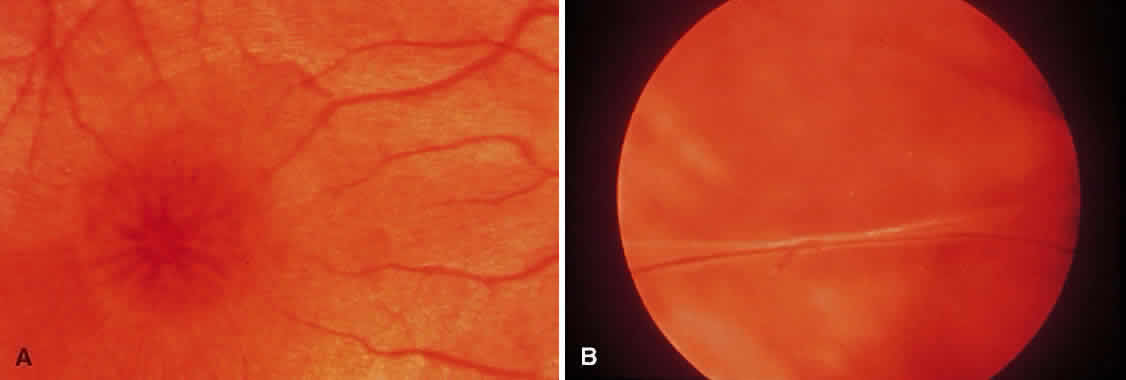
Fig. 8.
A.
Characteristic spokelike pattern of foveal change in X-linked juvenile retinoschisis.
B.
Peripheral retinoschisis with a vessel bordered by inner-wall holes on either side. (Courtesy of William Tasman, MD, Philadelphia, PA.)