|
|
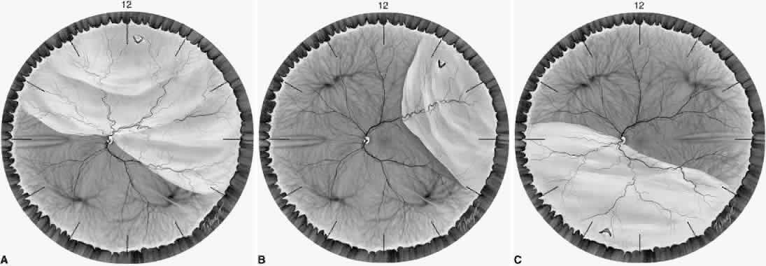
| Fig. 10. The configuration of a retinal detachment can help to localize the causative retinal break. A. A superior retinal detachment that crosses the 12-o'clock meridian. In total detachments or superior detachments that cross the midline, the primary hole is usually within 1 clock hour of the 12-o'clock meridian. If the detachment extends more inferiorly on either the nasal or temporal side, the causative break is usually on the same side of the 12-o'clock meridian. B. A superotemporal retinal detachment. The break lies near the superior edge of the detached retina. In superior nasal or temporal detachments, the hole lies within 1.5 clock hours of the highest border 98% of the time. C. An inferior retinal detachment. The break lies nasal to the optic nerve and the subretinal fluid extends higher nasally. In inferior detachments, the higher side indicates to which side of the disc an inferior hole lies 95% of the time. When an inferior detachment is bullous (not shown), the primary hole often lies above the horizontal meridian.93 |