Chapter 24
Retinitis Pigmentosa and Allied Retinal Diseases
ELIOT L. BERSON
Main Menu   Table Of Contents

Search

RETINITIS PIGMENTOSA
CHOROIDEREMIA
GYRATE ATROPHY OF THE CHOROID AND RETINA
OTHER RETINAL DEGENERATIONS
STATIONARY FORMS OF NIGHT BLINDNESS
GUIDELINES FOR CLINICAL EVALUATION OF PATIENTS WITH RETINITIS PIGMENTOSA AND ALLIED DISEASES
NIGHT VISION DEVICES
OTHER OPTICAL AIDS
GENETIC COUNSELING
PSYCHOLOGIC AND VOCATIONAL COUNSELING
SUNGLASSES
FUTURE DIRECTIONS
ACKNOWLEDGMENTS
REFERENCES

Considerable progress has been made in the understanding of retinitis pigmentosa and allied hereditary retinal diseases (Table 1). Medical treatments are now available for some of these disorders. This chapter provides a framework for diagnosing and managing patients with these conditions.


TABLE 1. Retinitis Pigmentosa and Some Allied Retinal Diseases

  1. Autosomal dominant forms of retinitis pigmentosa
  2. Autosomal recessive forms of retinitis pigmentosa
  3. Sex-linked (X-chromosome-linked) forms of retinitis pigmentosa
  4. Isolate (simplex) forms of retinitis pigmentosa
  5. Progressive cone-rod degeneration
  6. Atypical retinitis pigmentosa including sector, unilateral, and paravenous forms
  7. Some syndromes or diseases of which retinitis pigmentosa is a part:
    1. Bassen-Kornzweig syndrome (abetalipoproteinemia)
    2. Refsum disease
    3. Friedreich-like ataxia with retinitis pigmentosa
    4. Usher syndrome: types I, II, and III
    5. Laurence-Moon-Bardet-Biedl syndrome
    6. Kearns-Sayre syndrome
    7. Hereditary cerebroretinal degenerations including Batten disease
    8. Olivopontocerebellar atrophy
    9. Alström disease
    10. Cockayne syndrome

  8. Congenital amaurosis of Leber
  9. Choroideremia
  10. Gyrate atrophy of the choroid and retina
  11. Retinitis punctata albescens
  12. Stationary forms of night blindness


Back to Top
RETINITIS PIGMENTOSA
Retinitis pigmentosa is the name applied to a group of hereditary retinal diseases with a prevalence of about 1 in 4000 worldwide.1–6 An estimated 50,000 to 100,000 people are affected in the United States. Most cases are inherited by an autosomal dominant, autosomal recessive, or X-linked mode of transmission. This condition can also be transmitted by a digenic7 or mitochondrial mode.

Most patients have no associated systemic disease and, therefore, are considered cases of nonsyndromic retinitis pigmentosa. A few cases have associated nonocular disease (i.e., syndromic retinitis pigmentosa). The most common syndromic form is Usher syndrome. About 15% of all cases of retinitis pigmentosa have an associated partial hearing loss (Usher syndrome, type II) and another 2% to 6% have associated profound congenital deafness and vestibular ataxia (Usher syndrome, type I).

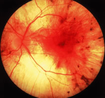
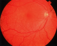
Patients with retinitis pigmentosa characteristically report difficulty with adaptation and night blindness in adolescence. They lose midperipheral and then far peripheral visual field in adulthood. As their condition progresses, they develop tunnel vision and a tendency to blue blindness. Most have reductions in central vision between ages 50 and 80. Signs on ophthalmoscopic examination include attenuated retinal arterioles, waxy pallor of the optic discs, and intraretinal bone spicule pigment around the midperiphery8–20 (Fig. 1). The bone spicule pigment is typically distributed 20 degrees to 40 degrees eccentric to the foveola in the zone where rods are normally in maximum concentration. Most show vitreous cells and a posterior vitreous detachment.14 Most patients develop posterior subcapsular cataracts in adulthood.21 An estimated 10% develop cystoid macular edema.22,23 Refractive errors such as astigmatism and myopia are common.24,25 Histologic studies of autopsy eyes have shown that loss of vision is due to degeneration of both rod and cone photoreceptor cells.26–34

Fig. 1. Fundus photograph of moderately advanced retinitis pigmentosa.

ELECTRORETINOGRAMS

In 1945 Karpe35 reported that patients with advanced retinitis pigmentosa have very small or nondetectable (less than 10 μV) electroretinograms (ERGs). Subsequently it has been shown that patients with early retinitis pigmentosa can have reduced but easily detectable ERG responses.36–39 Responses are not only reduced in size but are also delayed with respect to the time interval between stimulus flash onset and corresponding response peaks (i.e., b-wave implicit time).38–42 These ERG changes can be detected in some instances many years before diagnostic abnormalities are visible on fundus examination.41

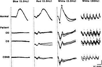
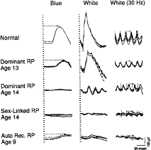
Figure 2 illustrates representative full-field ERGs from a normal subject and four children with early retinitis pigmentosa. Rod responses to single 0.5-Hz flashes of blue light (left column) are reduced and, when detectable, are delayed in b-wave implicit times. Cone responses to 30-Hz white flickering light (right column) are normal or reduced in amplitude and normal or delayed in b-wave implicit times. In most cases, cone b-wave implicit times are so delayed that a phase shift occurs between the stimulus flash onset (designated by the vertical lines) and the corresponding response peaks; each stimulus flash elicits the next-plus-one response in contrast to the normal. In mixed cone-rod responses to single 0.5-Hz flashes of white light (middle column), the cornea-negative a-wave generated by the photoreceptors is reduced in amplitude in all genetic types; this reflects the early involvement of the photoreceptors in this group of diseases.41

Fig. 2. Electroretinogram (ERG) responses for a normal subject and four patients with retinitis pigmentosa (ages 13, 14, 14, and 9). Responses were obtained after 45 minutes of dark adaptation to single flashes of blue light (left column) and white light (middle column). Responses (right column) were obtained to 30 cps (or 30 Hz) white flickering light. Calibration symbol (lower right corner) signifies 50 msec horizontally and 100 μV vertically. Rod b-wave implicit times in column 1 and cone implicit times in column 3 are designated with arrows. (From Berson EL. Retinitis pigmentosa and allied diseases: electrophysiologic findings. Trans Am Acad Ophthalmol Otolaryngol 1976;81: 659.)

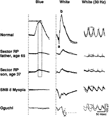
The reduced and delayed ERGs recorded from patients with widespread progressive forms of retinitis pigmentosa contrast with the reduced ERGs with normal b-wave implicit times observed in patients with self-limited sector retinitis pigmentosa. Figure 3 shows full-field ERGs from a father and son with dominantly inherited sector retinitis pigmentosa separated in age by almost 30 years; they have comparably reduced ERGs with normal b-wave implicit times. Patients with sector retinitis pigmentosa usually have patches of intraretinal pigment that are confined to one or two quadrants of the periphery of each fundus, with loss of peripheral rods and cones and consequent reductions in both rod and cone full-field ERG amplitudes. ERGs recorded from patients with sector retinitis pigmentosa are comparable to those recorded from patients who have large chorioretinal scars in the periphery.41

Fig. 3. Electroretinogram (ERG) responses of a normal subject and four patients with sector or stationary retinal disease. Horizontal arrows (column 1) designate range of normal rod b-wave implicit times, and vertical bar defining this range [mean ± standard deviation (SD)] has been extended through responses of patients with sector retinitis pigmentosa. Responses (middle column) from patient with Oguchi disease are interrupted by reflex blinking so the latter part cannot be illustrated. Cone implicit times in column 3 are designated with arrows. (From Berson EL. Retinitis pigmentosa and allied diseases: electrophysiologic findings. Trans Am Acad Ophthalmol Otolaryngol 1976;81:659.)

Full-field ERG testing can be used not only to identify which patients have widespread progressive forms of retinitis pigmentosa but also to determine which relatives are normal. Relatives of patients with retinitis pigmentosa, age 6 or older, with normal rod and cone ERG amplitudes and implicit times have not been observed to develop widespread progressive forms of retinitis pigmentosa at a later time.41–43

CARRIERS OF X-LINKED RETINITIS PIGMENTOSA

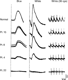
Females with the carrier state of X-linked retinitis pigmentosa can present with an area of bone spicule pigmentation in the periphery or an abnormal tapetal reflex in the macula. Less than 50% of carriers of child-bearing age show diagnostic findings on ophthalmoscopic examination, whereas more than 90% have abnormal ERGs.44 Abnormal ERGs in obligate carriers are either reduced in amplitude or delayed in cone b-wave implicit time, or both, in one or both eyes44,45 (Fig. 4). Daughters of obligate carriers can have either normal ERGs or abnormal ERGs similar to those recorded from obligate carriers.44

Fig. 4. Electroretinogram (ERG) responses from normal subject and four obligate carriers of sex-linked retinitis pigmentosa. Patient (Pt) 15, age 39; Pt 8, age 41; Pt 4, age 51; Pt 22, age 70. Stimulus onset is designated by vertical hatched lines for columns 1 and 2 and vertical shock artifacts for column 3. Cornea-positivity is upward deflection. Arrows in column 3 designate cone b-wave implicit times. (From Berson EL, Rosen JB, Simonoff EA. Electroretinographic testing as an aid in detection of carriers of X-chromosome-linked retinitis pigmentosa. Am J Ophthalmol 1979;87:460.)

Female carriers of X-linked retinitis pigmentosa can have a slowly progressive retinal degeneration, although the natural course remains to be defined. Some female carriers have had considerable loss of visual field and substantial reductions in ERG amplitudes by age 70.44

The abnormal ERGs of female carriers of X-linked retinitis pigmentosa contrast with the normal full-field ERG amplitudes and normal fundi observed in obligate female carriers of autosomal recessive disease.44 Carriers of X-linked retinitis pigmentosa have a 50% chance of having an affected son and a 50% chance of having a carrier daughter with each childbirth.

NATURAL COURSE

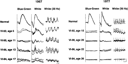
The ERG provides a quantitative measure of remaining retinal function in patients with retinitis pigmentosa. Figure 5 illustrates ERGs from affected patients followed over a 10-year period; responses become smaller as the condition progresses. Responses less than 10 μV are nondetectable with conventional recording techniques (i.e., without signal averaging). Patients with nondetectable conventional ERGs can still retain considerable visual function because most are not legally blind until ERG amplitudes decline to 0.05 μV or less.

Fig. 5. A. Electroretinograms recorded in 1967 for normal subject and four affected members from family with dominant retinitis pigmentosa with reduced penetrance. One to three responses to same stimulus are represented. Stimulus onset is designated by vertical hatched lines in columns 1 and 2 and vertical shock artifacts in column 3; cornea positivity is upward deflection; arrows in column 3 designate cone implicit times. Calibration symbol, lower right, designates 50 msec horizontally for columns 1 and 2 and 25 msec for column 3 and 50 μV vertically for column 1 and 100 μV vertically for columns 2 and 3. B. Electroretinograms recorded in 1977 for normal subject and four affected members from same family as in A for comparison with ERGs recorded in 1967. Calibration symbol (lower right corner) designates 60 msec horizontally and 100 μV vertically for all tracings. (From Berson EL, Simonoff EA. Dominant retinitis pigmentosa with reduced penetrance: further studies of the electroretinogram. Arch Ophthalmol 1979;97:1286.)

Signal averaging with a bipolar artifact reject buffer and narrow-bandpass filtering have extended the range of detectability of responses from affected patients 100-fold or more so that responses can be quantitated throughout almost the entire course of the disease. ERGs can now be recorded that are as low as 1 μV to 0.5-Hz flashes of white light (normal 350 μV) with signal averaging alone and, with bandpass filtering and signal averaging, as low as 0.05 μV to 30-Hz white flicker (normal equal to or greater than 50 μV). Full-field ERG function could be detected with at least one test criterion in 90% of an outpatient population with retinitis pigmentosa and a visual field diameter greater than 8 degrees. In an adult male with X-linked retinitis pigmentosa (whose ERGs could not be quantitated without signal averaging) computer-averaged 
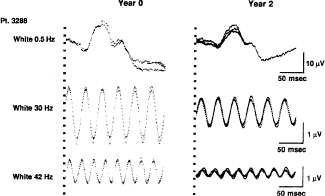
ERGs show changes in ERG function over a 2-year period46,47 (Fig. 6).

Fig. 6. Computer-averaged full-field electroretinograms from a 26-year-old man with X-linked retinitis pigmentosa obtained in response to 10-μsec flashes of white light at 16,000 foot Lamberts presented at 0.5 Hz (n = 64), 30 Hz (n = 256), and 42 Hz (n = 256) at base line (Year 0) and at 2-year follow-up (Year 2). Three consecutive response averages are superimposed in each case. Vertical broken lines denote flash onset for 0.5-Hz condition and onset of one of train of flashes for 30-Hz and 42-Hz conditions. (Berson EL, Sandberg MA, Rosner B et al. Natural course of retinitis pigmentosa over a three-year interval. Am J Ophthalmol 1985;99:240.)

Among 94 patients, ages 6 to 49 years, with the common forms of retinitis pigmentosa, full-field ERGs declined significantly over a 3-year interval in 66 of 86 patients (77%), with detectable responses at baseline. Patients lost on average 16% of remaining full-field ERG amplitude per year to single flashes of white light (95% confidence limits, 13.1% to 18.6%) and 18.5% of remaining amplitude per year to 30-Hz white flicker (95% confidence limits, 15.1% to 21.5%). Patients lost on average 5.2% of remaining foveal cone ERG amplitude per year, indicating that loss of retinal function was primarily extrafoveal in these patients. They lost on average 4.6% of remaining visual field area per year in the Goldmann perimeter with a V-4e white test light, whereas visual acuity and dark-adaptation thresholds remained relatively stable. Bone spicule pigment increased in 54% for whom comparisons could be make over a 3-year interval, suggesting that observation of increased pigmentation as a means of following this condition is not as sensitive as full-field ERG testing.46

Caution must be exercised in applying these population ERG results to predict longitudinal patterns in individual patients, because standard deviations derived from standard errors have revealed considerable variation around the mean for these patients.46 However, these results, describing the natural course on a quantitative basis, provide a frame of reference for planning interventions in similar populations to stabilize or slow the course of retinitis pigmentosa, particularly if monitored with full-field ERG testing.

MOLECULAR GENETIC STUDIES

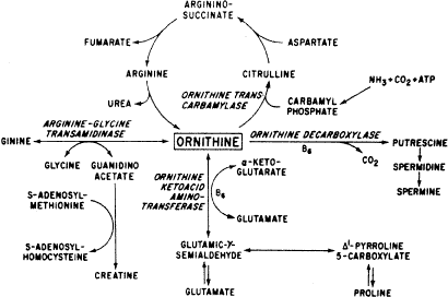
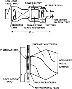
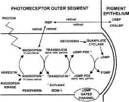
Substantial genetic heterogeneity exists among patients with retinitis pigmentosa with abnormalities in over 25 genes identified as of 2001; an updated list is maintained on the RetNet site (http://www.sph.uth.tmc.edu/Retnet/sum-dis.htm). Genes so far identified as causes of nonsyndromic retinitis pigmentosa, as well as Usher syndrome, are given in Table 2.48–87 Molecular genetic analyses have revealed defects in genes encoding proteins involved in the phototransduction cascade (Fig. 7), the retinoid cycle (Fig. 8), and structural components of photoreceptors, as well as in photoreceptor and retinal pigment epithelial proteins with unknown functions.


TABLE 2. Estimated Proportions of Cases of Retinitis Pigmentosa (RP) Caused by Identified Genes


Inheritance Pattern/GenePercent of all RP (including Usher Syndrome) 
Autosomal Dominant (ADRP) (-40% of all cases of retinitis pigmentosa)
1. rhodopsin (3q)10% (90/363 ADRP cases)48–51
2. peripherin/RDS (6p)3–4% (based on a survey of 227)52
3. RP1 (8q)2% (based on a survey of 187)53
4. NRL (14q)1%54
5. FSCN2 (17q)1.3% (4/120 ADRP cases in Japan)55
6. PRPC8 (17p)-1% (7/332 ADRP cases in UK)56
7. PRPF31 (19q)-3.2% (4/50 ADRP cases in UK)57
Autosomal Recessive (ARRP) (-50% of all cases of retinitis pigmentosa, including isolates)
1. rhodopsin (3q)0.4% (1/126 ARRP cases)58
8. rod PDEβ (4p)2% (4/92 ARRP cases)59–61
9. rod PDEα (5q)1% (3/173 ARRP cases)62,63
10. rod channel α (4p)1% (3/173 ARRP cases)64
11. myosin VIIa (11q)-2% (Usher IB)65
12. RPE65 (1p)0.7% (2/147 ARRP cases)66–68
13. CRALBP (15q)0.5% (3/324 ARRP cases plus isolates)69
14. TULP1 (6q)0.3% (1/162 ARRP cases)70
15. USH2A (1q)>10%71
16. ABCA4 (i.e., ABCR) (1p)<1% (a few cases have been reported)72,73
17. arrestin (2q)0.7% (3/120 ARRP cases in Japan; 0/85 in the U.S.)74,75
18. RGR (10q)<1%76
19. CRB1 (1q)? (10 families with ARRP)77
20.NR2E3 (15q)? 1%78
21. MERTCK (2q)? 1%79
22. LRAT (4q)~1% (3/267)80
23. harmonin (11p)? 1% (Usher IC)81
24. CDH23 (10q)? 1 % (Usher ID)92,83
25. USH3 (3q)? 1 % (Usher III)84
X-linked (XLRP) (~10% of all cases of retinitis pigmentosa)
26. RPGR (Xp21.1)~ 8%85,86
27. RP2 (Xp11.3)1% 
Digenic(only a few families described to date)
28. ROM1 (11q) peripherin/RDS (6p)(these two genes account for <1% of all cases)7,52
Mitochondrial (only one family, with Usher type III, described to date)
29. MTTS2<1%87

Note: Excluding syndromic RP except for Usher syndrome; genes currently identified account for 50% to 60% of cases of RP. Percentages are approximate and are based on the breakdown of RP cases according to genetic type reported by Fishman (Arch Ophthalmol 1978;96:822.), Macrae (Birth Defects: Original Article Series 1982;18:175), and Bunker et al. (Am J Ophthalmol 1984;97:357) and on the assumptions that all isolate cases are autosomal recessive and that Usher syndrome type I accounts for about 6% of all cases of RP. The values are calculated based on frequency of cases in a published series multiplied by the proportion of RP with that inheritance pattern (e.g., dominant rhodopsin mutations were found in 90/363 cases of ADRP; ADRP accounts for about 40% of all cases of RP; accordingly, the percentage of cases caused by dominant rhodopsin mutations is 90/363 = 24.8% × 0.4 = 9.92 ≈ 10%). A current listing of genes causing retinitis pigmentosa and allied hereditary retinal diseases can be accessed on the World Wide Web at: http://www.sph.uth.tmc.edu/Retnet/sum-dis.htm; Table prepared 9/15/01.


Fig. 7. Schematic representation of some proteins normally found in the rod photoreceptor outer segment, interphotoreceptor matrix, and retinal pigment epithelium. Genes encoding these proteins are considered candidate genes, because mutations in them could result in compromise of the phototransduction cascade or the mechanism by which vitamin A is transported between the photoreceptors and the pigment epithelium with consequent photoreceptor cell degeneration. IRBP, interphotoreceptor retinoid binding protein; CRBP, cellular retinol binding protein; CRALBP, cellular retinal binding protein; PDE, phosphodiesterase; cGMP, cyclic guanosine monophosphate. (Modified from Berson EL. Retinitis pigmentosa. The Friedenwald Lecture. Invest Ophthalmol Vis Sci 1993;34:1659.)

Fig. 8. Schematic diagram of the mammalian retinoid cycle in vision and the structure of the principal retinoids. Starting at the one o'clock position in the figure, the retinal pigment epithelium receives all-trans retinol (vitamin A) from the bloodstream or from photoreceptor cells when light bleaches cone or rod photopigments. In the retinal pigment epithelium, all-trans retinol is complexed with cellular retinol-binding protein (CRBP). Retinol is esterified to fatty acids, such as palmitic or stearic acid, by the enzyme lecithin:retinol acyltransferase (LRAT). The retinyl esters are the substrate for isomerohydrolase (isomerase), which produces 11-cis retinol. The diagram shows 11-cis retinol being oxidized to 11-cis retinal by the enzyme 11-cis retinol dehydrogenase. However, at this point there is an alternative pathway not shown in the diagram for simplicity, whereby 11-cis retinol is esterified and stored; the 11-cis retinyl esters are ultimately hydrolyzed to recreate 11-cis retinol, which is the substrate for 11-cis retinol dehydrogenase. In the retinal pigment epithelium, 11-cis retinal is complexed with cellular retinaldehyde-binding protein (CRALBP), whereas in the subretinal space it is complexed with interphotoreceptor retinoid binding protein (IRBP). On entering the cone and rod photoreceptors, 11-cis retinal functions as the chromophore in the cone opsins or rod opsin (rhodopsin). After exposure to light, the chromophore is converted to all-trans retinal. All-trans retinal dissociates from the opsins and, before leaving the photoreceptor cells, is reduced to all-trans retinol by all-trans retinol oxido-reductase. (Courtesy of Drs H Yamamoto, A Simon, U Eriksson, E Harris, EL Berson, and TP Dryja.)

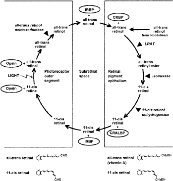
Of the more than 25 genes so far found to be causes of retinitis pigmentosa, some are known to play a role in the phototransduction cascade in rod photoreceptors (e.g., rhodopsin, the α- and β-subunits of rod cyclic guanosine monophosphate-phosphodiesterase (cGMP-PDE), the α-subunit of the rod cGMP gated channel, and arrestin). Other genes are thought to encode structural proteins in the outer segments (peripherin/RDS and ROM1). Others are involved in the recycling of vitamin A (CRALBP, RPE65, LRAT, and ABCA4). Some genes encode transcription factors (NRL and NR2E3) or proteins involved in the escort of opsin from the inner to the outer segment (RPGR and TULP1). The more than 25 genes (see Table 2) account for about 50% to 60% of cases of retinitis pigmentosa in the United States.

It should be noted that defects in the same gene can result in different phenotypes; for example, most rhodopsin mutations lead to retinitis pigmentosa, but some cause stationary night blindness. Defects in the ABCA4 gene can result in juvenile macular degeneration or a generalized cone-rod degeneration. Some mutations in the USH2A gene lead to retinitis pigmentosa with partial hearing loss, whereas another mutation in the same gene results in only retinitis pigmentosa. Variable clinical expression can exist among patients with the same gene defect, suggesting that factors other than the gene defect itself (e.g., diet, environment, modifier genes) affect the course of the disease with possible implications for therapy.

TREATMENT TRIALS

Many treatments have been attempted for the common forms of retinitis pigmentosa including various vitamins and minerals, vasodilators, tissue therapy with placental extract, cortisone, cervical sympathectomy, injections of a hydrolysate of yeast ribonucleic acid (RNA), ultrasound, transfer factor, dimethyl sulfoxide (DMSO), ozone, muscle transplants, and subretinal injections of fetal retinal cells.88–95 None of these has been shown to have proven therapeutic benefit. A study of patients evaluated before and after receiving electric stimulation, autotransfused ozonated blood, and ocular surgery in Cuba showed that this intervention provided no benefit and raised the possibility that this intervention was aggravating the course of the disease.95 None of these attempts at treatment was conducted with a randomized, controlled, double-masked protocol, which is necessary to avoid possible patient or examiner biases. Most of these studies were performed without electroretinographic data as an end point for evaluating efficacy, so that the amount of remaining retinal function could not be quantitated in an objective manner.

Claims of success with one or another treatment for patients with retinitis pigmentosa that are based solely on subjective reporting of improved visual function are to be interpreted with caution. Spontaneous fluctuations in visual acuity and visual field are well known in this condition. Given the slow course of retinitis pigmentosa without treatment, it will usually require several years to assess whether or not any proposed treatment has an effect on stabilizing or slowing the course of the disease. The problem of assessing treatments may be further complicated by the genetic heterogeneity of this condition and the stage of disease at which treatment is initiated.

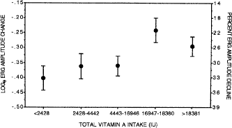
The common forms of retinitis pigmentosa have now yielded to treatment. In a randomized, controlled, double-masked trial among 601 patients ages 18 to 49, the course of retinal degeneration as monitored by the ERG was slower on average among patients taking a daily supplement of vitamin A palmitate, 15,000 IU/day, than among those not on this dose. Furthermore, the course appeared to be faster on average among patients taking a daily supplement of 400 IU/day of vitamin E than those not on this dose.96 Among a subset of 125 patients who could perform visual field testing with great precision, those on vitamin A showed a slower rate of decline of visual field than those in the control group.97 The optimal total intake of vitamin A palmitate in this study population appeared to be approximately 18,000 IU/day (i.e., 3000 IU of vitamin A in a regular diet and 15,000 IU/day of vitamin A as a supplement); higher intake did not provide greater benefit (Fig. 9). These findings have led to the recommendation that most adult patients with the common forms of retinitis pigmentosa should take a daily supplement of 15,000 IU of vitamin A in the palmitate form under the supervision of their ophthalmologist and avoid high-dose supplementation with vitamin E. For the average patient in the trial, it has been estimated that treatment with vitamin A could add 7 additional years of useful vision96; that is, the average patient with 1.3 μV of cone function who starts vitamin A at age 32 would retain useful vision until age 70 rather than age 63 if left untreated. For some patients with larger pretreatment ERGs, vitamin A supplementation could make the difference whereby these patients retain some vision for their entire lives.

Fig. 9. Mean ± standard error (SE) decline from baseline in 30-Hz electroretinogram (ERG) amplitude by total vitamin A intake (diet plus capsules) irrespective of randomization assignment for all patients in the higher amplitude cohort. The mean decline was calculated as the mean of screening and baseline minus the mean of all follow-up visits by quintile of total vitamin A intake averaged over all visits. Sample sizes were 69, 72, 74, 65, and 74 for the lowest to highest quintiles of total vitamin A intake. Vertical bars indicate SEs. (From Berson EL, Rosner B, Sandberg MA et al. A randomized trial of vitamin A and vitamin E supplementation for retinitis pigmentosa. Arch Ophthalmol 1993;111:761.)

Vitamin A may provide its benefit through the rescue of remaining cones, thereby, explaining how one supplement can help patients with many different rod-specific gene defects. The daily intake of a vitamin A supplement may provide protection against possible transient decreases in serum retinol concentrations that may adversely affect photoreceptor function. With respect to vitamin E, it has been hypothesized that a daily dose of 400 IU may adversely affect the course of retinitis pigmentosa at least in part by inhibiting the absorption or transport of vitamin A, because it was observed that the patients receiving vitamin E had slight but significant decreases in serum retinol concentration compared with those not receiving vitamin E.96

Because doses greater than 25,000 IU/day of preformed vitamin A can be associated with liver disease when taken over the long term, patients should have a fasting serum vitamin A and liver function profile before starting this treatment and annually thereafter while on this treatment. Beta-carotene is not predictably converted to preformed vitamin A and, therefore, should not be considered as a substitute for treatment with vitamin A palmitate. Because of the risk of birth defects associated with high-dose vitamin A supplementation, women who are pregnant or planning to become pregnant should not take this supplement. Because patients younger than age 18 were not included in this study, no formal recommendation can be made for patients with retinitis pigmentosa younger than this age.96,97 No toxic side effects have been observed in adults with retinitis pigmentosa in good general health on this dose of vitamin A who have been followed for up to 12 years.98

RETINITIS PIGMENTOSA ASSOCIATED WITH HEREDITARY ABETALIPOPROTEINEMIA

In 1950 Bassen and Kornzweig described an 18-year-old girl, born of first cousins, who had a malabsorption syndrome, a generalized retinal degeneration, a diffuse neuromuscular disease similar to Friedreich's ataxia, and a peculiar crenation of the red blood cells, now called acanthocytosis.99–101 In 1958 low serum cholesterol (less than 50 mg per dl) was observed.102 Soon thereafter, an absence of low-density plasma lipoproteins or so-called β-lipoproteins was found, and the term abetalipoproteinemia was assigned to this recessively inherited disorder.103–105 Other classes of lipoproteins have also been found to be abnormal.106

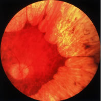
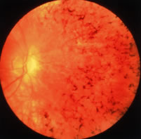
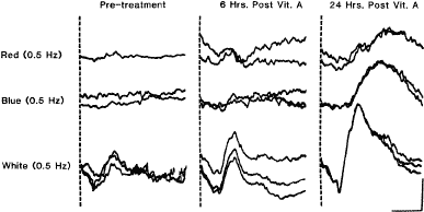
Patients with hereditary abetalipoproteinemia can assimilate fat into the intestinal mucosa, but a defect exists in its removal from this site because of the lack of chylomicra. Intestinal biopsies have revealed normal-sized villi filled with lipid droplets that are essentially triglycerides. Mutations in the gene encoding a microsomal triglyceride transfer protein have been found in patients with this condition.107 It appears that the liver and then the retina become depleted of vitamin A. Abnormal ERGs have been reported in a 15-month-old child108 and a 6-year-old patient109 in whom the fundi were still normal. The original case described by Bassen and Kornzweig showed multiple white dots in the early stages, but by age 31, the patient developed multiple areas of pigment epithelial cell atrophy. In other cases, the typical intraretinal pigment associated with retinitis pigmentosa has been noted in the retinal periphery.

Patients with this condition are treated with a low-fat diet and supplements of the fat-soluble vitamins A, E, and K. Vitamin A supplementation has been shown to restore elevated dark-adaptation thresholds and reduced ERG responses to normal in patients with the early stages110,111 (Fig. 10). More advanced cases have not responded, but in one such case in which the retina was examined after the death of the patient, widespread loss of photoreceptor cells was observed.112 Vitamin A therapy may not maintain retinal function over the long term, because patients have been reported in whom vitamin A levels have been restored to normal and yet the retinal degeneration has appeared to progress.113,114 Because these patients have low serum vitamin E levels, supplementation with vitamin E in addition to vitamin A has been advocated with reported stabilization of retinal function.115-19

Fig. 10. Full-field electroretinograms (ERGs) to red (top) and blue (middle) light, equal for rod vision, and brighter white stimulus (bottom) from patient with hereditary abetalipoproteinemia (dark-adapted). Responses in left column were obtained before vitamin A therapy, those in middle column at 6 hours, and those on right at 24 hours after vitamin A therapy. Two to three responses to same stimulus are superimposed. Light stimulus begins with each trace. Calibration (lower right) signifies 0.06 mV vertically and 60 msec horizontally. (From Gouras P, Carr RE, Gunkel RD. Retinitis pigmentosa in abetalipoproteinemia: Effects of vitamin A. Invest Ophthalmol Vis Sci 1971;10:790.)

RETINITIS PIGMENTOSA ASSOCIATED WITH REFSUM DISEASE

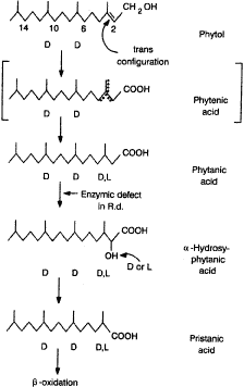
Refsum disease is a recessively inherited condition in which the patient accumulates exogenous phytanic acid.120,121 Findings include a peripheral neuropathy, ataxia, an increase in cerebrospinal fluid protein with a normal cell count, and retinitis pigmentosa. All have elevated serum phytanic acid. Some cases have anosmia, neurogenic impairment of hearing, electrocardiogram (EKG) abnormalities, and skin changes resembling ichthyosis. Patients show a granular fundus with areas of depigmentation around the periphery and have a subnormal ERG in the early stages or show more typical retinitis pigmentosa with a nondetectable ERG in more advances stages.122

A defect exists in the conversion of phytanic acid to alpha-hydroxy phytanic acid, specifically in the introduction of a hydroxyl group on the alpha carbon of phytanic acid123 (Fig. 11). The pathogenesis appears to involve accumulation of phytanic acid in a variety of tissues including the retinal pigment epithelium (RPE).124

Fig. 11. Phytanic acid, its immediate precursors and metabolites, and site of enzyme defect in Refsum disease (Rd). (From Eldjarn L, Stokke O, Try K. In: Vinken PJ, Bruyn GW (eds). Biochemical aspects of Refsum's disease and principles for the dietary treatment. Handbook of Clinical Neurology. Amsterdam, North-Holland, 1976:519–541.)

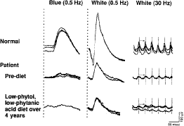
Treatment consists of restricting not only animal fats and milk products (i.e., foods that contain phytanic acid) but also green leafy vegetables containing phytol.125 Success of treatment depends on the patient maintaining his or her body weight; if body weight becomes reduced, phytanic acid is released from tissue stores, resulting in an increase of phytanic acid in serum and exacerbation of symptoms. Refsum126 has reported two patients whose serum phytanic acid levels were lowered to normal and who showed improvement in motor nerve conduction velocity, some relief of ataxia, and return of the cerebrospinal fluid protein to normal. Moreover, the retinitis pigmentosa and hearing impairment did not progress; one of these patients was followed for 10 years and the other for many years. One young adult with a mild form of this disorder has been followed on a low-phytol, low-phytanic acid diet for 4 years; full-field ERGs, reduced about 75% below normal before commencement of this diet, have remained about the same over this period42 (Fig. 12). Long-term effects of this diet on retinal function continue to be studied.

Fig. 12. Full-field ERGs from normal subject and patient with mild form of Refsum disease before and 4 years after treatment with low-phytol, low-phytanic acid diet. Pretreatment responses were recorded at age 31. (From Berson EL. Electroretinographic findings in retinitis pigmentosa. Jpn J Ophthalmol 1987;31:327.)

FREIDREICH-LIKE ATAXIA WITH RETINITIS PIGMENTOSA

Another rare recessively inherited form of ataxia associated with retinitis pigmentosa has recently been described. These patients present in adulthood with Friedreich-like ataxia, dysarthria, hyporeflexia, and decreased proprioceptive and vibratory sensation, as well as markedly decreased serum vitamin E levels. In later stages, patients can develop the fundus changes of retinitis pigmentosa and abnormal ERGs. Molecular genetic analysis has revealed a mutation in the α-tocopherol-transfer protein (α-TTP) gene. Oral administration of vitamin E restored serum vitamin E levels to normal and appeared to halt or slow the progression of the neurologic abnormalities and retinitis pigmentosa in three patients followed for 1, 4, and 10 years, respectively.127,128

USHER SYNDROME

About 15% to 20% of affected individuals with retinitis pigmentosa have associated hearing loss, sometimes referred to as Usher syndrome in recognition of the British ophthalmologist, C.H. Usher, who emphasized that this condition was recessively inherited.129,130 Patients with Usher syndrome type I typically have night blindness in the first or second decade, profound congenital deafness (i.e., greater than 70 dB loss of all frequencies) with unintelligible speech, and vestibular ataxia, whereas those with Usher syndrome type II usually report night blindness in the second to fourth decade, have a partial early onset hearing loss with intelligible speech, and do not show ataxia.131 Patients with Usher syndrome type II have been shown to have abnormalities in the cilia of sperm and abnormalities in the connecting cilia in many remaining photoreceptors in autopsy eyes.132,133 Patients with Usher syndrome type III have onset of hearing loss in midadulthood that can progress to profound deafness; some have vestibular ataxia.84,134

Molecular genetic analyses have revealed gene loci for Usher syndrome type I (further subdivided as types A-F), for Usher syndrome type II (further subdivided as types A-C), and for Usher syndrome type III. Patients with Usher syndrome type IB have mutations in a long-tailed, unconventional myosin designated as myosin VIIA.135 It has been estimated that mutations in the MYO7A gene on chromosome 11q account for approximately 75% of cases of Usher syndrome type I.136 Patients with Usher syndrome type IIA have been found to have mutations in the USH2A gene on chromosome 1q.137 This gene normally encodes a protein called usherin that has several laminin epidermal growth factor-like and fibronectin type III motifs; it is possibly involved in cell adhesion to Bruch's membrane. It has been estimated that mutations in the USH2A gene account for about 85% of cases of Usher syndrome type II.137 Some patients have been identified with autosomal recessive retinitis pigmentosa without hearing loss who have a mutation in the USH2A gene, Cys759Phe.138 Mutations in the USH3 gene on chromosome 3q have been identified in a population from Finland and Italy.84 The frequency of Usher syndrome type III in the United States is unknown.

About 2% to 6% of congenitally deaf children will develop Usher syndrome type I. If a child presents with profound deafness and a balance disorder manifested by late onset of walking usually after 15 months of age, the possibility of Usher syndrome type I should be considered.139 The diagnosis of retinitis pigmentosa as part of Usher syndrome can be made in early life with ERG testing. No tests of retinal function are yet available to identify carriers of Usher syndrome.

Patients with Usher syndrome appear to have a slowly progressive retinal degeneration. Rates of progression on a year-to-year basis remain to be defined. Patients with Usher syndrome type II may reasonably be treated with vitamin A palmitate 15,000 IU per day because these patients were included among those with autosomal recessive retinitis pigmentosa in a clinical trial of vitamin A for retinitis pigmentosa.96

LAURENCE-MOON-BARDET-BIEDL SYNDROME

The Laurence-Moon-Bardet-Biedl syndrome includes retinitis pigmentosa, mental retardation, polydactylism, truncal obesity, and hypogonadism as the most frequent features.140–147 Some authors have subdivided this syndrome into two disorders: polydactylism is found primarily in patients with the Bardet-Biedl form, whereas neurologic findings are found primarily in patients with the Laurence-Moon form.148,149 However, some patients have been described with both polydactylism and neurologic abnormalities and, therefore, would seem to qualify for inclusion in both syndromes.150,151 Variability of clinical expression is well known, and some patients may have this condition without mental retardation or polydactylism.152–156 Genetic heterogeneity exists as the condition has been linked to six distinct loci. Renal disease can be a part of this syndrome,157 and, therefore, patients should have their blood pressure and urine checked periodically, with appropriate treatment instituted for their hypertension and renal disease if detected.

Retinal degeneration is the most common feature of the Laurence-Moon-Bardet-Biedl syndrome, occurring in about 90% of cases.157 The macula is often involved early in this condition. The fundus in early life may be granular without pigment formation, so that the diagnosis is only established after electroretinographic testing.158 A child born with polydactylism to normal parents should be a suspect for this syndrome. Once this syndrome is identified in one individual, parents would know that they have a 25% chance with each succeeding childbirth of having another child with this condition. In some, presumably rare, instances a second abnormal gene locus is required and the chance of having a second affected child may be less than 25%.159 No treatment is known for the retinal degeneration that occurs as part of this syndrome.

CONGENITAL AMAUROSIS OF LEBER

Congenital amaurosis of Leber is usually an autosomal recessive disorder associated with severe reduction in vision near birth and very reduced ERGs.160–165 Patients characteristically have hyperopia and nystagmus, and fundus examination reveals granularity, white flecks, or intraretinal bone spicule pigment or some combination. When recessively inherited, parents would know that they have a 25% chance with each succeeding childbirth of having a child with this condition. Retardation of mental development has been reported in some patients, possibly secondary to visual impairment.164 Leber congenital amaurosis can occur in patients with a de novo mutation not present in the affected patient's parents; in this dominant form, the chance of having an affected child is 50%, possibly lower if the affected patient is a mosaic.166 Leber congenital amaurosis involving the retina and pigment epithelium should not be confused with Leber optic atrophy, a maternally inherited condition involving the optic nerve associated in some families with an abnormality in mitochondrial deoxyribonucleic acid (DNA).167,168

In 8 of 15 families with one form of this condition studied, mutations in the guanylate cyclase gene on chromosome 17p have been identified, suggesting that cGMP production in photoreceptors is abolished in this form. Consequently, photoreceptor excitation would be expected to be impaired due to closure of cGMP-gated cation channels with hyperpolarization of the plasma membrane. It has been proposed that the cGMP concentration in photoreceptor cells cannot be restored to the dark level, leading to a situation equivalent to constant light exposure during photoreceptor development.169 A model for this disease exists in the rd/rd chicken, which is functionally blind at hatching with an extinguished ERG, absent guanylate cyclase activity, and a mutation in the guanylate cyclase gene.170,171 Mutations in another gene, designated as RPE65, which is involved in the vitamin A cycle via the isomerase in the RPE, account for about 16% of cases of Leber congenital amaurosis in the United States;172,173 mutations in this gene are also a cause of about 2% of cases of autosomal recessive retinitis pigmentosa.173 Other genes found to be abnormal in Leber congenital amaurosis are cone-rod homeobox-containing gene (CRX), aryl hydrocarbon receptor-interacting protein-like 1 (AIPL1), tubby-like protein 1 (TULP1), homologous to crumbs protein in Drosophila (CRB1), and retinitis pigmentosa GTPase regulator interacting protein 1 (RPGRIP1). TULP1 encodes a protein thought to play a role in the escort of proteins in the inner segment. RPGRIP1 encodes a protein normally found in the photoreceptor connecting cilium; defects in this protein may interfere with transport of proteins from the inner to the outer segment. The functions of the proteins encoded by the other abnormal genes are not as well understood.

OTHER FORMS OF RETINITIS PIGMENTOSA

Mutations in the CRB1 gene77 have been observed in an unusual recessively inherited form of retinitis pigmentosa called preserved para-arteriolar retinal pigment epithelium (PPRPE). Like patients with typical Leber congenital amaurosis, these patients are highly hypermetropic. The condition is named for the relative preservation of the RPE adjacent to and underlying retinal arterioles in earlier stages; in later stages the RPE previously spared is lost. The degeneration associated with this condition appears to be rapidly progressive.174

Retinitis punctata albescens can be associated with some signs and symptoms characteristic of retinitis pigmentosa.175 Patients usually present with profound adaptational problems and gradual loss of peripheral vision. Examination with a direct ophthalmoscope reveals multiple punctate white deposits in the macula and around the midperiphery at the level of the pigment epithelium for which this condition was named. Patients have retinal arteriolar attenuation and some develop round areas of atrophy of the RPE in the midperiphery, as well as intraretinal bone spicule pigment in the midperiphery. This raises the possibility that this condition is a variant of retinitis pigmentosa.176,177 This form of retinal degeneration may affect the eyes of a patient asymmetrically; often these patients receive a neurologic evaluation in search of an intracranial abnormality before it is realized that the visual loss is due to a widespread retinal degeneration. Full-field ERGs of such patients are invariably abnormal with reductions in amplitude and delays in implicit times. The condition is usually slowly progressive, although the course appears to vary from one individual to another. This condition has been thought to be inherited by an autosomal recessive mode, but a dominant mode of transmission can occur; therefore, ERG testing of relatives of affected patients is recommended to help establish the mode of transmission. A null mutation in the peripherin/RDS gene has been reported in one family with this condition with a dominant mode of transmission;178 other families with an autosomal recessive mode of transmission have shown mutations in the cellular retinaldehyde binding protein (CRALBP) gene.179

Another atypical form of retinitis pigmentosa is designated as progressive cone-rod degeneration.156,180 Patients with this form typically present with reduced visual acuity, photophobia, color deficiency, and night deficiency and show signs of macular degeneration, retinal arteriolar attenuation, and, in some cases, intraretinal bone spicule pigment in the peripheral fundus. Dark-adaptation testing shows elevated final dark-adaptation thresholds across the fundus, whereas full-field ERGs show a profound loss of cone function and some reduction in rod function across the retina. Patients with the earlier stages of cone-rod degeneration typically have rod ERGs that are more than 30 times larger than cone ERGs. The condition is usually inherited by an autosomal recessive mode but can also be inherited by an autosomal dominant mode. Mutations have been found in the retina-specific ATP-binding cassette transporter (ABCA4) gene,181 the same gene that has been found to be abnormal in patients with juvenile macular degeneration (Stargardt) disease. No treatment is know for patients with cone-rod degeneration.

Other unusual forms of retinitis pigmentosa include those designated as paravenous or unilateral. The paravenous form is usually characterized by slightly reduced full-field ERGs with normal implicit times and intraretinal pigment and atrophy of the pigment epithelium confined to the distribution of the retinal veins in each eye. Unilateral retinitis pigmentosa is characterized by fundus changes of retinitis pigmentosa in one eye with no evidence of retinal degeneration in the other eye. Full-field ERGs are substantially reduced in the affected eye and normal in the fellow eye. Patients with unilateral retinitis pigmentosa do not develop retinitis pigmentosa in the fellow eye at a later time. In my experience, patients with either paravenous or unilateral disease have presented with a negative family history for retinal degeneration, suggesting that these forms of retinitis pigmentosa are not inherited.

Back to Top
CHOROIDEREMIA
Choroideremia is inherited by an X-linked mode. Young affected males typically have normal visual acuities, minimally increased dark-adaptation thresholds, and full visual fields to large test lights at a time when granularity and depigmentation of the RPE can be seen around the peripheral fundus. Full-field ERGs are reduced in amplitude and delayed in b-wave implicit time. In more advanced stages dark-adaptation thresholds are further increased and the visual fields become constricted; at this time ERGs are nondetectable and extensive choroidal atrophy and clumped pigment are visible around the peripheral fundus (Fig. 13). Men usually retain little, if any, central vision beyond the age of 60.182–185

Fig. 13. Fundus photograph of moderately advanced choroideremia.

Female carriers of this disease186,187 may demonstrate fundus changes that include patchy depigmentation of the RPE and coarse pigment granularity or even pigment clumps in the periphery. However, carriers typically retain normal visual acuities and normal final dark-adapted rod thresholds.187,188 Generally carriers have been thought to have a nonprogressive course,189 but carriers with severe disease have been described.190,191 In contrast to the carrier state of X-linked retinitis pigmentosa, ERGs of carriers of choroideremia usually are normal,184,185,190 although reduced amplitudes have been reported in some cases.183,185,192

The gene responsible for this condition, RAB escort protein 1 (REP-I), was identified in 1990.193 Many mutations in this gene have been described subsequently.194,195 It has been proposed that loss of the REP protein results in defects in RAB protein prenylation; the lack of prenylation in turn is thought to affect protein trafficking inside the RPE or choroidal cells triggering the degenerative process in this condition.196–198

Interfamilial and intrafamilial variability of clinical expression is known to exist in this disease. The natural course of choroideremia on a year-to-year basis remains to be defined. Correlations, if any, between specific gene defects and clinical expression of the disease remain to be found. No treatment is known.

Back to Top
GYRATE ATROPHY OF THE CHOROID AND RETINA
Gyrate atrophy of the choroid and retina is inherited by an autosomal recessive mode of transmission.199,200 Patients usually present in early adolescence with night deficiency and loss of peripheral vision. Ocular findings include myopia, constricted visual fields, elevated dark-adaptation thresholds, reduced ERG responses (Fig. 14), and chorioretinal atrophy distributed circumferentially around the peripheral fundus (Fig. 15). Abnormalities in electroencephalograms, muscle and hair morphology, and mitochondrial structure in the liver have also been reported.201–204 Patients develop cataracts in young adulthood and often require surgery. If untreated, enlargement, coalescence, and posterior extension of areas of atrophy can be seen205 and patients usually become virtually blind between the ages of 40 to 55 because of extensive chorioretinal atrophy.206

Fig. 14. Electroretinographic responses of four males with choroideremia. Cone flicker responses to white 30-cps flicker remained detectable in the oldest male when rod responses to blue light were not detectable. Normal responses are shown for comparison. Time of stimulus onset is designated by vertical hatched lines in columns 1 and 2 and vertical shock artifacts in column 3. Arrows and vertical bar on rod responses to blue light show range of normal b-wave implicit times. Responses to single flashes of white light show reduced a- and b-wave amplitudes in all males. Arrows on white flicker responses show delayed implicit times in the affected males. (From Sieving PA, Niffenegger AB, Berson EL. Electroretinographic findings in selected pedigrees with choroideremia. Am J Ophthalmol 1986;101:361.)

Fig. 15. Fundus photograph of moderately advanced gyrate atrophy of the choroid and retina.

Patients have plasma ornithine concentrations elevated ten- to twenty-fold207,208 because of a deficiency of ornithine-ketoacid-amino-transferase (OAT) activity (Fig. 16).209–211 Affected individuals cannot convert ornithine to pyrroline-5-carboxylic acid (PCA); this deficiency can be detected in extracts of cultured skin fibroblasts. Patients have virtually no OAT activity, and carrier parents have a partial deficiency. Plasma lysine212 and glutamate and glutamine,213 as well as serum and urine creatine,214 have also been found to be reduced. More than 60 mutations have been discovered in the OAT gene on chromosome 10 in affected patients.215–218

Fig. 16. Pathways of ornithine metabolism. (From Weleber RG, Kennaway GN, Buist NR. Gyrate atrophy of the choroid and retina. Approaches to therapy. Int Ophthalmol 1981;4:23.)

Because arginine is a precursor of ornithine and because arginine, but not ornithine, is a constituent of food protein, it has been suggested that dietary restriction of protein and arginine will reduce plasma ornithine levels in these patients.219 OAT-deficient mice produced by gene targeting develop a retinal degeneration over several months that is amenable to treatment postweaning with an arginine-restricted diet.220,221

The hyperornithinemia associated with human disease has been lowered toward normal with a low-protein, low-arginine diet in all cases so far studied219,222–225 and with vitamin B6 (300 to 500 mg/day) in some cases.205,226,227 However, extreme protein restriction (10 to 15 g/day) under supervision in the hospital has been difficult to achieve at home and therefore many patients have followed modified (20 to 35 g/day) protein restriction resulting in a slight decrease in their plasma ornithine levels. Some investigators have reported improvements in visual acuity, visual fields, dark-adaptation thresholds, and/or ERGs in patients with gyrate atrophy after onset of either the diet or vitamin B6,223,225,228–230 whereas others have not documented any significant improvement in visual function despite substantial reductions in plasma ornithine concentrations.224,231 Improvement in muscle morphology following creatine supplementation has been observed in several patients.232

Kaiser-Kupfer and coworkers233 have reported results of severe arginine restriction of two pairs of siblings younger than 10 years who were followed for 5 to 7 years. The plasma ornithine levels were reduced to approximately the normal range (106 and 121 μmol/L) in one pair of siblings and were twice the upper limit of normal (251 and 313 μmol/L) in the other pair. These younger siblings had significantly less atrophy than their elder siblings when they reached or approached the same age at which their elders began the diet. Thus, there is evidence that a low-protein, low-arginine diet can slow progression of the chorioretinal degeneration, but only a small number of patients have so far been studied.

The goal of treatment is to maintain plasma ornithine levels as close to normal as possible. Because some patients may respond to supplementation with pyridoxine (vitamin B6), all patients are initially given a trial of this vitamin to determine to what extent, if any, it will lower plasma ornithine levels. Both pyridoxine-responsive and nonresponsive patients are then placed on a low-protein, low-arginine diet. Biochemical control has been classified as good to excellent (less than 200 μmol/L), fair (200 to 400 μmol/L), and poor (greater than 400 μmol/L). In the management of children, expertise is required to be certain that growth and development remain normal while lowering ornithine levels with a low-protein diet. An arginine-free essential amino acid mixture [for example, Cyclinex™—1 or Cyclinex™—2 (Ross Laboratories), depending on the patient's age] is used to provide sufficient nitrogen and meet essential amino acid requirements. In adults, a low-protein diet is also likely to result in amino acid deficiency. Thus, adults should also be placed on an arginine-free essential amino acid mixture. Lysine supplementation may be necessary, depending on plasma levels. As a precaution, all patients are placed on a multivitamin preparation with minerals. In addition to a regular ocular examination, all patients on this treatment regimen should have their serum amino acid and protein levels monitored periodically.234

Back to Top
OTHER RETINAL DEGENERATIONS
If a patient presents with ophthalmoplegia, the Kearns-Sayre syndrome should be considered. Other abnormalities include ptosis, a diffuse disturbance of the RPE, ERGs that are usually reduced in amplitude, and respiratory distress. Some patients develop heart block and may require a pacemaker.235–238 The diagnosis is confirmed by the observation of ragged red fibers on muscle biopsy. This syndrome has been associated with mitochondrial DNA mutations; treatment of one patient with coenzyme Q10 and succinate resulted in clinical improvement of respiratory distress.238 The value, if any, of this treatment for retinal malfunction in this condition remains to be established.

Patients who present with a widespread loss of photoreceptor function with abnormal ERGs and symptoms of cerebral deterioration may have a cerebroretinal degeneration, grouped under the overall heading of neuronal-ceroid lipofuscinosis, or Batten disease. These diseases, which are recessively inherited, are characterized by accumulation of lipopigments (lipofuscin and ceroid) in neurons and other cell types. Batten disease can be subdivided into an infantile form (psychomotor deterioration by age 2, ataxia, microcephaly, granular inclusions on conjunctival biopsy), a late-infantile form (seizures, rapid mental deterioration in early childhood, curvilinear inclusions on conjunctival biopsy), a juvenile form (mental deterioration beginning around age 6, bull's eye maculopathy, fingerprint inclusions on conjunctival biopsy), and an adult-onset form (seizures, slow dementia, abnormal inclusions best seen on muscle biopsy).

Molecular genetic studies have revealed that the juvenile onset form of Batten disease results from defects in the CLN3 gene on chromosome 16p; most patients have a 1.2 kb deletion in this gene on at least one chromosome.239 The gene encodes a novel protein of unknown function. The mechanism of apoptosis appears to be involved in the demise of both cerebral neurons and photoreceptors.240 No treatment exists for the retinal degeneration associated with this condition.

Patients with olivopontocerebellar atrophy (more recently designated as SCA7, a form of spinocerebellar ataxia) can present with a history of tremors, ataxia, and dysarthria and show findings of oculomotor impairment and retinal degeneration. This condition is inherited by a dominant mode with variable penetrance. Some patients have retinal arteriolar attenuation, a diffuse disturbance of the RPE, and very reduced ERGs. Some patients may show only neurologic findings and have normal fundi and normal retinal function, whereas others have had abnormal ERGs with no diagnostic findings on fundus examination and no history of neurologic disease.241,242 Patients with this condition have been found to have expansions of CAG trinucleotide repeats on DNA analysis.243

Other rare syndromes associated with retinal degeneration include Alström disease and Cockayne syndrome. Patients with Alström disease have retinitis pigmentosa with profound loss of vision in the first decade and very reduced ERGs. Systemic findings include diabetes mellitus, obesity, deafness, renal failure, baldness, and hypogenitalism.244,245 Patients with Cockayne syndrome show extensive loss of vision by the second decade and have retinitis pigmentosa, dwarfism, deafness, mental deterioration, and premature aging.246 No treatments are known for these conditions.

Back to Top
STATIONARY FORMS OF NIGHT BLINDNESS
Stationary night blindness can be inherited by a autosomal dominant, autosomal recessive, or X-linked mode. Some patients can have a normal fundus appearance. Two forms, Oguchi disease and fundus albipunctatus, have characteristic fundus abnormalities. In the case of Oguchi disease the fundus has a golden-brown appearance under dark-adapted conditions that changes to a normal color under light-adapted conditions (i.e., the so-called Mizuo phenomenon). Patients with fundus albipunctatus have white deposits at the level of the RPE around the midperipheral fundus (Fig. 17). In both conditions the retinal vessels are normal in caliber. It is the normality or near-normality of the cone system in the full-field ERGs that allows separation of most stationary forms of night blindness from practically all forms of night blindness associated with the early stages of retinitis pigmentosa.

Fig. 17. Fundus photograph of fundus albipunctatus.

The previous classification of these diseases was based on the family of origin (e.g., Nougaret type), by the original description of the ERG waveforms associated with the condition (e.g., Riggs type or Schubert-Bornschein), or by the ophthalmologist who first described the disease (e.g., Oguchi disease). These diseases are now being reclassified by molecular genetic analysis.247

Missense mutations in three different genes encoding members of the rod phototransduction cascade have been reported to cause dominantly inherited stationary night blindness. Specifically, the mutations are the Ala292Glu and Gly90Asp mutations in rhodopsin,248,249 the His258Asp mutation in the β-subunit of rod phosphodiesterase,250 and the Gly38Asp mutation in the α-subunit of rod transducin.251 The latter mutation has only been seen in the descendants of Jean Nougaret. Although the Ala292Gly rhodopsin mutation apparently causes a complete loss of rod function (i.e., Riggs type),248 the other mutations cause an incomplete loss of rod function.251,252

Some evidence exists from psychophysical and ERG testing that the rods are constitutively active in the dark in some forms of stationary night blindness. This has been suggested for patients with the rhodopsin, Gly90Asp mutation.249 The ERG abnormalities in patients with the Gly38Asp mutation in the α-subunit of rod transducin can be simulated by light adapting the normal retina, compatible with the idea that these patients have mutant rod transducin that is constitutively active in the dark.252 Further evidence that these patients have rod photoreceptors despite their night blindness is provided by fundus reflectometry studies that have shown normal levels of rhodopsin and normal rhodopsin kinetics.253

Oguchi disease is a recessively inherited form of stationary night blindness. Following 1 hour of dark adaptation, patients with Oguchi disease have no rod b-wave in response to dim blue light flashes, a cornea-negative response to white light, and a normal cone response to 30-Hz white flicker. Following complete dark adaptation after 12 hours, some patients have a normal rod b-wave amplitude and normal rod b-wave implicit time but only in response to one or two flashes of light.254 Apparently the test flash used to elicit the ERG, although relatively dim, can be intense enough to light adapt the rod system. Molecular genetic analyses have shown a null mutation, Asn309(1-bp del), in the gene encoding arrestin in some patients of Japanese descent.255 Several null mutations have also been described in the gene encoding rhodopsin kinase in some patients of Eastern European descent.256 Both arrestin and rhodopsin kinase participate in the deactivation of photoactivated rhodopsin. The lack of either arrestin or rhodopsin kinase would be expected to have the same physiologic effect, namely the prolongation of photoactivated rhodopsin with a consequent abnormality in rod sensitivity.

Fundus albipunctatus is another form of recessively inherited stationary night blindness.257,258 Patients with fundus albipunctatus have a delay in rod visual pigment and foveal cone pigment regeneration, as monitored with fundus reflectometry.257 Changes in visual pigment levels parallel the prolonged cone and rod limbs of the dark-adaptation curve. Full-field ERGs have been found to be normal with respect to both cone and rod amplitudes and b-wave implicit times after full dark adaptation.258 The findings in fundus albipunctatus suggest an abnormality in visual pigment regeneration secondary to some abnormality in the relationship between the photoreceptors and the pigment epithelium. The composition of the yellow-white lesions and their relationship to the abnormality in visual pigment regeneration are not known. Patients with this condition have been observed to have defects in the gene encoding 11-cis retinol dehydrogenase.259

A form of stationary night blindness for which the precise gene defect remains unknown is congenital nyctalopia with myopia. This condition (i.e., Schubert-Bornschein type) is inherited by either an X-linked or autosomal recessive mode.260–262 The extent of the myopia is characteristically -3.5 to -14.5 diopters (D). Patients cannot attain normal dark-adapted rod thresholds and have fundus findings of myopia. Their rod ERG b-wave responses to dim blue light are nondetectable, and the patients have a characteristic cornea-negative response to white light in the dark-adapted state and normal or nearly normal cone responses to 30-Hz white flicker. The preservation of the a-wave from the rod photoreceptors with loss of the b-wave from neurons proximal to the photoreceptors suggests some abnormality in intraretinal transmission of the response from the rod photoreceptors to proximal retinal cells. ERG amplitudes are reduced in congenital nyctalopia with myopia compared with those from normal emmetropes; this probably occurs because of the known reductions of ERG amplitude seen in patients with moderate axial myopia as the only finding.263

The ERG abnormality in congenital nyctalopia with myopia has been reported as an acquired defect in patients with cutaneous malignant melanoma, who have an acute onset of night blindness with selective reduction in the rod b-wave response264 (Fig. 18). Development of an antibody to the melanoma that also reacts to retinal cells with an interruption in rod signal transmission has been considered as a probable mechanism to account for the sudden onset of night blindness in patients with this paraneoplastic retinopathy.

Fig. 18. Full-field electroretinographic responses from a normal subject, from the right (OD) and left (OS) eyes of a patient with malignant melanoma, and from a patient with congenital stationary night blindness with myopia (CSNB). Stimulus onset is designated by the vertical hatched lines in columns 1, 2, and 3 and the vertical lines superimposed on the responses in column 4. Two or three consecutive responses are illustrated and cornea positivity is an upward deflection. The peak of the cornea-negative a-wave, generated by photoreceptors, and the peak of the cornea-positive b-wave, generated by activity of cells proximal to the photoreceptors, are designated in the response of the normal subject to single flashes of white light. (Lower right) Calibration symbol designates 50 msec horizontally, 200 μV vertically for the top recording in column 3, and 100 μV vertically for all other tracings. Both patients lack the cornea-positive b-wave from the rod system in columns 1, 2, and 3 in contrast to the normal while retaining normal cone amplitudes (equal to or greater than 50 μV) in their responses to 30-Hz white flickering light in column 4. (From Berson EL, Lessell S. Paraneoplastic night blindness with malignant melanoma. Am J Ophthalmol 1988;106:307.)

Patients with congenital nyctalopia have been further subdivided into two groups designated as the complete or incomplete form of the condition based on dark-adaptation measures and ERG amplitudes. In the complete form, the rod a-wave is normal, whereas the rod b-wave is absent and responses are mediated by the cone system. In the incomplete form, rod ERG responses, although reduced in amplitude, are still measurable to dim blue light, and the cone ERG is also subnormal. Although patients with the complete form have moderate or high myopia, those with the incomplete form are thought to have mild myopia or slight hyperopia.265 Whereas the gene for the complete form remains unknown, a gene that encodes for a subunit of an L-type voltage-gated calcium channel has been found to be mutated in the incomplete form.266 The long-term course of patients with the incomplete form remains to be established.

It is of interest that stationary forms of night blindness, with impairment of rod photoreceptor function but no evidence of rod photoreceptor cell death, are not associated with pigment migration or attenuation of the retinal vessels, whereas these latter findings are characteristically seen in patients with rod photoreceptor cell degeneration and retinitis pigmentosa.267 Although stationary forms of night blindness are rare, it is important to identify these conditions when patients present with night blindness on a retinal basis, because the long-term prognosis in these patients is good, in contrast to the progressive disease seen in patients with night blindness associated with retinitis pigmentosa.

Back to Top
GUIDELINES FOR CLINICAL EVALUATION OF PATIENTS WITH RETINITIS PIGMENTOSA AND ALLIED DISEASES
Patients with known or suspected retinitis pigmentosa or an allied disease should be asked to provide a history of the age of onset of night blindness, visual field loss, and loss of visual acuity. Often symptoms of night blindness reflect problems in adaptation under dim photopic conditions, and, therefore, suggest cone malfunction. Most patients with hereditary disease develop these symptoms before age 50; patients with a sudden onset of night blindness, particularly after the age of 50, should be considered suspects for a paraneoplastic process (i.e., nonocular malignancy with associated retinal malfunction) or cancer-associated retinopathy.264,268,269 Difficulty when reading may reflect a widespread cone dysfunction or loss of cone function confined to the macula. Sometimes patients have difficulty tracking a line when reading as a result of a pericentral scotoma surrounding a small central visual field; this can be due to a generalized form of retinal degeneration or the early stages of a macular degeneration.

The medical history should include information on operations and illnesses, as well as current medications and nutritional supplements. Intestinal resection, pancreatic disease, or liver disease may have affected absorption and/or metabolism of vitamin A. Chronic intake of some medications (e.g., hydroxychloroquine, phenothiazines, etc.) may have compromised retinal function. A history should also include questions regarding hearing capability, because about 15% of patients with retinitis pigmentosa have a high-tone partial hearing loss, and about 5% have profound congenital deafness. A history of polydactylism, mental retardation, or renal disease raises the possibility of an associated retinal degeneration. A detailed family history is helpful to determine if other family members are affected with retinal degeneration and whether consanguinity exists in the parents, because this raises the likelihood of a hereditary disease.

The ocular examination should include a refraction because approximately 50% of patients with retinitis pigmentosa have 1 or more diopters of astigmatism in the less astigmatic eye.25 Uncorrected myopia can result in night blindness; therefore, it is important to confirm whether the patient has the correct prescription. Reading vision should be tested and lenses prescribed as needed so that the patient can read at least 8-point print. A visual field should then be performed with either the Goldmann perimeter or the Humphrey Field Analyzer. We recommend testing with both a small (e.g., I-4-e or II-4-e) and large (e.g., IV-4-e or V-4-e) white test light if the Goldmann perimeter is used, because many patients will not have sufficient function to detect the small light over years of follow-up; in this case, the large test light will be the basis for future comparisons. If the Humphrey Field Analyzer is used, we advise use of the size III or size V target using both the 30-2 and 60-2 programs to assess the central and the midperipheral visual fields; again, use of the larger test light insures that data is available for long-term comparisons. Tests of color vision should then be performed, preferably without dilation and under standard lighting conditions. In our experience, the Farnsworth panel D15 and the Ishihara plates are useful for initial screening for red and green color deficiencies; a tritan axis of confusion (i.e., acquired blue blindness) can occur in patients with retinitis pigmentosa as the condition becomes more advanced. Following these tests, applanation pressures are measured; elevated pressures (greater than 35 mm Hg) can result in diminished ERG amplitudes and must be borne in mind when interpreting the ERG as a measure of remaining retinal function.

Pupils are then maximally dilated with phenylephrine hydrochloride and cyclopentolate hydrochloride and the patient patched. After 30 to 45 minutes of dark adaptation, final dark-adaptation thresholds are measured, usually with an 11-degree white test light in the Goldmann-Weekers dark adaptometer, to determine whether the patient has impaired final rod thresholds. Several regions should be assessed to determine whether elevated rod thresholds, if present, are generalized or confined to a portion of the retina (as in sector retinitis pigmentosa). After dark-adaptation testing, full-field ERG testing can be performed. If ERG responses are detected without computer averaging, the test can be accomplished within 10 to 15 minutes per eye; if computer averaging is required, the test may take 20 to 30 minutes per eye. This sequence of evaluations helps to insure that the patients have their dark-adaptation and ERG testing before more extensive light exposure associated with ophthalmoscopy that might light adapt the patient and, thereby, affect the dark-adaptation and ERG measurements.

After dark-adaptation and ERG testing, patients are examined with pupils fully dilated, at which time the refractive error can be confirmed by retinoscopy and glasses modified as required. Slit lamp examination and direct and indirect ophthalmoscopy are performed; fine retinal deposits (e.g., those seen in retinitis punctata albescens) and slight retinal arteriolar attenuation may be appreciated with direct but not indirect ophthalmoscopy. If cataracts are present, a retinal acuity can be obtained using a retinal potential acuity meter or equivalent instrument to compare retinal acuity with distance acuity.

When X-linked disease is suspected, the patient's mother or other female relatives should be evaluated, if possible, with both a fundus examination and ERG testing to determine whether these relatives show signs of the carrier state and, if affected, how much retinal function is retained. Carriers of X-linked retinitis pigmentosa have abnormal full-field ERGs in one or both eyes in more than 90% of cases, whereas carriers of autosomal recessive disease have normal fundi and normal ERG amplitudes. A diagnosis of X-linked disease is an aid in determining visual prognoses in male patients with retinitis pigmentosa, because untreated males with X-linked disease characteristically become virtually blind by age 30 to 45, whereas untreated males with autosomal recessive disease usually become virtually blind by age 45 to 60.

Adult patients with the common form of retinitis pigmentosa should be advised that their condition is treatable with vitamin A palmitate.96,270,271 Before treatment is initiated, eligible patients should have a fasting serum vitamin A level and serum liver function profile. Information regarding available sources of the correct dose of vitamin A palmitate (i.e., 15,000 IU/day for adults) should be provided because this dose is often not available in pharmacies or nutritional outlets. Patients should be advised that higher doses provide no additional benefit and that doses equal to or greater than 25,000 IU/day are potentially toxic over the long term. Women should be advised not to take this dose of vitamin A if they are pregnant or planning to become pregnant because of the risk of birth defects associated with high-dose vitamin A supplementation. All patients with the common forms of retinitis pigmentosa should be advised of the possible adverse effect of high-dose (i.e., equal to or greater than 400 IU/day) vitamin E supplementation. Patients should be encouraged to eat a regular diet without selecting foods high in vitamin A and to have annual checks of their fasting serum vitamin A level and serum liver function profile by their internist while on this dose of vitamin A.

As part of the initial evaluation, every effort should be made not only to establish the diagnosis and explain the genetic implications, if any, but also to offer referral to psychologic, social, and community resources where appropriate. Follow-up examinations are usually recommended every 2 years to help determine the course of the disease as monitored by visual acuity, visual fields, and ERGs; to adjust glasses and provide low vision aids as required; and to determine whether cataract surgery is indicated. In the case of retinitis pigmentosa, cataract surgery is usually deferred until a patient cannot read with either eye. A trial of dilating drops taken at dusk (e.g., homatropine 2%) may be made in an effort to determine whether the patient can meet his or her visual needs without surgery. Cataract extraction has the greatest potential for improving vision among those patients who have cataracts that are large enough to impair ophthalmoscopic visualization of the fundus, full-field ERGs that are detectable with computer averaging (as a measure of remaining foveal and extrafoveal retinal function), and a retinal acuity that is better than distance acuity.

Acetazolamide has been reported to be useful for patients with retinitis pigmentosa with cystoid macular edema; some patients on this drug have shown an improvement in visual acuity, whereas others have claimed subjective improvement in the absence of measurable changes in visual acuity. The value of this treatment remains to be established.272–274

If Bassen-Kornzweig syndrome or Refsum disease is suspected in a patient with autosomal recessive retinitis pigmentosa younger than age 40, serum lipoprotein electrophoresis or serum phytanic acid should be assessed after fasting for 12 hours. If nutritional deficiency or intestinal malabsorption is suspected, a fasting serum vitamin A level should be obtained. A serologic test for syphilis should be performed if this diagnosis is suspected, because retinitis pigmentosa has been associated with this disease. A fasting serum vitamin E level should be considered in patients with the signs of retinitis pigmentosa who also report the onset of ataxia in adulthood in search of low serum vitamin E levels because some success has been reported with vitamin E supplementation for this rare disorder.127,128

Every effort should be made to answer the patient's questions faithfully and to provide a clear description of not only what abnormalities exist but also how much function remains. Discussion of results of the ocular examination with the patient, and with the patient's family if indicated, must be individualized for each case. The measurement of any remaining ERG function is of advantage in this regard, because the patient can be shown in positive terms what function he or she possesses, and computer averaging and narrow bandpass filtering is particularly important, because most patients with remaining vision have detectable cone ERGs with use of this specialized technology (which can detect responses as low as 0.05 μV) but will show nondetectable responses with conventional full-field ERG systems (which can detect responses only as low as 10 μV). It is important to impress on the patient that these conditions in general progress at a very slow rate. Remarks such as “Your vision may last for 2 years or 30 years” should be avoided; the patient usually hears only the more severe prognosis and does not understand that in most instances he or she may retain vision for 20 to 40 years or more after the initial evaluation, particularly in the case of retinitis pigmentosa or choroideremia. It is also helpful to learn what the patient believes he or she was told previously so that any misconceptions can be addressed. For example, some patients believe that retinal dystrophy and degeneration are “different diseases” and erroneously conclude that there is no agreement regarding the diagnosis.

It is very important to provide literature to the patient at the time of the initial visit, because most patients cannot assimilate all the information given to them verbally. Printed information describing their condition, patterns of inheritance, reasons why they should be followed over time by their ophthalmologist, the significance of molecular genetic studies, and so forth should be available for distribution in the office. Patients may wish to receive a copy of the results of their ocular examination and a copy of their ERG waveforms because part of this report can be useful in showing the patient how much retinal function remains.

When a child is diagnosed with retinitis pigmentosa or an allied retinal degeneration, the parents usually request a full written report. With the parents' agreement, the patient should be provided with sufficient verbal information at the time of the examination to allow the child to understand his or her own visual symptoms. For example, if the child is night deficient, then the child should be told to be careful engaging in activities under conditions of dim illumination. If the child has decreased peripheral vision, then the child should be advised that it may be difficult if not unsafe to play certain sports (e.g., baseball or hockey). A child may well be legally blind because of loss of acuity and yet still be able to read with magnifying lenses; if this child has sufficient visual field, he or she may well be able to perform in a regular school. Both the child and the parents benefit by knowing how much visual function remains rather than how much has been lost; conclusions about long-term visual prognosis should be deferred until the child has been examined several times over 2-year intervals. In the case of children aged 6 to 18 with the common forms of retinitis pigmentosa, reduced doses of vitamin A palmitate adjusted for age and weight may be considered on a trial basis with the agreement of the parents and follow-up by the ophthalmologist and pediatrician.

Back to Top
NIGHT VISION DEVICES
Night vision devices are available commercially that amplify light sufficiently to allow patients with retinitis pigmentosa, choroideremia, stationary night blindness, and allied diseases to use their cones to function under scotopic (starlight or moonlight conditions) or under dim photopic conditions that exist at night near streetlights or in a slightly darkened room.275,276 These instruments contain bright point source protection so that a patient can view a dimly illuminated scene in the presence of automobile headlights or street lights.

Fig. 19 illustrates a schematic model of a hand-held monocular pocketscope that is about 4 in. long. The instrument provides a patient with a 40-degree field of view and gives the patient his or her best daylight vision at night in one eye; it has a green display. This monocular device can be head mounted. Monocular devices are most useful in patients with a best corrected vision of better than 20/200 and a visual field diameter of greater than 20 degrees in at least one eye. Candidates considering a monocular device should also have good mobility in daylight with one eye patched. Patients who depend heavily on a far temporal crescent for mobility are not good candidates; nor are patients who, because of a central scotoma, cannot view the output screen in these devices. The patient should be advised to assess the value of the device for routine activities before making a decision as to whether or not to obtain it.

Fig. 19. (Top) Schematic diagram of a night-vision pocketscope. The pocketscope contains a light-emitting diode to provide supplementary illumination within 3 meters and is powered by two AA batteries. It weighs 13.8 oz. and is 4.5 in. long by 2 in. wide by 2.25 in. high. (Bottom) Single-stage image intensifier tube, 3 cm in length, contained in the night vision pocketscope. (From Berson EL, Rabin AR, Mehaffey L. Advances in night vision technology: A pocketscope for patients with retinitis pigmentosa. Arch Ophthalmol 1973;90:427.)

A new generation night-vision device is presently being field tested. This binocular electro-optical device (about 1.25 in. thick) is worn like a pair of glasses. This instrument uses digital amplification and has a black and white display. It is connected to a powerpack that can be carried in the patient's pocket or attached to a belt. This instrument has a 40-degree field of view. It remains to be determined whether this binocular night vision aid will be more helpful than a handheld monocular device for patients with retinitis pigmentosa and other night blinding retinal disorders.

Back to Top
OTHER OPTICAL AIDS
Some patients with retinitis pigmentosa with decreased visual acuity can benefit from a closed circuit television system that magnifies the print, particularly when reading material can be viewed as white text on a black background to reduce glare. Magnifying lenses put into a frame for glasses are also helpful. Patients with difficulty tracking a line may find use of a ruler or cutout window to be of value. Patients with reduced night vision should be encouraged to carry a penlight. Bifocals are to be avoided in patients with very constricted visual fields, because the lower segment may cut the visual field such that the patient finds them less than useful. In my experience, field wideners have had limited if any value for patients with retinitis pigmentosa and allied conditions.
Back to Top
GENETIC COUNSELING
In families with dominantly inherited retinitis pigmentosa (i.e., three consecutive generations with father-to-son transmission), each affected patient has a 1 in 2 chance with each childbirth of having a child of either sex with this condition. In families with autosomal recessive retinitis pigmentosa (i.e., at least two comparably affected female siblings or a male and female sibling comparably affected with normal parents or an isolate case with a family history of consanguinity), an affected patient has a 1 in 80 chance of marrying a carrier with this condition and the carrier has a 1 in 2 chance of passing on the abnormal gene; therefore, the likelihood of a patient with an autosomal recessive form of retinitis pigmentosa having an affected child is about 1 in 160 with each childbirth (1/80 × 1/2). All offspring of males or females with autosomal recessive retinitis pigmentosa are carriers of this condition; carriers enjoy normal vision but have about a 1/320 chance of having an affected child with each childbirth (i.e., 1 in 80 chance of marrying a carrier and, if married to a carrier, 1 in 4 chance of having an affected child; 1/80 × 1/4 = 1/320). If a male has X-linked retinitis pigmentosa or choroideremia, all of his sons will be normal and all of his daughters will be carriers; carriers of X-linked disease have a 50% chance of having an affected son and a 50% chance of having a carrier daughter with each childbirth. Patients with isolate (simplex) retinitis pigmentosa (i.e., no known affected family members) can be considered to be recessive, although exceptions exist. In the case of Usher syndrome type I, Usher syndrome type II, Leber congenital amaurosis, or other autosomal recessive diseases, once a couple has one affected child they have a 1 in 4 chance with each succeeding childbirth of having another affected child.

In families with a single affected male, retinitis pigmentosa may be inherited by an autosomal recessive or X-linked mode; ERG testing and fundus examination of the mother can help to determine whether or not she is a carrier of X-linked disease. A careful family history and examination of relatives of an affected patient with ERG testing can aid in establishing this mode of transmission. It is important to recognize that retinitis pigmentosa may skip generations and nonetheless be transmitted by a dominant mode; the dominant mode with variable penetrance should be suspected in patients who have large cone ERGs with substantial delays in implicit time under age 20.

A digenic mode of inheritance should be considered if a pedigree with multiple affected members is inconclusive as to whether the disease is transmitted by an autosomal recessive or autosomal dominant mode. In digenic transmission, two unlinked mutations, neither of which results in retinitis pigmentosa by itself, cause this disease in patients who have both mutations. Patients affected with digenic retinitis pigmentosa can have asymptomatic parents but have a 25% chance of having an affected child with each birth. The discovery of this mode of transmission provides one explanation on a molecular genetic basis for the pattern of inheritance described clinically as dominant retinitis pigmentosa with reduced or incomplete penetrance.7

Some young people may have retinitis pigmentosa and yet be asymptomatic and show no diagnostic findings on a routine ocular examination. By age 30, more than 90% of patients can be diagnosed with the ophthalmoscope, but younger than age 30, ERG testing is indicated if the patient wants to be certain whether or not he or she is affected. The ERG identifies those who are affected and those who are normal; in families with retinitis pigmentosa, patients age 6 and older with normal ERGs would not be expected to develop this condition at a later time.

Recent advances with molecular genetic techniques undoubtedly will facilitate genetic counseling; for example, about 25% of patients with autosomal dominant retinitis pigmentosa in the United States have rhodopsin gene mutations detectable through analysis of leukocyte DNA; patients identified through analysis of DNA would have an ocular examination including ERG testing to confirm the diagnosis and to determine the amount of remaining retinal function, because variable clinical expression at a given age is known to exist among patients with the same rhodopsin gene mutation. It is important to establish the correct genetic type in families with retinitis pigmentosa, not only for the sake of providing accurate genetic counseling but also as a guideline in establishing long-term visual prognoses of affected patients, because patients with dominantly inherited disease generally retain vision longer than those with autosomal recessive or X-linked disease.

Back to Top
PSYCHOLOGIC AND VOCATIONAL COUNSELING
Patients with progressive retinal degenerations for which no treatment is known often need psychologic counseling to help them adjust to their loss of vision over time. Expressions of anger, frustration, despair, or depression are normal reactions in such patients at the time of diagnosis, and they, therefore, may need professional advice to help them come to terms with their condition and its prognosis. Parents of affected patients may feel profound guilt when they learn about the genetic implications of the disease and may also need psychologic counseling. A support group where they can talk to others who have similar problems may provide benefit to these individuals. Mobility training should be encouraged for patients with visual fields less than 20 degrees; those patients with active lifestyles and in good systemic health may benefit from use of a guide dog. Patients may benefit from guidance to help them select an appropriate vocation, consistent not only with their present vision but which can also be pursued even when their vision fails. It is very important that the patient be advised to return every 1 to 2 years to see an ophthalmologist. Patients with these diseases need periodic contact with their ophthalmologist to receive information on the course of their disease, particularly if they are being treated with vitamin A; to obtain, wherever possible, optical aids to maximize use of remaining vision; and, in some cases, to receive psychologic support.
Back to Top
SUNGLASSES
It is generally accepted that patients with hereditary retinal degenerations should avoid excessive exposure to bright sunlight until more is known about whether or not light stress aggravates the course of these conditions.277 Two patients with retinitis pigmentosa wore an opaque scleral contact lens over one eye for 8 to 10 hours per day for 5 years but showed comparable degeneration in both eyes; this would suggest that this type of protection from light did not alter the course of their disease.278 Some patients express a preference for orange photochromic sunglasses (e.g., Corning CPF 550) with tinted side shields or dark amber sunglasses (e.g., NoIR brand worn over an existing prescription) with tinted side shields. However, no particular type of sunglasses has been shown to slow the course of retinal degeneration. Until more is known, patients should be advised to select sunglasses for outdoor use that provide maximal comfort without compromising vision.
Back to Top
FUTURE DIRECTIONS
Molecular genetic analyses of leukocyte DNA provide an opportunity to determine the causes of different forms of retinitis pigmentosa and allied retinal diseases. This approach allows detection of asymptomatic affected patients and allows study of the functional consequences of gene abnormalities in early stages. It is also known that mutations in the same gene can result in different phenotypes (e.g., peripherin/RDS mutations can produce retinitis pigmentosa, retinitis punctata albescens, and a retinal degeneration apparently limited to the macula). More research should help determine to what extent the category of mutation (i.e. null, missense, etc.) or the part of the protein affected by the mutation can be related to severity at a given age and rates of progression of retinal degeneration. Laboratory studies should also help to reveal the mechanisms by which abnormalities within a given gene lead to different clinical presentations.

Mutant human gene constructs have now been injected into mouse eggs to create transgenic models of human retinal degenerations; genes of interest have also been “knocked-out” to produce animal models of retinal degeneration. These models are being used to define pathogenesis as well as to search for treatments. Gene therapy with an RPE65 gene construct has been used successfully to restore retinal function in an RPE65 canine model of retinal degeneration.279 Mutant genes can also be synthesized in the laboratory and their expression studied in a variety of in vitro systems.280–283 Antibodies produced to abnormal gene products may be applied to autopsy eyes of animal models, as well as to autopsy eyes of patients with retinal degenerations, to reveal the amount and distribution of the mutant gene product in remaining photoreceptor cells and thereby help to clarify pathogenesis.

Disease progression in most patients with retinitis pigmentosa and allied diseases can be followed objectively throughout almost the entire course with ERG testing using computer averaging and narrow bandpass filtering. The variability of clinical severity at a given age seen among patients with the same gene defects suggests that some other factor(s) other than the gene defects alone (e.g., light, diet, environment, or modifier genes) may be involved in the clinical expression of some of these diseases.284 Possible therapies may be discovered through risk factor analyses of subgroups of patients with known gene defects followed over a period of years. Wherever possible, randomized, controlled, double- masked trials should be conducted to assess the clinical value of proposed therapies.

Back to Top
ACKNOWLEDGMENTS
Supported in part by grant EY00169 from the National Eye Institute and in part by a center grant from the Foundation Fighting Blindness.
Back to Top
REFERENCES

1. Ammann F, Klein D, Franceschetti A. Genetic and epidemiological investigation of pigmentary degeneration of the retina and allied disorders in Switzerland. J Neurol Sci 1965;2:183

2. Boughman JA, Conneally PM, Nance WE. Population genetic studies of retinitis pigmentosa. Am J Hum Genet 1980;32:223

3. Hu DN. Genetic aspects of retinitis pigmentosa in China. Am J Med Genet 1982;12:51

4. Jay M. On the hereditary of retinitis pigmentosa. Br J Ophthalmol 1982;66:405

5. Bundey S, Crews SJ. Wishes of patients with retinitis pigmentosa concerning genetic counseling. J Med Genet 1982;19:317

6. Bunker CH, Berson EL, Bromley WC et al. Prevalence of retinitis pigmentosa in Maine. Am J Ophthalmol 1984;97:357

7. Kajiwara K, Berson EL, Dryja TP. Digenic retinitis pigmentosa due to mutations at the unlinked peripherin/RDS and ROM1 loci. Science 1994;264:1604

8. Donders FC. Beiträge zur pathologischen Anatomie des Auges. Albrecht von Graefes Arch Klin Exp Ophthalmol 1855;1:139; 1857;2:106

9. Leber T. Die Pigmentdegeneration der Netzhaut und mit ihr verwandte Erkrankungen. In: Graefe Saemisch-Hess: Handbuch der Gesamten Augenheilkunde. Vol VII, 2nd ed. Leipzig, Germany, W Engelmann, 1916:1025–1076

10. Bell J. Retinitis pigmentosa and allied diseases of the eye. In: Pearson K (ed). Treasury of Human Inheritance. Vol II. London, Cambridge University Press, 1922:1–28

11. Franceschetti A, François J, Babel J. Les Héredodégenerescences Chorio-rétiniennes. Paris, Masson & Cie, 1963

12. Deutman AF. Rod cone dystrophy: Primary hereditary, pigmentary retinopathy, retinitis pigmentosa. In: Krill AE, Archer DB (eds). Krill's Hereditary Retinal and Choroidal Disease. Vol II, Clinical Characteristics. Hagerstown, Maryland. Harper & Row, 1977:479–576

13. Marmor MF, Aguirre G, Arden G et al. Retinitis pigmentosa, a symposium on terminology and methods of examination. Ophthalmology 1983;90:126

14. Pruett RC. Retinitis pigmentosa: clinical observations and correlations. Tr Am Ophth Soc 1983;81:693

15. Heckenlively JR, RP cone-rod degeneration. Tr Am Ophth Soc 1987;85:438

16. Heckenlively JR. Retinitis Pigmentosa. Philadelphia, JB Lippincott, 1988

17. Newsome DA. Retinitis pigmentosa, Usher's syndrome, and other pigmentary retinopathies. In: Newsome DA (ed). Retinal Dystrophies and Degenerations. New York, Raven Press, 1988:161

18. Berson EL. Retinitis pigmentosa: the Friedenwald lecture. Invest Ophthalmol Vis Sci 1993;34:1659

19. Berson EL. Retinitis pigmentosa and allied diseases. In: Albert DM, Jakobiec FA (eds). Principles and Practice of Ophthalmology. 2nd ed. Philadelphia, WB Saunders, 2000:2262–2290

20. Weleber RG, Gregory-Evans K. Retinitis pigmentosa and allied disorders. In: Ryan SJ (ed). Retina. 3rd ed. St Louis, Mosby, 2001:362–460

21. Heckenlively JR. The frequency of posterior subcapsular cataract in the hereditary retinal degenerations. Am J Ophthalmol 1982;93:733

22. Fetkenhour CL, Choromokos E, Weinstein J et al. Cystoid macular edema in retinitis pigmentosa. Trans Am Acad Ophthalmol Otolaryngol 1977;83:515

23. Fishman GA, Maggiero JM, Fishman M. Foveal lesions seen in retinitis pigmentosa. Arch Ophthalmol 1977;95:1993

24. Sieving PA, Fishman GA. Refractive errors of retinitis pigmentosa patients. Br J Ophthalmol 1978;62:163

25. Berson EL, Rosner B, Simonoff EA. Risk factors for genetic typing and detection in retinitis pigmentosa. Am J Ophthalmol 1980;89:763

26. Kolb H, Gouras P. Electron microscopic observations of human retinitis pigmentosa dominantly inherited. Invest Ophthalmol Vis Sci 1974;13:487

27. Szamier RB, Berson EL. Retinal ultrastructure in advanced retinitis pigmentosa. Invest Ophthalmol Vis Sci 1977;16:947

28. Szamier RB, Berson EL, Klein R et al.. Sex-linked retinitis pigmentosa: Ultrastructure of photoreceptors and pigment epithelium. Invest Ophthalmol Vis Sci 1979;18:145

29. Meyer KT, Heckenlively JR, Spitznas M et al. Dominant retinitis pigmentosa: A clinicopathologic correlation. Ophthalmology 1982;89:1414

30. Santos-Anderson RM, Tso MOM, Fishman GA. A histopathologic study of retinitis pigmentosa. Ophthal Paediatr Genet 1982;1:151

31. Gartner S, Henkind P. Pathology of retinitis pigmentosa. Ophthalmology 1982;89:1425

32. Bunt-Milam AH, Kalina RE, Pagon RA. Clinical-ultrastructural study of a retinal dystrophy. Invest Ophthalmol Vis Sci 1983;24:458

33. Rodrigues MM, Wiggert B, Hackett J et al. Dominantly inherited retinitis pigmentosa. Ultrastructure and biochemical analysis. Ophthalmology 1985;92:1165

34. Flannery JG, Farber DB, Bird AC et al. Degenerative changes in a retina affected with autosomal dominant retinitis pigmentosa. Invest Ophthalmol Vis Sci 1989;30:191

35. Karpe G. The basis of clinical electroretinography. Acta Ophthalmol 1945;24(Suppl):84

36. Goodman G, Gunkel RD. Familial electroretinographic and adaptometric studies in retinitis pigmentosa. Am J Ophthalmol 1958;46(No 3, Pt 2):142

37. Gouras P, Carr RE. Electrophysiological studies in early retinitis pigmentosa. Arch Ophthalmol 1964;72:104

38. Berson EL, Gouras P, Gunkel RD. Rod responses in retinitis pigmentosa, dominantly inherited. Arch Ophthalmol 1968;80:58

39. Berson EL, Gouras P, Gunkel RD et al. Dominant retinitis pigmentosa with reduced penetrance. Arch Ophthalmol 1969;81:226

40. Berson EL, Gouras P, Hoff M. Temporal aspects of the electroretinogram. Arch Ophthalmol 1969;81:207

41. Berson EL. Retinitis pigmentosa and allied diseases: Electrophysiologic findings. Trans Am Acad Ophthalmol Otolaryngol 1976;81:659

42. Berson EL. Electroretinographic findings in retinitis pigmentosa. Jpn J Ophthalmol 1987;31:327

43. Berson EL. Retinitis pigmentosa, the electroretinogram, and Mendel's laws. Trans Pa Acad Ophthalmol Otolaryngol 1973;26:109

44. Berson EL, Rosen JB, Simonoff EA. Electroretinographic testing as an aid in detection of carriers of X-chromosome-linked retinitis pigmentosa. Am J Ophthalmol 1979;87:460

45. Fishman GA, Weinberg AB, McMahon TT. X-linked recessive retinitis pigmentosa: Clinical characteristics of carriers. Arch Ophthalmol 1986;104:1329

46. Berson EL, Sandberg MA, Rosner B et al. Natural course of retinitis pigmentosa over a three-year interval. Am J Ophthalmol 1985;99:240

47. Andréasson SOL, Sandberg MA, Berson EL. Narrow-band filtering for monitoring low-amplitude cone electroretinograms in retinitis pigmentosa. Am J Ophthalmol 1988;105:500

48. Vaithinathan R, Berson EL, Dryja TP. Further screening of the rhodopsin gene in patients with autosomal dominant retinitis pigmentosa. Genomics 1994;21:461

49. Sheffield VC, Fishman GA, Beck JS et al. Identification of novel rhodopsin mutations associated with retinitis pigmentosa by GC-clamped denaturing gradient gel electrophoresis. Am J Hum Genet 1991;49:699

50. Inglehearn CF, Keen TJ, Bashir R et al. A completed screen for mutations of the rhodopsin gene in a panel of patients with autosomal dominant retinitis pigmentosa. Hum Mol Genet 1992;1:41

51. Bunge S, Wedemann H, David D et al. Molecular analysis and genetic mapping of the rhodopsin gene in families with autosomal dominant retinitis pigmentosa. Genomics 1993;17:230

52. Dryja TP, Hahn LB, Kajiwara K et al. Dominant and digenic mutations in the peripherin/RDS and ROMI genes in retinitis pigmentosa. Invest Ophthalmol Vis Sci 1997;38:1972

53. Pierce EA, Quinn T, Meehan T et al. Mutations in a gene encoding a new oxygen-regulated photoreceptor protein cause dominant retinitis pigmentosa. Nature Genet 1999;22:248

54. Bessant DAR, Payne AM, Mitton KP et al. A mutation in NRL is associated with autosomal dominant retinitis pigmentosa. Nature Genet 1999;21:355

55. Wada Y, Abe T, Takeshita T et al. Mutation of human retinal fascin gene (FSCN2) causes autosomal retinitis pigmentosa. Invest Ophthalmol Vis Sci 2001;42:2395

56. McKie AB, McHale JC, Keen TJ et al. Mutations in the pre-mRNA splicing factor gene PRPC8 in autosomal dominant retinitis pigmentosa (RP13). Hum Mol Genet 2001;10:1555

57. Vithana EN, Abu-Safieh L, Allen MJ et al. A human homolog of yeast pre-mRNA splicing gene, PRP31, underlies autosomal dominant retinitis pigmentosa on chromosome 19q13.4 (RP11). Mol Cell 2001;8:375

58. Rosenfeld PJ, Cowley GS, McGee TL et al. A null mutation in the rhodopsin gene causes rod photoreceptor dysfunction and autosomal recessive retinitis pigmentosa. Nature Genet 1992;1:209

59. McLaughlin MEE, Ehrhart TL, Berson EL et al. Mutation spectrum of the gene encoding the β subunit of rod phosphodiesterase among patients with autosomal recessive retinitis pigmentosa. Proc Natl Acad Sci U S A 1995;92:3249

60. Danciger M, Blaney J, Gao YQ et al. Mutations in the PDE6B gene in autosomal recessive retinitis pigmentosa. Genomics 1995;30:1

61. Veske A, Orth U, Rüther K et al. Mutations in the gene for the β-subunit of rod photoreceptor cGMP-specific phosphodiesterase (PDEB) in patients with retinal dystrophies and dysfunctions. In: Anderson RE (ed). Degenerative Diseases of the Retina. New York, Plenum Press, 1995:313–322

62. Dryja TP, Rucinski D, Chen SH et al. Frequency of mutations in the gene encoding the α subunit of rod cGMP-phosphodiesterase in autosomal recessive retinitis pigmentosa. Invest Ophthalmol Vis Sci 1999;40:1859

63. Meins M, Janecke A, Marschke C et al. Mutations in PDE6A, the gene encoding the α-subunit of rod photoreceptor cGMP-specific phosphodiesterase, are rare in autosomal recessive retinitis pigmentosa. In: Anderson RE (ed). Degenerative Diseases of the Retina. New York, Plenum Press, 1995:237–244

64. Dryja TP, Finn JT, Peng Y-W et al. Mutations in the gene encoding the α-subunit of the rod cGMP-gated channel in autosomal recessive retinitis pigmentosa. Proc Natl Acad Sci U S A 1995;92:10177

65. Adato A, Weil D, Kalinski H et al. Mutation profile of all 49 exons of the human myosin VIIA gene, and haplotype analysis, in Usher IB families from diverse origins. Am J Hum Genet 1997;61:813

66. Gu SM, Thompson DA, Srikumari CRS et al. Mutations in RPE65 cause autosomal recessive childhood-onset severe retinal dystrophy. Nature Genet 1997;17:194

67. Morimura H, Fishman GA, Grover SA et al. Mutations in the RPE65 gene in patients with autosomal recessive retinitis pigmentosa or Leber congenital amaurosis. Proc Natl Acad Sci U S A 1998;95:3088

68. Marlhens F, Bareil C, Griffoin JM et al. Mutations in RPE65 cause Leber's congenital amaurosis. Nature Genet 1997;17:139

69. Morimura H, Berson EL, Dryja TP. Recessive mutations in the RLBPI gene encoding cellular retinaldehyde-binding protein in a form of retinitis punctata albescens. Invest Ophthalmol Vis Sci 1999;40:1000

70. Hagstrom SA, North MA, Nishina PM et al. Recessive mutations in the gene encoding the tubby-like protein TULPI in patients with retinitis pigmentosa. Nature Genet 1998;18:174

71. Eudy ID, Weston MD, Yao S et al. Mutation of a gene encoding a protein with extracellular matrix motifs in Usher syndrome type IIa. Science 1998;280:1753

72. Martinez-Mir A, Paloma E, Allikmets R et al. Retinitis pigmentosa caused by a homozygous mutation in the Stargardt disease gene ABCR. Nature Genet 1998;8:11

73. Cremers; FPM, van de Pol DJR, Van Driel M et al. Autosomal recessive retinitis pigmentosa and cone/rod dystrophy caused by splice site mutations in the Stargardt's disease gene ABCR. Hum Mol Genet 1998;7:355

74. Nakazawa M, Wada Y, Tarnai M. Arrestin gene mutations in autosomal recessive retinitis pigmentosa. Arch Ophthalmol 1998;116:498

75. Sippel KC, DeStefano JD, Berson EL et al. Evaluation of the human arrestin gene in patients with retinitis pigmentosa and stationary night blindness. Invest Ophthalmol Vis Sci 1998;39:665

76. Morimura H, Saindelle-Ribeaudeau F, Berson EL et al. Mutations in RGR, encoding a light-sensitive opsin homologue, in patients with retinitis pigmentosa. Nature Genet 1999;23:393

77. Den Hollander AI, ten Brink JB, de Kok YJ et al. Mutations in a human homologue of Drosophila crumbs cause retinitis pigmentosa (RP12). Nature Genet 1999;23:217

78. Haider NB, Jacobson SG, Cideciyan AV et al. Mutation of a nuclear receptor gene, NR2E3, causes enhanced S-cone syndrome, a disorder of retinal cell fate. Nature Genet 2000;24:127

79. Gal A, Li Y, Thompson DA et al. Mutations in MERTK, the human orthologue of the RCS rat retinal dystrophy gene, cause retinitis pigmentosa. Nature Genet 2000;26:270

80. Thompson DA, Li Y, McHenry CL et al. Mutations in the gene encoding lecithin retinol acyltransferase are associated with early-onset severe retinal dystrophy. Nature Genet 2001;28:123

81. Verpy E, Leibovici M, Zwaenepoel I et al. A defect in harmonin, a PDZ domain-containing protein expressed in the inner ear sensory hair cells, underlies Usher syndrome type 1C. Nature Genet 2000;26:51

82. Bolz H, von Brederlow B, Ramirez A et al. Mutation of CDH23, encoding a new member of the cadherin gene family, causes Usher syndrome type 1D. Nature Genet 2001;27:108

83. Bork JM, Peters LM, Riazuddin S et al. Usher syndrome ID and nonsyndromic autosomal recessive deafness DFNB12 are caused by allelic mutations of the novel cadherin-like gene CDH23. Am J Hum Genet 2001;68:26

84. Joensuu T, Hämäläinen R, Yuan B et al. Mutations in a novel gene with transmembrane domains underlie Usher syndrome type 3. Am J Hum Genet 2001;69:673

85. Meindl A, Dry K, Herrmann K et al. A gene (RPGR) with homology to the RCC I guanine nucleotide exchange factor is mutated in X-linked retinitis pigmentosa (RP3). Nature Genet 1996;13:35

86. Schwahn U, Lenzner S, Dong J et al. Positional cloning of the gene for X-linked retinitis pigmentosa 2. Nature Genet 1998;19:327

87. Mansergh FC, Millington-Ward S, Kennan A et al. Retinitis pigmentosa and progressive sensorineural hearing loss caused by a C12258A mutation in the mitochondrial MTTS2 gene. Am J. Hum Genet 1999;64:971

88. Biro I. Therapeutic experiments in cases of retinitis pigmentosa. Br J Ophthalmol 1939;23:332

89. Gordon DM. The treatment of retinitis pigmentosa with special reference to the Filatov method. Am J Ophthalmol 1947;30:565

90. Campbell DA, Harrison R, Tonks EL. Retinitis pigmentosa: Vitamin A serum levels in relation to clinical findings. Exp Eye Res 1964;3:412

91. Chatzinoff A, Nelson E, Stahl N et al. Eleven-CIS vitamin A in the treatment of retinitis pigmentosa: A negative study. Arch Ophthalmol 1968;80:417

92. Bergsma DR, Wolf ML. A therapeutic trial of vitamin A in patients with pigmentary retinal degeneration: A negative study. In: Landers MA, Wolbarsht ML, Laties AM, et al. (eds). Retinitis Pigmentosa. New York, Plenum, 1976

93. Katznelson LA, Khoroshilova-Maslova IP, Eliseyeva RF. A new method of treatment of retinitis pigmentosa/pigmentary abiotrophy. Ann Ophthalmol 1990;22:167

94. Duke-Elder S, Dobrie JH. System of ophthalmology. In: Duke-Elder S (ed). Diseases of the Retina. Vol X. St Louis, CV Mosby, 1967

95. Berson EL, Remulla JFC, Rosner B et al. Evaluation of patients with retinitis pigmentosa receiving electric stimulation, ozonated blood, and ocular surgery in Cuba. Arch Ophthalmol 1996;114: 560

96. Berson EL, Rosner B, Sandberg MA et al. A randomized trial of vitamin A and vitamin E supplementation for retinitis pigmentosa. Arch Ophthalmol 1993;111:761

97. Berson EL. Treatment of retinitis pigmentosa with vitamin A. Proceedings of the Fernström Symposium on Tapetoretinal Degenerations, Lund, Sweden. Digital J Ophthalmol 1998; http://www.djo.harvard.edu/meei/OA/RPB/RPB.html

98. Sibulesky L, Hayes KC, Pronczuk A et al. Safety of <7500 RE (<25000 IU) vitamin A daily in adults with retinitis pigmentosa. Am J Clin Nutr 1999;69:656

99. Bassen FA, Kornzweig AL. Malformation of the erythrocytes in a case of atypical retinitis pigmentosa. Blood 1950;5:381

100. Singer K, Fisher B, Perlstein MA. Acanthrocytosis: A genetic erythrocytic malformation. Blood 1952;7:577

101. Druez G. Un nouveau cas d'acanthocytose; Dysmorphie érythrocytaire congénitale avec rétinite, troubles nerveux et stigmates dégenératifs. Rev Hématol 1959;14:3

102. Jampel RS, Falls HF. Atypical retinitis pigmentosa, acanthocytosis and heredodegenerative neuromuscular disease. Arch Ophthalmol 1956;59:818

103. Salt HB, Wolff OH, Lloyd JK et al. On having no beta-lipoprotein. A syndrome comprising A-beta-lipoproteinemia, acanthocytosis and steatorrhea. Lancet 1960;2:325

104. Druez G, Lamy M, Frézal J et al. L'acanthocytose: Ses rapports avec l'absence congénitale de betalipoproteines. Presse Med 1961;69:1546

105. Di-George AM, Mabry CC, Auerbach VH. A specific disorder of lipid transport (acanthocytosis). Treatment with intravenous lipids. Am J Dis Child 1961;102:580

106. Scanu AM, Aggerbeck LP, Kruski AW et al. A study of the abnormal lipoproteins in abetalipoproteinemia. J Clin Invest 1974;53:440

107. Narcisi TME, Shoulders CC, Chester SA et al. Mutations of the microsomal triglyceride-transfer-protein gene in abetalipoproteinemia. Am J Hum Genet 1995;57:1298

108. Bach C, Polonovski J, Polonovski C et al. L'absence congenitale de β-lipoproteines: Une nouvelle observation. Arch Fr Pediatr 1967;25:1093

109. Lamy M, Frézal J, Polonovski J et al. Congenital absence of beta-lipoproteins. Pediatrics 1963;31:277

110. Carr RE. Abetalipoproteinemia and the eye. Birth Defects 1976;12:385

111. Gouras P, Carr RE, Gunkel RD. Retinitis pigmentosa in abetalipoproteinemia: Effects of vitamin A. Invest Ophthalmol Vis Sci 1971;10:784

112. von Sallmann L, Gelderman AH, Laster L. Ocular histopathologic changes in a case of a-beta-lipoproteinemia (Bassen-Kornzweig syndrome). Doc Ophthalmol 1969;26:451

113. Wolff OH, Lloyd JK, Tonks EL. A-beta-lipoproteinämia with special reference to the visual defect. Exp Eye Res 1964;3:439

114. Bohlmann HG, Thiede H, Rosenstiel K et al. A-β-lipoproteinämie bei drei Geschwistern. Dtsch Med Wochenschr 1972;97: 892

115. Muller DR, Harries JT, Lloyd JK. Vitamin E therapy in abetalipoproteinaemia. Arch Dis Child 1970;45:715

116. Bishara S, Merin S, Cooper M et al.. Combined vitamin A and E therapy prevents retinal electrophysiological deterioration in abetalipoproteinemia. Br J Ophthalmol 1982;66:767

117. Runge P, Muller DP, McAllister J et al. Oral vitamin E supplements can prevent the retinopathy of abetalipoproteinemia. Br J Ophthalmol 1986;70:166

118. Schaefer EJ. Diagnosis and management of ocular abnormalities in abetalipo-proteinemia. In: Zrenner E, Krastel H, Goebel HH (eds). Research in Retinitis Pigmentosa, Advances in Biosciences. Vol LXII. Oxford, Pergamon Journals Ltd, 1987:579–581

119. Rader DJ, Brewer HB Jr. Abetalipoproteinemia. New insights into lipoprotein assembly and vitamin E metabolism from a rare genetic disease. JAMA 1993;270:865

120. Refsum S. Heredopathia atactica polyneuritiformis: A familial syndrome not hitherto described. Acta Psychiatr Neurol Scand 1946;38(Suppl):1

121. Klenk E, Kahlke W. Über das Vorkommen der 3,7,11,15-tetramethylhexadecansäure (Phytansäure) in den Cholesterinestern und anderen Lipoidfraktionen der Organe bei einem Krankheitsfall unbekannter Genese: Verdacht auf Heredopathia atactica polyneuritiformis (Refsum Syndrome). Hoppe Seylers Z Physiol Chem 1963;333:133

122. Berson EL. Nutrition and retinal degenerations: Vitamin A, taurine, ornithine and phytanic acid. Retina 1982;2:236

123. Eldjarn L, Stokke O, Try K. α-Oxidation of branched chain fatty acids in man and its failure in patients with Refsum's disease showing phytanic acid accumulation. Scand J Clin Lab Invest 1966;18:694

124. Toussaint D, Danis P. An ocular pathologic study of Refsum's syndrome. Am J Ophthalmol 1971;72:342

125. Eldjarn L, Stokke O, Try K. Biochemical aspects of Refsum's disease and principles for the dietary treatment. In: Vinken PJ, Bruyn GW (eds). Handbook of Clinical Neurology. Amsterdam, North-Holland, 1976:519–541

126. Refsum S. Heredopathia atactica polyneuritiformis, phytanic acid storage disease. Refsum's disease: A biochemically well-defined disease with a specific dietary treatment. Arch Neurol 1981;38:605

127. Yokota T, Shiojiri T, Gotoda T et al. Retinitis pigmentosa and ataxia caused by a mutation in the gene for the a-tocopherol-transfer protein. N Engl J Med 1996;335:1770

128. Yokota T, Shiohiri T, Gotoda T et al. Friedreich-like ataxia with retinitis pigmentosa caused by the His101Gln mutation of the α-tocopherol transfer protein gene. Ann Neurol 1997;41:826

129. Usher CH. On the inheritance of retinitis pigmentosa, with notes of cases. R Lond Ophthalmol Hosp Rep 1914;19:130

130. Usher CH. The Bowman lecture: On a few hereditary eye affections. Trans Ophthal Soc UK 1935;55:164

131. Fishman GA, Kumar A, Joseph ME et al. Usher's syndrome: Ophthalmic and neuro-otologic findings suggesting genetic heterogeneity. Arch Ophthalmol 1983;101:1367

132. Hunter DG, Fishman GA, Mehta RS et al. Abnormal sperm and photoreceptor axonemes in Usher's syndrome. Am J Ophthalmol 1986;104:385

133. Berson EL, Adamian M. Ultrastructural findings in an autopsy eye from a patient with Usher's syndrome, type II. Am J Ophthalmol 1992;114:748

134. Sankila EM, Pakarinen L, Kaariainen H et al. Assignement of an Usher syndrome type III (USH3) gene to chromosome 3q. Hum Mol Genet 1995;4:93

135. Weil D, Blanchard S, Kaplan J et al. Defective myosin VIIA gene responsible for Usher syndrome type Ib. Nature 1995;374:60

136. Levy G, Levi-Acobas F, Blanchard S et al. Myosin VIIA gene: Heterogeneity of the mutations responsible for Usher syndrome type IB. Hum Mol Genet 1997;6:111

137. Eudy JD, Weston MD, Yao S et al. Mutation of a gene encoding a protein with extracellular matrix motifs in Usher syndrome type IIa. Science 1995;47:122

138. Rivolta C, Sweklo EA, Berson EL et al. Missense mutation in the USH2A gene: Association with recessive retinitis pigmentosa without hearing loss. Am J Hum Genet 2000;66:1975

139. Vernon M. Usher's syndrome—deafness and progressive blindness. Clinical cases, prevention, theory and literature survey. J Chron Dis 1969;22:133

140. Laurence JZ, Moon RC. Four cases of “retinitis pigmentosa,” occurring in the same family, and accompanied by general imperfections of development. Ophthalmic Rev 1866;2:32(old series)

141. Hutchinson J. Slowly progressive paraplegia and disease of the choroids with defective intellect and arrested sexual development in several brothers and a sister. Arch Surg (Lond) 1900;11:118

142. Bardet G. Sur un syndrome d'obésité congénitale avec polydactylie et rétinite pigmentaire (contribution à l'étude des formes cliniques de l'obésité hypophysaire). These de Paris, Amédée Legrand, 1920

143. Biedl A. Ein Geschwisterpaar mit adiposo-genitaler Dystrophie. Dtsch Med Wochenschr 1922;48:1630

144. Luz MF, Marques MNT, Jorge E et al. The Laurence-Moon-Bardet-Biedl syndrome. Metabolic Pediatr System Ophthalmol 1985;8:15

145. Ehrenfeld EN, Rowe H, Auerbach E. Laurence-Moon-Bardet-Biedl syndrome in Israel. Am J Ophthalmol 1970;70:524

146. Sorsby A, Avery H, Cockayne EA. Obesity, hypogenitalism, mental retardation, polydactyly, and retinal pigmentation; the Laurence-Moon-Biedl syndrome. Q J Med 1939;8:51(new series)

147. Stiggelbout W. The Bardet-Biedl syndrome; including Hutchinson-Laurence-Moon syndrome. In: Vinken PJ, Bruyn GW (eds). Handbook of Clinical Neurology. Vol XIII, Neuroretinal Degenerations. Amsterdam, North-Holland, 1972:380–412

148. Schachat AP, Maumenee IH. Bardet-Biedl syndrome and related disorders. Arch Ophthalmol 1982;100:285

149. Campo RV, Aaberg TM. Ocular and systemic manifestations of the Bardet-Biedl syndrome. Am J Ophthalmol 1982;94:750

150. Björk A, Lindblom U, Wadensten L. Retinal degeneration in hereditary ataxia. J Neurol Neurosurg Psychiatry 1956;19:186

151. Rizzo J, Berson EL, Lessell S. Retinal and neurological findings in the Laurence-Moon-Bardet-Biedl phenotype. Ophthalmology 1986;93:1452

152. Ammann F. Investigations cliniques et génétiques sur le syndrome de Bardet-Biedl en Suisse. J Genet Hum 1970;18(Suppl):287

153. Stanescu B, Evrard P, Michiels J et al. Electroretinographic changes in a case of spino-cerebellar degeneration (SCD). Metab Pediatr Ophthalmol 1980;4:221

154. Klein D, Ammann F. The syndrome of Laurence-Moon-Bardet-Biedl and allied diseases in Switzerland; Clinical, genetic, and epidemiological studies. J Neurol Sci 1969;9:479

155. Froment J, Bonnet P, Colrat A. Hérédo-dégénérations rétinienne et spino-cérébelleuse. Variantes ophthalmoscopiques et neurologiques présentées par trois générations successives. J Med Lyon 1937; 8:153

156. Berson EL, Gouras P, Gunkel RD. Progressive cone-rod degeneration. Arch Ophthalmol 1968;80:68

157. Bell J. The Laurence-Moon Syndrome. In: Penrose LS (ed). The Treasury of Human Inheritance. Vol V, part III. Cambridge, England, University Press, 1958:51–96

158. Krill AE, Folk E, Rosenthal IM. Electroretinography in the Laurence-Moon-Biedl syndrome: An aid in diagnosis of the atypical case. Am J Dis Child 1961;102:205

159. Katsanis N, Ansley SJ, Badano JL et al. Triallelic inheritance in Bardet-Biedl syndrome, a Mendelian recessive disorder. Science 2001;293:2256

160. Leber T. Über Retinitis pigmentosa und angeborene Amaurose. Graefes Arch Clin Exp Ophthalmol 1869;15:1

161. Franceschetti A, Dieterlé P. Importance diagnostique et prognostique de l'électrorétinogramme (ERG) dans les dégénérescenses tapéto-rétiniennes avec rétrécissement du champ visuel et héméralopie. Confinia Neurol 1954;4:184

162. François J. Leber's congenital tapetoretinal degeneration. Int Ophthalmol Clin 1968;8:929

163. Noble KG, Carr RE. Leber's congenital amaurosis. A retrospective study of 33 cases and a histopathological study of one case. Arch Ophthalmol 1978;96:818

164. Nickel B, Hoyt CS. Leber's congenital amaurosis: Is mental retardation a frequent associated defect? Arch Ophthalmol 1982;100:1089

165. Heher KL, Traboulsi EI, Maumenee IH. The natural history of Leber's congenital amaurosis: Age-related findings in 35 patients. Ophthalmology 1992;99:241

166. Rivolta C, Berson EL, Dryja TP. Dominant Leber congenital amaurosis, cone-rod degeneration, and retinitis pigmentosa caused by mutant versions of the transcription factor CRX. Hum Mutat 2001;18:488

167. Wallace DC, Singh G, Lott MT et al. Mitochondrial DNA mutation associated with Leber's hereditary optic neuropathy. Science 1988;242:1427

168. Newman NJ, Lott MT, Wallace DC. The clinical characteristics of pedigrees of Leber's hereditary optic neuropathy with the 11778 mutation. Am J Ophthalmol 1991;111:750

169. Perrault I, Rozet JM, Calvas P et al. Retinal-specific guanylate cyclase gene mutations in Leber's congenital amaurosis. Nature Genet 1996;14:461

170. Lee NR, Ulshafer RJ, Cohen RJ. cGMP in the rd chicken retina. Invest Ophthalmol Vis Sci 1987;28(Suppl):344

171. Semple-Rowland SL, Buczylko J, Palczewski K et al. Abnormal expression of GCAP1 and photoreceptor guanylate cyclase in the rd chicken retina. Invest Ophthalmol Vis Sci 1996;37(Suppl):3729

172. Morimura H, Fishman GA, Grover SA et al. Mutations in the RPE65 gene in patients with autosomal recessive retinitis pigmentosa or Leber congenital amaurosis. Proc Natl Acad Sci U S A 1998;95:3088

173. Gu S, Thompson DA, Srikumari CRS et al. Mutations in RPE65 cause autosomal recessive childhood-onset severe retinal dystrophy. Nature Genet 1997;17:194

174. Heckenlively JR. Preserved para-arteriole retinal pigment epithelium (PPRPE) in retinitis pigmentosa. Br J Ophthalmol 1982;66:26

175. Mooren A. Fünf Lustren ophthalmologischer Wirksamkeit. Wiesbaden, Bergmann, 1882

176. Kurozumi T, Ohara M, Yasui T et al. A clinical observation on retinitis punctata albescens and degeneration pigmentosa retinae sine pigmento in brothers. J Clin Ophthalmol 1963;17:177

177. Krill AE. Incomplete rod-cone degenerations. In: Krill AD (ed) with special assistance of Archer DB. Krill's Hereditary Retinal and Choroidal Diseases. Vol II, Clinical Characteristics. Hagerstown, Maryland, Harper & Row, 1977:577–643

178. Kajiwara K, Sandberg MA, Berson EL et al. A null mutation in the human peripherin/RDS gene in a family with autosomal dominant retinitis punctata albescens. Nature Genet 1993;3:208

179. Maw MA, Kennedy B, Knight A et al. Mutation of the gene encoding cellular retinaldehyde-binding protein in autosomal recessive retinitis pigmentosa. Nature Genet 1997;17:198

180. Birch DG, Anderson JL, Fish GE. Yearly rates of rod and cone functional loss in retinitis pigmentosa and cone-rod dystrophy. Ophthalmology 1999;106:258

181. Martinez-Mir A, Paloma E, Allikmets R et al. Retinitis pigmentosa caused by a homozygous mutation in the Stargardt disease gene ABCR. Nature Genet 1998;18:11

182. McCulloch C. Choroideremia. A clinical and pathologic review. Trans Am Ophthalmol Soc 1969;67:142

183. Pameyer JK, Waardenburgh PJ, Henkes HE. Choroideremia. Br J Ophthalmol 1960;44:724

184. Bounds GW Jr, Johnston TL. The electroretinogram in choroideremia. Am J Ophthalmol 1955;39:166

185. Sieving PA, Niffenegger AB, Berson EL. Electroretinographic findings in selected pedigrees with choroideremia. Am J Ophthalmol 1986;101:361

186. Goedbloed J. Mode of inheritance of choroideremia. Ophthalmologica 1942;104:308

187. McCulloch C, McCulloch RJP. A hereditary and clinical study of choroideremia. Trans Am Acad Ophthalmol 1948;52:160

188. Sorsby A. Ophthalmic Genetics. 2nd ed. New York, Appleton-Century-Crofts, 1970

189. Kurstjens JH. Choroideremia and gyrate atrophy of the retina and choroid. Doc Ophthalmol 1965;19:1

190. Fraser GR, Friedmann AI. Choroideremia in a female. Br Med J 1968;2:732

191. Jacobson J, Stephens G. Hereditary choroidoretinal degeneration. Study of a family including electroretinography and adaptometry. Arch Ophthalmol 1962;67:321

192. Harris GS, Miller JR: Choroideremia. Arch Ophthalmol 1968;80:423

193. Cremers FPM, van de Pol DJR, van Kerkhoff LPM et al. Cloning of a gene that is rearranged in patients with choroideraemia. Nature 1990;347:674

194. Van den Hurk JA, Schwartz M, van Bokhoven H et al. Molecular basis of choroideremia (CHM): Mutations involving the Rab escort protein-1 (REP-1) gene. Hum Mutat 1997;9:110

195. Nesslinger N, Mitchell G, Strasberg P et al. Mutation analysis in Canadian families with choroideremia. Ophthalmic Genet 1996;17:47

196. Seabra MC, Brown MS, Goldstein JL. Retinal degeneration in choroideremia: Deficiency of Rab geranylgeranyl transferase. Science 1993;259:377

197. Seabra, MC. New insights into the pathogenesis of choroideremia: A tale of two REPs [Editorial]. Ophthalmic Genet 1996;17:43

198. Seabra MC, Brown MS, Slaughter CA et al. Purification of component A of Rab Geranylgeranyl transferase: Possible identity with the choroideremia gene product. Cell 1992;70:1049

199. Fuchs E. Über zwei der Retinitis pigmentosa verwandte Krankheiten (Retinitis punctata albescens und atrophia gyrata choroideae et retinae). Arch Augenheilkd 1896;32:111

200. Takki K, Simell O. Genetic aspects in gyrate atrophy of the choroid and retina with hyperornithinaemia. Br J Ophthalmol 1974;58:907

201. McCullough C, Marliss EB. Gyrate atrophy of the choroid and retina with hyperornithinemia. Am J Ophthalmol 1975;80:1045

202. Sipilä I, Simell O, Rapola J et al. Gyrate atrophy of the choroid and retina with hyperornithinemia: Tubular aggregates and type 2 fiber atrophy in muscle. Neurology 1979;29:996

203. Kennaway NG, Weleber RG, Buist NRM. Gyrate atrophy of the choroid and retina with hyperornithinemia: Biochemical and histologic studies and response to vitamin B6. Am J Hum Genet 1980;32:529

204. Kaiser-Kupfer MI, Kuwabara T, Askanas V et al. Systemic manifestations of gyrate atrophy of the choroid and retina. Ophthalmology 1981;88:302

205. Berson EL, Schmidt SY, Shih VE. Ocular and biochemical abnormalities in gyrate atrophy of the choroid and retina. Ophthalmology 1978;85:1018

206. Takki KK, Milton RC. The natural history of gyrate atrophy of the choroid and retina. Ophthalmology 1981;88:292

207. Simell O, Takki K. Raised plasma-ornithine and gyrate atrophy of the choroid and retina. Lancet 1973;1:1031

208. Takki K. Gyrate atrophy of the choroid and retina associated with hyperornithinaemia. Br J Ophthalmol 1974;58:3

209. Valle D, Kaiser-Kupfer M, Del Valle LA. Gyrate atrophy of the choroid and retina: Deficiency of ornithine aminotransferase transformed lymphocytes. Proc Natl Acad Sci U S A 1977;74:5159

210. Kennaway NG, Weleber RG, Buist NRM. Gyrate atrophy of the choroid and retina: Deficient activity of ornithine ketoacid aminotransferase in cultured skin fibroblasts. N Engl J Med 1977;297:1180

211. Trijbels JMF, Sengers RCA, Bakkeren JAJM et al. L-Ornithine-ketoacid-transaminase deficiency in cultured fibroblasts of a patient with hyperornithaemia and gyrate atrophy of the choroid and retina. Clin Chem Acta 1977;79:371

212. Berson EL, Schmidt SY, Rabin AR. Plasma amino-acids in hereditary retinal disease: Ornithine, lysine, and taurine. Br J Ophthalmol 1976;60:142

213. Arshinoff SA, McCulloch JC, Matuk Y et al. Amino-acid metabolism and liver ultrastructure in hyperornithinemia with gyrate atrophy of the choroid and retina. Metabolism 1979;28:979

214. Sipilä I. Inhibition of arginine-glycine amidinotransferase by ornithine: A possible mechanism for the muscular and chorioretinal atrophies in gyrate atrophy of the choroid and retinal with hyperornithinemia. Biochim Biophys Acta 1980;613:79

215. Valle D, Simell O. The hyperornithinaemias. In: Scriver C, Beaudet A, Sly W et al. (eds). The Metabolic and Molecular Bases of Inherited Disease. New York, McGraw-Hill, 1995:1147–1185

216. Mitchell G, Brody LC, Looney J et al. An initiator codon mutation in ornithine δ-aminotransferase causing gyrate atrophy of the choroid and retina. J Clin Invest 1988;81:630

217. Michaud J, Brody LC, Steel G et al. Strand-separating conformational polymorphism analysis: Efficacy of detection of point mutations in the human ornithine-δ-aminotransferase gene. Genomics 1992;13:389

218. Brody LC, Mitchell GA, Obie C et al. Ornithine δ-aminotransferase mutations in gyrate atrophy: Allelic heterogeneity and functional consequences. J Biol Chem 1992;267:3302

219. Valle D, Walser M, Brusilow SW et al. Gyrate atrophy of the choroid and retina: Amino acid metabolism and correction of hyperornithinemia with an arginine-restricted diet. J Clin Invest 1980;65:371

220. Wang T, Lawler AM, Steel G et al. Mice lacking ornithine-δ-amino-transferase have paradoxical neonatal hypoornithinaemia and retinal degeneration. Nature Genet 1995;11:185

221. Wang T, Milam AH, Valle D. Correction of ornithine accumulation prevents retinal degeneration in a mouse model of gyrate atrophy of the choroid and retina. Am J Hum Genet 1996;59(Suppl):A15(70)

222. Stoppolini G, Prisco F, Santinelli R et al. Hyperornithinemia and gyrate atrophy of the choroid and retina. Helv Paediatr Acta 1978;33:429

223. Kaiser-Kupfer MI, deMonasterio FM, Valle D et al. Gyrate atrophy of the choroid and retina: Improved visual function following reduction of plasma ornithine by diet. Science 1980;210:1128

224. Berson EL, Shih VE, Sullivan PL. Ocular findings in patients with gyrate atrophy on pyridoxine and low-protein, low arginine diets. Ophthalmology 1981;88:311

225. McInness RR, Arshinoff SA, Bell L et al. Hyperornithinaemia and gyrate atrophy of the retina: Improvement of vision during treatment with a low-arginine diet. Lancet 1981;1:513

226. Weleber RG, Kennaway NG, Buist NRM. Vitamin B6 in management of gyrate atrophy of the choroid and retina. Lancet 1978;2:1213

227. Kaiser-Kupfer MI, Valle D, Bron AJ. Clinical and biochemical heterogeneity in gyrate atrophy. Am J Ophthalmol 1980;89:219

228. Kaiser-Kupfer MI, deMonasterio FM, Valle D et al. Visual results of a long-term trial of a low-arginine diet in gyrate atrophy of the choroid and retina. Ophthalmology 1981;88:307

229. Weleber RG, Kennaway NG. Clinical trial of vitamin B6 for gyrate atrophy of the choroid and retina. Ophthalmology 1981;88:316

230. Weleber RG, Kennaway GN, Buist NR. Gyrate atrophy of the choroid and retina. Int Ophthalmol 1981;4:23

231. Berson EL, Hanson AH, Rosner B et al. A two-year trial of low-protein, low-arginine diets of vitamin B6 for patients with gyrate atrophy. In: Cotlier E, Maumenee I, Berman E (eds). Birth Defects: Original Article Series. New York, AR Liss, 1982:209–218

232. Sipila I, Rapola J, Simell O et al. Supplemental creatine as a treatment for gyrate atrophy of the choroid and retina. N Engl J Med 1981;304:867

233. Kaiser-Kupfer MI, Caruso RC, Valle D. Gyrate atrophy of the choroid and retina. Long-term reduction of ornithine slows retinal degeneration. Arch Ophthalmol 1991;109:1539

234. Acosta PB, Yannicelli S. Protocol 16—Gyrate atrophy of the choroid and retina. Nutrition support of infants, children, and adults with Cyclinex™—1 and Cyclinex™—2 amino acid-modified medical foods. In: The Ross Metabolic Formula System. Nutrition Support Protocols. Columbus, Ross Laboratories, 1993:359–392

235. Kearns TP, Sayre GP. Retinitis pigmentosa, external ophthalmoplegia, and complete heart block: Unusual syndrome with histologic study in one of two cases. Arch Ophthalmol 1958;60:280

236. Kearns TP. External ophthalmoplegia, pigmentary degeneration of the retina, and cardiomyopathy: A newly recognized syndrome. Tr Am Ophth Soc 1965;63:559

237. Eagle RC, Hedges TR, Yanoff M. The atypical pigmentary retinopathy of Kearns-Sayre syndrome: A light and electron microscopic study. Ophthalmology 1982;89:1433

238. Shoffner JM, Lott MT, Voljavec AS et al. Spontaneous Kearns-Sayre/chronic external ophthalmoplegia plus syndrome associated with a mitochondrial DNA deletion: A slip-replication model and metabolic therapy. Proc Natl Acad Sci U S A 1989;86:7952

239. The International Batten Disease Consortium. Isolation of a novel gene underlying Batten disease, CLN3. Cell 1995;82:949

240. Munroe PB, Mitchison HM, O'Rawe AM et al. Spectrum of mutations in the Batten disease gene, CLN3. Am J Hum Genet 1997;61:310

241. Traboulsi EI, Maumenee IH, Green WR et al. Olivopontocerebellar atrophy with retinal degeneration: A clinical and ocular histopathologic study. Arch Ophthalmol 1988;106:801

242. To KW, Adamian M, Jakobiec FA et al. Olivopontocerebellar atrophy with retinal degeneration: An electroretinographic and histopathologic investigation. Ophthalmology 1993;100:15

243. Del-Favero J, Krols L, Michalik A et al. Molecular genetic analysis of autosomal dominant cerebellar ataxia with retinal degeneration (ADCA type II) caused by CAG triplet repeat expansion. Hum Mol Genet 1998;7:177

244. Alström CH, Hallgren B, Nilsson LB. Retinal degeneration combined with obesity, diabetes mellitus and neurogenous deafness: A specific syndrome (not hitherto described) distinct from the Laurence-Moon-Bardet-Biedl syndrome—Clinical, endocrinological, and genetic examination based on a large pedigree. Acta Psychiatr Neurol Scand 1959;34(Suppl 129):1

245. Millay RH, Weleber RG, Heckenlively JR. Ophthalmologic and systemic manifestations of Alström's disease. Am J Ophthalmol 1986;102:482

246. Pearce WG. Ocular and genetic features of Cockayne's syndrome. Can J Ophthalmol 1972;7:435

247. Dryja TP. Molecular genetics of Oguchi disease, fundus albipunctatus, and other forms of stationary night blindness: LVII Edward Jackson Memorial Lecture. Am J Ophthalmol 2000;130:547

248. Dryja TP, Berson EL, Rao VR et al. Heterozygous missense mutation in the rhodopsin gene as a cause of congenital stationary night blindness. Nature Genet 1993;4:280

249. Sieving PA, Richards JE, Naarendorp F et al. Dark-light: Model for nightblindness from the human rhodopsin Gly—90→Asp mutation. Proc Natl Acad Sci U S A 1995;92:880

250. Gal A, Orth U, Baehr W et al. Heterozygous missense mutation in the rod cGMP phosphodiesterase b-subunit gene in autosomal dominant stationary night blindness. Nature Genet 1994;7:64

251. Dryja TP, Hahn LB, Reboul T et al. Missense mutation in the gene encoding the α subunit of rod transducin in the Nougaret form of congenital stationary night blindness. Nature Genet 1996;13:358

252. Sandberg MA, Pawlyk BS, Arnaud B et al. Rod function in the Nougaret form of stationary night blindness (SNB). Invest Ophthalmol Vis Sci 1997;38(Suppl):712

253. Carr RE, Ripps H, Siegel IM et al. Rhodopsin and the electrical activity of the retina in congenital night blindness. Invest Ophthalmol 1966;5:497

254. Gouras P. Electroretinography: Some basic principles. Invest Ophthalmol 1970;9:557

255. Fuchs S, Nakazawa M, Maw M et al. A homozygous 1-base pair deletion in the arrestin gene is a frequent cause of Oguchi disease in Japanese. Nature Genet 1995;10:360

256. Yamamoto S, Sippel KC, Berson EL et al. Defects in the rhodopsin kinase gene in the Oguchi form of stationary night blindness. Nature Genet 1997;15:175

257. Carr RE, Ripps H, Siegel IM. Visual pigment kinetics and adaptation in fundus albipunctatus. In: Documenta Ophthalmologica Proceedings Series, 9, Eleventh ISCERG Symposium, Bad Nauheim, West Germany. The Hague: W Junk BV, 1974:193–204

258. Marmor MF. Defining fundus albipunctatus. In: Lawwill T (ed). Documenta Ophthalmologica Proceedings Series, 13, Fourteenth ISCERG Symposium, Louisville, Kentucky, May 10–14, 1976. The Hague: W Junk BV, 1976:227–234

259. Yamamoto H, Simon A, Eriksson U et al. Mutations in the gene encoding 11-cis retinol dehydrogenase cause delayed dark adaptation and fundus albipunctatus. Nat Genetics 1999;22:188

260. Schubert G, Bornschein H. Beitrag zur Analyse des Menschlichen Elektroretinogramms. Ophthalmologica 1952;123:396

261. Carr RE. Congenital stationary nightblindness. Trans Am Ophthalmol Soc 1974;72:448

262. Goodman G, Bornschein H. Comparative electroretinographic studies in congenital night blindness and total color blindness. Arch Ophthalmol 1957;58:174

263. Hill DA, Arbel KF, Berson EL. Cone electroretinograms in congenital nyctalopia with myopia. Am J Ophthalmol 1974;78:127

264. Berson EL, Lessell S. Paraneoplastic night blindness with malignant melanoma. Am J Ophthalmol 1988;106:307

265. Miyake Y, Yagasaki K, Horiguchi M et al. Congenital stationary night blindness with negative electroretinogram: A new classification. Arch Ophthalmol 1986;104:1013

266. Strom TM, Nyakatura G, Apfelstedt-Sylla E et al. An L-type calcium-channel gene mutated in incomplete X-linked congenital stationary night blindness. Nature Genet 1998;19:260

267. Berson EL. Electrical phenomena in the retina. In: Hart WM Jr (ed). Adler's Physiology of the Eye. 9th ed. St Louis, CV Mosby, 1992:641–707

268. Thirkill CE, FitzGerald P, Sergott RC et al. Cancer-associated retinopathy (CAR syndrome) with antibodies reacting with retinal, optic-nerve and cancer cells. N Engl J Med 1989;321:1589

269. Keltner JL, Thirkill CE, Tyler NK et al. Management and monitoring of cancer-associated retinopathy. Arch Ophthalmol 1992;110:48

270. Massof RW, Finkelstein D. Supplemental vitamin A retards loss of ERG amplitude in retinitis pigmentosa [Editorial]. Arch Ophthalmol 1993;111:751

271. Correspondence relating to reference (96). Arch Ophthalmol 1993;111:1456

272. Cox SN, Hay E, Bird AC. Treatment of chronic macular edema with acetazolamide. Arch Ophthalmol 1988;106:1190

273. Fishman GA, Gilbert LD, Fiscella RG et al. Acetazolamide for treatment of chronic macular edema in retinitis pigmentosa. Arch Ophthalmol 1989;107:1445

274. Chen JC, Fitzke FW, Bird AC. Long-term effect of acetazolamide in a patient with retinitis pigmentosa. Invest Ophthalmol Vis Sci 1990;31:1914

275. Berson EL, Rabin AR, Mehaffey L. Advances in night vision technology: A pocketscope for patients with retinitis pigmentosa. Arch Ophthalmol 1973;90:427

276. Berson EL. Retinitis pigmentosa and allied diseases: Some aspects of diagnosis, pathogenesis, and management. In: Committee on Vision, National Research Council, Night Vision: Current Research and Future Directions. Washington DC, National Academy Press, 1987:41–56

277. Newsome DA, Berson EL, Bonner R et al. Possible role of optical radiation in retinal degenerations. In: Waxler M, Hitchins VM (eds). Optical Radiation and Visual Health. Boca Raton, Florida, CRC Press, 1986:89–101

278. Berson EL. Light deprivation and retinitis pigmentosa. Vision Res 1980;20:1179

279. Acland GM, Aguirre GD, Ray J et al. Gene therapy restores vision in a canine model of childhood blindness. Nature Genet 2001;28:92

280. Oprian DD, Molday RS, Kaufman RJ et al. Expression of a synthetic bovine rhodopsin gene in monkey kidney cells. Proc Natl Acad Sci U S A 1987;84:8874

281. Khorana HG, Knox BE, Nasi E et al. Expression of a bovine rhodopsin in Xenopus oocytes: Demonstration of light dependent ionic currents. Proc Natl Acad Sci U S A 1988;85:7917

282. Liu X, Garriga P, Khorana HG. Structure and function in rhodopsin: Correct folding and misfolding in two point mutants in the intradiscal domain of rhodopsin identified in retinitis pigmentosa. Proc Natl Acad Sci U S A 1996;93:4554

283. Garriga P, Liu X, Khorana HG. Correct folding and misfolding in point mutants at and in proximity to the site of the retinitis pigmentosa mutation Leu-125→Arg in the transmembrane helix C. Proc Natl Acad Sci U S A 1996;93:4560

284. Berson EL, Sandberg MA, Dryja TP. Search for correlations between severity of retinitis pigmentosa and primary mutations. Proceedings of the Fernström Symposium on Tapetoretinal Degenerations, Lund, Sweden. Dig J Ophthalmol 1998;4(2) http://www.djo.harvard.edu/meei/OA/RPA/RPA.html

Back to Top