|
|
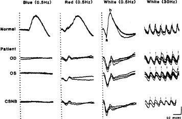
| Fig. 18. Full-field electroretinographic responses from a normal subject, from the right (OD) and left (OS) eyes of a patient with malignant melanoma, and from a patient with congenital stationary night blindness with myopia (CSNB). Stimulus onset is designated by the vertical hatched lines in columns 1, 2, and 3 and the vertical lines superimposed on the responses in column 4. Two or three consecutive responses are illustrated and cornea positivity is an upward deflection. The peak of the cornea-negative a-wave, generated by photoreceptors, and the peak of the cornea-positive b-wave, generated by activity of cells proximal to the photoreceptors, are designated in the response of the normal subject to single flashes of white light. (Lower right) Calibration symbol designates 50 msec horizontally, 200 μV vertically for the top recording in column 3, and 100 μV vertically for all other tracings. Both patients lack the cornea-positive b-wave from the rod system in columns 1, 2, and 3 in contrast to the normal while retaining normal cone amplitudes (equal to or greater than 50 μV) in their responses to 30-Hz white flickering light in column 4. (From Berson EL, Lessell S. Paraneoplastic night blindness with malignant melanoma. Am J Ophthalmol 1988;106:307.) |