Chapter 53
Ocular Developmental Anomalies
SCOTT E. OLITSKY and LEONARD B. NELSON
Main Menu   Table Of Contents

Search

PART 1: THE RELATIONSHIP OF OCULAR ANOMALIES TO EVENTS IN DEVELOPMENT
STAGE 1: EMBRYOGENESIS
STAGE 2: ORGANOGENESIS
STAGE 3: DIFFERENTIATION
PART 2: DEVELOPMENTAL OCULAR ANOMALIES
ANOPHTHALMIA
CYCLOPIA AND SYNOPHTHALMIA
CONGENITAL CYSTIC EYE
MICROPHTHALMOS
TYPICAL COLOBOMAS (OF THE IRIS, RETINA, CHOROID, AND OPTIC NERVE)
LENS ANOMALIES
DEVELOPMENTAL CATARACTS
RETINAL DYSPLASIA
FALCIFORM RETINAL FOLDS
SCLERAL ANOMALIES
CORNEAL ANOMALIES
POSTERIOR KERATOCONUS
DERMOIDS
CONGENITAL HEREDITARY ENDOTHELIAL DYSTROPHY
ANTERIOR SEGMENT DYSGENESIS
POSTERIOR EMBRYOTOXON
AXENFELD-RIEGER SYNDROME
PETERS' ANOMALY
IRIDOCORNEAL ENDOTHELIAL SYNDROME
CONGENITAL GLAUCOMA
ANOMALIES OF THE IRIS
ANOMALIES OF THE VITREOUS
OTHER ANOMALIES OF THE HYALOID SYSTEM
ANOMALIES OF THE ZONULES
OPTIC NERVE ANOMALIES
ANOMALIES OF THE EYELID AND LACRIMAL APPARATUS
REFERENCES

The clinical identification of an ocular developmental anomaly should stimulate curiosity concerning the etiology and pathogenesis of the condition. Etiology refers to the cause of the anomaly (e.g., a mutation, a teratogen, and so forth); pathogenesis refers to the mechanisms involved in the evolution of the anomaly. Because the mechanisms producing these anomalies are often related to a mishap in a particular stage of normal development, pathogenesis is best studied once normal ocular development is understood.

Mann1 divided the causes of developmental anomalies into four categories:

  Category 1: This includes those anomalies attributed only to a mutation inherited by the fetus.
  Category 2: This includes a genetic predisposition to malformations when exposed to certain environmental influences. Without this predisposition, the specific environmental influence alone would not produce maldevelopment. Conversely, without the genetic predisposition, the specific environmental factor would not be capable of inducing an abnormal morphology.
  Category 3: This includes defects induced by environmental factors that interfere with the development of a genetically normal zygote. These factors may be infectious, chemical, or physical and are referred to as teratogens.
  Category 4: This includes spontaneous mutations that result in ocular abnormalities that are not related to any known environmental agent.

Categories 2 and 3 indicate that genetic and environmental influences may interact to produce morphologic abnormalities. The same morphologic abnormalities may result from either the genetic or the environmental influence or from a variable mixture of both. When considering a structural abnormality, therefore, it may be impossible to determine accurately the proportionate contributions of the genetic and environmental factors that lead to the production of the defect in question.

Because pathogenesis must be understood in the context of normal ocular development, a frame of reference is useful for discussion purposes. It is therefore customary to refer to “stages” of development. Of course ocular development is a continuous process and does not proceed in discrete stages; however, staging remains a useful approach and will be used in this chapter. Three stages of fetal development will be addressed: embryogenesis, organogenesis, and differentiation.

In the initial stage, embryogenesis, the three germinal layers of the developing fetus—ectoderm, mesoderm, and endoderm—become organized. Defects during this period result in widespread somatic structural alterations, and the conceptus rarely proceeds to term. It is rare, therefore, for an event that occurs in embryogenesis to result in an isolated ocular anomaly. In organogenesis, the germinal layers throughout the fetus become organized into the general architectural patterns of the various organs. Therefore, many of the ocular anomalies arising from a mishap during this stage involve the entire globe (e.g., anophthalmia and microphthalmos with cyst). During the final period, differentiation, the characteristic substructures of each organ are developed; thus, abnormalities occurring during this period affect specific ocular structures. Specific structural ocular defects attributed to an event that occurs during this period may result from local growth retardation (e.g., microcornea), from the failure of embryologic structures to atrophy (e.g., persistent hyperplastic primary vitreous), or from an alteration in differentiation (e.g., retinal dysplasia).

There are two reasons to consult this chapter: (1) for an overview of ocular maldevelopment in the organized framework of embryology, and (2) as a reference for obtaining specific information about a particular entity. The remainder of this chapter is, therefore, divided into two parts. Part 1, The Relationship of Ocular Anomalies to Events in Development, is an outline of ocular developmental anomalies related to the periods of embryogenesis, organogenesis, and differentiation. Part 2, Developmental Ocular Anomalies, indexes and discusses ocular anomalies on the basis of the anatomic location of the defect. For rapid reference, a reader interested in a particular defect may turn directly to Part 2 for the discussion of an entity without first reading Part 1. The interested reader, however, should later return to Part 1 for a more complete grasp of the significance of the defect under study.

Back to Top
PART 1: THE RELATIONSHIP OF OCULAR ANOMALIES TO EVENTS IN DEVELOPMENT
Back to Top
STAGE 1: EMBRYOGENESIS
After fertilization, the ovum begins a rapid series of mitotic divisions, forming a solid ball of cells, the morula. These cells rearrange and organize around a central fluid-filled cavity, the blastocyst. The cells of the blastocyst divide, and those cells that will eventually produce the actual embryo accumulate at one pole, forming the embryoblast. These cells further differentiate into two layers, the epiblast and hypoblast. The two layers divide the central cavity of the blastocyst to form the amniotic cavity and the yolk sac. Central epiblast cells now invaginate between the two-layered embryoblast to form a trilayered structure. The central cells of this structure differentiate to form the mesodermal layer, which fills in the area between the epiblast and hypoblast cells. The three definitive germ cell layers—ectoderm, mesoderm, and endoderm—have now been produced, and they will give rise to all other structures of the developing embryo. The period of embryogenesis, therefore, spans the events from fertilization through the organization of germinal layers.

Cephalad ectoderm differentiates into neural ectoderm, which then forms the neural plate; this, in turn, eventually develops into the head and brain. This neural ectoderm expands to form bilateral elevations called neural folds, which grow toward the midline and eventually fuse, producing the neural tube. Fusion begins in the central portion and continues anteriorly and posteriorly until only a small opening at either end remains. A malformation of the forebrain and mesodermal structures in the midline during the development of the neural plate may result in fusion of the structures that will eventually become the optic vesicles. The condition in which there is complete fusion of the elements of the two eyes (optic vesicles) is referred to as cyclopia; partial fusion is called synophthalmia. The ocular anomalies produced during embryogenesis are merely reflections of protean systemic anomalies that are incompatible with life.

Back to Top
STAGE 2: ORGANOGENESIS
The process of organogenesis begins with the segregation and arrangement of the primitive tissues to form the general pattern of the organs that will eventually develop. This process largely involves the movement of masses of tissues to the places where differentiation will occur.

During the third week of development, the optic pits or sulci appear as depressions or invaginations on the inner surface on each side of the anterior neural folds. As the neural folds approach each other, neural crest cells migrate from the neural ectoderm at its junction with the surface ectoderm. The neural crest cells migrate underneath the surface ectoderm and spread throughout the embryo, including the region of the optic pits. These cells are important precursors to many of the major structures of the eye, including corneal and iris stroma, ciliary muscle, choroid, sclera, and bone. Gradually the primary optic vesicle forms as a spheric outpouching from the optic pit. The development of the optic vesicle from the optic sulcus occurs on approximately day 25. The optic vesicle is in contact with the surface ectoderm that will differentiate into the lens via the lens placode.

Primary anophthalmia results when the optic pit does not develop properly; it is not associated with a generalized fetal abnormality.2 One type of secondary anophthalmia involves an arrest in development after the appearance of the optic vesicle.2 Anophthalmia may also occur when the optic vesicle forms but subsequently degenerates.2 If the tip of the optic vesicle makes contact with the surface ectoderm over less than the normal area, a perfectly formed but microphthalmic eye results.3 On approximately day 28, the primary optic vesicle begins to invaginate to form the optic cup. If development proceeds to the outgrowth of an optical vesicle but becomes arrested before its invagination, a condition known as congenital cystic eye will result.4

Invagination of the optic vesicle occurs from the sides and below, simultaneously forming a groove along the underside of the optic cup and optic stalk known as the fetal (choroidal) fissure. Immediately adjacent to the fetal fissure, tissue primarily of neural crest origin proliferates and forms the hyaloid system of vessels. The fetal fissure begins to fuse centrally, and this fusion extends both anteriorly and posteriorly. A typical coloboma occurs when the margins of the fissure fail to fuse anywhere along the line that extends from the optic disc to the inferonasal border of the pupillary margin.5 Another defect that results from an anomalous closure of the fetal fissure is microphthalmos with cyst formation.2 Optic nerve aplasia has also been related to anomalous closure of the ventral fissure.

Back to Top
STAGE 3: DIFFERENTIATION
The events of organogenesis result in the framework of the developing eye. The specific components of the eye are developed by the process of differentiation.

LENS DIFFERENTIATION

Between days 24 and 26, the surface ectoderm adjacent to the optic vesicle thickens, forming the lens plate (placode). The lens placode invaginates to form the lens vesicle, which has a hollow center, between days 32 and 33 (Fig. 1). The lens vesicle consists of a single layer of cells whose apices are directed toward the center. After the lens vesicle has detached from the surface ectoderm, it is surrounded by a basal lamina, which will become the lens capsule.

Fig. 1. The eye at 4 weeks. The lens vesicle is about to separate from surface ectoderm. The optic vesicle is an invaginated cup. Note the relationship of the vascularized primary vitreous to the lens. (Courtesy of Irene H. Maumenee, MD)

Primary aphakia results when the lens plate is absent.6 A lens that forms but subsequently degenerates and largely disappears leads to secondary aphakia.6 If the lens plate forms normally, cuboidal cells line the anterior part of the lens vesicle. The posterior portion of the vesicle is lined by columnar cells that form the primary fibers. These fibers fill the lens vesicle, obliterating its cavity, and create an arc across the lens anterior to the equator. The anterior lens epithelial cells remain cuboidal and become the permanent lens epithelium that, through mitosis, gives rise to future cortical lens fibers later in life.

By the seventh week of development, secondary fibers begin to proliferate in both directions from the equatorial region of the lens. The secondary fibers interlace anteriorly to produce an upright Y suture and posteriorly to produce an inverted Y suture. The lens continues to grow throughout a person's life through the proliferation of secondary lens fibers. As the fibers are laid down, the different optical densities of the fibers produce four zones of discontinuity that can be observed clinically with a slit lamp:

  1. The embryonic nucleus, an optically clear central area formed in embryonic life (during months 1 to 3) from the primary lens fibers, which retain embryonic transparency
  2. The fetal nucleus, formed from secondary fibers from the third to eighth month of fetal life
  3. The infantile nucleus, formed from the last weeks of fetal life through puberty
  4. The adult nucleus, formed after puberty in adult life

Abnormalities of the lens that occur during the stage of differentiation include alterations in size (e.g., microspherophakia), in shape (e.g., anterior and posterior lenticonus), and in the lens fibers themselves (e.g., developmental cataracts).

The developing lens receives its blood supply from multiple branches of the hyaloid artery. These cover the anterior and posterior aspect of the lens and anastomose with vessels within the mesenchyme tissue that covers the lens, forming the pupillary membrane. This hyaloidal vascular system is called the tunica vasculosa lentis. Its greatest development occurs at 9 weeks' gestation, starts to regress by the end of the third month, and is usually complete by the eighth month. Failure of this system to regress leads to a persistent pupillary membrane. Mittendorf's dot is also a failure of complete regression and represents that part of the hyaloid artery that attached to the posterior lens capsule.

RETINAL DIFFERENTIATION

When the optic vesicle collapses, the neural ectoderm folds onto itself to produce the optic cup, which has a double layer of neural ectoderm. The optic cup will differentiate into the neurosensory retina (inner layer of neural ectoderm) and retinal pigment epithelium (outer layer). Further development of the sensory retina may be divided into four stages of progressive differentiation: the initial differentiation into zones, the organization into temporary layers, the migration and differentiation of cells, and the final organization of layers. Disorganized differentiation may result in the histologic picture of retinal dysplasia.7

VITREOUS DIFFERENTIATION

The primary vitreous forms between the inner layer of the optic cup and the developing lens at approximately 6 weeks. The secondary vitreous is derived from the retina and surrounds the primary vitreous. It is composed of very fine, densely packed fibrils that are arranged in an orderly manner at right angles to the retinal surface. Condensation of the fibrils forms the walls of the canal of Cloquet, which is a conduit for the hyaloid artery. The hyaloid artery supplies the vascular network surrounding the lens. At the end of the third month, the hyaloid artery atrophies and retracts with the primary vitreous to form a narrow and constricted zone in the central vitreous cavity (Figs. 2 and 3). The atrophy begins centrally and proceeds anteriorly and posteriorly. Incomplete atrophy may result in anterior hyaloid remnants associated with the lens or in posterior remnants associated with the optic nerve head. An example of an anterior remnant is Mittendorf's dot. Bergmeister's papilla may be considered a posterior remnant of the hyaloid system.

Fig. 2. The eye at 12 weeks. The cornea is separated from the more anterior fused eyelids. A membrane covers the pupil (pupillary membrane). Note the relationship of the scleral spur to the anterior chamber: the angle is incompletely formed. The lens vesicle is now obliterated. The secondary vitreous is avascular, whereas the primary vitreous is vascularized. Note the vascular arcades on the posterior surface of the lens (posterior tunica vasculosa lentis). (Courtesy of Irene H. Maumenee, MD)

Fig. 3. The eye at 4 months. Note the changed proportions of secondary and primary vitreous. (Courtesy of Irene H. Maumenee, MD)

The third period of vitreous formation begins at the end of the third month. The tertiary vitreous begins as an accumulation of collagen fibers between the equator of the lens and the optic cup and will eventually differentiate into the vitreous base and lens zonules.

SCLERAL DIFFERENTIATION

The sclera is derived from tissue of neural crest origin. Most of the cornea is derived from the same mesenchymal tissue with the exception of the corneal epithelium, which is derived from surface ectoderm. The anterior sclera forms first as a condensation of this tissue and is continuous with the cornea. Condensation proceeds posteriorly until the 12th week of development, at which time the sclera surrounds the optic nerve.

CORNEAL DIFFERENTIATION

The lens vesicle detaches from the overlying surface ectoderm during the sixth week of gestation. During the seventh week, a single layer of neural crest cells invades the region between the surface ectoderm and the lens vesicle. This single layer of cells will differentiate into the corneal endothelium. A second wave of neural crest cells migrates between the primitive endothelium and surface ectoderm to form fibroblasts. These fibroblasts will eventually differentiate into keratoblasts that will produce the collagen fibrils and extracellular matrix of the corneal stroma.8–10 Initially these neural crest cells occupy the space that will become the anterior chamber. They are the precursor cells that will develop into the corneal stroma, corneal endothelium, anterior iris stroma, and many of the structures of the iridocorneal angle. The surface ectoderm will give rise to the corneal epithelium. The anterior chamber is formed when these neural crest cells separate from the tunica vasculosa lentis and pupillary membrane, which overlie the anterior lens surface. Therefore, the anterior chamber is almost entirely derived from neural crest cells, not mesoderm as was once believed. The vascular endothelium is now thought to be the only component of the anterior segment that is derived from mesoderm.

The corneal epithelium is derived from surface ectoderm, whereas the remainder of the cornea is derived from neural crest cells, as described previously. The corneal stroma gives rise to Bowman's membrane. Therefore, Bowman's membrane is of neural crest origin and is a condensation of anterior corneal stroma. It is not the basement membrane of the corneal epithelium and is not derived from the same precursor cells. Descemet's membrane is present by the sixth month of gestation and the cornea begins to become transparent at about this time.

IRIS AND CILIARY BODY DIFFERENTIATION

During the third month of development, the anterior rim of the optic cup, which is composed of neural ectoderm, begins growing rapidly in a curve around the anterior surface of the lens. The iris pigmented and nonpigmented epithelium will develop from this anterior advancing edge of the optic cup. The iris sphincter and dilator muscles develop from these epithelial layers and are the only muscles of the body that are derived from neural ectoderm. The inner epithelial layer begins to develop folds, which are the early ciliary processes. The ciliary body epithelium also develops from the anterior portion of the optic cup. The tertiary vitreous forms from an extracellular matrix that is produced by the ciliary epithelium and will eventually become the lens zonules. The iris stroma develops from the neural crest cells, which migrate between the lens vesicle and surface ectoderm.

IRIDOCORNEAL ANGLE DIFFERENTIATION

Traditionally, the study of the development of the anterior chamber and angle has concentrated on the role of mesenchyme. It is now known that neural crest cells are the major contributor to the development of these structures. There continues to be some controversy, however, regarding the mechanism by which the angle is formed. There are conflicting opinions as to whether the angle structures are formed as a result of simple cleavage of the iris root from the cornea, or whether a more active growth process is required for normal angle development.

OPTIC NERVE DIFFERENTIATION

The optic nerve develops within the substance of the optic stalk. The nerve fibers of the developing optic nerve are composed of axons of the ganglion cells in the retina. The nerve fibers completely fill the optic stalk by the end of the seventh week. Myelination of the nerve fibers starts at the chiasm at about 7 months and progresses anteriorly toward the eye. Myelination normally stops at the lamina cribrosa approximately 1 month after birth. Myelinated nerve fibers occur if the myelination process continues past the lamina and most likely represents ectopic myelination, not a structural defect in the lamina cribrosa.

EYELID AND LACRIMAL APPARATUS DIFFERENTIATION

The eyelids develop from both surface ectoderm and neural crest cells. Surface ectoderm gives rise to the epidermis, cilia, and conjunctival epithelium, while the deeper structures of the lids, including the dermis and tarsus, are derived from neural crest cells. The orbicularis and levator muscles develop from mesoderm. The eyelids begin to form by 6 weeks and grow to cover the eye. The upper and lower folds fuse at approximately 12 weeks and gradually start to separate at 6 months' gestation.

The lacrimal apparatus is formed by contributions from the lateral nasal and maxillary processes as well as from the lids. During the third month, the lacrimal canaliculus is formed by the lysis of a central core of cells. The lacrimal punctum opens onto the lid margin just before the lids separate, usually during the seventh month. The lower end of the nasolacrimal duct is frequently separated from the cavity of the inferior meatus at birth by a membrane. This membrane consists of the opposed mucosal linings of the nasal fossa and the lower end of the duct. Obstruction of the nasolacrimal duct is usually caused by defective canalization or persistence of the nasal mucosal membrane.11

Back to Top
PART 2: DEVELOPMENTAL OCULAR ANOMALIES
Back to Top
ANOPHTHALMIA
Anophthalmia (Fig. 4) is the congenital absence of the eye. It is a rare entity and occurs in approximately 1 in 20,000 births.12 Anophthalmia can be divided into three types: (1) primary, which results from an isolated failure of optic pit development (i.e., the remainder of the neural tube may be unaffected); (2) secondary, which results from a complete suppression or abnormality of the forebrain, in which case the anophthalmia is merely a component of neural malformation; and (3) consecutive, or degenerative, which results from regression of a formed but rudimentary optic vesicle.

Fig. 4. Bilateral clinical anophthalmia. No eye is apparent in either orbit. (Courtesy of Robison D. Harley, MD)

Because anophthalmia may be difficult to distinguish clinically from profound microphthalmos, removal of the orbital contents with serial histologic examination may be necessary to establish the diagnosis.13 A small congenital cystic eye (see later discussion) may also give the clinical appearance of anophthalmia. Because few cases are confirmed histologically, the term clinical anophthalmia is used to describe the condition in which an eye appears to be absent. Radiologic examination may be helpful in attempting to distinguish anophthalmia from severe microphthalmos or when searching for a small cystic eye within the orbit. An MRI appears to be superior to a CAT scan for this purpose.14 Systemic malformation syndromes occur in most patients with clinical anophthalmia, and unilateral cases often show abnormalities of the contralateral eye.12,15

Most cases of anophthalmia occur sporadically, although autosomal-dominant, autosomal-recessive, and sex-linked-recessive transmissions have been reported.16–22 The causes of developmental failure of the optic vesicle or vesicle regression may include environmental factors, such as radiation or anoxia.4 Animal models of anophthalmia suggest that suppression of the optic vesicles may result from the introduction of a variety of teratogenic factors, such as lithium, trypan blue, or actinomycin, at the time the optic vesicle is beginning to appear.4

Back to Top
CYCLOPIA AND SYNOPHTHALMIA
Cyclopia (total fusion of the optic vesicles) and synophthalmia (partial fusion of the optic vesicles) represent degrees of an anomaly that prohibits the development of separate eyes. They occur in approximately 1 in 100,000 births.23 Two mechanisms can account for either of these developmental malformations: an early defect in which a single bilobed midline region fails to separate completely or a later loss of the midline territory, which leads to fusion of the ocular fields that had been previously divided. Because the prosencephalon is responsible for the development of separate eyes, cyclopia and synophthalmia are anomalies that reflect profound neural maldevelopments that are incompatible with life.

The cyclopic eye usually demonstrates multiple internal abnormalities, such as the presence of retinal rosettes, the absence of the retinal pigment epithelium at the posterior pole, and abnormal choroidal development.24 Synophthalmia is characterized by graded degrees of ocular fusion.25 Except for the anterior structures of the eye, such as the lens and anterior uvea, the fused eyes may share a single structure (e.g., a common optic nerve).25 In most cases, the retina may show a coloboma of various degrees, rosette formation, and a paucity of ganglion cells.4

Teratogenetic agents and chromosomal abnormalities both may cause cyclopia and synophthalmia. The teratogenetic periods in the human are probably 2½ weeks for cyclopia and 3 weeks for synophthalmia.26 A deletion on the long arm of chromosome 7 has been implicated as an etiologic factor in arresting prosencephalon development at an early stage, leading to the development of cyclopia.27 Cyclopia has been reported in association with trisomy 4.28

Back to Top
CONGENITAL CYSTIC EYE
Congenital cystic eye results when the optic vesicle fails to invaginate.2 When invagination does not occur, the development of the entire globe is arrested, and the ectodermal elements (what will become the cornea and lens placode) do not differentiate. Instead of a globe, the orbit contains a cyst, which may be either unicameral or subdivided into several loculi.29 If the congenital cystic eye is very small, it may be confused with clinical anophthalmia (see earlier discussion). Congenital cystic eyes may also be confused clinically with the condition of microphthalmos with cyst, in which the cyst is so large that it dwarfs the severely microphthalmic component.
Back to Top
MICROPHTHALMOS
Microphthalmos is a general term used to describe an eye that is smaller than normal. It can result from an insult that may occur at various developmental stages. An early insult may lead to a primary decrease in the size of the optic vesicle. Because the growth of the sclera and choroid appears to depend on intraocular pressure, insults that occur later in development that affect proper maintenance of the intraocular pressure may lead to a secondary decrease in the size of the fetal eye.30 An example of this can be seen in colobomatous microphthalmos, in which failure or late closure of the embryonic fissure prevents establishment of normal fetal intraocular pressure (see later discussion). Microphthalmos occurs in many forms and can be divided into three basic types: (1) pure, (2) simple, and (3) complex.

PURE MICROPHTHALMOS (NANOPHTHALMOS)

Pure microphthalmos, also called nanophthalmos, refers to a small eye containing a lens of “normal” volume. It is thought to result from an arrest in ocular development after the fetal fissure has closed.2 The eye is small in its overall dimensions, but shows no other gross defects. Because the size of the lens is normal, patients with nanophthalmic eyes have a shallow anterior chamber and pronounced hypermetropia (more than 10 diopters).31 Therefore, these eyes are prone to attacks of angle-closure glaucoma.32 Thickening of the sclera around the exits of the vortex veins may contribute to retinal and choroidal effusion.33,34 Scleral thickening is a result of abnormal interlacing of the scleral fibers, which may be secondary to abnormal glycosaminoglycan metabolism.35–37 In early life the visual acuity is usually normal, although macular hypoplasia may account for poor vision in some younger patients. Poor vision later in life is often due to the secondary complications of this disorder. Nanophthalmos is generally bilateral, and these eyes are usually deeply set in shallow orbits.

Most affected individuals are members of pedigrees that demonstrate consanguinity. The mode of inheritance, therefore, is presumed to be autosomal recessive.4 Only a few pedigrees compatible with autosomal-dominant inheritance have been reported.38,39

SIMPLE MICROPHTHALMOS

Simple microphthalmos is the term used to describe a small eye that is otherwise normal. Approximately 50% of patients with simple microphthalmos have an associated systemic developmental abnormality. These eyes have a short axial length and are therefore moderately hyperopic. The level of hyperopia is less than that seen in nanophthalmos (usually less than + 10.00 diopters).40 Most patients have normal vision. The complications that are seen with nanophthalmos do not occur in this disorder.

Most cases of simple microphthalmos are sporadic and occur bilaterally. Simple microphthalmos represents a retardation in growth that occurs once the primary optic vesicle has formed and invaginated.2 It has been caused in animal experiments by radiation, mechanical stimulation, and chemicals. Microphthalmos may result from intrauterine toxoplasmosis or rubella, and it may occur as a component of an ocular-genetic syndrome such as Norrie's syndrome or oculodentodigital dysplasia.41–45 It may also be associated with systemic disorders, such as fetal alcohol syndrome, myotonic dystrophy, and achondroplasia.

COMPLEX MICROPHTHALMOS

The term complex microphthalmos denotes a small, deformed eye. One or more malformations may occur as an isolated ocular entity or as part of an associated systemic disorder. Associated ocular deformities can include anterior segment maldevelopment, hyperplastic primary vitreous, and other abnormalities of the lens, vitreous, or retina. Complex microphthalmos may be unilateral or bilateral, and the vision can range from normal to no light perception, depending on the severity of the associated defects. Both sporadic and hereditary forms are known.

Microphthalmos with coloboma is one specific type of complex microphthalmos and is caused by an incomplete closure of the embryonic fissure. It may also occur as both a sporadic and hereditary form. It may be associated with a number of systemic diseases, including the CHARGE* association. Recognition of this entity when evaluating a child with microphthalmos is important because the heart defects in the CHARGE association can be lethal. Autosomal-dominant colobomatous microphthalmos is a well-known and important disorder. It occurs without an associated systemic abnormality and has variable expressivity and penetrance. Family members of affected patients should receive appropriate genetic counseling.


coloboma, heart defect, atresia cochonal, retarded growth, genital hypoplasia, ear anomalies.

Microphthalmos with cyst results from proliferation of neuroectoderm at the edge of a persistent embryonic fissure. The cavity of the cyst is continuous with the interior of the microphthalmic globe.2,46 Rudimentary optic nerve fibers can be seen within the cyst wall.47 The microphthalmic eye itself is often overshadowed by the larger orbital cyst (Fig. 5).48 Cases of extreme microphthalmos may be confused with anophthalmia.49,50 The anterior segment of the microphthalmic eye may appear normal, although it commonly displays marked disorganization and iridocorneal adhesions. The lens is frequently cataractous and dislocated.51 The retina is usually detached, disorganized, and gliotic.52 No consistent -association has been documented between mi-crophthalmos with cyst and a specific systemic anomaly. Systemic disorders reported with microphthalmos with cyst include central nervous -system defects (e.g., meningoencephalocele, hydrocephalus) and cardiac, urogenital, facial, and skeletal abnormalities.53–55

Fig. 5. The structure visible on the right side is the cyst component of microphthalmos with cyst. (AFIP #220948; courtesy of Torrence A. Makley, Jr, MD)

Back to Top
TYPICAL COLOBOMAS (OF THE IRIS, RETINA, CHOROID, AND OPTIC NERVE)
The term coloboma is employed in ophthalmology to refer to any notch, gap, or fissure in any of the ocular structures, whether it be congenital or acquired.2,5 Technically, therefore, a surgical iridectomy is a type of iris coloboma, but for the remainder of this discussion, the term coloboma will refer only to congenital defects.

Colobomas may occur anywhere along the embryonic fissure and can therefore affect the iris, ciliary body, choroid, retina, and optic nerve. The embryonic fissure begins to close at the equator of the eye sometime around the fifth week of gestation. Closure of the fissure then proceeds in an anterior and posterior direction. Because of this, colobomas may be found at the two ends of the embryonic fissure, the iris and optic nerve, with normal tissue between them.

Iris colobomas can be designated as typical or atypical, depending on where they are located. Typical iris colobomas are located inferonasal to the pupillary border; thus, they reflect the incomplete closure of the embryonic fissure.2 Iris colobomas (Fig. 6) may be associated with flattening of the exposed portion of the lens, gaps in the zonular fibers, and localized cortical lens opacities.2 The lens notch is commonly referred to as a lens coloboma. This is not a true coloboma because there is no focal absence of lens material and the lens does not develop from the embryonic fissure. Rather, it results from the absence of zonules and zonular traction in the region of the ciliary body coloboma. Atypical iris colobomas are not located in the inferonasal sector, and they are not considered to be related to defective embryonic fissure closure. Atypical iris colobomas are therefore not associated with posterior uveal or optic nerve colobomas.

Fig. 6. Typical iris coloboma. Note the inferonasal location of the iris defect.

A chorioretinal coloboma appears through the ophthalmoscope as a glistening white defect with distinct margins, often outlined by irregular pigment clumps.2 Although a coloboma may occasionally be flat and smooth, the floor of the defect is usually uneven and usually bulges posteriorly.2 An intercalary membrane of undifferentiated neurosensory retina may cover the defect.5 Breaks in this membrane have been reported to have caused retinal detachment. When a hemorrhage is found in the coloboma, a break in the intercalary membrane may be present.56

In optic nerve coloboma (Fig. 7) the optic disc is enlarged, frequently oval vertically, and excavated.57 The colobomatous defect may involve the entire disc or just the inferior portion. The excavated region is decentered inferiorly and may extend to involve the choroid and retina. When it does, the eye is often microphthalmic.57 When the entire disc is involved, the inferior region is excavated to a greater extent than the remainder of the disc, confirming its colobomatous nature. The retinal vessels may radiate from the disc in a spokelike fashion, with fewer bifurcations than normal.57 An association between optic nerve colobomas and basal encephaloceles has been reported58,59; however, examination of the literature reveals only a few photographically documented cases, and the reported association may be because of its confusion with morning glory disc anomaly.60,61

Fig. 7. Optic nerve coloboma.

Vision in the presence of a colobomatous defect can range from normal to no light perception. The degree of vision loss is related to the location of the defect and the presence of any associated abnormalities. Isolated iris colobomas do not usually affect visual acuity, whereas optic nerve colobomas almost always lead to some reduction in vision. If a chorioretinal coloboma is located in the posterior pole, an associated staphyloma may cause disturbance of the macula with resultant poor vision.

Isolated colobomatous anomalies are most commonly inherited in an autosomal-dominant manner with highly variable penetrance and expressivity.62 Some cases of families with probable autosomal-recessive transmission have been reported.60 Because colobomas may be associated with a variety of systemic anomalies and heritable disorders (Table 1), affected individuals should be examined systemically and a careful family history should be taken. A thorough genetic evaluation should be performed when indicated.


TABLE 1. Coloboma and Associated Disorders

  Chromosomal Abnormalities
  Trisomy 13
  Triploidy
  Cat-eye syndrome
  4p-
  11p-
  11q-
  Trisomy 18
  Klinefelter's syndrome
  Systemic Disorders
  CHARGE association
  Lenz's syndrome (microphthalmos)
  Goltz's syndrome (focal dermal hypoplasia)
  Basal cell nevus syndrome
  Meckel's syndrome
  Warburg's syndrome
  Aicardi's syndrome
  Goldenhar's syndrome
  Rubinstein-Taybi syndrome
  Linear sebaceous nevus syndrome


Back to Top
LENS ANOMALIES

CONGENITAL APHAKIA

Congenital aphakia, a very rare anomaly, can be divided into two types: primary, in which a lens failed to develop; and secondary, in which a lens developed to some degree, but either was resorbed or extruded through a corneal perforation before or during birth.4,6 Primary congenital aphakia has been characterized histologically by aplasia of the anterior segment.6 This defect may result from anomalous invagination of the optic vesicle, which disturbs the normal relationship between the optic vesicle and the surface ectoderm. The lack of surface ectodermal induction may lead to widespread anterior segment anomalies. The abnormal optic vesicle invagination may also be accompanied by defects in the closure of the fetal fissure, and therefore by typical colobomas.6

Secondary congenital aphakia is more common than the primary type.4 The lens is usually represented by a wrinkled capsule. Several attempts have been made to explain secondary aphakia. One theory suggests that the lens may develop normally, but that the capsule is thinner than usual and ruptures. Intrauterine inflammations may also lead to degeneration of the lens and to subsequent capsular shrinkage, which is sometimes accompanied by mesenchymal infiltration of the lens remnant.2

MICROSPHEROPHAKIA

Microspherophakia is a bilateral condition in which the lens is usually small and spheric.2 Following pupillary dilation, the lens edge may be identified with a slit lamp. The small size and spheric shape of the lens may result in significant myopia. The elongated zonules may permit the lens to move forward, increasing the area of iridolenticular contact.63 This mechanism may lead to pupillary block. Miotics may increase the pupillary block, and mydriatics may relieve it, a paradox that Urbanek64 termed inverse glaucoma and that others have confirmed exists.65,66 Microspherophakia has been reported in association with such systemic entities as Marfan's syndrome, homocystinuria, Alport's syndrome, Klinefelter's syndrome, and Weill-Marchesani syndrome.67–70 It may occur as an isolated anomaly transmitted as either an autosomal-dominant or autosomal-recessive condition.71 The lens is typically spheric before the fifth or sixth month of fetal life, so an arrest in lenticular development at this stage would explain uncomplicated microspherophakia.

ANTERIOR LENTICONUS

Anterior lenticonus is a rare bilateral condition in which the anterior surface of the lens bulges centrally. It may be accompanied by lens opacities or other eye anomalies and is a prominent feature of Alport's syndrome.72,73 Examination by retinoscopy or by ophthalmoscopy shows a dark area resembling an oil droplet in the center of the lens. The increased curvature of the central area may cause high myopia.4

Histologic examination of an anterior capsule removed from a patient with anterior lenticonus showed it to be one third the normal thickness, having multiple capsular dehiscences containing fibrillar material and vacuoles.74 The capsular fragility observed in this condition may be a result of a mutation in the gene that encodes for one or more chains in type IV collagen. This mutation has been documented in patients with Alport's syndrome.75

POSTERIOR LENTICONUS

Posterior lenticonus, which occurs more commonly than anterior lenticonus, is an ectasia of the posterocentral surface of the lens. It is often associated with progressive lens opacities of the posterior cortex. The majority of cases are unilateral, and the condition is found more frequently in females than in males. When no lens opacities are present, the clinical appearance of posterior lenticonus resembles the oil droplet appearance of anterior lenticonus. The characteristic localized posterior bulge can be seen with a slit lamp.4 The etiology of posterior lenticonus is unknown. Because the zonules exert traction on the lens and determine its shape, focal defective zonular traction along the posterior lens capsule may account for abnormal curvature in the area.

Back to Top
DEVELOPMENTAL CATARACTS
Developmental cataracts may be classified according to the location (e.g., nuclear, polar) or configuration (e.g., coralliform, blue dot) of the opacity.

Progress has been made in determining the etiology of many congenital cataracts. The various factors that might cause these lens defects include hereditary factors, infectious agents, metabolic abnormalities, toxins, endocrinologic effects, and ionizing radiation.76 Despite a meticulous medical history and a thorough clinical and laboratory workup, the cause of a congenital cataract in any particular patient can still escape identification. Because they may develop at any time during the life of the fetus, lens opacities often make excellent markers for timing developmental defects. In general, developmental aberrations affecting the primary lens fibers may cause central lens opacities. If the secondary fibers are opacified, a clinical opacity restricted to a zone (zonular cataract) may occur.4

Back to Top
RETINAL DYSPLASIA
Hunter and Zimmerman77 suggested the term retinal dysplasia to describe a nonspecific result of disturbed retinal growth and differentiation. Retinal dysplasia is generally seen in patients with other systemic abnormalities, most commonly trisomy 13–15. The histopathologic hallmark of retinal dysplasia is the rosette, several types of which have been described.78

Rosette formation may result from the exaggeration of the normal developmental process, which causes an overgrowth of the cells of the inner layer of the optic cup during closure of the embryonic fissure.79 Mann2 observed that retinal dysplasia could be produced by exposure to x-rays during the period of active differentiation of the inner layer of the optic cup. Orderly retinal morphogenesis may depend on the organizing influence of the adjacent pigment epithelium. When this layer is lacking, the neurosensory retina may be disorganized.80

Reese and co-workers81,82 applied the term retinal dysplasia to a specific syndrome consisting of bilateral retinal defects and other ocular defects associated with multiple systemic anomalies. This description, however, appeared before chromosomal analysis was available, and many of the cases that they described may have actually been examples of trisomy 13.83–85 Retinal dysplasia has also been associated with cyclopia, Norrie's disease, congenital progressive oculoacousticocerebral degeneration (inherited as a sex-linked recessive), chromosome 18 deletion defect, retinoblastoma, and persistent hyperplastic primary vitreous (PHPV).54,86–88 As the syndromes that are associated with retinal dysplasia become better known, this term will eventually become a purely histologic description, rather than a clinical entity.

Back to Top
FALCIFORM RETINAL FOLDS
The falciform retinal fold extends from the optic disc to the ciliary body, usually in the inferotemporal quadrant.2 It may be bilateral and symmetrically distributed, and it is typically associated with persistent remnants of the hyaloid artery. The falciform fold usually occurs in an otherwise healthy eye, although it may cause a tentlike detachment of the retina.89–91 The pathogenesis of this anomaly may involve traction on the retina, which is caused by persistence of hyaloid vessels and by elements of the primary vitreous that become attached to the inner layer of the optic cup. Falciform retinal folds may also occur secondary to retinopathy of prematurity (ROP), foreign bodies, and granulomatous inflammation.
Back to Top
SCLERAL ANOMALIES

BLUE SCLERA

Blue sclera is considered to result from an arrest in scleral maturation. Congenital blue sclera frequently occurs in premature infants as a normal finding. Blue sclera, brittle bones, and deafness form the clinically diagnostic triad of osteogenesis imperfecta, a disorder transmitted in an autosomal-dominant pattern.92,93 Blue sclera may also be found in other ocular and systemic disorders (Table 2).


TABLE 2. Systemic Disorders in Which Blue Sclera May Be Seen

  Crouzon's syndrome
  Ehlers-Danlos syndrome
  Phenylketonuria
  Pseudoxanthoma elasticum
  Werner's syndrome
  Hypophosphatasia
  Albright's syndrome (hereditary osteodystrophy)
  Hallermann-Streiff syndrome
  Pyknodysostosis
  Turner's syndrome


Back to Top
CORNEAL ANOMALIES

SCLEROCORNEA

In sclerocornea, the limbus is clinically indistinct, and opaque tissue extends into the cornea (Fig. 8). The curvature of the cornea is often flatter, similar to the sclera.4,94 Fine blood vessels arising from the conjunctiva may extend superficially over the peripheral cloudy cornea and terminate in arcades, leaving an avascular central area that may be opaque to clear.95 This condition is usually bilateral. In approximately 50% of the cases reported, a dominant or recessive inheritance has been described.95 Sclerocornea has been reported in association with numerous systemic abnormalities, including limb deformities, craniofacial defects, and genitourinary disorders.95,96 Histologic studies have demonstrated collagen fibers that resemble scleral collagen in their distribution and size. Bowman's membrane is usually absent, and Descemet's membrane and the endothelial layer are attenuated or absent.97,98

Fig. 8. Sclerocornea. (Courtesy of James J. Reidy, MD)

CORNEA PLANA

Up to the fourth month of fetal life, the cornea and sclera have the same radius of curvature. If some factor arrests the relative decrease in the corneal radius of curvature during the fourth or fifth month, cornea plana results.4 In cornea plana, the increase in the corneal radius of curvature leads to flattening of the cornea. Because of the flattening of the cornea, the anterior chamber becomes shallow. The limbus is obscured by “sclerization” of the peripheral cornea, so that clinically the cornea appears to be small.99 Anterior segment anomalies frequently occur, including iris coloboma and congenital cataracts.100 Two types of cornea plana exist, an autosomal-dominant and autosomal-recessive form.99 A defect on the long arm of chromosome 12 has been linked to both types.101,102

MICROCORNEA

The term microcornea describes a cornea that measures less than 11 mm in diameter.4 This finding usually occurs in otherwise normal eyes, but it may be seen in eyes with crowded anterior segments.103 Microcornea is inherited in either an autosomal-dominant or autosomal-recessive manner.104 It may be caused as a result of overgrowth of the tips of the optic cup.

MEGALOCORNEA

Megalocornea is a nonprogressive symmetric condition, characterized by an enlarged cornea (greater than 12 mm in diameter) and an anterior segment that has no evidence of previous or concurrent ocular hypertension.99 The cornea itself is clear, and there is no increase in corneal thickness. A frequent complication is the development of lens opacities in adult life.105 Although all modes of inheritance have been described, the X-linked recessive mode is the most common, which is why this disorder is most commonly seen in males.99,106 Ocular abnormalities associated with megalocornea include myopia and Krukenberg's spindle. Congenital glaucoma and megalocornea may occur in the same family.107,108 Systemic abnormalities that may be associated with megalocornea include Marfan's syndrome, craniosynostosis, and Alport's syndrome.109 The cause of the enlargement of the cornea and the anterior segment is unknown.4 Possible explanations to explain the etiology include a defect in the growth of the optic cup and an arrest of congenital glaucoma. Recently, the region on the X chromosome responsible for this disorder has been identified.110

Back to Top
POSTERIOR KERATOCONUS
Posterior keratoconus is a rare corneal disorder characterized by a localized craterlike defect on the posterior corneal surface, with the concavity facing toward the anterior chamber (Fig. 9).111 The anterior corneal surface is normally sculpted. Vision may be normal or slightly reduced. Reduced vision may be secondary to scattering of light from the irregular posterior surface, or to a steepening of the anterior cornea in the region of the posterior defect.112 Posterior keratoconus may be either unilateral or bilateral. It is frequently nonfamilial, although it has been described in a parent and sibling in three families and in siblings in another family.111,113,114

Fig. 9. Posterior keratoconus. A. Direct illumination. B. Slit-lamp image demonstrating the concave defect. (Courtesy of James J. Reidy, MD)

Histopathologically, Descemet's membrane is present but may show thinning, abnormal anterior banding, and posterior excrescences.111,115 The endothelium is intact but displays cytoplasmic vacuolation and attenuation adjacent to the posterior excrescences.111 Several theories have been proposed to explain the pathogenesis of congenital posterior keratoconus. Mann suggested that posterior keratoconus is caused by an arrest of corneal development and the resulting persistence of embryonal characteristics.116 Greene117 explained the origin of posterior keratoconus as the result of delayed separation of the lens from the cornea.

Back to Top
DERMOIDS
Epibulbar dermoids are choristomas.118 They are often present at birth and may increase in size with age. They occur most frequently in the lower temporal quadrant (Fig. 10). Most commonly they straddle the limbus and extend into the peripheral cornea. Rarely, they may be confined entirely to the cornea or conjunctiva.118 Epibulbar dermoids may cause a visual disturbance by encroaching on the visual axis or by contributing to the development of astigmatism.

Fig. 10. Bilateral epibulbar dermoids. (AFIP #1433457; courtesy of Marshall M. Parks, MD)

The dermoid usually appears as a well-circumscribed, rounded or oval, gray or pinkish yellow mass with a dry surface from which short hairs may protrude. It may affect only the superficial layers of the cornea, although full-thickness involvement is not uncommon. Associated ocular anomalies include eyelid and iris colobomas, microphthalmos, and retinal and choroidal defects. Thirty percent of dermoids are associated with systemic abnormali-ties.4 Many of the associated anomalies involve developmental defects of the first brachial arch (e.g., vertebral anomalies, dystoses of the facial bones, dental anomalies, Goldenhar's syndrome). Epibulbar dermoids are found in 75% of cases of Goldenhar's syndrome.119

The pathogenesis of epibulbar dermoids is disputed and may vary from case to case. One theory is that a dermoid may be formed when remnants of the plica semilunaris become implanted at a particular part of the cornea or conjunctiva.4 The only place in the ocular adnexa where cartilage can normally be found is in the semilunar fold.120 Therefore, the finding of cartilage lends support to the aforementioned theory, at least in cases of epibulbar dermoids that contain cartilage.

Back to Top
CONGENITAL HEREDITARY ENDOTHELIAL DYSTROPHY
Congenital hereditary endothelial dystrophy (CHED) is characterized clinically by diffuse bilateral corneal opacification, which is visible early in life.121,122 CHED must be considered in the differential diagnosis of congenital corneal opacities. The corneal clouding varies from a mild haze to a milky ground-glass appearance.99 CHED may be inherited in either an autosomal-dominant or autosomalrecessive manner. With recessive inheritance, corneal clouding is observed at birth, and nystagmus is frequently present.123 A dominantly inherited form comes to clinical attention in the first or second year of life because of photophobia and epiphora; nystagmus is uncommonly seen with this form.123 Both forms display diffuse edema and increased corneal thickness. Mild epithelial edema is common, but gross bullous keratopathy is rare.121,124,125 A fibroblastlike transformation of the endothelium during corneal differentiation may be the cause of this disorder. The observed distribution of collagen in the posterior portion of affected corneas supports this theory.126,127
Back to Top
ANTERIOR SEGMENT DYSGENESIS
Anterior segment dysgenesis refers to a wide spectrum of developmental anomalies that may affect the cornea, iris, lens, and angle. Until recently, these disorders were referred to as anterior chamber cleavage syndromes or mesodermal dysgenesis.128 Because it is now known that neural crest cells, not mesoderm, are responsible for the majority of anterior segment development and that the simple concept of anterior segment “cleavage” may not be valid, this new term is more appropriate. In general, anterior segment dysgenesis represents an arrest in late gestational development. This developmental arrest may result in retained primitive endothelium, failure of the peripheral iris to migrate posteriorly, or failure of the corneal endothelium to differentiate fully. Posterior embryotoxon, Axenfeld-Rieger syndrome, and congenital iris ectropion may be secondary to retained primitive corneal endothelium. Congenital glaucoma may occur as a result of an arrest in the posterior migration of the peripheral iris and Peters' anomaly, and the iridocorneal endothelial (ICE) syndromes may represent an arrest in the final differentiation of the corneal endothelium.129
Back to Top
POSTERIOR EMBRYOTOXON
Axenfeld first used the term posterior embryotoxon to describe a patient with a white line in the cornea, with strands of iris extending from the peripheral iris to this line. Posterior embryotoxon is now used to describe a prominent, anteriorly displaced Schwalbe's line. On slit lamp evaluation, posterior embryotoxon appears as an irregular, white line just anterior to the limbus. Although the line may extend for 360°, it is frequently incomplete and seen most often temporally. Posterior embryotoxon occurs in approximately 10% of normal eyes. Because it is not associated with other ocular defects, when it occurs as an isolated condition, it may represent a normal anatomic variation.
Back to Top
AXENFELD-RIEGER SYNDROME
Ocular manifestations of the Axenfeld-Rieger syndrome include the presence of posterior embryotoxon and iris strands that attach to Schwalbe's line (Fig. 11). If the iris is normal, the condition is termed Axenfeld's anomaly. If iris defects are present, then it is classified as Rieger's anomaly. Iris findings in Rieger's anomaly may range from mild hypoplasia to full-thickness hole formation (Fig. 12). Glaucoma develops in approximately 50% to 60% of patients with Axenfeld-Rieger syndrome. When Rieger's anomaly is associated with systemic anomalies, it is known as Rieger's syndrome. Developmental abnormalities seen in Rieger's syndrome commonly affect the teeth and facial bones. These defects may include microdontia, hypodontia, and maxillary hypoplasia. Originally described as separate clinical entities, Axenfeld's and Rieger's anomaly are now considered to be variations in the spectrum of the same developmental disorder.

Fig. 11. Gonioscopic view of the iris processes in Axenfeld-Rieger syndrome. (Courtesy of James J. Reidy, MD)

Fig. 12. Rieger's syndrome. (Courtesy of James J. Reidy, MD)

A late arrest in the development of anterior chamber structures derived from neural crest cells is responsible for the ocular defects seen in Axenfeld-Rieger syndrome. Contracture of an abnormally retained primitive endothelial layer on the surface of the iris and anterior chamber angle, deposition of basement membrane by these endothelial cells, and incomplete development of the trabecular meshwork and Schlemm's canal are thought to be responsible for the iris changes, prominent Schwalbe's line, and glaucoma that are seen in this disorder. The systemic malformations seen in Rieger's syndrome are also secondary to a defect in neural crest cell development.

Back to Top
PETERS' ANOMALY
Peters' anomaly is a central corneal opacity that is present at birth (Fig. 13). It is often associated with iridocorneal adhesions that extend from the iris collarette to the border of the corneal opacity.130,131 In approximately one half of patients, there are other ocular abnormalities, which may include cataracts, glaucoma, and microcornea.131–134 Up to 80% of cases may be bilateral, and 60% are associated with systemic malformations that may affect any major organ system.132,135 Some authors have divided Peters' anomaly into two types130,136:

  Type I: a mesodermal or neuroectodermal form that shows no associated lens changes
  Type II: a surface ectodermal form that does show associated lens changes

Fig. 13. Central corneal opacity in an infant with Peters' anomaly. (Courtesy of James J. Reidy, MD)

Histologic findings include a focal absence of Descemet's membrane and corneal endothelium in the region of the opacity.133 Peters' anomaly may be caused by an incomplete migration and differentiation of the precursor cells of the central corneal endothelium and Descemet's membrane, or by a defective separation of the lens vesicle from the surface ectoderm.137,138

Back to Top
IRIDOCORNEAL ENDOTHELIAL SYNDROME
The ICE syndrome is a single disease entity with a wide spectrum of features, including essential iris atrophy, Chandler's syndrome, and iris-nevus (Cogan-Reese) syndrome. The variable contribution of each of these findings was the original cause for the separate categorization of these entities. The clinical findings common to each of these disorders include corneal endothelial abnormalities, glaucoma, and iris abnormalities, such as atrophy and hole formation. These disorders are typically unilateral, affect females more commonly than males, and become symptomatic in middle adulthood.

Iris atrophy, corectopia, and hole formation are the prominent findings of essential iris atrophy. Endothelialization of the anterior chamber produces peripheral anterior synechiae and trabecular meshwork dysfunction with secondary glaucoma. Contraction of the synechiae lead to the corectopia and traction holes. Iris changes are mild in Chandler's syndrome. Corneal endothelial dysfunction, which leads to corneal edema with normal, or only modestly elevated, intraocular pressure, is the hallmark of this disorder. Iris “nevi” are the classic finding seen in Cogan-Reese syndrome. These so-called nevi are actually areas of normal iris that have not been abnormally covered with corneal endothelium. Iris atrophy and hole formation may be seen late in the disease process.

A developmental abnormality in the corneal endothelium is believed to be responsible for the findings seen in ICE syndrome. A late arrest in the final differentiation of the endothelium may allow these cells to remain capable of multiplying and migrating. This late arrest may affect only a small subpopulation of endothelial cells, which then gradually proliferate and replace the normal endothelium. This would explain why affected patients do not show symptoms for several decades.138

Back to Top
CONGENITAL GLAUCOMA
In congenital glaucoma, developmental anomalies at the anterior chamber angle obstruct the drainage of aqueous humor, causing a rise in intraocular pressure. Nearly 75% of cases are unilateral, and most cases are seen in boys. The majority of cases are sporadic, although when a hereditary pattern is present, it is usually autosomal recessive.139

The symptoms of childhood glaucoma vary, but frequently include epiphora, photophobia, and blepharospasm. The earliest sign may be an enlarged cornea (greater than 12 mm in diameter) with or without corneal edema. Mild bilateral cases may present late because parents often view their child's large eyes as attractive rather than abnormal. Horizontally oriented ruptures in Descemet's membrane may appear in the lower half of the cornea and may be clinically detected as Haab's striae. In contrast, striae caused by birth trauma are usually unilateral and oriented vertically or diagonally.140 The entire globe may enlarge (buphthalmos) until the patient is 3 years old. Beyond this age, the sclera and cornea are resistant to diffuse stretching, although focal staphylomas may develop, especially at the limbus or over the ciliary body (intercalary staphyloma). Gonioscopy often shows a flat, anterior iris insertion, and histologic examination may reveal an anterior displacement of the ciliary body and iris base. These features are seen during fetal angle development.

The pathogenesis of congenital glaucoma is not yet fully understood. Barkan141 and Worst142 proposed that a continuous cellular layer or membrane obstructs the outflow of aqueous humor. Histologic examination of affected eyes has not confirmed the presence of a “Barkan's membrane.” Current theories regarding the pathogenesis of congenital glaucoma now centers around neural crest cell migration and development. Because neural crest cells are critical in the development of the trabecular meshwork, an abnormality in their migration or induction may lead to the developmental angle anomalies seen clinically in congenital glaucoma.143,144

Back to Top
ANOMALIES OF THE IRIS

CORECTOPIA

Corectopia, or displacement of the pupil, is usually bilateral and symmetric.4 Strands of glistening white tissue may be visible at the margin of the displaced pupil. Corectopia rarely occurs as an isolated anomaly; it is usually accompanied by ectopia lentis (ectopia lentis et pupillae), and the lens and pupil are commonly dislocated in opposite directions.145 The apparent confusion about the existence of isolated corectopia resulted because most of the original reports appeared in the latter half of the 19th century, before biomicroscopy was available to detect subtle lens subluxations.145 Ectopia lentis et pupillae is transmitted as an autosomal-recessive disorder, and consanguinity is common.146

POLYCORIA

In polycoria, there is more than one opening in the iris as a result of local hypoplasia of the iris stroma and pigment epithelium. In true polycoria, each opening has its own sphincter mechanism. True polycoria is very rare, and most cases involving multiple iris openings are more accurately termed pseudopolycoria. Pseudopolycoria may be acquired or congenital. Acquired cases may be observed in progressive corectopia, Axenfeld-Rieger syndrome, ICE syndrome, or after surgery. Congenital pseudopolycoria represents an abnormality in neural crest cell migration.

CONGENITAL IRIS ECTROPION

In congenital iris ectropion, posterior iris pigment epithelium is present at the pupillary margin and on the anterior iris stroma. This condition is often referred to as “ectropion uvea.” This term should be avoided, however, because the posterior iris epithelium is derived from neural ectoderm and is not part of the uvea. Associated anterior segment abnormalities may include a smooth, cryptless iris surface, high iris insertion, angle dysgenesis, and glaucoma.147–149 Congenital iris ectropion may be thought of as a neurocristopathy in which a developmental arrest late in gestation is thought to result in retention of primitive iris endothelium, which leads to the clinical and histologic findings observed in this disorder.129,150

ANIRIDIA

The term aniridia is really a misnomer, because iris tissue is usually present, although it is hypoplastic.151 Two thirds of the cases are dominantly transmitted with a high degree of penetrance. The other one third of cases are sporadic and are considered to be the products of mutations. The condition is bilateral in 98% of all affected patients, regardless of the means of transmission, and is found in approximately 1 in every 50,000 people.151

Aniridia is a panocular disorder and should not be thought of as an isolated iris defect. Macular and optic nerve hypoplasia are commonly present and lead to decreased vision and sensory nystagmus. Optic nerve hypoplasia may be caused by fewer neurons traveling from the hypoplastic macular area.152 The visual acuity is measured as 6/60 (20/200) in most patients, although occasionally the vision may be better.153 Other ocular deformities are common and include lens opacities, corneal pannus, and glaucoma development.

The association between aniridia and Wilms' tumor is now well known. Miller and associates154 found aniridia in 6 of 440 cases of Wilms' tumor (ratio, 1:73); however, in one fifth of sporadic aniridic patients a Wilms' tumor may develop.155 Of particular interest is the association between a partial deletion of the short arm of chromosome 11 and cases of aniridia, genitourinary anomalies, and mental retardation156; Wilms' tumor is more common among these patients. It is thought that only patients with sporadic aniridia are at risk of Wilms' tumor development, although there has been a case report of Wilms' tumor occurring in a patient with familial aniridia.157–159

The gene for aniridia had been localized to the 11p13 region.160–162 This gene may be involved in properly directing the interactions between the optic cup, surface ectoderm, and neural crest cells during early formation of the iris and other ocular structures.163

PERSISTENT PUPILLARY MEMBRANE

During the fifth to sixth month, the central arcades that make up the pupillary membrane partly atrophy, leaving the minor vascular circle to supply the iris. These arcades and the associated mesodermal membrane may persist in a variety of forms that are collectively called persistent pupillary membrane.1 These membranes are nonpigmented strands of obliterated vessels that cross the pupil and may secondarily attach to the lens or cornea.

CONGENITAL MICROCORIA

Microcoria (congenital miosis) describes a small pupil (less than 2 mm in diameter) that does not react to light or accommodation and that dilates poorly, if at all, in response to mydriatics.4 The condition may be unilateral or bilateral. In bilateral cases, the degree of miosis may be different in each eye.164 The iris stroma is stretched, with no circular folds. The eye may be otherwise normal or may demonstrate other abnormalities, including microcornea, megalocornea, and other developmental deformities of the anterior chamber.165,166 Microcoria may occur secondary to contracture of material on the pupillary margin from remnants of the tunica vasculosa lentis or malformation of the dilator muscle.167,168 Congenital microcoria is usually transmitted as an autosomal-dominant disorder, although it may occur sporadically.4

CONGENITAL MYDRIASIS

In congenital mydriasis, the pupils are dilated and fixed, but otherwise normal. Trauma, pharmacologic mydriasis, and neurologic disorders should be considered in the differential diagnosis. In many cases of congenital mydriasis, there are abnormali-ties of the central iris structures; in such cases, the condition may be considered a form or aniridia.

Back to Top
ANOMALIES OF THE VITREOUS

PERSISTENT HYPERPLASTIC PRIMARY VITREOUS

PHPV is frequently noted at birth or shortly thereafter as a microphthalmic eye, often with a white pupillary reflex. PHPV is therefore a part of the differential diagnosis of retinoblastoma. Although 90% of cases are unilateral, a Mittendorf's dot or other developmental anomaly of the anterior vitreous may be observed in the contralateral eye.169,170 Elongated ciliary processes may be seen, and the anterior chamber may become progressively shallow. Late complications include swelling of the lens with cataract formation, as well as pupillary block with angle-closure glaucoma. If not treated, this condition may progress to phthisis bulbi. PHPV is caused by an abnormal regression and hyperplasia of the primary vitreous (see Part 1). Retinal astrocytes and glial cells from the optic nerve head are responsible for the hyperplastic component of PHPV. These cells may also be the source of the fibrous component of the PHPV membrane.171,172 Adipose tissue, smooth muscle, and cartilage may be found within the retrolental plaque.173

Back to Top
OTHER ANOMALIES OF THE HYALOID SYSTEM
Bergmeister's papilla is seen on ophthalmoscopy as an epipapillary or peripapillary white-to-gray membrane or glial cyst.174,175 It may result from failure of the posterior hyaloid system to regress (see Part 1). Histologically, a glial membrane protrudes from the disc, and frequently a central vascular core is present.176 Mittendorf's dot, a faint opacity noted on the inferonasal aspect of the posterior lens capsule, is a landmark of the embryonic hyalolenticular attachment.176
Back to Top
ANOMALIES OF THE ZONULES
The zonular fibers provide balanced support and maintain the shape and position of the lens. In cases of focal defect (e.g., ciliary body coloboma), absence or defective formation of the zonule may lead to a corresponding notch in the area the zonule would normally occupy. If the zonular absence is pronounced, the lens may be displaced toward the opposite side (Fig. 14). Ectopia lentis may occur (1) as an isolated phenomenon; (2) in association with a displaced pupil; and (3) as a component of systemic syndromes with mesodermal anomalies and skeletal defects (e.g., Marfan's syndrome and WeillMarchesani syndrome) or amino acidurias (e.g., homocystinuria, hyperlysinemia, sulfite oxidase deficiency).177

Fig. 14. Ectopia lentis. Note the inferior lens edge in the pupillary space. (Courtesy of Irene H. Maumenee, MD)

Back to Top
OPTIC NERVE ANOMALIES

OPTIC NERVE APLASIA

Optic nerve aplasia is a rare congenital anomaly that is typically unilateral.4 The optic nerve, retinal ganglion cells, and retinal blood vessels are absent.178 A vestigial dural sheath usually connects with the sclera in a normal position, but no neural tissue is present within this sheath. Optic nerve aplasia typically occurs sporadically in an otherwise healthy person. A wide variety of ocular abnormalities may occur, but colobomas are the most frequently associated finding.178

OPTIC NERVE HYPOPLASIA

Hypoplasia of the optic nerve is a nonprogressive condition characterized by a subnormal number of optic nerve axons with normal mesodermal elements and glial supporting tissue.178,179 This condition may be associated with systemic anomalies that most commonly involve the central nervous system. Protean central nervous system defects (e.g., hydranencephaly, anencephaly) or more focal lesions compatible with continued development of the patient (e.g., septo-optic dysplasia with endocrine dysfunction) may accompany optic nerve hypoplasia, but unilateral or bilateral optic nerve hypoplasia may be seen without any concomitant defects.180–184 Clinical recognition of a yellowish peripapillary halo, the so-called double-ring sign, may facilitate the diagnosis of optic nerve hypoplasia, especially if the suspected nerve is compared with the contralateral normal nerve. The double-ring sign is not present in all cases. Bilateral, subtle hypoplasia may be difficult to diagnose from the appearance of the disc alone because no comparison with a contralateral uninvolved eye is possible. Despite the problems involved, it is important to establish the diagnosis because (1) proper diagnosis eliminates confusion with optic atrophy or glaucoma; (2) recognition of this abnormality may explain the cause of decreased vision in a patient unresponsive to amblyopia therapy; and (3) endocrine function should be watched closely in patients with optic nerve hypoplasia.

The etiology of optic nerve hypoplasia remains unclear. Originally, it was thought that it represented a primary failure of retinal ganglion cell differentiation185; however, this theory does not account for the frequent central nervous system defects seen in patients with hypoplastic optic nerves. Early gestational injuries to midline central nervous system structures, with secondary axonal injury or disruption of normal neuronal guidance mechanisms that affect both optic nerve and cerebral neurons, have recently been suggested to account for these commonly associated disorders.186–190

MORNING GLORY DISC ANOMALY

Morning glory disc anomaly is a rare congenital defect of the optic nerve. Kindler191 first introduced this descriptive term because of its resemblance to the morning glory flower. The affected eye shows a large, funnel-shaped, staphylomatous defect in the posterior pole, which involves the optic disc and peripapillary retina. The disc is markedly enlarged and may appear to be elevated or recessed within the central portion of the staphylomatous defect. White glial tissue is present in the central part of the disc. The retinal vessels are abnormal, appearing at the peripheral disc and coursing over the elevated pink rim in a radial fashion.182,191,192 Most cases of morning glory disc are unilateral. Females are affected twice as often as males.192 Visual acuity is usually severely reduced, and retinal detachment occurs in approximately one third of involved eyes.192 The association between basal encephaloceles and morning glory disc anomaly has been well established.193–198

The etiology of morning glory disc anomaly has been debated in the literature. Some authors have considered it to represent a form of optic nerve coloboma, whereas others have argued that it is a primary mesenchymal abnormality.198–200 Recently, it has been proposed that an abnormal, funnel-shaped enlargement of the distal optic stalk at its junction with the primitive optic vesicle is the initial embryologic defect that leads to the development of this disorder.192

PIT OF THE OPTIC DISC

Optic pits have been described as craterlike holes within the substance of the optic nerve head.201 Usually these defects are unilateral and situated in the lower temporal quadrant of the disc. Only one pit is usually found in a disc. The affected disc is enlarged (megalopapilla) in 80% of cases, and the defect does not extend to the margin of the disc.176 Histopathologically, the pit is filled with rudimentary retinal tissue associated with glial elements, nerve fibers, and pigment epithelium.202 The lamina cribrosa is defective in the region of the pit. Some pits may extend into the subarachnoid space. If the patient is asymptomatic, the condition may be an incidental finding on ophthalmoscopy. Temporally located optic pits may be associated with a serous detachment of the macula; there may occasionally be secondary macular edema and a macular hole.176,203–205 The source of the intraretinal fluid remains controversial.

The theories of the pathogenesis of optic nerve pits have been reviewed thoroughly.206 Many authors attribute these defects to atypical colobomas involving the optic nerve. Unlike typical colobomas, however, optic pits do not always occur in the region of the embryonic fissure, are unlikely to be inherited, and are not seen in association with iris or retinal colobomas. Therefore, an alternate theory attributes these pits to disturbances in the development of the primitive papilla.206

Back to Top
ANOMALIES OF THE EYELID AND LACRIMAL APPARATUS

CRYPTOPHTHALMOS

In cryptophthalmos, a sheet of skin covers the eye.4 This sheet is continuous with the skin of the periorbital area. The horizontal fissure and lashes are absent (Fig. 15). This skin may be the most anterior surface of the underlying globe, with the skin substituting for or fusing with underlying corneal tissue. The globe is usually microphthalmic, with the lens defective or absent and the anterior chamber usually shallow or even nonexistent.207 Fraser's syndrome is inherited in an autosomal-recessive manner, and cryptophthalmos is present in 85% of affected children. In those cases in which cryptophthalmos is present, 72% have bilateral involvement. Other features of this disorder include mental retardation, cardiac anomalies, genitourinary anomalies, cleft lip and palate, ear and nose anomalies, and syndactyly.208,209

Fig. 15. Unilateral cryptophthalmos. Note the continuous sheet of skin covering the right eye. (Courtesy of Dario F. Savino, MD, Hospital Universitario de Caracas Venezuela)

The pathogenetic mechanisms responsible for cryptophthalmos are unknown. Among the possible explanations are (1) primary failure of mesodermal and ectodermal differentiation, resulting in the absence of eyelid folds; (2) intrauterine inflammation, producing fusion of the eyelids to the globe; and (3) normal eyelid fold development with maldifferentiation of the conjunctiva, resulting in symblepharon.2,4,207,209

EPICANTHUS

Epicanthus is characterized by a crescentic fold of skin that extends from the side of the nose to the lower lid and partially covers the inner canthus.210 When present, epicanthus is invariably bilateral, although the extent to which it develops in each eye may be markedly different. Epicanthus occurs most commonly as an isolated finding with an autosomal-dominant mode of inheritance.211 The association between epicanthal folds and trisomy 21 is well known, but the epicanthal fold may also be seen as a component of other syndromes (Table 3). The epicanthal fold is normally present in fetal life from the third to sixth month. If this normal fetal structure fails to regress, clinical epicanthus is seen.


TABLE 3. Systemic Disorders Associated With Epicanthus

  Chromosomal Disorders
  Turner's syndrome
  Klinefelter's syndrome
  Deletion of the short term arm of chromosome 5
  Deletion of the long arm of chromosome 13
  Deletion of the short arm of chromosome 18
  Trisomy 10q+
  Trisomy 14q+
  Trisomy 18
  Trisomy 21
  Trisomy 22
  Nonchromosomal systemic disorders
  Ehlers-Danlos syndrome
  Smith-Lemli-Opitz syndrome
  Cerebrohepatorenal (Zellweger) syndrome


EYELID COLOBOMAS

An eyelid coloboma is characterized by a triangular defect, with the base of the notch directed primarily at the margin.4 Usually the defect in the upper lid is between the inner and middle third of the lid, whereas in the lower lid the defect is usually located between the middle and outer third of the lid. Colobomas of the upper lid are often found in Goldenhar's syndrome.119 When eyelid colobomas and epibulbar dermoids are found concomitantly, the lid notch often fits over the dermoid. Lid colobomas may result from the defective fusion of temporal and nasal waves of mesodermal tissue.4 In addition, experiments suggest that ischemia of the rapidly differentiating lid complex could cause infarction of the part of the eyelid that is farthest from the principal blood supply and result in a notch.

CONGENITAL PTOSIS

Congenital ptosis (blepharoptosis) is a common anomaly characterized by an inability to elevate a drooping eyelid. The deformity may be minimal and remain almost unnoticeable. In marked cases, however, the skin fold normally present at the site of the levator attachment to the skin may be absent (Fig. 16). It occurs more commonly as a unilateral condition, and it is inherited as an autosomal-dominant disorder with relatively high penetrance.212–215 Congenital ptosis is caused by abnormal development of the levator muscle involving a deficiency of striated muscle fibers. Fibrous replacement of these fibers occurs and leads to the typical finding of limited downward excursion of the eyelid (lid lag).216

Fig. 16. Unilateral congenital ptosis. Note the absence of a skin fold in the left upper lid. (Courtesy of Robison D. Harley, MD)

ANKYLOBLEPHARON

Ankyloblepharon is characterized by fusion of the lid margins over a portion of their length, producing a shortening of the palpebral fissure.4 Most commonly, the lids are fused at the outer canthus (external ankyloblepharon); more rarely, the inner canthus is involved (internal ankyloblepharon). Both internal and external ankyloblepharon probably result from faulty or incomplete lid separation in the sixth or seventh month of fetal development.4 Although ankyloblepharon is usually a sporadic finding, an autosomal-dominant transmission has been reported.

CONGENITAL NASOLACRIMAL DUCT OBSTRUCTION

Obstruction of the nasolacrimal duct is seen in approximately 2% to 4% of otherwise normal infants. Symptoms may include excessive tearing and recurrent or persistent conjunctivitis and dacryocystitis. Congenital nasolacrimal duct obstruction is thought to be caused by a failure of the column of epithelial cells that form the nasolacrimal duct to canalize completely. The most common site of obstruction is at the mucosal entrance into the nose under the inferior turbinate (valve of Hasner).217

Back to Top
REFERENCES

1. Mann I: The Development of the Human Eye. New York, Grune & Stratton, 1969

2. Mann I: Developmental Abnormalities of the Eye, 2nd ed. Philadelphia, JB Lippincott, 1957

3. Coulombre AJ: Regulation of ocular morphogenesis. Invest Ophthalmol 8:25, 1969

4. Duke-Elder S, Cook C: Normal and abnormal development. In Duke-Elder S (ed): System in Ophthalmology, Vol 3, Part 2, Congenital Deformities. St Louis, CV Mosby, 1963

5. Pagon RA: Ocular coloboma. Surv Ophthalmol 25:223, 1981

6. Manschot WA: Primary congenital aphakia. Arch Ophthalmol 69:71, 1963

7. Fulton AB, Craft JL, Howard RO et al: Human retinal dysplasia. Am J Ophthalmol 85:690, 1978

8. Hay ED: Development of the vertebrate cornea. Int Rev Cytol 63:263, 1980

9. Johnson MC, Noden DM, Hazelton RD et al: Origins of avian ocular and periocular tissues. Exp Eye Res 29:27, 1979

10. Zinn KM, Mockel-Pohl S: Fine structure of the developing cornea. Int Ophthalmol Clin 15:19, 1975

11. Peterson RA, Robb RM: The natural course of congenital obstruction of the nasolacrimal duct. J Pediatr Ophthalmol Strabismus 15:246, 1978

12. Clementi M, Turolla L, Mammi I et al: Clinical anophthalmia: An epidemiological study in northeast Italy based on 368,256 consecutive births. Teratology 46:551, 1992

13. Sassani JW, Yanoff M: Anophthalmos in an infant with multiple congenital anomalies. Am J Ophthalmol 83:43, 1977

14. Daxecker F, Felber S: Magnetic resonance imaging features of congenital anophthalmia. Ophthalmologica 206:139, 1993

15. O'Keefe M, Webb M, Pashby RC et al: Clinical anophthalmos. Br J Ophthalmol 71:635, 1987

16. Zeiter MS: Congenital microphthalmos: A pedigree of four affected siblings and an additional report of 44 sporadic cases. Am J Ophthalmol 55:910, 1963

17. Bianchine JW: A family with microphthalmia, anophthalmia and concomitant oligophrenia. Birth Defects 7:205, 1971

18. Pearce WG, Nigam W, Rootman J: Primary anophthalmos: Histological and genetic features. Can J Ophthalmol 9:141, 1974

19. Joseph R: A pedigree of anophthalmos. Br J Ophthalmol 41:541, 1957

20. Roberts JAF: Sex-linked microphthalmia sometimes associated with mental deficiency. Br Med J 2:1213, 1937

21. Fryns JP, Legius E, Moerman P et al: Apparently new “anophthalmia-plus” syndrome in sibs. Am J Med Genet 58:113, 1995

22. Graham CA, Redmond RM, Nevin NC: X-linked clinical anophthalmos: Localization of the gene to Xq27–Xq28. Ophthalmic Paediatr Genet 12:43, 1991

23. Kallen B, Castilla EE, Lancaster PA et al: The cyclops and the mermaid: An epidemiological study of two types of rare malformation. J Med Genet 29:30, 1992

24. Gartner S: Cyclopia. Arch Ophthalmol 37:220, 1947

25. Torczynski E, Jakobiec FA, Johnston MC et al: Synophthalmia and cyclopia: A histopathologic, radiographic and organogenetic analysis. Doc Ophthalmol 44:311, 1977

26. O'Rahilly R, Muller F: Interpretation of some median anomalies as illustrated by cyclopia and symmelia. Teratology 40:409, 1989

27. Lurie IW, Kirillova IA, Nedzved MK et al: Which brain defects accompany cyclopia?. Genet Counsel 3:127, 1992

28. Van Allen MI, Ritchie S, Toi A et al: Trisomy 4 in a fetus with cyclopia and other anomalies. Am J Med Genet 46:193, 1993

29. Dolfus MA, Marx P, Langlois J et al: Congenital cystic eyeball. Am J Ophthalmol 66:504, 1968

30. Bard JBL, Ross ASA: The morphogenesis of the ciliary body of the avian eye. Dev Biol 92:87, 1982

31. Cross HE, Yoder F: Familial nanophthalmos. Am J Ophthalmol 81:300, 1976

32. Kimbrongh RL, Trempe CS, Brockhurst RJ et al: Angle-closure glaucoma in nanophthalmos. Am J Ophthalmol 88:572, 1979

33. Brockhurst RJ: Vortex vein decompression for nanophthalmic uveal effusion. Arch Ophthalmol 98:1987, 1980

34. Trelstad RL, Silbermann NN, Brockhurst RJ: Nanophthalmic sclera ultrastructure of histochemical and biochemical observations. Arch Ophthalmol 100:1935, 1982

35. Yue BY, Duvall J, Goldberg MF et al: Nanophthalmic sclera: Morphologic and tissue culture studies. Ophthalmology 93:534, 1986

36. Kawamura M, Tajima S, Azuma N et al: Biochemical studies of glycosaminoglycans in nanophthalmic sclera. Graefes Arch Clin Exp Ophthalmol 233:58, 1995

37. Shiono T, Shoji A, Mutoh T et al: Abnormal sclerocytes in nanophthalmos. Graefes Arch Clin Exp Ophthalmol 230:348, 1992

38. Syogren T, Carsson T: Microphthalmos and anophthalmos with or without coincident oligophrenia: A clinical and genetic statistical study. Acta Psychiatr Scand 56(suppl):1, 1949

39. Leydhecker F: Eine Familie mit Mikrophthalmia congenita. Graefes Arch Ophthalmol 139:790, 1938

40. Weiss AH, Kousseff BG, Ross EA: Simple microphthalmos. Arch Ophthalmol 107:1625, 1989

41. Warburg M: Norrie's disease: A congenital progressive oculoacousticocerebral degeneration. Acta Ophthalmol 89:1, 1966

42. Gorlin RJ, Meskin LM, Geme JW: Oculodentodigital dysplasia. J Pediatr 63:69, 1963

43. Sugar MS, Thompson JP, Davis JD: The oculodentodigital dysplasia syndrome. Am J Ophthalmol 44:1448, 1966

44. Kung HW: Hypoplastic anemia with multiple congenital defects. Pediatrics 10:286, 1952

45. Silver HK, Blair WC, Kempe CH: Fanconi syndrome. Am J Dis Child 83:14, 1952

46. Aristikartis M: A case report of bilateral microphthalmos with cysts. Arch Ophthalmol 82:480, 1969

47. Lieb W, Rochels R, Gronemeyer U: Microphthalmos with colobomatous orbital cyst: Clinical, histological, immunohistological, and 
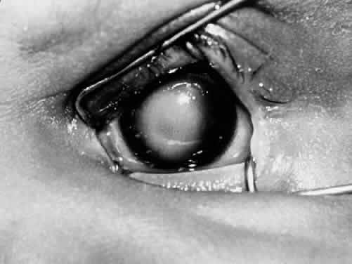
electronmicroscopic findings. Br J Ophthalmol 74:59, 1990

48. Waring GO, Roth AM, Rodrigues MM: Clinicopathologic correlation of microphthalmos with cyst. Am J Ophthalmol 82:714, 1976

49. Lim G, Bejec LC: Congenital cystic microphthalmos. Philippine J Ophthalmol 5:93, 1973

50. Weyman MF: Microphthalmos with cyst formation. Am J Ophthalmol 8:214, 1925

51. Kok-van Alphen CC, Manshot WA, Frederiks E et al: Microphthalmos and orbital cyst. Ophthalmologica 167:389, 1973

52. Makley TA Jr, Battles M: Microphthalmos with cyst: Report on two cases in the same family. Surv Ophthalmol 13:200, 1969

53. Ladenheim J, Metrick S: Congenital microphthalmos with cyst formation. Am J Ophthalmol 41:1059, 1956

54. Yanoff M, Ballian LR, Niederer BS: Ocular and cerebral abnormalities in chromosome 18 deletion defect. Am J Ophthalmol 70:391, 1970

55. von Hippel E: Über die Beyrehunder von Mikrophthalmos mit Unterlidcyste zu allgemeinen Missbildungen besonders zum Lindauschen Symptomenkomplex. Graefes Arch Ophthalmol 132:25, 1934

56. Jesberg DO, Schepans CL: Retinal detachment associated with coloboma of the choroid. Arch Ophthalmol 65:163, 1961

57. Jensen PE, Kalina RE: Congenital anomalies of the optic disk. Am J Ophthalmol 82:27, 1976

58. Francois J: Colobomatous malformations of the ocular globe. Int Ophthalmol Clin 8:797, 1968

59. Pollock JA, Newton TH, Hoyt WF: Transsphenoidal and transethmoidal encephaloceles: A review of clinical and roentgen features in 8 cases. Radiology 90:442, 1968

60. Corbett JJ, Savino PJ, Schatz NJ et al: Cavitary developmental defects of the optic disc: Visual loss associated with optic pits and colobomas. Arch Neurol 37:210, 1980

61. Streletz LJ, Schatz NJ: Transsphenoidal encephalocele associated with colobomas of the optic disc and hypopituitary dwarfism. In Smith JL, Glaser JS (eds): Neuro-Ophthalmology Symposium of the University of Miami and the Bascom Palmer Eye Institute, Vol 7, pp 78–86. St. Louis, CV Mosby, 1973

62. Cross HE: Ocular colobomas. In Bergsma D (ed): Birth Defects Compendium, p 791. New York, Alan R Liss, 1979

63. Meyer SJ, Holstein T: Spherophakia with glaucoma and brachydactyly. Am J Ophthalmol 24:247, 1941

64. Urbanek J: Glaucoma juvenile inversion. Z Augenheilkd 77:171, 1930

65. Blaxter PL: Spherophakia. Trans Ophthalmol Soc UK 88:621, 1969

66. Willi M, Kut K, Cother E: Pupillary block glaucoma in the Marchesani syndrome. Arch Ophthalmol 90:504, 1973

67. Cross HE, Jensen AD: Ocular manifestations in the Marfan syndrome and homocystinuria. Am J Ophthalmol 75:405, 1973

68. Schar E: Renal disease, inner ear deafness and ocular changes: A new heredofamilial syndrome. Arch Intern Med 97:627, 1956

69. Bessiere E, Riviera J, Leuret JP: Le rebellar, an association of Klinefelter's disease and congenital anomalies, comptodactyly, microphakia. Bull Soc Ophthalmol Fr 62:197, 1962

70. Jensen AD, Cross HE, Paton D: Ocular complications in the Weill-Marchesani syndrome. Am J Ophthalmol 77:261, 1974

71. Johnson VP, Grayson M, Christian JC et al: Dominant microspherophakia. Arch Ophthalmol 85:534, 1971

72. Arnott EJ, Crawford MD, Toghill PJ: Anterior lenticonus and Alport's syndrome. Br J Ophthalmol 50:390, 1966

73. Chugh KS, Sakhuja V, Agarwal A et al: Hereditary nephritis (Alport's syndrome): Clinical profile and inheritance in 28 kindreds. Nephrol Dial Transplant 8:690, 1993

74. Streeten BW, Robinson MR, Wallace R et al: Lens capsule abnormalities in Alport's syndrome. Arch Ophthalmol 105:1693, 1987

75. Cheong HI, Kashtan CE, Kim Y et al: Immunohistologic studies of type IV collagen in anterior lens capsules of patients with Alport syndrome. Lab Invest 70:553, 1994

76. Francois J: Congenital Cataracts. Assen, Charles C Thomas, 1963

77. Hunter WS, Zimmerman LE: Unilateral retinal dysplasia. Arch Ophthalmol 74:23, 1965

78. Lahav M, Albert DM, Wyand S: Clinical and histopathologic classification of retinal dysplasia. Am J Ophthalmol 75:648, 1973

79. Seefelder R: Zur Frage der Netzhautanomalien in sonst normalen fötalen menschlichen Augen. Graefes Arch Ophthalmol 73:216, 1909

80. Silverstein AM, Osburn BI, Prendergast RA: The pathogenesis of retinal dysplasia. Am J Ophthalmol 72:13, 1971

81. Reese AB, Blodi FC: Retinal dysplasia. Am J Ophthalmol 33:23, 1950

82. Reese AB, Straatsma BR: Retinal dysplasia. Am J Ophthalmol 45:199, 1958

83. Miller M, Robbins J, Fishman R et al: A chromosomal anomaly with multiple ocular defects including retinal dysplasia. Am J Ophthalmol 55:901, 1963

84. Cogan DG, Kuwabara T: Ocular pathology of the 13–15 trisomy syndrome. Arch Ophthalmol 72:246, 1964

85. Zimmerman LE, Font RL: Congenital malformations of the eye. JAMA 196:684, 1966

86. Fradkin AH: Norrie's disease: Congenital progressive oculoacousticocerebral degeneration. Am J Ophthalmol 72:947, 1971

87. Howard RO, Bey WR, Albert DM et al: Retinoblastoma and chromosome abnormality. Arch Ophthalmol 92:490, 1974

88. Haddad R, Font RL, Reeser F: Persistent hyperplastic primary vitreous: A clinicopathologic study of 62 cases and review of the literature. Surv Ophthalmol 23:123, 1976

89. van Sien S, Sullivan WB: Congenital retinal fold: A case report. Am J Ophthalmol 39:643, 1955

90. Guerry D: Congenital retinal folds: Report of two cases. Am J Ophthalmol 27:1132, 1944

91. Anderson SR: A typical falciform retinal fold. Acta Ophthalmol 30:325, 1952

92. Dessoff J: Blue sclerotics, fragile bones and deafness. Arch Ophthalmol 12:60, 1934

93. Ruedman AD: Osteogenesis imperfecta congenita and blue sclera. Arch Ophthalmol 49:6, 1953

94. Francois J: Affections of the cornea. In: Heredity in Ophthalmology. St. Louis, CV Mosby, 1961

95. Howard RO, Abrahams IW: Sclerocornea. Am J Ophthalmol 71:1254, 1971

96. Goldstein JE, Cogan DG: Sclerocornea and associated congenital anomalies. Arch Ophthalmol 67:761, 1962

97. Kanai A, Wood TC, Polack FM et al: The fine structure of sclerocornea. Invest Ophthalmol Vis Sci 10:687, 1971

98. Petroutsos G, Patey A, Savoldelli M et al: Sclerocornee: Etude ultrastruccturale et morphologique. J Fr Ophthalmol 6:769, 1983

99. Kenyon KR, Fogle JA, Grayson M: Dysgeneses, dystrophies, and degenerations of the cornea. In Duane TD (ed): Clinical Ophthalmology. Philadelphia, JB Lippincott, 1982

100. Kokott W: Cornea plana und mikrocornea periplana. Klin Monatsbl Augenheilkd 98:372, 1937

101. Tahvanainen E, Villanueva AS, Forsius H et al: Dominantly and recessively inherited cornea plana congenita map to the same small region of chromosome 12. Genome Res 6:249, 1996

102. Tahvanainen E, Forsius H, Karila E et al: Cornea plana congenita gene assigned to the long arm of chromosome 12 by linkage analysis. Genomics 26:290, 1995

103. Lowe RF: Aetiology of anatomical basis of primary angle-closure glaucoma: Biometrical comparisons between normal eyes and eyes with primary angle-closure glaucoma. Br J Ophthalmol 54:161, 1970

104. Grayson M: Diseases of the Cornea. St. Louis, CV Mosby, 1979

105. Vail DT: Adult hereditary anterior megalophthalmus sine glaucoma: A definite disease entity, with special reference to extraction of the cataract. Arch Ophthalmol 6:39, 1931

106. Meire FM, Bleeker WE, Oehler M et al: X-linked megalocornea: Ocular findings and linkage analysis. Opthalmic Paediatr Genet 12:153, 1991

107. Francois J: La gonioscopie, III, Coloboma de l'iris. Adv Ophthalmol 4:44, 1955

108. Malbran E, Dodds R: Megalocornea and its relation to congenital glaucoma. Am J Ophthalmol 49:908, 1960

109. Stephenson WV: Anterior megalophthalmos and arachnodactyly. Am J Ophthalmol 8:315, 1954

110. Mackey DA, Buttery RG, Wise GM et al: Description of X-linked megalocornea with identification of the gene locus. Arch Ophthalmol 109:829, 1991

111. Krachmer JH, Rodrigues MM: Posterior keratoconus. Arch Ophthalmol 96:1867, 1978

112. Mannis MJ, Lightman J, Plotnik RD: Corneal topography of posterior keratoconus. Cornea 11:351, 1992

113. Jacobs HB: Posterior conical cornea. Br J Ophthalmol 41:31, 1957

114. Haney W, Falls HF: The occurrence of congenital keratoconus posticus circumscriptus. Am J Ophthalmol 52:53, 1961

115. Wolter JR, Haney W: Histopathology of keratoconus posticus circumscriptus. Arch Ophthalmol 69:357, 1963

116. Butler TH: Keratoconus posticus. Trans Ophthalmol Soc UK 50:551, 1930

117. Greene PB: Keratoconus posticus circumscriptus: Report of a case. Arch Ophthalmol 34:432, 1945

118. Benjamin SN, Allen HF: Classification for limbal dermoid choristomas and branchial arch anomalies. Arch Ophthalmol 87:305, 1972

119. Baum JL, Feingold M: Ocular aspects of Goldenhar's syndrome. Am J Ophthalmol 75:250, 1973

120. Hogan MJ, Zimmerman LE: Ophthalmic Pathology. Philadelphia, WB Saunders, 1962

121. Maumenee AE: Congenital hereditary corneal dystrophy. Am J Ophthalmol 50:1114, 1960

122. Kenyon KR, Maumenee AE: Further studies of congenital hereditary endothelial dystrophy of the cornea. Am J Ophthalmol 76:419, 1973

123. Judisch GF, Maumenee IH: Clinical differentiation of recessive congenital hereditary endothelial dystrophy and dominant hereditary endothelial dystrophy. Am J Ophthalmol 85:606, 1978

124. Arffa RC: Disorders of the endothelium. In Grayson's Diseases of the Cornea, 2nd ed. St. Louis, Mosby-Year Book, 1991

125. Ostler HB: Diseases of the cornea. In: Diseases of the External Eye and Adnexa: A Text and Atlas. Baltimore, Williams & Wilkins, 1993

126. Bahn CF, Falls HF, Varley GA et al: Classification of corneal endothelial disorders based on neural crest origin. Ophthalmology 91:558, 1984

127. Sekundo W, Marshall GE, Lee WR et al: Immunoelectron labelling of matrix components in congenital hereditary endothelial dystrophy. Graefes Arch Clin Exp Ophthalmol 232:337, 1994

128. Alkemade PPH: Dysgenesis Mesodermalis of the Iris and the Cornea. Assen, Charles C Thomas, 1969

129. Wilson ME: Congenital iris ectropion and a new classification for anterior segment dysgenesis. J Pediatr Ophthalmol Strabismus 27:48, 1990

130. Townsend WM: Congenital anomalies of the cornea. In Kaufman HE, Barron BA, McDonald MB, Waltman SR (eds): The Cornea. New York, Churchill Livingstone, 1988

131. Waring GO, Rodrigues MM, Laibson PR: Anterior-chamber cleavage syndrome: A stepladder classification. Surv Ophthalmol 20:3, 1975

132. Yue BY, Baum JL, Silbert JE: Synthesis of glycosaminoglycans by cultures of normal human corneal endothelial and stromal cells. Invest Ophthalmol Vis Sci 17:523, 1978

133. Townsend WM, Font RL, Zimmerman LE: Congenital corneal leukomas: II. Histopathologic findings in 19 eyes with central defect in Descemet's membrane. Am J Ophthalmol 77:192, 1974

134. Traboulsi EI, Maumenee IH: Peters' anomaly and associated congenital malformations. Arch Ophthalmol 110: 1739, 1992

135. Yang LLH, Lambert SR, Fernhoff PM, Stulting RD: Peters' anomaly: Associated congenital malformations and etiology. Invest Ophthalmol Vis Sci 36(suppl):S41, 1995

136. Wilson FM: Congenital anomalies. In Smolin G, Thoft RA (eds): The Cornea: Scientific Foundations and Clinical Practice, 2nd ed, p 457. Boston, Little, Brown & Co, 1987

137. Pearce WG, Tripathi RC, Morgan G: Congenital endothelial corneal dystrophy: clinical, pathological and genetic study. Br J Ophthalmol 53:577, 1969

138. Bourne WM: Partial corneal involvement in the iridocorneal endothelial syndrome. Am J Ophthalmol 94:774, 1982

139. Kolker AE, Hetherington J Jr: Becker-Shaffer's Diagnosis and Therapy of the Glaucomas. St. Louis, CV Mosby, 1970

140. Hetherington J: Congenital glaucoma. In Duane TD (ed): Clinical Ophthalmology, Vol 3, Chap 51. Philadelphia, JB Lippincott, 1982

141. Barkan O: Pathogenesis of congenital glaucoma. Am J Ophthalmol 40:1, 1955

142. Worst J: The cause and treatment of congenital glaucoma. Trans Am Acad Ophthalmol Otolaryngol 68:766, 1964

143. Kupfer C, Kaiser-Kupfer MI: Observations on the development of the anterior chamber angle with reference to the pathogenesis of congenital glaucoma. Am J Ophthalmol 88:424, 1979

144. Tripathi BJ, Tripathi RC: Neural crest origin of human trabecular meshwork and its implications for the pathogenesis of glaucoma. Am J Ophthalmol 107:583, 1989

145. Cross HE: Ectopia lentis et pupillae. Am J Ophthalmol 88:381, 1979

146. Nelson LB, Spaeth GL, Nowinski TS et al: Aniridia: A review. Surv Ophthalmol 28:621, 1984

147. Dowling JL, Albert DM, Nelson LB et al: Primary glaucoma associated with iridotrabecular dysfunction and ectropion uvae. Ophthalmology 92:912, 1985

148. Quigley HA, Stanish FS: Unilateral congenital iris pigment epithelial hyperplasia associated with late onset glaucoma. Am J Ophthalmol 86:182, 1978

149. Ritch R, Forbes M, Hetherington J et al: Congenital ectropion uvae with glaucoma. Ophthalmology 91:326, 1984

150. Shields MB: Axenfeld-Rieger syndrome: A theory of mechanism and distinction from the iridocorneal endothelial syndrome. Trans Am Ophthalmol Soc 81:736, 1983

151. Shaw MW, Falls HF, Neel JV: Congenital aniridia. Am J Hum Genet 12:389, 1960

152. Layman PR, Anderson RA, Flynn JT: Frequent occurrence of hypoplastic optic discs in patients with aniridia. Am J Ophthalmol 77:513, 1974

153. Elsas FJ, Maumenee IH, Kenyon KR et al: Familial aniridia with preserved ocular function. Am J Ophthalmol 83: 718, 1977

154. Miller RW, Fraameni JF, Manning MD: Association of Wilms' tumor with aniridia, hemihypertrophy and other congenital malformations. N Engl J Med 170:922, 1964

155. Fraumeni JF: The aniridia-Wilms' tumor syndrome. Birth Defects 5:198, 1969

156. Riccardi VM, Sujansky E, Smith AC et al: Chromosomal imbalance in the aniridia-Wilms' tumor association: 11p interstitial deletion. Pediatrics 61:604, 1978

157. DiGeorge AM, Harley RD: Association of aniridia, Wilms' tumor and genital abnormalities. Arch Ophthalmol 75: 796, 1966

158. Flanagan JC, DiGeorge AM: Sporadic aniridia and Wilms' tumor. Am J Ophthalmol 67:558, 1969

159. Fraumeni JF, Glass AG: Wilms' tumor and aniridia. JAMA 206:825, 1968

160. Francke U, Holmes LB, Atkins L et al: Aniridia-Wilms' tumor association: Evidence for specific deletion of 11P13. Cytogenet Cell Genet 24:185, 1979

161. Francke U, Riccardi VM, Hittner HM et al: Interstitial deletion (11P) as a cause of the aniridia-Wilms' tumor association: Band localization and a hereditable basis (abstr). Am J Hum Genet 30:81A, 1978

162. Hittner HM, Riccardi VM, Francke U: Aniridia caused by hereditable chromosome 11 deletion. Ophthalmology 86:1173, 1979

163. Ton CCT, Hirvonen H, Miwa H et al: Positional cloning and characterization of a paired box—and homeobox—containing gene from the aniridia region. Cell 67:1059, 1991

164. Cresswell TM: A case of congenital miosis. Br J Ophthalmol 8:278, 1924

165. Polomeno RC, Milot J: Congenital miosis. Can J Ophthalmol 14:43, 1979

166. Veirs ER, Brown W: Congenital miosis. Arch Ophthalmol 65:59, 1961

167. Lambert SR, Amaya L, Taylor D: Congenital idiopathic microcoria. Am J Ophthalmol 106:590, 1988

168. Holth S, Berner O: Congenital miosis or pinhole pupils owing to developmental faults of the dilator muscle. Br J Ophthalmol 7:401, 1923

169. Jensen OA: Persistent hyperplastic primary vitreous. Acta Ophthalmol 46:418, 1968

170. Pruett RC, Schepens CL: Posterior hyperplastic primary vitreous. Am J Ophthalmol 69:535, 1970

171. Wolter JR, Flaherty NW: Persistent hyperplastic vitreous. Am J Ophthalmol 47:491, 1959

172. Manschot WA: Persistent hyperplastic primary vitreous. Arch Ophthalmol 59:188, 1958

173. Font RL, Yanoff M, Zimmerman LE: Intraocular adipose tissue and persistent hyperplastic primary vitreous. Arch Ophthalmol 82:43, 1969

174. Bullock JO: Developmental vitreous cysts. Arch Ophthalmol 91:83, 1974

175. Francois J: Prepapillary cyst developed from remnants of the hyaloid artery. Br J Ophthalmol 34:365, 1950

176. Apple DJ, Rabb MF, Walsh PM: Congenital anomalies of the optic disc. Surv Ophthalmol 27:3, 1982

177. Nelson LB, Maumenee IH: Ectopia lentis. Surv Ophthalmol 27:143, 1982

178. Frisen L, Holmegaard L: Spectrum of optic nerve hypoplasia. Br J Ophthalmol 62:7, 1978

179. Hoyt CS, Billson FA: Optic nerve hypoplasia: Changing perspectives. Aust NZ J Ophthalmol 14:325, 1986

180. Manshot WA: Eye findings in hydroencephaly. Ophthalmologica 162:151, 1971

181. Ellenberger C Jr, Runyon TE: Holoprosencephaly with hypoplasia of the optic nerves, dwarfism and agenesis of the septum pellucidum. Am J Ophthalmol 70:960, 1970

182. Hoyt WF, Kaplan SL, Grumbach MM et al: Septo-optic dysplasia and pituitary dwarfism. Lancet 1:893, 1970

183. Harris RJ, Haas L: Septo-optic dysplasia with growth hormone deficiency (de Morsier syndrome). Arch Dis Child 47:973, 1972

184. Edwards WC, Layden WE: Optic nerve hypoplasia. Am J Ophthalmol 70:950, 1970

185. Scheie HG, Adler FH: Aplasia of the optic nerve. Arch Ophthalmol 26:61, 1941

186. Barkovich AJ, Lyon G, Evrard PL: Formation, maturation, and disorders of white matter. AJNR 13:447, 1992

187. Brodsky MC: Septo-optic dysplasia: A reappraisal. Semin Ophthalmol 6:227, 1991

188. Hatten ME, Mason CA: Mechanisms of glial guided neuronal migration in vitro and in vivo. Experientia 46:907, 1990

189. Hoyt CS, Good WV: Do we really understand the difference between optic nerve hypoplasia and atrophy? Eye 6:210, 1992

190. Skarf B, Hoyt CS: Optic nerve hypoplasia in children. Arch Ophthalmol 102:255, 1984

191. Kindler P: Morning glory syndrome: Unusual congenital optic disk anomaly. Am J Ophthalmol 69:376, 1970

192. Pollock S: The morning glory disc anomaly: Contractile movement, classification, and embryogenesis. Doc Ophthalmol 65:439, 1987

193. Beyer WB, Quencer RM, Osher RH: Morning glory syndrome: A functional analysis including fluorescein angiography, ultrasonography, and computerized tomography. Ophthalmology 89:1362, 1982

194. Caprioli J, Lesser RL: Basal encephalocele and morning glory syndrome. Br J Ophthalmol 67:349, 1983

195. Goldhammer Y, Smith JL: Optic nerve anomalies in basal encephaloceles. Arch Ophthalmol 93:115, 1975

196. Hope-Ross M, Hohnston SS: The morning glory syndrome associated with sphenethmoidal encephalocele. Ophthalmic Paediatr Genet 2:147, 1990

197. Koenig SP, Naidich TP, Lissner G: The morning glory syndrome associated with sphenoidal encephalocele. Ophthalmology 89:1368, 1982

198. Traboulsi EU, O'Neill JF: The spectrum in the morphology of the so called “morning glory disc anomaly.” J Pediatr Ophthalmol Strabismus 25:93, 1988

199. Gardner TW, Zaparackas ZG, Naidach TP: Congenital optic nerve colobomas: CT demonstration. J Comput Assist Tomogr 8:95, 1984

200. Mafee MF, Jampol LM, Langer BG, Tso MOM: Computed tomography of optic nerve colobomas, morning glory anomaly, and colobomatous cyst. Radiol Clin North Am 25:693, 1987

201. Greear JN Jr: Pits or crater like holes in the optic disk. Arch Ophthalmol 28:467, 1942

202. Ferry AP: Macular detachment associated with congenital pit of the optic nerve head. Arch Ophthalmol 70:346, 1963

203. Sugar HA: An explanation for the acquired macular pathology associated with congenital pits of the optic disc. Am J Ophthalmol 57:833, 1964

204. Gass JD: Serous detachment of the macula secondary to optic pit of the nerve head. Am J Ophthalmol 67:821, 1969

205. Brockhurst R: Optic pits and posterior retinal detachment. Trans Am Ophthalmol Soc 73:264, 1975

206. Kranenburg EW: Craterlike holes in the optic disc and central serous retinopathy. Arch Ophthalmol 64:912, 1960

207. Francois J: Syndrome malformatif avec cryptophthalmic. Acta Genet Med Gemellol 18:18, 1969

208. Sugar HS: The cryptophthalmos-syndactyly syndrome. Am J Ophthalmol 66:897, 1968

209. Waring GO, Shields JA: Partial unilateral cryptophthalmos with syndactyly, brachycephaly and renal anomalies. Am J Ophthalmol 79:437, 1975

210. Goldberger E: Epicanthus and its variants among Caucasians. Arch Ophthalmol 16:506, 1936

211. Usher CH: The Bowman lecture: On a few hereditary eye affections. Trans Ophthalmol Soc UK 55:164, 1935

212. Berke RN: Congenital ptosis: A classification of two hundred cases. Arch Ophthalmol 41:188, 1949

213. Briggs H: Hereditary congenital ptosis with report of 64 cases conforming to the mendelian rule of dominance. Am J Ophthalmol 2:408, 1919

214. Roden FH, Barkan H: Hereditary congenital ptosis: Report of a pedigree and review of literature. Am J Ophthalmol 18:213, 1935

215. Rank BK, Thomson JA: The genetic approach to hereditary congenital ptosis. Aust NZ J Surg 28:274, 1959

216. Berke RN, Wadsworth JAC: Histology of levator muscle in congenital and acquired ptosis. Arch Ophthalmol 53:413, 1955

217. Cassidy JV: Developmental anatomy of the nasolacrimal duct. Arch Ophthalmol 47:141, 1952

Back to Top