|
|
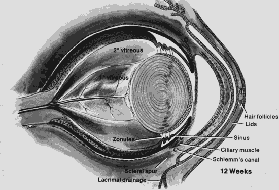
| Fig. 2. The eye at 12 weeks. The cornea is separated from the more anterior fused eyelids. A membrane covers the pupil (pupillary membrane). Note the relationship of the scleral spur to the anterior chamber: the angle is incompletely formed. The lens vesicle is now obliterated. The secondary vitreous is avascular, whereas the primary vitreous is vascularized. Note the vascular arcades on the posterior surface of the lens (posterior tunica vasculosa lentis). (Courtesy of Irene H. Maumenee, MD) |