Chapter 19
Glaucoma
JOSEPH W. SASSANI
Main Menu   Table Of Contents

Search

INCIDENCE
CLASSIFICATION
NORMAL OUTFLOW
IMPAIRED OUTFLOW
VARIABLE OUTFLOW
TISSUE CHANGES CAUSED BY ELEVATED INTRAOCULAR PRESSURE
REFERENCES

The glaucomas are a diverse group of disorders that damage the optic nerve, resulting in characteristic optic nerve head cupping and visual field loss. The intraocular pressure (IOP) in such persons is presumed to be inconsistent with normal optic nerve function and is usually elevated above normal values. Nevertheless, IOP elevation need not be present to make the diagnosis of glaucoma. Patients with characteristic optic nerve cupping and visual field loss in the presence of normal IOP are diagnosed as having normal-pressure (low-pressure) glaucoma. Conversely, patients with elevated IOP in the absence of detectable secondary abnormalities in ocular structure and function are classified as ocular hypertensives.

The glaucomas are, therefore, a group of syndromes that require for their diagnosis characteristic abnormalities in ocular structure and function rather than merely an elevated IOP. To simplify terminology and to comply with conventional usage, “glaucoma” will be used in the singular in this chapter.

Back to Top
INCIDENCE
Glaucoma is the leading cause of blindness in the United States and is responsible for 80,000 of the 500,000 legally blind people in this country.1 Two million Americans have glaucoma and 5 million have elevated IOP.1 The worldwide incidence of glaucoma has been estimated by various authors as between 0.47% and 8%.2–4 The variation in these estimates may in part reflect differences in sampling technique and racially determined factors.

There is a prominent racial influence on the incidence of glaucoma. In one urban U.S. population, the prevalence of primary open-angle glaucoma among blacks was four to five times higher than among whites.5 Further data on the incidence of glaucoma will be presented as individual syndromes are discussed.

Back to Top
CLASSIFICATION
The following is a useful classification of glaucoma, constructed with reference to the anatomy of the anterior chamber angle as well as to specific pathophysiologic and etiologic factors:
  1. Normal outflow
    1. Hypersecretion glaucoma

  2. Impaired outflow
    1. Congenital glaucoma (developmental)
    2. Primary glaucoma
      1. Angle-closure
        1. With pupillary block
        2. Without pupillary block

      2. Open-angle

    3. Secondary glaucoma
      1. Angle-closure
        1. With pupillary block
        2. Without pupillary block

      2. Open-angle


  3. Variable outflow
    1. Normal-pressure (low-pressure) glaucoma

Back to Top
NORMAL OUTFLOW

HYPERSECRETION GLAUCOMA

Hypersecretion glaucoma is rare and is said to have no antecedent cause.6 It is characterized by a normal outflow facility accompanied by elevated IOP, presumably secondary to increased production of aqueous humor. It affects mainly middle-aged women, particularly those having neurogenic systemic hypertension. The histology of the trabecular meshwork is said to be normal. Some authors question the existence of hypersecretion glaucoma as a discrete entity.

Back to Top
IMPAIRED OUTFLOW

EMBRYOLOGIC DEVELOPMENT AND GLAUCOMA

Traditionally, it has been thought that much of the anterior segment of the globe is derived from mesoderm, the primary component of the middle embryologic layer. Recent studies suggest that the cells of the neural crest, which arise at the margins of the embryologic neural plate just before the closure of the neural tube, contribute large portions of the eye, orbit, and head, thereby functioning as mesoderm in this region.7–17 These studies demonstrate that specific ocular tissues that may be derived from the neural crest include corneal stroma and endothelium, anterior iris stroma, iris melanocytes, ciliary body, sclera, intraocular vascular pericytes, and portions of the trabecular meshwork, including endothelium. The following description of the embryology of the anterior segment is derived from these studies.

The neural crest cells are believed to move into the developing eye in three waves that pass between the surface ectoderm and the lens. The first wave of cells differentiates into the corneal endothelium by the eighth developmental week and produces the trabecular meshwork and its endothelium. The corneal endothelium is responsible for the production of Descemet's membrane. The second wave infiltrates between the corneal epithelium and endothelium to form keratocytes. The third wave produces the iris stroma. Subsequently, the iris pigment epithelium is derived from neural ectoderm. The endothelium of Schlemm's canal and vascular endothelium are derived from vascular mesoderm and not from neural crest.

Neural crest cells also contribute to important structures of the orbit and head, including the meninges, pituitary gland, bones and cartilage of the upper face, dental papillae, and connective tissue supporting cells of the orbit.

The widespread contributions of neural crest-derived cells in the embryology of the eye have given neural crest a central role in the pathogenesis of several forms of glaucoma. Further, abnormalities in neural crest cell migration and differentiation may explain many of the developmental abnormalities of the face and head associated with various glaucoma syndromes. Table 1 attempts to correlate anterior segment disorders with the presumed abnormality in neural crest cell function. This table will be referenced later in this chapter as several glaucoma syndromes are discussed.


TABLE 19-1. Classification of Anterior Segment Disorders Based on Neural Crest Origins


Cell AbnormalitySecondary Disorder
Deficient neural crest formation (brain-eye-face malformations) 
Abnormal crest cell migrationCongenital glaucoma
 Posterior embryotoxin
 Axenfeld's anomaly
 Rieger's syndrome
 Peters' syndrome
 Sclerocornea
Abnormal crest cell proliferationIris nevus syndrome
 Chandler's syndrome
 Essential iris atrophy
Abnormal crest cell terminal induction (final differentiation)Congenital hereditary endothelial dystrophy
 Posterior polymorphous dystrophy
 Fuchs' combined dystrophy
Acquired abnormalitiesMetaplasia
 Abiotrophy
 Proliferation
(Modified from Bahn CF, Falls HF, Varley GA, et al: Classification of corneal endothelial disorders based on neural crest origin. Ophthalmology 91:558, 1984)


CONGENITAL GLAUCOMA

The term congenital glaucoma designates glaucoma that is detected in the neonatal and infantile period. The topic has been reviewed extensively by deLuise and Anderson, and their work has been most helpful in the preparation of this section.18 The incidence of congenital glaucoma is approximately 1 in 10,000 births.19 Usually it is sporadic; however, autosomal inheritance has been reported.18,20 It is bilateral in 64% to 88% of cases,21,22 and most affected persons are boys (60% to 70%).18 It is diagnosed within the first year of life in 86% of cases, with 40% diagnosed at birth and 34% between birth and 6 months of age.18

There have been many theories relating to its pathogenesis. The surface of the trabecular meshwork in congenital glaucoma has been described as having a peculiar sheen on gonioscopic examination. This finding has suggested to some authors the presence of an abnormal mesodermally derived membrane, Barkan's membrane, covering the surface of the trabecular meshwork and preventing the efflux of aqueous. The effectiveness of goniotomy surgery and the ineffectiveness of miotics in the treatment of congenital glaucoma have been cited in support of this theory. There is, however, little histologic proof for this theory.

Except in the anterior chamber cleavage syndromes, absence of Schlemm's canal is probably rare as an etiology of congenital glaucoma. Compression and obliteration of Schlemm's canal is most often seen as a secondary finding in advanced cases in which there is marked distortion of normal anterior segment anatomy.

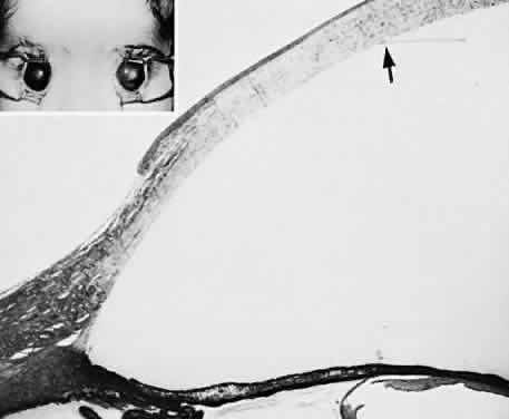
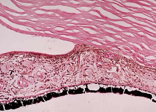
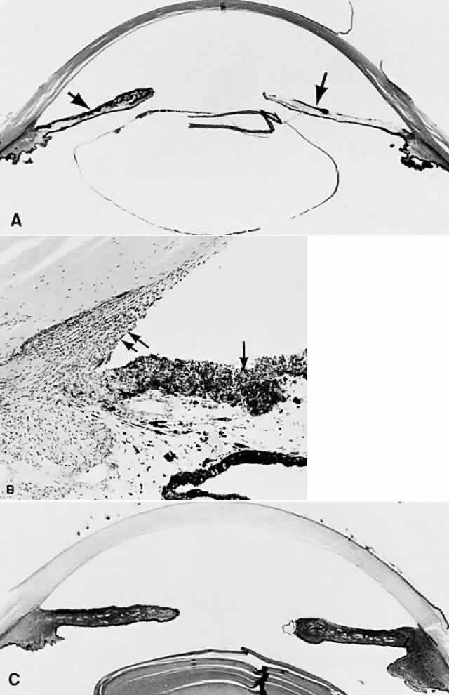
Until recently, the anterior chamber angle was thought to arise through a process of cleavage of mesodermal tissue. Arrest in this process was said to result in an “embryonic” configuration of the anterior chamber angle (Fig. 1). This immature angle is characterized histologically by an anterior insertion of the iris root on the ciliary body, anteriorly displaced ciliary processes, insertion of the ciliary meridional muscles into the trabecular meshwork instead of into (or over) the scleral roll (when more prominent, as in the older adult, the scleral roll is called the scleral spur), and the presence of mesenchymal tissue in the anterior chamber angle.18 More severe abnormalities in this cleavage process were thought to result in the anterior chamber cleavage syndromes: Axenfeld's, Rieger's, and Peters' syndromes.23 Rieger's syndrome may be associated with facial, dental, otologic, dermatologic, and neurologic disorders, including pituitary abnormalities, and also may be associated with trisomy of chromosome 13 or with partial trisomy 16q (see also Chap. 9 for further information on these syndromes). The anterior cleavage syndromes now are recognized as being caused by abnormalities in neural crest development.24

Fig. 1. “Embryonic configuration” of the anterior chamber angle typically seen in congenital glaucoma. It is not possible to determine on a histopathologic basis whether the person had glaucoma (hematoxylin-eosin, × 25).

Of particular public health importance is the recently recognized association of Peters' syndrome and fetal alcohol syndrome, which is secondary to maternal alcohol ingestion early in the first trimester of pregnancy. It is the most common known cause of mental retardation in the United States.25 Affected infants have a characteristic appearance that includes microcephaly, microphthalmia and other ocular anomalies, a poorly developed philtrum, short palpebral fissures, a thin upper lip, and flattening of the upper maxillary area. Other associated findings are growth retardation and central nervous system and intellectual abnormalities.26,27 Approximately 90% of children with fetal alcohol syndrome have ocular abnormalities,28 which include optic nerve hypoplasia,28–30 tortuosity of retinal blood vessels,28,29 strabismus,28,31 microphthalmos,32 and ptosis.28 Fetal alcohol syndrome also may be associated with DiGeorge syndrome.33

Although an “embryonic” anterior chamber angle is found in congenital glaucoma, it is also seen in age-matched persons who do not have the disorder (see Fig. 1). Further, difficulties in interpreting histologic sections in this age group are compounded by the few truly meridional sections encountered in the usual serial sectioning of the globe. Thus, sectioning artifact enhances the appearance of an “embryonic” configuration in the anterior chamber angle.

The recognition of the contribution of neural crest-derived cells in the embryology of the anterior chamber angle may provide the means for unifying several glaucoma syndromes, particularly those associated with congenital glaucoma.

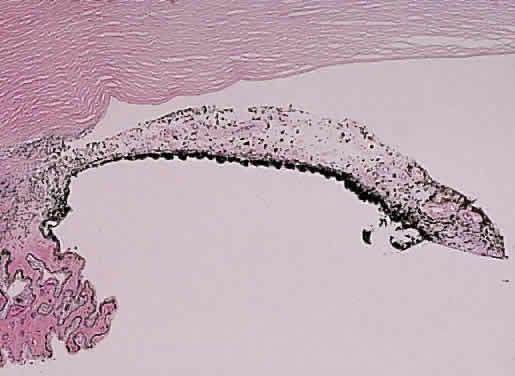
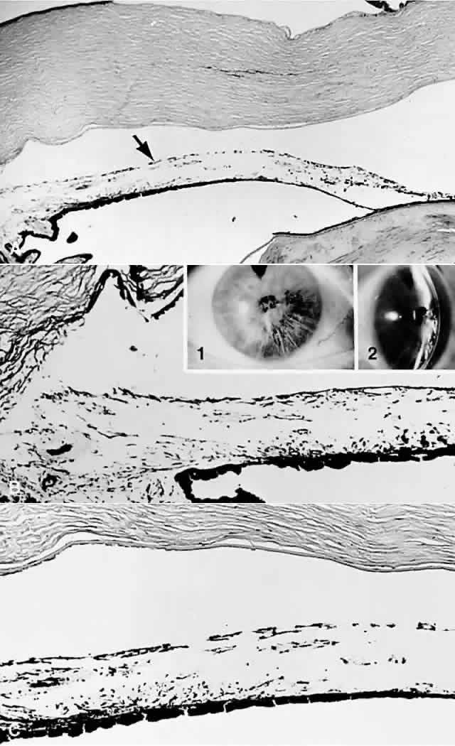
A syndrome of primary glaucoma associated with iridotrabecular dysgenesis and ectropion uveae has been described.34 In these patients, the iris pigment epithelium proliferates onto the anterior iris stroma in utero so that congenital ectropion uveae is present (Fig. 2). Glaucoma may be present in infancy or may develop later in life. Histologically, the central corneal stroma is normal; however, there is posterior embryotoxin (prominence and anterior displacement of Schwalbe's ring) with iris processes. Migration and hyerplasia of the pigment epithelium onto the anterior iris surface is accompanied by endothelialization of the iris surface peripheral to the area where pigment epithelium is present. Angle closure with iris adherent to a normally differentiated trabecular meshwork also is seen. The iris stroma is hypoplastic. The relation of this syndrome to the anterior chamber cleavage syndromes and to the iridocorneal endothelial (ICE) syndrome has not been established. This syndrome may bridge the groups of disorders characterized in Table 1 as secondary to abnormal crest cell migration and proliferation.

Fig. 2. Left, Slit-lamp photograph of congenital ectropion uveae. Right, Gonioscopic appearance of dysgenesis of filtration angle in congenital ectropion uveae. (Dowling JL Jr, Albert DM, Nelson LB, et al: Primary glaucoma associated with iridotrabecular dysgenesis and ectropion uveae. Ophthalmology 92:912, 1985)

Congenital ectropion uveae may be found in patients with neurofibromatosis, or it may be associated with other syndromes, including Prader-Willi syndrome.35–39 Table 2 outlines other ocular disorders that may be associated with congenital glaucoma. The possible relation of abnormalities in neural crest cell migration to the developmental disorders in Table 2 remains to be established.


TABLE 19-2. Ocular Disorders Associated With Congenital Glaucoma

  Corneal anomalies

  Sclerocornea
  Microcornea
  Megalocornea
  Corneal staphyloma


  Corneal (ectodermal-mesodermal) dysgenesis

  Axenfeld's anomaly
  Rieger's syndrome
  Peters' syndrome
  Iridotrabecular dysgenesis-ectropion uveae syndrome


  Microphthalmos

  Simple
  Associated


  Iris anomalies

  Aniridia
  Colobomata
  Polycoria, microcoria


  Lens anomalies

  Congenital aphakia
  Lenticonus
  Lentiglobus
  Spherophakia
  Ectopia lentis

  Homocystinuria
  Marfan syndrome
  Weill-Marchesani syndrome
  Ehlers-Danlos syndrome


(Modified from Bardelli AM, Hadjistilianou T: Congenital glaucoma associated with other abnormalities in 150 cases. Glaucoma 9:10, 1987)


Congenital glaucoma is a common feature of several systemic disorders, summarized in Table 3. The multiple ocular anomalies and systemic disorders associated with congenital glaucoma suggest the probability that multiple factors may result in impaired aqueous outflow.


TABLE 19-3. Systemic Disorders Associated With Congenital Glaucoma

  Chromosomal defects

  10p-
  Partial trisomy 16q
  Ring chromosome 6
  Trisomy 13


  Inborn errors of metabolism

  Homocystinuria
  Hurler's syndrome
  Lowe's syndrome
  Refsum's syndrome


  Infectious

  Rubella


  Phakomatoses

  Neurofibromatosis
  Nevus of Ota
  Sturge-Weber syndrome


  Pluriformative syndromes

  Coffin-Siris syndrome
  Down's syndrome
  Hallermann-Streiff-François syndrome
  Marafan syndrome
  Meyer-Schwickerath syndrome
  Pierre Robin syndrome
  Rubenstein-Taybi syndrome
  Weill-Marchesani syndrome


(Modified from Bardelli AM, Hadjistilianou T: Congenital glaucoma associated with other abnormalities in 150 cases. Glaucoma 9:10, 1987)


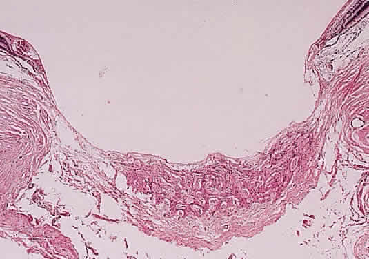
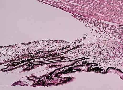
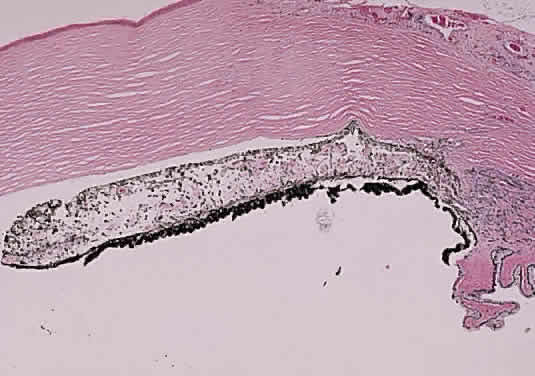
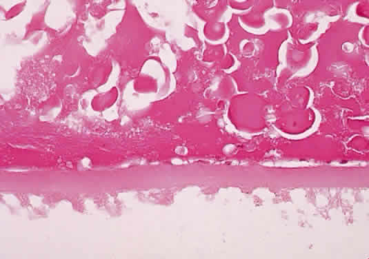
Prolonged elevation of IOP results in characteristic histopathologic changes in the eyes of children 10 years of age or younger. These effects are independent of the specific etiology of the IOP elevation. Ocular stretching, particularly at the corneoscleral limbal region, results in limbal ectasia and the clinical appearance of buphthalmos (Fig. 3). If the thinned and stretched limbus is also lined by uvea (e.g., with peripheral anterior synechiae), a limbal staphyloma is present. When this area extends posteriorly to involve the ciliary body region of sclera, it is called an intercalary staphyloma.

Fig. 3. Congenital glaucoma with enlargement, stretching, and thinning of the cornea and sclera, especially in the limbal region (limbal ectasia). Note the deep anterior chamber and the rupture in Descemet's membrane (arrow). Fibrosis of the iris root and trabecular meshwork is present. (Inset) Markedly enlarged corneas in a child with bilateral congenital glaucoma. (Main figure, H&E, × 8; inset, clinical)

The stretching of the entire anterior segment of the globe is also reflected in corneal enlargement. The 95% range of normal corneal diameters is as follows: age 1 month, 9.4 to 11 mm; 6 months, 10.5 to 11.7 mm; 12 months, 10.8 to 12 mm.40 A corneal diameter of 11 mm or less is found in only 24% of patients with primary congenital glaucoma younger than 3 months of age, and in only 9% of those older than 3 months of age so afflicted.41

Progressive corneal enlargement ruptures Descemet's membrane (Haab's striae). These breaks, which are usually horizontal in the central area and concentric toward the limbus, are generally in the lower half of the cornea and are usually associated with corneal edema (see Fig. 3). As in glaucoma in the adult, optic nerve cupping is a characteristic and diagnostic finding in congenital glaucoma. In congenital glaucoma, however, cupping is an early finding and is frequently reversible in its early stages.

Late manifestations of congenital glaucoma include fibrosis of the iris root and trabecular meshwork, disappearance of Schlemm's canal, and generalized atrophy of the ciliary body, choroid, and retina.

PRIMARY GLAUCOMA

Angle-Closure Glaucoma (Narrow Angle; Acute Congestive)

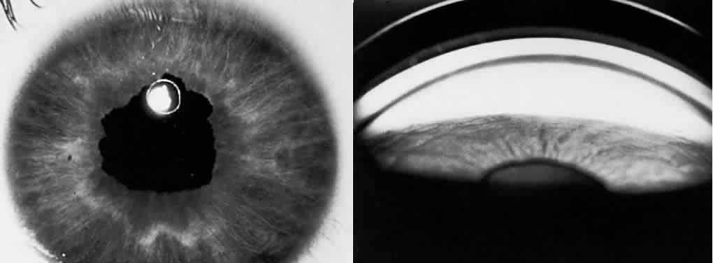
Primary angle-closure glaucoma (PACG) results from an anatomically anomalous anterior segment of the globe (Fig. 4). This configuration predisposes to the apposition of the iris to the surface of the filtering trabecular meshwork. Occlusion of the filtering trabeculum by the iris results in obstruction to aqueous outflow and subsequent IOP elevation, which may be precipitous (Fig. 5). Congdon and associates42 recently extensively reviewed the epidemiology of PACG. The Bedford Glaucoma Survey detected 10 cases of PACG among 5941 persons for an incidence of 0.17%.43 There is, however, considerable racial variation in the incidence of the disorder and its specific manifestations. Acute PACG has been noted to be uncommon among American blacks, in whom subacute or chronic angle-closure glaucoma is more often noted.42 A similar rarity in acute angle closure has been noted in African blacks.44 In contrast, Inuits may have the highest incidence of PACG, with at least 3% of those older than 40 years affected.42,45,46

Fig. 4. Photomicrograph illustrating an anatomically narrow anterior chamber angle configuration in a “crowded” anterior segment most characteristic of hyperopic persons (hematoxylin-eosin, × 10).

Fig. 5. Peripheral anterior synechiae seen in patients with untreated angle closure or treatment delay and angle closure (hematoxylin-eosin, × 10).

PACG may result from either a classic pupillary block/iris bombé mechanism or from a plateau iris, which is a nonpupillary block mechanism. Eyes that display a pupillary block mechanism have a shallow anterior chamber, increased lens thickness, and decreased axial length.47–52 The resulting congenital anatomic predisposition to angle closure is compounded by further lens thickening resulting from advancing age. In such persons, when the pupil is dilated to the midposition, resistance to the passage of aqueous between the lens and iris results in a ballooning forward of the iris ( bombé), permitting the iris to contact the filtering trabecular meshwork and closing the filtering angle.

In other patients, the anterior chamber is of near-normal depth centrally; however, the peripheral iris has an unusual contour and an extremely anterior insertion on the ciliary body. Dilating the pupil in these patients causes the iris to be drawn up toward the filtering trabeculum, in theory, without pupillary block precipitating the event.53,54 Such patients are said to have a plateau iris configuration to their filtering angle. Recently, the ultrasound biomicroscope has demonstrated the presence of anteriorly placed ciliary processes in patients with plateau iris.55,56

PACG has also been described in association with several syndromes and systemic disorders. Ritch57 has emphasized the significance of angle closure in some patients with pseudoexfoliation (PXE) syndrome, who also may exhibit extremely elevated IOP in the absence of angle closure. An occludable angle has been reported in 9.3% of PXE patients.58 Angle closure has been found in association with childhood cystinosis, in which the typical crystals were noted histologically to thicken the iris and were presumed to precipitate the attack in that manner.59 Iridoschisis60 and Fuchs' dystrophy also are associated with angle closure.61,62

Before an attack of PACG, the trabecular meshwork is normal. After repeated attacks (often at a subclinical level), the anterior chamber angle, although open, may become damaged (sclerosed or fibrotic), simulating chronic simple (open-angle) glaucoma. Similarly, patients with normal-tension (low-tension) glaucoma should be examined carefully to rule out the possibility that subclinical attacks of angle-closure glaucoma have resulted in optic nerve damage, although interval IOPs may return to normal levels.

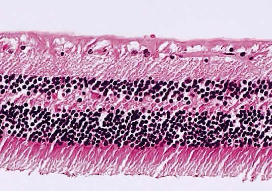
When IOP rises suddenly, as in acute angle-closure glaucoma, swelling of the iris root and occlusion of the anterior arterial circle of the iris or of one or more of its branches may result in occlusion of the arterial supply to the iris stroma and secondary iris necrosis. Subsequently, segmental iris atrophy, usually in its upper half, may be seen. Extreme atrophy occasionally results in a full-thickness iris hole that may cure the pupillary block. Histologically, there are marked atrophy of the stromal layer of the iris and varying degrees of pigment epithelial atrophy.

Iris sphincter ischemia during the period of acute IOP elevation compounds the attack by fixing the pupil in the mid-dilated position and may render it unresponsive to topical miotics given to break the attack. Later, secondary dilator and sphincter muscle atrophy may result in an irregular pupil. This muscular atrophy or iris necrosis can be demonstrated histologically (Fig. 6). Glaukomflecken are anterior subcapsular, tiny gray-white lens opacities on slit-lamp examination after acute angle-closure glaucoma (see Fig. 6).63 They probably result from interference with lens epithelial cell metabolism, either as a direct pressure effect or through alterations in the normal constituents of the aqueous during the angle-closure attack. Histologically, small areas of epithelial cell necrosis are seen accompanied by tiny areas of adjacent subcapsular cortical degeneration.

Fig. 6. A. After an acute attack of angle-closure glaucoma, the iris is necrotic. The clinical triad comprises irregular pupil, segmental iris atrophy (the iris from only the 5- to 9-o'clock positions is relatively normal), and glaukomflecken, tiny white anterior subcapsular lens opacities (inset). B. In this area, there is near-total iris necrosis (hematoxylin-eosin, × 10).

Visualization of the optic nerve during an attack of angle-closure glaucoma frequently is limited because of corneal edema. If the cornea is cleared with topical glycerol, however, optic disc edema often is present. In experimental acute elevation of IOP, blockage of axonal transport within the optic nerve can be demonstrated, even in the presence of hyperbaric oxygenation. Mechanical compression of axonal fibers with blockade of axoplasmic flow at the level of the lamina cribrosa has been postulated as the cause for this finding, rather than optic nerve head ischemia.

Untreated angle-closure glaucoma results in the formation of peripheral anterior synechiae in which the peripheral iris is permanently adherent to the trabecular meshwork, resulting in chronic angle-closure glaucoma (see Fig. 5) (more information on this topic is given later in this chapter).

The treatment of PACG is to make an opening in the iris, either through a cutting procedure (iridectomy) or with the aid of a laser (iridotomy).64 The goal of these procedures is to create a pressure-relief communication between the anterior and posterior chambers of the eye so as to prevent the buildup of pressure behind the iris, thereby eliminating iris bombé. If peripheral anterior synechiae are forming, or if iridotomy fails to correct the tendency to angle closure, and plateau iris is suspected, laser gonioplasty may be required.65–67

Open-Angle Glaucoma (Chronic Simple Glaucoma; Idiopathic)

Primary open-angle glaucoma (POAG) is a bilateral condition that may be asymmetric both in time of onset and in severity when each eye is compared. By definition, the filtration angle is open to gonioscopic examination. The diagnosis is to some extent one of exclusion because secondary causes must be ruled out before the diagnosis is made. The incidence of the disorder shows wide racial and ethnic variation. Its overall incidence is probably 1% to 3% and increases with increasing patient age: by age 75 years, 5% of the population is affected.68 It is documented to be a more severe problem among American blacks, of whom more than 11% are affected by age 80 years.5 Conversely, it is found less frequently than PACG in Inuits and Asians.

POAG is considered a genetically determined disorder. A positive family history for POAG is found in 50% of patients with POAG.69 The exact mode of inheritance is not definitely established for most glaucomas; however, some juvenile and adult open-angle glaucomas have been mapped to the long arm of chromosome 1.70–72

The nature of the obstruction to aqueous outflow in POAG is not known, although it is commonly believed to be located in the area of the juxtacanalicular connective tissue adjacent to Schlemm's canal.73–78 Unfortunately, few early cases of POAG have been examined histologically. One must be cautious in interpreting specimens from more advanced cases because changes may reflect secondary effects resulting from elevated IOP or from the medication used to treat it. Even greater caution must be used in interpreting specimens removed at surgery for glaucoma: the possibility of surgically induced artifact must be added to “end stage” and previous medical therapy as obfuscating factors. Thus, reports of “sclerosis” of the trabecular meshwork, compression of Schlemm's canal, or changes in the number of macrovacuoles in the endothelial lining of Schlemm's canal may reflect “end stage” changes or artifacts related to fixation.

The relation of changes in the trabecular extracellular matrix (including acid mucopolysaccharides, glycoproteins, glycosaminoglycans, and collagen fibrils) to the pathophysiology of POAG remains to be determined.79–81 A significant decrease in the number of trabecular endothelial cells, when compared to age-matched controls, has been reported in patients with POAG.82,83 This finding can be viewed as an acceleration of the usual age-related decline in the number of trabecular endothelial cells.83,84 Similarly, in POAG, some authors have reported histologic evidence for premature accentuation of the usual age-related development of the scleral spur, characterized by compaction of the uveal meshwork against the scleral roll, “hyalinization” of the adjacent ciliary muscle, and atrophy of the iris root.84 The latter changes are on occasion accompanied by proliferation of the endothelial cells lining Schlemm's canal into its lumen.

In the normal person, an age-related decrease in aqueous facility of outflow is accompanied by a parallel decrease in aqueous production. The net result is a quantitative balance between aqueous production and drainage. POAG can be viewed as an imbalance in the usual parallel relation of these aging changes. From this perspective, it is easy to speculate that POAG itself may really represent a multifactoral problem with a final common pathway of elevated IOP in most patients.

SECONDARY ANGLE-CLOSURE GLAUCOMA

Secondary angle-closure glaucoma (SACG) can be subdivided in a manner similar to PACG as follows:

  1. SACG with pupillary block
    1. Untreated PACG
    2. Phacogenic
      1. Phacomorphic
      2. Secondary to lens subluxation or dislocation

    3. Posterior synechiae induced
      1. Inflammatory
      2. Aphakic or pseudophakic pupillary block
      3. Ciliary block (malignant glaucoma)


  2. SACG without pupillary block
    1. Secondary to sheetlike cellular or vascular proliferations in the anterior segment
      1. Neovascular glaucoma (rubeosis iridis)
      2. ICE syndrome
      3. Epithelial downgrowth
      4. Stromal ingrowth
      5. Endothelialization of the anterior chamber angle

    2. Secondary to anterior displacement of anterior segment structures
      1. Postoperative failure of formation of the anterior chamber
      2. Tumor or cyst related
      3. Retinopathy of prematurity
      4. Persistent hyperplastic primary vitreous

    3. Miscellaneous
      1. Iridoschisis


Untreated PACG

Repeated attacks of PACG may produce peripheral anterior synechiae and SACG. Even without the formation of synechiae, repeated subclinical attacks of PACG may damage the trabecular meshwork, giving rise to a secondary open-angle glaucoma (SOAG). Histologically, peripheral anterior synechiae, which may be broad-based, may be seen (see Fig. 5).

Phacomorphic Phacogenic Glaucoma

Phacomorphic angle-closure glaucoma usually is seen in the final stages of lens cortex liquefaction. Sudden osmotically induced lens swelling displaces the iris anteriorly, inducing pupillary block. To a more modest degree, the lens probably plays a contributory role in inducing pupillary block in most cases of PACG. Occasionally, a more gradual chronic angle closure with peripheral anterior synechiae formation can result from lens-induced anterior displacement of the iris. Depending on the asymmetry of cataract formation in a given patient, there may be a significant difference in the anterior chamber depth when each eye is compared in phacomorphic glaucoma.

Phacomorphic Glaucoma Secondary to Lens Subluxation or Dislocation

Lens subluxation or dislocation may result from several causes. Blunt trauma may rupture zonules. Several syndromes and systemic disorders are associated with abnormal lens mobility. These disorders include Marfan syndrome, homocystinuria, and Weill-Marchesani (microspherophakia) syndrome.85–97 Ectopia lentis is present in up to 56% of patients with aniridia.98 Of particular importance to the clinician is the significant incidence of lens subluxation in PXE syndrome, which was discussed in the section on PACG.

In all of these disorders, an abnormally mobile lens may shift position, block the pupil, and precipitate angle closure. The small lens in the Weill-Marchesani syndrome may dislocate into the anterior chamber, causing an unusual inverse pupillary block configuration.

The clinician may be consulted for fluctuating vision by patients with unusual lens motility. It is particularly important, therefore, to be sensitive to the possibility of homocystinuria, because such patients may die from surgical and anesthetic complications.99–103 The early diagnosis of homocystinuria also is important because treatment with vitamins and dietary measures can be helpful in reducing complications if treatment is instituted early in infancy.104

Recently, genetically determined abnormalities in fibrillin expression and in microfibrillar structure have been found in Marfan syndrome.105–107

Posterior Synechiae-Induced Angle-Closure Glaucoma

Posterior synechiae usually are inflammatory adhesions between the iris and the lens. Less commonly, such adhesions may form between the iris and vitreous face (aphakic pupillary block) or between the iris and an intraocular lens (pseudophakic pupillary block). All such glaucomas result from iris bombé secondary to adhesions preventing the egress of aqueous from the posterior to the anterior chamber. All are treated initially by pupillary dilatation to try to break adhesions, or by laser iridotomy or laser pupillary mydriasis if pharmacologic pupillary dilatation fails. Obviously, pupillary dilatation might be contraindicated in the presence of an iris-plane intraocular lens.

CILIARY BLOCK (MALIGNANT) GLAUCOMA. Ciliary block (malignant) glaucoma is a dreaded complication of cataract extraction, iridectomy, and filtration surgery, particularly that after PACG, although “spontaneous” attacks have been reported.108–110 Its onset may follow shortly after surgery or may lag for many years.111 Malignant glaucoma was extensively reviewed by Luntz and Rosenblatt in 1987,112 by Levene in 1986,113 and by Shaffer and Hoskins in 1978.114 Initially, it was hoped that the avoidance of traditional surgical iridectomy through the use of laser iridotomy would prevent malignant glaucoma associated with PACG. Nevertheless, unilateral and even bilateral attacks have been reported after laser iridotomy.115–117 Malignant glaucoma has resulted from laser suture lysis after filtration surgery118,119 and YAG laser posterior capsulotomy after cataract surgery.120 It is ironic that the latter procedure often is used to treat pseudophakic malignant glaucoma.121–124 Melamed and colleagues125 noted malignant glaucoma after glaucoma seton surgery. An usual form of malignant glaucoma has accompanied keratomycotic infections.126–128

In ciliary block glaucoma, the lens or vitreous face becomes incarcerated in the ciliary ring, forcing misdirection of aqueous into the vitreous compartment. A cycle of increasing compression of the lens or vitreous face into the ciliary ring and anterior iris displacement, followed by increasing misdirection of aqueous into the vitreous compartment, is established.129,130 Ultrasound biomicroscopy has confirmed the pathophysiology of this entity.131–133 Changes or abnormalities in vitreous permeability also may play some role in its pathogenesis.134

Treatment of malignant glaucoma is directed at providing access to the anterior chamber for aqueous trapped behind the lens within the vitreous. Such therapy includes cycloplegia, hyperosmolar agents, and vitreous face disruption. Some laser techniques already have been discussed. Pars plana vitrectomy may be helpful if less invasive measures are unsuccessful.135–137

Neovascular Glaucoma (Rubeosis Iridis)

Neovascular glaucoma is characterized by the proliferation of a fibrovascular membrane on the anterior iris and onto the trabecular meshwork (Fig. 7). In the initial stages, as the sheet of tissue proliferates over the trabecular meshwork surface, neovascular glaucoma is a SOAG. Later, myofibroblasts within this fibrovascular membrane contract, drawing the iris into contact with the trabeculum and closing the filtration angle through the creation of peripheral anterior synechiae.138

Fig. 7. Neovascularization in the anterior chamber angle region, with formation of a secondary “pseudoangle” anterior to a peripheral anterior synechia (hematoxylin-eosin, × 33).

In a review of 208 patients with neovascular glaucoma, Brown and associates139 found retinal venous obstructive disease in 36.1%, diabetic retinopathy in 32.2%, and carotid artery obstructive disease in 12.9%. They concluded that 97% of involved eyes they studied demonstrated extensive retinal ischemia. In such cases, the ischemic retina is believed to produce a vasoproliferative substance that stimulates and supports vascular proliferation. Other causes of neovascular glaucoma include chronic retinal disease, uveal melanoma, longstanding retinal detachment, previous ocular injury, or Fuchs' heterochromic iridocyclitis (FHI).

The fragile vessels produced by neovascularization are prone to bleed, creating a source of spontaneous hyphema and compounding the secondary glaucoma. Because of its association with iris neovascularization and secondary hyphema, retinoblastoma must be excluded in any child with “spontaneous” hyphema.140,141 Other diagnoses to be considered in a child with spontaneous hyphema include child abuse, medulloepithelioma, iris juvenile xanthogranuloma, pseudogliomas, and blood dyscrasia.142–148

Iridocorneal Endothelial Syndrome

ICE syndrome encompasses and unifies the disorders previously categorized as Chandler's syndrome, iris nevus (Cogan-Reese) syndrome, and essential iris atrophy.149–153 Although certain patients may exhibit one or more features of a specific entity, including iris holes, endothelial dystrophy, iris surface nodules, and corectopia,154–156 there is enough overlap between them that they seem to represent different manifestations of the same or very similar developmental abnormalities.149–153 For clarification, each entity will be described in its classic form; however, a given patient may display elements of more than one of these disorders in the same eye.

Table 4 compares Axenfeld-Rieger and ICE syndromes and posterior polymorphous dystrophy. Posterior polymorphous dystrophy may show characteristics similar to the ICE syndrome such as endothelial abnormalities on specular microscopy, corneal edema, iridocorneal adhesions, endothelialization of the anterior chamber angle, and glaucoma. The corneal endothelium in posterior polymorphous dystrophy, however, displays epithelial-like characteristics, in contrast to the ICE syndrome.157 Nevertheless, some authors also have suggested epithelial characteristics for the cells in Chandler's syndrome.158,159 Posterior polymorphous dystrophy also differs from the ICE syndrome in that the former is usually familial, is bilateral, and tends not to be progressive.157


TABLE 19-4. Distinctions Among Axenfeld-Rieger (A-R) and Iridocorneal Endothelial (ICE) Syndromes and Posterior Polymorphous Dystrophy (PPMD)


CharacteristicsA-R SyndromeICE SyndromePPMD
LateralityBilateralUnilateralBilateral
Age of presentationBirthYoung adultBirth
Sex predilectionNoneWomenNone
FamilialFrequentlyRarelyTypically
Associated nonocular disordersFrequentNoNo
Corneal edemaNoFrequentOccasional
Corneal endotheliumNormalAbnormalAbnormal
Origin of membraneRetention of primordial tissueProliferation from abnormal corneal endotheliumProliferation from abnormal corneal endothelium
Mechanism of secondary glaucomaMaldevelopment of aqueous outflow systemOutflow obstruction by membrane or peripheral synechiaeEither maldevelopment (as in A-R) or membrane induced (as in ICE)
(Modified from Shields MB, Buckley E, Klintworth GK, et al: Axenfeld-Rieger syndrome. A spectrum of development disorders. Surv Ophthalmol 29:387, 1985)


Table 1 provides a possible differentiation of Axenfeld-Rieger and ICE syndromes and posterior polymorphous dystrophy on the basis of the hypothetical underlying abnormality in neural crest cell embryology or activity. Viewed from this perspective, Axenfeld-Rieger syndrome would be related to abnormal neural crest cell migration, the ICE syndrome to abnormal crest cell proliferation, and posterior polymorphous dystrophy to abnormal crest cell terminal induction or differentiation. Nevertheless, the specific causes for the abnormal behavior of corneal endothelium present in these syndromes have not been established with certainty. Alvarado and associates160–163 have suggested that herpes simplex virus infection plays a causative role in the pathogenesis of the ICE syndrome.

Essential iris atrophy is usually unilateral and most often affects women in the third decade of life. The initial clinical finding is the formation of a peripheral anterior synechia that distorts the pupil in the direction of the synechia. There is progressive pupillary distortion, and ectropion uveae develops. (Ectropion uveae refers to the migration of the posterior pigment epithelium of the iris onto the anterior iris surface, usually as the result of traction from contracting iris surface membranes.) The iris findings may lead to a misdiagnosis of iris melanoma.164

Characteristically, full-thickness iris holes develop in the quadrant opposite the distorted pupil. The etiology of the iris holes is not known. Because they develop in the iris least involved with endothelial proliferation, it is tempting to postulate a “splinting effect” by endothelial proliferation protecting the involved iris from the traction-induced holes seen in the segment of iris not covered by endothelium. A vascular etiology for the iris holes has been suggested and disputed.165–167

Peripheral anterior synechia formation progresses circumferentially, closing the filtration angle and resulting in intractable glaucoma. Iris nodules may appear late in the disorder and are a clinical marker for iris endothelialization. They display a progressive change in color from yellow initially to dark brown when larger.

Histologically, the iris stroma is atrophic. Eventually, full-thickness iris holes involve the pigment epithelium. As in all three of the components of the ICE syndrome, endothelial proliferation is present. In essential iris atrophy, endothelialization of the iris surface and filtration angle is a characteristic finding. The endothelium then deposits a new basement membrane beneath the original endothelium and Descemet's membrane.168

Chandler's syndrome is almost always unilateral. The glaucoma is usually mild; however, poor endothelial function results in corneal edema even with modest pressure elevation. Iris surface abnormalities and peripheral anterior synechiae are least prominent in this syndrome. Stromal thinning is limited and holes are rare. Histologically, endothelialization of the anterior chamber angle and anterior iris may be present.

The iris nevus syndrome, like the other components of the ICE syndrome, most often is unilateral and is most often seen in women. Commonly described features of the iris nevus syndrome include peripheral anterior synechiae and associated iris stromal atrophy, a matted appearance to the iris stroma accompanied by a whorl-like iris surface displaying loss of crypts, and the presence of fine iris nodules.156–170 Iris nodules are not covered by the proliferating endothelium. Other associated findings include ectropion uveae, heterochromia, and secondary glaucoma. Histologically, the iris surface contains a diffuse nevus and is covered by an endothelial proliferation and ectopic Descemet's membrane that also covers the anterior chamber angle.

Epithelial Downgrowth

Epithelial downgrowth is an uncommon complication of anterior segment surgery. It was noted in 0.06% of 45,500 anterior segment surgical procedures reported by Terry and colleagues from the Massachusetts Eye and Ear Infirmary.171 Over the past decade, the incidence of the disorder at that institution was 0.08%, with decreased vision, red eye, and pain being the most common presenting complaints.172 Epithelial downgrowth is probably more common after keratoplasty (0.27%) than after cataract surgery (0.091% to 0.11%).173 It is, however, a dreaded complication with a poor visual prognosis.

Typically, epithelial downgrowth presents as a horizontally oriented area of endothelial haze that progressively migrates inferiorly on the posterior cornea.174 This finding may be accompanied by chronic inflammation and ocular hypotony progressing to intractable glaucoma. Corneal striae and stromal vascularization may be seen overlying the area of corneal involvement. Epithelium may proliferate on the anterior iris before it is visible elsewhere;175 however, eventually iris surface architecture effacement is seen. Histologically, a multilayered epithelial sheet, which often has characteristics suggesting a conjunctival origin, is seen to proliferate on the posterior cornea, angle structures, and iris surface, and it may even involve the vitreous face and ciliary body.176–178

Poor wound apposition, fistulas, postoperative hypotony, and nonspecific postoperative complications such as inflammation and hemorrhage are all thought to contribute to the establishment of an epithelial beach-head within the anterior chamber.171–181 Sassani and associates confirmed earlier work by Cameron and colleagues and others that emphasized the importance of healthy endothelium in inhibiting epithelial migration, thereby acting as a defense against epithelial downgrowth.182–185

There can be several mechanisms for the glaucoma in epithelial downgrowth. Desquamating epithelial cells may plug the trabecular meshwork, resulting in SOAG.171 If the epithelial sheet causes posterior synechiae, pupillary block angle-closure glaucoma can be produced. Ultimately, secondary angle closure without pupillary block results from progressive synechial closure of the filtering angle by the proliferating epithelial sheet.172

Specular microscopy can be of assistance in the diagnosis of epithelial downgrowth,186,187 and laser photocoagulation of the iris surface can be helpful in delineating the extent of epithelial membrane proliferation. Cytologic examination of aqueous or vitreous fluids from aspirates or of scrapings from the corneal endothelial surface or from the iris surface is confirmatory.

The clinical progression of epithelial downgrowth to intractable glaucoma may be gradual although inexorable. The clinical course of the patient must be weighed against the radical therapy usually required to treat the disorder.

Stromal Ingrowth

Stromal ingrowth most frequently is seen after failed corneal transplants;188,189 however, it may follow other surgical procedures, such as cataract surgery, especially if they have been complicated by vitreous loss and incarceration.190,191 Although many of these membranes represent direct proliferation of keratocytes through a dehiscence in Descemet's membrane, some retrocorneal fibrous membranes may represent fibrous metaplasia of corneal endothelium.192,193 Histologically, a fibrous membrane is seen to extend from a corneal wound to proliferate over the posterior cornea. Peripheral anterior synechiae result in SACG.

Endothelialization of the Anterior Chamber Angle

Endothelialization of the anterior chamber angle is seen on histologic examination in 20% of enucleated eyes.194 Endothelial proliferation extends over anterior segment structures, including the iris and the anterior chamber angle (or pseudoangle in the presence of peripheral anterior synechiae, which are noted frequently) (Fig. 8). Endothelialization may be found accompanying rubeosis iridis.195 In three such cases, the endothelial cells exhibited myoblastic features.195

Fig. 8. Endothelialization of pseudoangle anterior to peripheral anterior synechia. The anterior iris surface is covered by proliferated corneal endothelium.

Postoperative Failure of Anterior Chamber Reformation

Postoperative “flat” anterior chamber is seen in various surgical procedures, particularly those performed for glaucoma, in which overfiltration and wound leaks are common causes.196–199 Such overfiltration or wound leaks are particularly likely to occur if adjunctive antimetabolites are used.200–202 The advent of viscoelastic agents and releasable sutures has decreased the frequency of these complications.203–208 Less commonly, malignant glaucoma may simulate simple delayed chamber formation.209–212

Persistent flat anterior chamber may result in peripheral anterior synechiae that may be very broad. Corneal-lens contact usually requires immediate surgical reformation of the anterior chamber to prevent the rapid formation of cataract.213,214 Persistent hypotony secondary to aqueous loss also may lead to choroidal “detachment,” which can further contribute to anterior chamber shallowing.215

Iris and Ciliary Body Cysts

Cysts of the iris and anterior ciliary body, particularly if multiple, may displace the iris lens diaphragm anteriorly, resulting in angle closure.216 Angle closure in the case of multiple cysts may be acute. The cysts may be idiopathic or acquired, such as those associated with trauma or late syphilitic interstitial keratitis.217–219 Unlike acquired cysts, primary cysts tend to be stationary, although important exceptions occur.219,220 The latter lesions also accounted for 38% of lesions misdiagnosed as iris melanoma.164 Late syphilitic interstitial keratitis also may be associated with angle closure secondary to peripheral anterior synechia formation.217,221,222 Recently, the ultrasound biomicroscope has been demonstrated to be useful in the evaluation of cystic lesions of the iris.223

Intraocular Tumors

Intraocular tumors, if large, may displace the iris-lens diaphragm anteriorly in a manner similar to that of iris and ciliary body cysts.224 This displacement is compounded by retinal detachment, if present. Posterior synechiae also may result in pupillary block angle closure. Eventually, peripheral anterior synechiae may form. Large tumors also may be associated with angle closure secondary to rubeosis iridis, or on a phacomorphic basis secondary to cataract formation. Tumors that infiltrate the iris (e.g., diffuse iris melanoma) may close the angle secondary to iris thickening.

Retinopathy of Prematurity and Persistent Hyperplastic Primary Vitreous

Hartnett and associates225 have stated that unsuspected glaucoma may be a cause of decreased vision in severe retinopathy of prematurity. Progressive anterior segment crowding may lead to angle closure in these patients.226 They found peripheral anterior synechiae of at least 180° in 12% of 27 eyes in 17 infants with stage IV or V retinopathy of prematurity.227 Numerous other angle abnormalities also were noted on gonioscopic examination of these infants. Even adults with a history of retinopathy of prematurity should have periodic examinations to exclude the development of additional ocular abnormalities, particularly glaucoma.226

Persistent hyperplastic primary vitreous is a congenital ocular disorder characterized by microphthalmos, cataract, and leukocoria.228 Progressive cataract formation also may contribute to angle closure, and repeated hemorrhages may result in organization and iridocorneal adhesions.

Iridoschisis

Iridoschisis affects persons in the seventh decade. It is bilateral and without sex predilection. Unlike the ICE syndrome, the pupil is not displaced (Fig. 9); rather, a separation develops within the iris stroma that elevates it, particularly in its inferior half. Initially, the stromal fibers maintain their pupillary and peripheral attachments. Later, they become detached at one end, which floats free in the anterior chamber. Eventually, glaucoma develops in 50% of affected patients secondary to peripheral anterior synechia formation. Histologic examination of the involved iris has demonstrated stromal atrophy; however, no vascular or neural abnormalities have been documented.229 Thus, the specific etiology of this disorder is not known. Its relation to PACG was discussed earlier in this chapter.60 It also may be familial, or it may follow trauma, keratoconus, syphilitic interstitial keratitis, or microphthalmos.230–235

Fig. 9. Iridoschisis. (A) Anterior iris stromal layers (arrow) are separated widely from the deeper layers. Note the bullous keratopathy. (B and C) Anterior stromal layers are separated from the deeper layers extending from (B) the iris root toward (C) papillary iris. (B, insets) Clinical appearance in another case as viewed (1) directly and (2) with a slit beam. (A, H&E, × 16; B, PAS × 40; inset 1, clinical; inset 2, slit beam; C, H&E, × 40)

SECONDARY OPEN-ANGLE GLAUCOMA

In SOAG, the angle is open anatomically but is functioning poorly for reasons other than POAG. The classification scheme for SOAG that follows is somewhat arbitrary; nevertheless, it may be helpful to conceptualize these entities, at least in their pure form. It is beyond the scope of this chapter to discuss all the causes of SOAG cited, so only selected topics will be described.

SOAG can be subclassified as follows:

  1. SOAG caused by cells or debris in the angle
    1. Hyphema
    2. Uveitis
    3. Pigmentary glaucoma
    4. Pseudoexfoliation
    5. Hemolytic and “ghost cell” glaucoma
    6. Phacolytic glaucoma
    7. Nondenatured lens material-induced glaucoma
    8. Melanomalytic or melanocytomalytic glaucoma
    9. Tumor seeding of the trabecular meshwork
    10. Schwartz-Matsuo syndrome

  2. SOAG caused by damaged outflow channels
    1. Previous uveitis
    2. Blunt trauma
    3. Repeated hyphema
    4. Siderosis and hemosiderosis oculi
    5. Repeated attacks of acute angle-closure glaucoma
    6. Early rubeosis or other anterior segment cellular proliferative disorder

  3. SOAG caused by corneoscleral and extraocular disease
    1. Interstitial keratitis
    2. Orbital venous thrombosis
    3. Encircling element after retinal disease
    4. Retrobulbar mass
    5. Leukemia
    6. Mediastinal mass

  4. SOAG secondary to miscellaneous causes
    1. Steroid-induced glaucoma
    2. Alpha-chymotrypsin glaucoma
    3. Glaucomatocyclitic crisis (Posner-Schlossman syndrome)
    4. Fuchs' heterochromic iridocyclitis

SOAG Caused by Cells or Debris in the Angle

HYPHEMA.

Hyphema refers to blood in the anterior chamber. The most common causes of this condition are anterior segment neovascularization (rubeosis iridis) and ocular trauma. Rubeosis iridis was discussed as a cause of SACG. As in all the syndromes involving cellular proliferations in the anterior segment that may result in SACG, rubeosis iridis, in its early stages, may go through a period of SOAG in which the trabeculum may be coated by the cellular proliferating membrane, which has not yet resulted in synechial angle closure.

One must be extremely cautious when presented with a case of apparently spontaneous hyphema in a child. Most frequently, such cases represent occult trauma, particularly child abuse.148,236 Other important causes of spontaneous hyphema in children include juvenile xanthogranuloma,237 retinoblastoma,140,238,239 and medulloepithelioma.146,240

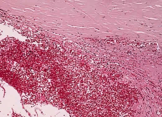
When the globe is struck forcefully, the cornea is compressed, transmitting the concussive force to the aqueous humor. The shock wave thus generated is propagated through the eye and may give rise to open-angle glaucoma through several mechanisms. The force may cause scarring within the trabecular meshwork, resulting in SOAG. The ciliary body face may be lacerated, tearing the anterior arterial circle and resulting in hyphema. Recession of the anterior chamber angle is a marker for such severe trauma. Initially, the sheer mass of blood may block aqueous outflow through the trabecular meshwork. Later, as the clot undergoes dissolution, red blood cell debris and the macrophages that engulf it also may plug the trabeculum, resulting in hemolytic glaucoma (Fig. 10). In a similar fashion, degenerating “ghost” red blood cells from a vitreous hemorrhage may block the trabeculum after they wash into the anterior chamber through a disrupted vitreous face.241,242 Degenerating hemoglobin releases iron, which acts as an enzymatic poison to the trabecular meshwork, further contributing to the possibility of SOAG. Finally, organization of the hyphema may result in SACG.

Fig. 10. Red blood cells and their products of degeneration obstruct the trabecular meshwork in hyphema (hematoxylin-eosin, × 40).

Several days after the hyphema from the initial injury, spontaneous rebleeding may cause the entire cycle to be repeated in the presence of an already compromised aqueous outflow mechanism.243–246 Spontaneous rebleeding may be more common in blacks.247–252 Such rebleeding is associated with a poorer visual prognosis, particularly in children.253 Scarring of the trabeculum may follow recurrent hyphema. Patients who have sickle cell disease may be at particular risk for increased IOP from hyphema because sickled red blood cells pass poorly through the trabecular meshwork.254–256 The damage caused by the increased IOP may be compounded by the tendency for these patients to develop visual loss secondary to ischemic optic neuropathy during such pressure rises.257

Histologically, free blood and clot are seen initially. Later, debris, disrupted red blood cells, and macrophages that may contain hemosiderin and other blood breakdown products are present. In “ghost cell” glaucoma, these more rigid cell remnants appear to obstruct the trabecular meshwork mechanically.241,242

The most severe form of hyphema is the “black ball,” characterized by its darkened color. Histologic examination of the clot from such patients reveals the clots to be composed of concentric layers of fibrin lacking fibroblastic or neovascular proliferations.258

Elevated IOP at the time of hyphema contributes to corneal blood staining; however, this entity has been reported in the presence of relatively low IOP.259 Corneal blood staining represents hemoglobin debris that has traversed the corneal endothelium to reside within the stroma. Histologic examination of involved corneas also has demonstrated intracellular and extracellular hemoglobin particles, keratocyte degeneration, and endothelial cell loss.260,261 Light toxicity exacerbated by the presence of porphyrins has been postulated as contributing to keratocyte and endothelial damage.262,263

UVEITIS. Aqueous production is usually decreased in the presence of iritis, iridocyclitis, or pars planitis. As a result, IOP may be normal or even decreased, even if aqueous outflow is decreased. Later, as aqueous production resumes with appropriate anti-inflammatory therapy, glaucoma may ensue, reflecting damage to the aqueous drainage mechanism.264

There are several mechanisms through which uveitis may lead to glaucoma. SACG resulting from posterior synechia-caused iris bombé was discussed under SACG. Inflammatory cells and debris may plug the trabecular meshwork and trabeculitis may decrease its function, resulting in SOAG. Topical or systemic steroids used in the treatment of uveitis also may cause SOAG in “steroid responders.” Finally, recurrent or chronic inflammation may cause trabecular scarring, thereby decreasing aqueous outflow.264

Two specific “inflammatory” syndromes that result in SOAG will be discussed—glaucomatocyclitic crisis and Fuchs' heterochromic iridocyclitis. SOAG also may result from low-grade chronic anterior uveitis in oculodermal melanocytosis. The melanocytosis itself also may contribute to aqueous outflow obstruction.

Glaucomatocyclitic Crisis (Posner-Schlossman Syndrome). The original designation of this syndrome as the “syndrome of glaucomatocyclitic crises” by Adolph Posner and Abraham Schlossman highlights its characteristic recurrent nature.265,266 It is usually found as a unilateral condition in adults in the third through fifth decades. During acute episodes, patients may experience a rise in IOP to 40 to 60 mmHg. If pressure is sufficiently elevated, halos around light and decreased vision may result from corneal edema. Usually, discomfort is minimal and the involved eye does not appear inflamed on external examination. The pupil often is mildly dilated during the attack. The duration of the episode may be hours to weeks.

A few fine keratic precipitates may be seen initially on slit-lamp examination. Later, the precipitates can resemble mutton fat. Little or no anterior chamber reaction is usually noted, and the angle appears normal on gonioscopic examination. During the acute episode, aqueous outflow facility is decreased and aqueous production has been reported to be elevated. After the acute episode, the IOP may decrease to below normal levels. Some patients have a persistent elevation of IOP and decreased outflow facility. Although optic nerve damage is not characteristic, cupping may occur and filtration surgery is occasionally required. An association between this syndrome and POAG is probable.267,268 A developmental anomaly of the anterior chamber angle and abnormal prostaglandin production have been postulated to be causative for Posner-Schlossman syndrome.269–272 Herpes simplex virus also has been suggested as a factor in the development of the syndrome.273

Light and electron microscopic examination of a trabeculectomy specimen from a patient with recurrent episodes of this disorder revealed numerous mononuclear cells within the trabecular meshwork, as well as erythrocytes.274 The latter were thought to be secondary to the surgery. Giant vacuoles were not seen in the inner wall of Schlemm's canal, even though the preoperative IOP was 60 mmHg. The authors postulated that the macrophages might obstruct aqueous outflow; however, they might also represent a secondary response to the longstanding episode that preceded surgery.

Decreased corneal endothelial cell count has been demonstrated in patients with recurrent attacks of Posner-Schlossman syndrome.275

Fuchs' Heterochromic Iridocyclitis. In 1906, Fuchs276 summarized previously reported cases of heterochromia (difference in iris color between eyes) associated with cataract and reported 38 additional cases. Fuchs' heterochromic iridocyclitis (FHI), the syndrome he defined,277–279 is characterized by iris stromal atrophy, which usually causes the involved iris to be hypochromic relative to the fellow eye. As the condition worsens, increased visibility of the iris pigment epithelium may make the involved iris appear darker. Mild iritis with fine keratic precipitates, iris muscle atrophy, and mydriasis also are seen.

A recent review of 89 eyes of 77 patients noted that the most frequent presenting complaint was decreased vision (96.1%); only 18.2% complained of pain.280 FHI was diagnosed in 50.6% at presentation. Clinical heterochromia was found in 70.1% and was bilateral in 15.6% of patients. Cataract was present in 73%, with 40.4% having already had cataract surgery. Glaucoma was diagnosed in 15.7% at presentation and in 21.3% at review, by which time 9% of the total had required filtration surgery.280

The glaucoma in FHI can be very difficult to control. La Hey and associates281 reported 30 patients with glaucoma (27%) among 111 patients with FHI. Filtration surgery was required in 22 of these 30 patients (73%).

Vascular anomalies of the iris have been noted, including iris perfusion defects and leakage of iris vessels, on fluorescein angiography in FHI.282 Such findings have suggested anterior segment ischemia as one causative factor in FHI.282 Indolent iris neovascularization may be present and may give rise to the filiform hemorrhage seen during cataract surgery in these patients. Such hemorrhage usually is self-limited and of no clinical significance. Iris and filtering angle neovascularization, combined with trabeculitis, is thought to result in the associated glaucoma.283 Histologically, hyalinized blood vessels have been observed in iris specimens from affected patients.284 This finding supports the vascular theory for the origin of this condition. Various inflammatory and immunologic abnormalities also have been detected in iris specimens from patients with FHI.285,286 Abnormal melanocytes with few, small, and immature melanin granules accompanying iris plasma cell infiltration, and abnormal adrenergic innervation were noted on one electron microscopic study of two iris specimens.287

Intractable glaucoma has been reported as a consequence of secondary posterior capsulotomy in one patient with this disorder.288 The eye had been free of inflammation and glaucoma for 27 years before the procedure.

Pigment Dispersion Syndrome (Pigmentary Glaucoma)

The pigment dispersion syndrome (PDS) is found most commonly in young adult male myopes.289,290 It was found in 10% of white and black subjects with and without glaucoma.291 In some black patients, the syndrome may involve older, hyperopic women who may not demonstrate iris transillumination and who tend to have a flatter iris insertion into the ciliary body.292 The syndrome is called pigmentary glaucoma when it is accompanied by glaucoma. Migliazzo and colleagues293 noted progression to glaucoma in 35% of their series of PDS patients with ocular hypertension over a mean follow-up period of 17 years.

The syndrome is characterized by depigmentation of iris pigment epithelium, producing characteristic iris transillumination, which is demonstrable on slit-lamp examination at the junction of the inner two thirds with the outer one third of the iris (Fig. 11). These areas of depigmentation are accompanied by a band of increased granular iris pigmentation overlying the ring of increased iris transillumination. The finding is most easily seen in blue irises but may be seen fairly easily in brown. This band is presumed to be caused by many pigment-laden macrophages within this area of iris stroma. It has been postulated that the distribution of iris depigmentation corresponds to areas where bundles of zonules chafe the iris pigment epithelium. Iridodonesis may be present. Gonioscopic examination reveals that the trabecular meshwork is heavily pigmented. An increased incidence of retinal detachment is also found in PDS, suggesting the possibility of an underlying pigment epithelial defect.

Fig. 11. Pigment dispersion syndrome. (A) Extensive increased iris transillumination is present mainly in the middle third of the iris. (B) Goniscopic view shows marked deposition of pigment in the anterior chamber angle. (C) Melanin pigment granules present in the corneal endothelial cytoplasm are responsible for the Krukenberg's spindle seen clinically (inset). (A, clinical; B, gonioscopy; C, 1.5-μ section, PD, × 300; inset, clinical)

Another typical slit-lamp finding in PDS is the presence of a Krukenberg's spindle, a vertically oriented column of pigment phagocytosed by central and inferior corneal endothelium (see Fig. 11). Pigment may be found within the aqueous after pupillary dilatation or after exercise. Such pigment release may be accompanied by an acute rise in IOP. Significant deposits of pigment also are found on the iris surface, lens, and zonules and within the trabecular meshwork.

The pathophysiology of glaucoma associated with some PDS seems to be related to an “inverse pupillary block” mechanism in which posterior bowing of the iris results in chafing of its posterior surface on bundles of zonules or on ciliary processes, thereby releasing pigment.294–299 However, some have questioned this mechanism.300 The ultrasound biomicroscope has provided strong evidence for this mechanism in a subset of patients who exhibit the characteristic concave iris contour.299–304

Increased IOP in some PDS patients caused by pigment released during exercise is well recognized but not universally accepted.305,306 Posterior iris concavity also tends to increase with exercise in some patients.307 It also may be exacerbated by cataract formation.308 Prophylactic or therapeutic iridotomy has been suggested for patients with posterior iris bowing.297–303 Iris chafing as a cause of pigment dispersion also has been reported in pseudophakia.309–311

Some authors have cited the frequent occurrence of anomalous iris processes in pigmentary glaucoma as evidence that a congenitally anomalous anterior chamber angle is the cause for the glaucoma.299 Ultrasound biomicroscopic examination demonstrated such processes in 75% of untreated PDS patients.299 Other authors believe that the PDS gene may merely be located close to a gene for POAG so that they tend to be inherited together.

Argon laser trabeculoplasty appears to be successful in PDS, with a life-table analysis indicating a success rate of 80% at 1 year, 62% at 2 years, and 45% at 6 years after treatment in one study.312

The incidence of retinal detachment is increased in PDS.313 In one study of 60 PDS patients, lattice retinal degeneration was found in 20%, full-thickness retinal breaks were present in 11.7%, and asymptomatic retinal detachments were found in 3.3% (two patients).314

Histologically, the posterior layer of iris pigment epithelium atrophies in areas that correspond to the foci of iris transillumination.315 The dilator muscle may be atrophic, hypertrophic, or absent. The adjacent iris stroma contains large numbers of pigment-filled macrophages. Neuroepithelial melanin granules are widely distributed within the endothelium of the cornea (corresponding to the Krukenberg's spindle) (see Fig. 11) and within the trabecular meshwork; however, endothelial cell counts have been reported to be similar to those in control patients.316,317

Johnson318 was unable to find differences in trabecular cellularity or morphology in nine normal human eye anterior segments examined for pigment-associated differences in trabecular cellularity or morphology. Epstein and colleagues319 were unable to produce long-term IOP elevation by infusing melanin granules into monkey eyes, and they concluded that factors other than, or in addition to, pigment particle accumulation in the trabecular meshwork must be involved in the mechanism of human pigmentary glaucoma. Similarly, Murphy and associates320 reported that 3.5% of the pigment in PDS was in the juxtacanalicular tissue and 96.5% in the corneoscleral and uveoscleral tissue in the specimens they examined. They concluded that the development of glaucoma in PDS cannot be directly attributed to pigment accumulation in the juxtacanalicular tissue. Clinically, however, increased pigment dispersion, as evidenced by an increase in iris transillumination, an increase in corneal pigmentation, or the appearance of pigment granules on the surface of the lens in the pupil, has been associated with worsening of glaucoma in PDS.321

Alvarado and Murphy322 concluded that trabecular cul-de-sacs provide a major portion of aqueous outflow resistance, and that reduction in the trabecular cul-de-sac area accounts for a major portion of the increase in aqueous outflow resistance in PDS. They also stated that macrophages were largely responsible for clearing the trabecular meshwork of pigment and debris.

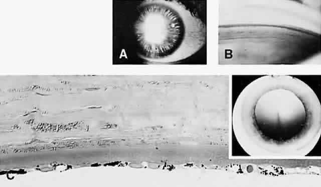
Pseudoexfoliation Glaucoma

PXE refers to granular or frostlike material deposited on the lens surface in a characteristic pattern (Fig. 12). The term was first used by Georgiana Dvorak-Theobald323 to distinguish the condition from “true exfoliation,” which is a splitting off of layers of the lens capsule that results most commonly from occupational exposure to infrared irradiation. Typical PXE material has been found in the conjunctiva of patients with “preclinical” PXE324 and in PXE patients.325,326 Similar deposits have been noted in the wall of a short posterior ciliary artery in the orbit,327 in eyelid skin,328 and in other extraocular structures, such as extraocular rectus and oblique muscles, vortex veins, and the optic nerve sheaths, in PXE patients.329,330 Abnormalities of the cornea in PXE have been reported.331 An abnormal blood-ocular barrier in PXE is supported by the findings of increased laser flare-cell measurements in these patients,332 and by the fibrinoid reaction sometimes found in PXE patients after cataract surgery.333,334 The systemic nature of the disorder has been confirmed by the finding of typical material in skin biopsy specimens335 and in the skin, heart, lung, liver, kidney, and meninges at autopsy.336,337

Fig. 12. Pseudoexfoliation material is seen on the surface of the lens capsule (hematoxylin-eosin, × 80).

Increased anterior chamber angle pigmentation has been reported in PXE.338 A pigmented line, Sampaolesi's line, is found anterior to Schwalbe's line and may cause errors in estimation of the width of the anterior chamber angle on gonioscopic examination.339 As was stated earlier in this chapter, Ritch and others57,334,340,341 have emphasized the significance of angle closure in some patients with PXE, which also may exhibit extremely elevated IOP in the absence of angle closure. An occludable angle was reported in 9.3% of PXE patients in one study.58

Peripheral and generalized iris transillumination also is typical of PXE.342,343 Generalized iris transillumination has been presented as evidence of vascular insufficiency in these patients.343 Other characteristic clinical findings in PXE are pupillary ruff defects, iris sphincter transillumination, a characteristic whorl-like pattern of particulate pigment deposition on the iris sphincter, particulate pigment deposition on the peripheral iris and trabecular meshwork, and exfoliation material accumulation on the zonules and ciliary body.344,345

There appears to be a considerable racial variation in the incidence of PXE.346–351 Forsius351 reported a variation in prevalence of PXE worldwide of 0% in Eskimos to 21% in Finns over 60 years of age. Aasved352 noted a prevalence of PXE of 4%, 4.7%, and 6.3%, respectively, in Birmingham, Bonn, and Bergen. These differences were not statistically significant. There is an increased incidence of cataract and glaucoma among PXE patients in the Eastern Mediterranean area of Turkey.353 The percentage of PXE patients with glaucoma was 34.3%, and the prevalence of PXE in glaucoma patients was 46.9%. There is a particularly high incidence of PXE in African Bantus, in whom there is a 6.4% prevalence in the age group 30 to 39.354 In contrast, the Framingham Eye Study noted a prevalence of only 0.6% for persons ages 52 to 64.355 The prevalence increased to only 5% in those 75 to 85 years. There appears to be a higher incidence in women. There also is an increased prevalence among Navajo Indians356 and among the male Spanish American population of New Mexico.357 The prevalence of PXE syndrome has been reported to be decreased in some areas of the southeastern United States.358,359 PXE has been reported in association with PDS.360 The prevalence of PXE is decreased in persons with background diabetic retinopathy and is further decreased in those with proliferative diabetic retinopathy.361

Considerable variation in the prevalence of glaucoma in the PXE syndrome also has been reported. Ringvold and associates362 found a 30% prevalence of glaucoma and a 4.2% prevalence of ocular hypertension in those with PXE among 1941 persons older than 64 years. PXE increased in prevalence up to age 75 to 79 years and declined thereafter. Jones and colleagues357 noted that the incidence of glaucoma associated with PXE in New Mexico was 22 times higher. Wollensak and associates363 even questioned whether there is an increased incidence of glaucoma in PXE.

Herry and colleagues364 studied 680 patients with PXE followed from 1940 to 1980. Of these patients, 347 initially had normal IOP. The cumulative probability of an eye with PXE developing elevated IOP was 5.3% in 5 years and increased to 15.4% in 10 years. In a study of 100 consecutive patients in whom PXE was an incidental finding, 78% had normal IOP, 15% were ocular hypertensives, and 7% had glaucoma.365,366 A retrospective study of 206 eyes in 164 patients with PXE found that 65.5% of the eyes that initially were normotensive remained normotensive; however, 34.5% developed ocular hypertension or glaucoma.367

Glaucoma associated with PXE may be more difficult to control and may be associated with more severe optic nerve damage than POAG.352,357,368,369 Henke and Naumann370 also noted an increased prevalence of PXE among 455 enucleated eyes (3.4%), and postulated that PXE may cause increased secondary changes leading to the necessity for enucleation. PXE was found in 87.8% of those undergoing trabeculectomy in a Greek population.371 The severity of glaucoma in PXE may be associated with the patient's blood group.372 Elastosis of the optic nerve head has been reported in PXE compared to POAG.373 One might postulate that such changes could contribute to increased IOP sensitivity in these patients.

The cause of the glaucoma in PXE is not certain. Some authors postulate a plugging of the trabecular meshwork by the PXE material or by pigment released from the iris pigment epithelium.339,374–377 The trabecular meshwork and its endothelium also may undergo secondary and degenerative changes that may compound the simple mechanical effects of the material deposited within it.378,379 It has been suggested that PXE is closely associated with a gene for POAG, and that the PXE is not directly responsible for the glaucoma associated with the disorder.380 Congenital anterior chamber angle anomalies (goniodysgenesis) have been postulated to contribute to the glaucoma in PXE.381

Fragmented zonular fibers have been noted in an electron microscopic study of anterior segments in PXE,382 and increased zonular friability has been reported.383 It is not surprising, therefore, that an increased incidence of cataract surgery complications has been found in PXE.384–387 Naumann385 noted an incidence of vitreous loss of 1.8% in those without PXE and 9% in those with PXE among 1205 cataract extractions. Poor pupillary dilatation also increases the chances of complications during cataract surgery in patients with PXE.385

Morrison and Green378 reviewed the light microscopic findings in PXE and noted that the material is only loosely adherent to most anterior segment structures. It is firmly adherent, however, to the equatorial lens capsule, the posterior epithelium of the iris, and the nonpigmented ciliary epithelium, where the characteristic material (fibrils embedded in an amorphous matrix) is present within epithelial cells and associated with a disorganized, reduplicated basement membrane.378

Streeten and others388–396 have contributed greatly to our knowledge of the composition of the material deposited in PXE and have suggested a similarity to elastic-like material. The fibrils are 20 to 30 nm thick with 10-nm subunits. They may be 800 to 900 nm long and have a characteristic 50-nm periodicity. The material has been produced in cultured tissues from PXE patients.397 The deposition of a precapsular layer composed of microfibrils, amorphous material, and granular inclusions has been cited as a possible precursor to PXE.398

In their review of the light microscopic findings in PXE, Morrison and Green378 noted that glycosaminoglycans, which may comprise the interfibrillar portion of the material, have been found in PXE. They also noted that other studies have demonstrated the similarities between PXE material and zonules, and that the fibrils are related to the microfibrillar portion of elastin. This latter material has the staining properties of oxytalan, the microfibrillar component of elastic tissue.399,400 A similarity to amyloid material also has been postulated by some;401 however, this concept is not widely supported.378 Takei and Mizuno382 proposed that PXE material may represent degenerated zonular fibers. Glycoconjugates of exfoliative material have been characterized.402 Actually, various abnormal materials probably are produced as part of PXE material. Based on immunohistochemical studies, Schlotzer-Schrehardt and colleagues403 have postulated that PXE material is a multicomponent expression of a disordered extracellular matrix synthesis, including the incorporation of the major noncollagenous basement membrane components. Additional histologic findings in this syndrome are discussed in the chapter on the lens.

Phacogenic SOAG

The lens may give rise to SOAG in several ways. Leaking lens protein from a mature lens may incite a marked histiocyte response. This finding was confirmed by Flocks and associates,404 who examined eyes enucleated because of glaucoma associated with hypermature cataracts. They noted that macrophages, swollen with phagocytosed lens material, can plug the trabecular meshwork, resulting in aqueous outflow obstruction. They coined the term “phacolytic glaucoma” to designate the severe rise in IOP that is characteristic of the entity. Similar findings were reported by Yanoff and Scheie405 based on anterior chamber aspirates after needling procedures for soft cataracts. Alternatively, heavy-molecular-weight proteins from a leaking mature lens or fragments of cortical material may themselves block outflow through the trabecular meshwork.406–408 Epstein408 noted that macrophages are not invariably found in phacolytic glaucoma. Both cholesterol crystals and calcium oxalate crystals have been found in phacolytic glaucoma.409,410

SOAG Caused by Uveal Malignant Melanoma

Yanoff224 noted that 20% of 96 eyes containing uveal malignant melanomas presented clinically with glaucoma. Approximately 7% to 10% of enucleated eyes have been reported to contain melanomas that were unsuspected clinically.411–415 Unsuspected melanomas were 10 times more prevalent in persons presenting with glaucoma than in control patients.224 The pathophysiology of the glaucoma included peripheral anterior synechia formation with secondary angle closure, and cellular obstruction of an open anterior chamber angle (Fig. 13). Melanoma should be ruled out in any adult with a blind, painful eye having opaque media.

Fig. 13. Open-angle glaucoma secondary to uveal melanomas. (A and B) Seeding of the iris melanoma onto the anterior surface of the iris (arrows). Melanoma cells also cover the angle recess and trabecular meshwork (B, double arrow). (C) Ring melanoma involves the root of the iris and the anterior ciliary body over a full 360 degrees, with invasion of the anterior chamber angle structures. (A, H&E, × 7; B, H&E, × 100; C, H&E, × 7)

Melanomalytic glaucoma represents mechanical obstruction of the trabecular meshwork by macrophages and debris from a necrotic uveal melanoma.416–418 A similar process has been reported in association with necrotic iris melanocytomas.419 Ring melanoma may result in glaucoma secondary to tumor infiltration of the anterior chamber angle structures (see Fig. 13).420

The mechanisms for angle-closure glaucoma secondary to uveal malignant melanomas were discussed earlier in this chapter. There are several mechanisms for SOAG in these lesions.

Schwartz-Matsuo Syndrome

In general, IOP is lowered in eyes with retinal detachment. Occasionally, however, such eyes present with an acute elevation in IOP, the characteristic finding in Schwartz-Matsuo syndrome.421,422 These cases also typically are accompanied by the presence of cells in the anterior chamber. The cells have been characterized as photoreceptor outer segments.

Back to Top
VARIABLE OUTFLOW

NORMAL-TENSION (LOW-TENSION) GLAUCOMA

Normal-tension glaucoma (NTG) is characterized by typical glaucomatous optic nerve head cupping and visual field loss at an IOP that is within the statistically normal range.423–426 NTG is a diagnosis of exclusion; the clinician must rule out simulating optic nerve lesions, “burned-out” POAG, wide diurnal swings in IOP, and systemic medication masking POAG. Nevertheless, at least 30% of glaucoma patients may be classified as having NTG.2,4,68

NTG patients must be divided into two groups, according to Levene.425 The first group is really a subset of POAG, with peak pressures near 24 mmHg. These patients can be demonstrated to benefit most from pressure control. The second group have pressures consistently well within the normal range, but they exhibit the optic nerve head cupping and visual field loss usually associated with glaucoma. When compared to POAG, this latter group more often exhibits early central visual field loss and decreased visual acuity, exhibits disproportionate cupping in relation to the degree of field loss, has a higher incidence of monocular cases, is found more often in females, is associated with optic disc splinter hemorrhages, and is poorly responsive to therapy directed at lowering IOP.

Each of the characteristics of NTG cited above is disputed by one or more authors. In particular, patients in whom a definite history of one or more hemodynamic crises can be elicited tend to have a better prognosis and stable visual fields.427 Various studies have tried to associate NTG with systemic and local hemodynamic factors, such as orthostatic hypotension, carotid occlusive disease, blood viscosity, migraine, diabetes, and particularly the regulation of optic nerve head blood flow.428–443 Thus, most authors associate NTG with some factor affecting blood circulation. At least some cases of NTG may be caused by calcification, dilatation, and ectasia of the carotid artery into the optic canal.444 Others have questioned a hemodynamic cause for NTG.445

On histologic examination of one pair of NTG eyes, Quigley and colleagues446 noted compression and distortion of the lamina cribrosa similar to that observed in POAG. The cause for these changes was not demonstrable.

Back to Top
TISSUE CHANGES CAUSED BY ELEVATED INTRAOCULAR PRESSURE
Corneal clarity depends on the regular arrangement and spacing of the corneal stromal collagen fibers and on the relative dehydration of the cornea in comparison to other biologic tissues. This relative corneal dehydration is dependent on the ability of the corneal endothelium to remove water from the corneal stroma against the opposing force of IOP. In effect, the cornea is a leaking boat and its endothelium a bilge pump. Depending on the health of the corneal endothelium, an IOP can be reached beyond which the endothelium can no longer maintain the relative stromal dehydration. A “steamy” or edematous corneal stroma results.

Histologically, corneal edema is reflected in a loss of the usual stromal “clefting” artifact and in edema of the basal layer of the corneal epithelium. Eventually, the epithelium may separate from Bowman's layer as bubbles or bullae. Rupture of these bullae exposes corneal nerves, resulting in severe pain, and exposes the cornea to secondary infection and ulceration, a common prelude to enucleation for a blind and painful eye. Chronic corneal edema can result in the formation of a degenerative corneal pannus, hypertrophy of corneal nerves, and corneal vascularization. Atrophy of the epithelium and endothelium also may be seen.

Chronic glaucoma may produce “scarring” (sclerosis) of the anterior chamber angle structures. Secondary endothelialization of the angle is often seen, particularly after trauma.194 A similar endothelialization may occur over a pseudoangle formed by peripheral anterior synechiae and is frequently seen in enucleated, glaucomatous eyes.

Pigment dispersed from within the iris pigment epithelium eventually is phagocytosed by macrophages within the iris stroma, or by endothelial cells of the cornea or trabecular meshwork. Other degenerative changes within the iris secondary to glaucoma include stromal fibrosis and atrophy of the muscular elements. Ectropion uveae was already discussed as being secondary to proliferating cellular membranes on the iris surface, as in the ICE syndrome or in rubeosis iridis.

Damage to the ciliary body is evidenced by atrophy of the pars plicata, and fibrosis and hyalinization of the fibrovascular cores of its ciliary processes.

Glaukomflecken were discussed as a complication of acute angle-closure glaucoma. Cataract also is a frequent sequela of filtration surgery for glaucoma.

Although limbal thinning is characteristic of congenital glaucoma, scleral thinning may accompany prolonged elevated IOP in the adult. If lined by uvea, the term “staphyloma” is applied to thinned, ectatic sclera.

Atrophy of the inner retina, involving primarily the nerve fiber and ganglion cell layers, permits the histologic identification of glaucomatous retinal damage (Fig. 14). Further, Quigley and colleagues446–448 have reported selective loss of large ganglion cells in retinal atrophy secondary to glaucoma. Nevertheless, there is no significant loss of photoreceptors.449 An increased loss of magnocellular tissue at the level of the lateral geniculate has been reported in glaucoma.450,451

Fig. 14. Glaucomatous retinal atrophy has resulted from the loss of retinal ganglion cells (hematoxylin-eosin, × 100).

Optic nerve cupping is the histologic hallmark of glaucoma (Fig. 15). It is the final common pathway reflecting optic nerve damage from the myriad of syndromes that make up the glaucomas.452–455 Cupping is the reflection of the loss of axons located in the neuroretinal rim.456 It is the larger optic nerve axons that are the first to be damaged in glaucoma.457,458 Glaucomatous optic nerve cupping may be partially reversible, even in adults.459

Fig. 15. Severe glaucomatous optic nerve cupping has resulted in marked ectasia of the lamina, which nearly resulted in “buttonholing” of the posterior globe during enucleation (hematoxylin-eosin, × 10).

The mechanism through which elevated IOP results in optic nerve injury is the center of considerable controversy. The two main theories relating to this issue are the mechanical theory and the vascular theory.

The mechanical theory postulates a posterior bowing of the lamina cribrosa caused by IOP elevation.460–468 The mechanical distortion of the lamina is believed to obstruct axoplasmic flow within optic nerve fibers at the level of the lamina cribrosa, producing fiber death.464 Histologic studies of eyes from POAG patients and from monkeys in whom elevated IOP has been created experimentally demonstrate similar patterns of optic nerve fiber loss in the superior and inferior quadrants of the nerve.469 The pattern of fiber loss also corresponds to the common patterns of visual field loss in glaucoma. The fibers in these quadrants have been shown to be least supported in their passage through the lamina cribrosa.

The vascular theory contends that elevated IOP produces optic nerve head ischemia, resulting in nerve fiber death.460,470 Fluorescein angiographic and optic nerve blood flow studies do not support this theory.471 For example, in optic nerves damaged by glaucoma, capillary numbers are not significantly reduced.463 Moreover, hyperbaric oxygenation does not protect against the effects of IOP elevation.

It is unlikely that astrocyte loss is primarily responsible for glaucomatous optic nerve injury.469

The preceding theories are not necessarily mutually exclusive. Further, multiple factors may contribute to the final common pathway that we call glaucoma. In a given patient, one factor may predominate, whereas in a second patient another factor may be more important. Regardless of causes or local factors, all the glaucomas present a similar histologic picture of optic nerve cupping and ganglion cell loss.

As clinicians are aware, most cases of glaucoma are asymptomatic until quite advanced. This clinical impression has been confirmed histologically. Microscopic examination of the optic nerve of an eye with ocular hypertension and a normal visual field suggested that 40% of the optic nerve axons might be lost without developing a typical visual field defect.472

The pathophysiologic mechanism leading to ganglion cell loss is not known, but apoptosis may play a role.473,474

Optic nerve head hemorrhages often are the harbinger of progression of optic nerve damage in glaucoma.475,476 Changes in some of the connective tissues making up the lamina also are seen in response to glaucoma.476–478

Occasionally, cystic spaces filled with hyaluronic acid are present posterior to the lamina cribrosa. This pathologic change is called Schnabel's cavernous optic atrophy. It has been presumed that vitreous is forced through the atrophic optic nerve head into the substance of the scleral portion of the optic nerve in such cases. Similar findings, however, have been reported in nonglaucomatous eyes containing primary or metastatic melanomas.

Back to Top
REFERENCES

1. Vision research: A national plan 1994-1998. NIH, National Eye Institute, 1993

2. Bengtsson B: The prevalence of glaucoma. Br J Ophthalmol 65:46, 1981

3. Bengtsson BO: Incidence of manifest glaucoma. Br J Ophthalmol 73:483, 1989

4. Hollows FC, Graham PA: Intra-ocular pressure, glaucoma, and glaucoma suspects in a defined population. Br J Ophthalmol 50:570, 1966

5. Tielsch JM, Sommer A, Katz J, Royall RM, Quigley HA, Javitt J: Racial variations in the prevalence of primary open-angle glaucoma. The Baltimore Eye Survey [see comments]. JAMA 266:369, 1991

6. Becker B, Keskey GG, Christensen RE: Hypersecretion glaucoma. Arch Ophthalmol 56:180, 1956

7. Kupfer C, Kaiser-Kupfer MI: New hypothesis of developmental anomalies of the anterior chamber associated with glaucoma. Trans Ophthalmol Soc UK 98:213, 1978

8. Kupfer C, Kaiser-Kupfer MI: Observations on the development of the anterior chamber angle with reference to the pathogenesis of congenital glaucomas. Am J Ophthalmol 88:424, 1979

9. Johnston MC, Noden DM, Hazelton RD, Coulombre JL, Coulombre AJ: Origins of avian ocular and periocular tissues. Exp Eye Res 29:27, 1979

10. Beauchamp GR, Knepper PA: Role of the neural crest in anterior segment development and disease. J Pediatr Ophthalmol Strabismus 21:209, 1984

11. Cook CS: Experimental models of anterior segment dysgenesis. Ophthalmic Paediatr Genet 10:33, 1989

12. Tripathi BJ, Tripathi RC: Neural crest origin of human trabecular meshwork and its implications for the pathogenesis of glaucoma [see comments]. Am J Ophthalmol 107:583, 1989

13. Tripathi B, Tripathi R, eds: Embryology of the anterior segment of the human eye. St. Louis, CV Mosby, 1989

14. Kaiser-Kupfer MI: Neural crest origin of trabecular meshwork cells and other structures of the anterior chamber [editorial]. Am J Ophthalmol 107:671, 1989

15. Doran RM: Anterior segment malformations: Aetiology and genetic implications [editorial]. Br J Ophthalmol 75:579, 1991

16. Williams DL: A comparative approach to anterior segment dysgenesis. Eye 7:607, 1993

17. Ozanics V, Jakobiec FA, eds: Prenatal development of the eye and its adnexa. Philadelphia, JB Lippincott, 1994

18. deLuise VP, Anderson DR: Primary infantile glaucoma (congenital glaucoma). Surv Ophthalmol 28:1, 1983

19. Miller SJH: Genetic aspects of glaucoma. Trans Ophthalmol Soc UK 81:425, 1962

20. Ritch R, Shields MB, Krupin T: The glaucomas. St. Louis, CV Mosby, 1989

21. Duke-Elder S: Congenital deformities. In: Duke-Elder S, ed. System of ophthalmology. St. Louis, CV Mosby, 1969: 548–565

22. Moller PM: Goniotomy and congenital glaucoma. Acta Ophthalmol 55:436, 1977

23. Waring GO, Rodrigues MM, Laibson PR: Anterior chamber cleavage syndrome. A stepladder classification. Surv Ophthalmol 20:3, 1975

24. Shields MB, Buckley E, Klintworth GK, Thresher R: Axenfeld-Rieger syndrome. A spectrum of developmental disorders. Surv Ophthalmol 29:387, 1985

25. Abel EL, Sokol RJ: Incidence of fetal alcohol syndrome and economic impact of FAS-related anomalies. Drug Alcohol Depend 19:51, 1987

26. Rosett H: A clinical perspective of the fetal alcohol syndrome. Alcohol Clin Exp Res 4:119, 1980

27. Miller M, Israel J, Cuttone J: Fetal alcohol syndrome. J Pediatr Ophthalmol Strabismus 18:6, 1981

28. Stromland K: Ocular abnormalities in the fetal alcohol syndrome. Acta Ophthalmol Suppl (Copenh) 171:1, 1985

29. Stromland K: Ocular involvement in the fetal alcohol syndrome. Surv Ophthalmol 31:277, 1987

30. Stromland K, Pinazo-Duran MD: Optic nerve hypoplasia: Comparative effects in children and rats exposed to alcohol during pregnancy. Teratology 50:100, 1994

31. Altman B: Fetal alcohol syndrome. J Pediatr Ophthalmol 13:255, 1976

32. Weiss AH, Kousseff BG, Ross EA, Longbottom J: Simple microphthalmos. Arch Ophthalmol 107:1625, 1989

33. Ammann AJ, Wara DW, Cowan MJ, Barrett DJ, Stiehm ER: The DiGeorge syndrome and the fetal alcohol syndrome. Am J Dis Child 136:906, 1982

34. Dowling JL Jr, Albert DM, Nelson LB, Walton DS: Primary glaucoma associated with iridotrabecular dysgenesis and ectropion uveae. Ophthalmology 92:912, 1985

35. Ritch R, Forbes M, Hetherington J Jr, Harrison R, Podos SM: Congenital ectropion uveae with glaucoma. Ophthalmology 91:326, 1984

36. Hertzberg R: Congenital ectropion uveae and glaucoma. Aust NZ J Ophthalmol 13:45, 1985

37. Levin H, Ritch R, Barathur R, Dunn MW, Teekhasaenee C, Margolis S: Aniridia, congenital glaucoma, and hydrocephalus in a male infant with ring chromosome 6. Am J Med Genet 25:281, 1986

38. Futterweit W, Ritch R, Teekhasaenee C, Nelson ES: Coexistence of Prader-Willi syndrome, congenital ectropion uveae with glaucoma, and factor XI deficiency. JAMA 255:3280, 1986

39. Candaele C, Lefebvre A, Meire F, Kestelyn P: Congenital ectropion uveae with glaucoma. Bulletin de la Societe Belge d'Ophtalmologie 249:131, 1993

40. Kiskis AA, Markowitz SN, Morin JD: Corneal diameter and axial length in congenital glaucoma. Can J Ophthalmol 20:93, 1985

41. Morin JD, Merin S, Sheppard RW: Primary congenital glaucoma—a survey. Can J Ophthalmol 9:17, 1974

42. Congdon N, Wang F, Tielsch JM: Issues in the epidemiology and population-based screening of primary angle-closure glaucoma. Surv Ophthalmol 36:411, 1992

43. Bankes JLK, Perkins ES, Tsolakis S, Wright JE: Bedford glaucoma survey. Br Med J 1:791, 1968

44. Alper MG, Laubach JL: Primary angle-closure glaucoma in the American Negro. Arch Ophthalmol 79:663, 1968

45. Arkell SM, Lightman DA, Sommer A, Taylor HR, Korshin OM, Tielsch JM: The prevalence of glaucoma among Eskimos of northwest Alaska. Arch Ophthalmol 105:482, 1987

46. Van Rens GH, Arkell SM, Charlton W, Doesburg W: Primary angle-closure glaucoma among Alaskan Eskimos. Documenta Ophthalmologica 70:265, 1988

47. Lowe RF: Central corneal thickness. Ocular correlations in normal eyes and those with primary angle-closure glaucoma. Br J Ophthalmol 53:824, 1969

48. Lowe RF: Corneal radius and ocular correlations. Am J Ophthalmol 67:864, 1969

49. Lowe RF: Causes of shallow anterior chamber in primary angle-closure glaucoma. Ultrasonic biometry of normal and angle-closure glaucoma eyes. Am J Ophthalmol 67:87, 1969

50. Lowe RF: Acute angle closure glaucoma and the crystalline lens. Aust J Ophthalmol 1:89, 1973

51. Lowe RF, Clark BA: Radius of curvature of the anterior lens surface. Correlations in normal eyes and in eyes involved with primary angle-closure glaucoma. Br J Ophthalmol 57:471, 1973

52. Lowe RF, Clark BA: Posterior corneal curvature. Correlations in normal eyes and in eyes involved with primary angle-closure glaucoma. Br J Ophthalmol 57:464, 1973

53. Godel V, Stein R, Feiler-Ofry V: Angle-closure glaucoma. Postoperative acute glaucoma after phenylephrine eyedrops. Am J Ophthalmol 65:552, 1968

54. Lowe RF: Plateau iris. Aust J Ophthalmol 9:71, 1981

55. Pavlin CJ, Ritch R, Foster FS: Ultrasound biomicroscopy in plateau iris syndrome. Am J Ophthalmol 113:390, 1992

56. Wand M, Pavlin CJ, Foster FS: Plateau iris syndrome: Ultrasound biomicroscopic and histologic study [letter]. Ophthalmic Surg 24:129, 1993

57. Ritch R: Exfoliation syndrome and occludable angles. Trans Am Ophthalmol Soc 92:845, 1994

58. Gross FJ, Tingey D, Epstein DL: Increased prevalence of occludable angles and angle-closure glaucoma in patients with pseudoexfoliation [see comments]. Am J Ophthalmol 117:333, 1994

59. Wan WL, Minckler DS, Rao NA: Pupillary-block glaucoma associated with childhood cystinosis. Am J Ophthalmol 101:700, 1986

60. Salmon JF, Murray AD: The association of iridoschisis and primary angle-closure glaucoma. Eye 6:267, 1992

61. Pitts JF, Jay JL: The association of Fuchs's corneal endothelial dystrophy with axial hypermetropia, shallow anterior chamber, and angle closure glaucoma [see comments]. Br J Ophthalmol 74:601, 1990

62. Loewenstein A, Goyer O, Hourvitz D, Lazar M: The association of Fuch's corneal endothelial dystrophy with angle closure glaucoma [letter; comment]. Br J Ophthalmol 75:510, 1991

63. Brini A, Flament J: Cataracta glaucomatosa acuta. Exp Eye Res 16:19, 1973

64. Laser peripheral iridotomy for pupillary-block glaucoma. Am Acad Ophthalmol 101:1749, 1994

65. Wand M: Argon laser gonioplasty for synechial angle closure. Arch Ophthalmol 110:363, 1992

66. Weiss HS, Shingleton BJ, Goode SM, Bellows AR, Richter CU: Argon laser gonioplasty in the treatment of angle-closure glaucoma [see comments]. Am J Ophthalmol 114:14, 1992

67. Lim AS, Tan A, Chew P, et al: Laser iridoplasty in the treatment of severe acute angle closure glaucoma. Intl Ophthalmol 17:33, 1993

68. Klein BE, Klein R, Sponsel WE, et al: Prevalence of glaucoma. The Beaver Dam Eye Study. Ophthalmology 99:1499, 1992

69. Shin DH, Becker B, Kolker AE: Family history in primary open-angle glaucoma. Arch Ophthalmol 95:598, 1977

70. Lichter PR: Genetic clues to glaucoma's secrets. The L. Edward Jackson Memorial Lecture. Part 2. Am J Ophthalmol 117:706, 1994

71. Richards JE, Lichter PR, Boehnke M, et al: Mapping of a gene for autosomal dominant juvenile-onset open-angle glaucoma to chromosome Iq. Am J Hum Genet 54:62, 1994

72. Morissette J, Cote G, Anctil JL, et al: A common gene for juvenile and adult-onset primary open-angle glaucomas confined on chromosome 1q. Am J Hum Genet 56:1431, 1995

73. Teng CC, Paton RT, Katzin HM: Primary degeneration in the vicinity of the chamber angle as an etiologic factor in wide-angle glaucoma. Am J Ophthalmol 40:619, 1955

74. Teng CC, Katzin HM, Chi HH: Primary degeneration in the vicinity of the chamber angle as an etiologic factor in wide-angle glaucoma: Part II. Am J Ophthalmol 43, 1957

75. Ashton N: Doyne Memorial Lecture: The exit pathway of the aqueous. Trans Ophthalmol Soc UK 80:397, 1960

76. Fine BS: Observations on the drainage angle in man and rhesus monkey: A concept of the pathogenesis of chronic simple glaucoma: A light and electron microscopic study. Invest Ophthalmol 3:609, 1964

77. Tripathi RC: Aqueous outflow pathway in normal and glaucomatous eyes. Br J Ophthalmol 56:157, 1972

78. Alvarado JA, Yun AJ, Murphy CG: Juxtacanalicular tissue in primary open angle glaucoma and in nonglaucomatous normals. Arch Ophthalmol 104:1517, 1986

79. Rohen JW: Why is intraocular pressure elevated in chronic simple glaucoma? Anatomical considerations. Ophthalmology 90:758, 1983

80. Tawara A, Varner HH, Hollyfield JG: Distribution and characterization of sulfated proteoglycans in the human trabecular tissue. Invest Ophthalmol Vis Sci 30:2215, 1989

81. Yun AJ, Murphy CG, Polansky JR, Newsome DA, Alvarado JA: Proteins secreted by human trabecular cells. Glucocorticoid and other effects. Invest Ophthalmol Vis Sci 30:2012, 1989

82. Alvarado J, Murphy C, Juster R: Trabecular meshwork cellularity in primary open-angle glaucoma and nonglaucomatous normals. Ophthalmology 91:564, 1984

83. Grierson I: What is open angle glaucoma? Eye 1:15, 1987

84. Fine BS, Yanoff M, Stone RA: A clinicopathologic study of four cases of primary open-angle glaucoma compared to normal eyes. Am J Ophthalmol 91:88, 1981

85. Nelson LB, Maumenee IH: Ectopia lentis. Surv Ophthalmol 27:143, 1982

86. Goldberg MF: Clinical manifestations of ectopia lentis et pupillae in 16 patients. Trans Am Ophthalmol Soc 86:158, 1988

87. al-Salem M: Autosomal recessive ectopia lentis in two Arab family pedigrees. Ophthal Paediatr Genet 11:123, 1990

88. Colley A, Lloyd IC, Ridgway A, Donnai D: Ectopia lentis et pupillae: The genetic aspects and differential diagnosis. J Med Genet 28:791, 1991

89. Edwards MJ, Challinor CJ, Colley PW, et al: Clinical and linkage study of a large family with simple ectopia lentis linked to FBN1. Am J Med Genet 53:65, 1994

90. Bjerrum K, Kessing SV. Congenital ectopia lentis and secondary buphthalmos likely occurring as an autosomal recessive trait. Acta Ophthalmol 69:630, 1991

91. Meire FM: Hereditary ectopia lentis. A series of 10 cases of ectopia lentis et pupillae. Bulletin de la Societe Belge d'Ophtalmologie 241:25, 1991

92. Reichel E, Wiggs JL, Mukai S, et al: Oxycephaly, bilateral ectopia lentis, and retinal detachment. Ann Ophthalmol 24:97, 1992

93. Verloes A, Hermia JP, Galand A, Koulischer L, Dodinval P: Glaucoma-lens ectopia-microspherophakia-stiffness-shortness (GEMSS) syndrome: A dominant disease with manifestations of Weill-Marchesani syndromes. Am J Med Genet 44:48, 1992

94. Bawle E, Quigg MH: Ectopia lentis and aortic root dilatation in congenital contractural arachnodactyly. Am J Med Genet 42:19, 1992

95. Noble KG, Bass S, Sherman J: Ectopia lentis, chorioretinal dystrophy and myopia. A new autosomal recessive syndrome. Documenta Ophthalmol 83:97, 1993

96. Lonnqvist L, Child A, Kainulainen K, Davidson R, Puhakka L, Peltonen L: A novel mutation of the fibrillin gene causing ectopia lentis. Genomics 19:573, 1994

97. Cruysberg JR, Pinckers A: Ectopia lentis et pupillae syndrome in three generations. Br J Ophthalmol 79:135, 1995

98. Nelson LB, Spaeth GL, Nowinski TS, Margo CE, Jackson I: Aniridia. A review. Surv Ophthalmol 28:621, 1984

99. Regenbogen L, Ilie S, Elian I: Homocystinuria—a surgical and anaesthetic risk. Metab Pediatr Ophthalmol 4:209, 1980

100. Favre JP, Becker F, Lorcerie B, Dumas R, David M: Vascular manifestations in homocystinuria. Ann Vasc Surg 6:294, 1992

101. Lieberman ER, Gomperts ED, Shaw KN, Landing BH, Donnell GN: Homocystinuria: Clinical and pathologic review, with emphasis on thrombotic features, including pulmonary artery thrombosis. Persp Pediatr Pathol 17:125, 1993

102. Mandel H, Brenner B, Berant M, et al: Coexistence of hereditary homocystinuria and factor V Leiden—effect on thrombosis. N Engl J Med 334:763, 1996

103. Lowe S, Johnson DA, Tobias JD: Anesthetic implications of the child with homocystinuria [see comments]. J Clin Anesthesia 6:142, 1994

104. Burke JP, Bowell R, Naughten ER, et al. Ocular complications in homocystinuria—early and late treated. Br J Ophthalmol 73:427, 1989

105. Kielty CM, Davies SJ, Phillips JE, Jones CJ, Shuttleworth CA, Charles SJ: Marfan syndrome: Fibrillin expression and microfibrillar abnormalities in a family with predominant ocular defects. J Med Genet 32:1, 1995

106. Tsipouras P, Del Mastro R, Sarfarazi M, et al: Genetic linkage of the Marfan syndrome, ectopia lentis, and congenital contractural arachnodactyly to the fibrillin genes on chromosomes 15 and 5. The International Marfan Syndrome Collaborative Study. N Engl J Med 326:905, 1992

107. Kainulainen K, Karttunen L, Puhakka L, Sakai L, Peltonen L: Mutations in the fibrillin gene responsible for dominant ectopia lentis and neonatal Marfan syndrome. Nature Genetics 6:64, 1994

108. Fanous S, Brouillette G: Ciliary block glaucoma: Malignant glaucoma in the absence of a history of surgery and of miotic therapy. Can J Ophthalmol 18:302, 1983

109. Manku MS: Spontaneous bilateral malignant glaucoma. Aust NZ J Ophthalmol 13:249, 1985

110. Gonzalez F, Sanchez-Salorio M, Pacheco P: Simultaneous bilateral “malignant glaucoma” attack in a patient with no antecedent eye surgery or miotics. Eur J Ophthalmol 2:91, 1992

111. Mills DW, Willis NR: Malignant glaucoma—the long and short of it. Can J Ophthalmol 513:86, 1978

112. Luntz MH, Rosenblatt M: Malignant glaucoma. Surv Ophthalmol 32:73, 1987

113. Levene RZ: Current concepts of malignant glaucoma. Ophthalmic Surg 17:515, 1986

114. Shaffer RN, Hoskins HD Jr: Ciliary block (malignant) glaucoma. Ophthalmology 85:215, 1978

115. Robinson A, Prialnic M, Deutsch D, Savir H: The onset of malignant glaucoma after prophylactic laser iridotomy. Am J Ophthalmol 110:95, 1990

116. Cashwell LF, Martin TJ: Malignant glaucoma after laser iridotomy [see comments]. Ophthalmology 99:651, 1992

117. Aminlari A, Sassani JW: Simultaneous bilateral malignant glaucoma following laser iridotomy. Graefes Arch Clin Exp Ophthalmol 231:12, 1993

118. DiSclafani M, Liebmann JM, Ritch R: Malignant glaucoma following argon laser release of scleral flap sutures after trabeculectomy. Am J Ophthalmol 108:597, 1989

119. Macken P, Buys Y, Trope GE: Glaucoma laser suture lysine. Br J Ophthalmol 80:398, 1996

120. Mastropasqua L, Ciancaglini M, Carpineto P, Lobefalo L, Gallenga PE: Aqueous misdirection syndrome: A complication of neodymium: YAG posterior capsulotomy. J Cataract Refractive Surg 20:563, 1994

121. Brown RH, Lynch MG, Tearse JE, Nunn RD: Neodymium-YAG vitreous surgery for phakic and pseudophakic malignant glaucoma. Arch Ophthalmol 104:1464, 1986

122. Liu Y, Yang W, Li S: Neodymium:YAG laser therapy in aphakic pupillary block glaucoma and aphakic malignant (ciliovitreal block) glaucoma. Yen Ko Hsueh Pao [Eye Science] 6:11, 1990

123. Melamed S, Ashkenazi I, Blumenthal M: Nd-YAG laser hyaloidotomy for malignant glaucoma following one-piece 7 mm intraocular lens implantation. Br J Ophthalmol 75:501, 1991

124. Little BC: Treatment of aphakic malignant glaucoma using Nd:YAG laser posterior capsulotomy. Br J Ophthalmol 78:499, 1994

125. Melamed S, Cahane M, Gutman I, Blumenthal M: Postoperative complications after Molteno implant surgery. Am J Ophthalmol 111:319, 1991

126. Chin GN, Goodman NL: Aspergillus flavus keratitis. Ann Ophthalmol 10:415, 1978

127. Lass JH, Thoft RA, Bellows AR, Slansky HH: Exogenous Nocardia asteroides endophthalmitis associated with malignant glaucoma. Ann Ophthalmol 13:317, 1981

128. Kuriakose T, Thomas PA: Keratomycotic malignant glaucoma. Indian J Ophthalmol 39:118, 1991

129. Chandler PA, Simmons RJ, Grant WM: Malignant glaucoma. Medical and surgical treatment. Am J Ophthalmol 66:495, 1968

130. Weiss DI, Shaffer RN: Ciliary block (malignant) glaucoma. Trans Am Acad Ophthalmol Otolaryngol 76:450, 1972

131. Tello C, Chi T, Shepps G, Liebmann J, Ritch R: Ultrasound biomicroscopy in pseudophakic malignant glaucoma [published erratum appears in Ophthalmology 100(12):1747, 1993]. Ophthalmology 100:1330, 1993

132. Trope GE, Pavlin CJ, Bau A, Baumal CR, Foster FS: Malignant glaucoma. Clinical and ultrasound biomicroscopic features. Ophthalmology 101:1030, 1994

133. Park M, Unigame K, Kiryu J, Kondo T: Management of a patient with pseudophakic malignant glaucoma; role of ultrasound biomicroscopy [letter]. Br J Ophthalmol 80:676, 1996

134. Epstein DL, Hashimoto JM, Anderson PJ, Grant WM: Experimental perfusions through the anterior and vitreous chambers with possible relationships to malignant glaucoma. Am J Ophthalmol 88:1078, 1979

135. Momoeda S, Hayashi H, Oshima K: Anterior pars plana vitrectomy for phakic malignant glaucoma. Japan J Ophthalmol 27:73, 1983

136. Lynch MG, Brown RH, Michels RG, Pollack IP, Stark WJ: Surgical vitrectomy for pseudophakic malignant glaucoma. Am J Ophthalmol 102:149, 1986

137. Harbour JW, Rubsamen PE, Palmberg P: Pars plana vitrectomy in the management of phakic and pseudophakic malignant glaucoma. Arch Ophthalmol 114:1073, 1996

138. John T, Sassani JW, Eagle RCJ: The myofibroblastic component of rubeosis iridis. Ophthalmology 90:721, 1983

139. Brown GC, Magargal LE, Schachat A, Shah H: Neovascular glaucoma. Etiologic considerations. Ophthalmology 91: 315, 1984

140. Margo CE, Zimmerman LE: Retinoblastoma: The accuracy of clinical diagnosis in children treated by enucleation. J Pediatr Ophthalmol Strabismus 20:227, 1983

141. Byrnes GA, Shields CL, Shields JA, De PP, Eagle RJ: Retinoblastoma presenting with spontaneous hyphema and dislocated lens. J Pediatr Ophthalmol Strabismus 30:334, 1993

142. Ghose S, Kishore K, Patil ND, Saxena R: Spontaneous hyphema in an infant with Christmas disease. Can J Ophthalmol 28:40, 1993

143. Moazed K, Albert D, Smith TR: Rubeosis iridis in “pseudogliomas.”Surv Ophthalmol 25:85, 1980

144. Thieme R, Lukassek B, Keinert K. (Problems in juvenile xanthogranuloma of the anterior uvea [author's transl]). Klin Monatsbl Augenheilkunde 176:893, 1980

145. Witmer R, Landolt E. (Juvenile nevoxanthogranuloma of the iris). Klin Monatsbl Augenheilkunde 176:658, 1980

146. Canning CR, McCartney AC, Hungerford J: Medulloepithelioma (diktyoma). Br J Ophthalmol 72:764, 1988

147. Gartner S, Henkind P: Neovascularization of the iris (rubeosis iridis). Surv Ophthalmol 22:291, 1978

148. Tseng SS, Keys MP: Battered child syndrome simulating congenital glaucoma. Arch Ophthalmol 94:839, 1976

149. Campbell DG, Shields MB, Smith TR: The corneal endothelium and the spectrum of essential iris atrophy. Am J Ophthalmol 86:317, 1978

150. Shields MB, Campbell DG, Simmons RJ: The essential iris atrophies. Am J Ophthalmol 85:749, 1978

151. Eagle RJ, Font RL, Yanoff M, Fine BS: Proliferative endotheliopathy with iris abnormalities. The iridocorneal endothelial syndrome. Arch Ophthalmol 97:2104, 1979

152. Shields MB: Proliferative endotheliopathy with iris abnormalities. The iridocorneal endothelial syndrome. Arch Ophthalmol 97:2104, 1979

153. Shields MB: Progressive essential iris atrophy, Chandler's syndrome, and the iris nevus (Cogan-Reese) syndrome: A spectrum of disease. Surv Ophthalmol 24:3, 1979

154. Quigley HA, Forster RF: Histopathology of cornea and iris in Chandler's syndrome. Arch Ophthalmol 96:1878, 1978

155. Shields MB, McCracken JS, Klintworth GK, Campbell DG: Corneal edema in essential iris atrophy. Ophthalmology 86:1533, 1979

156. Eagle RJ, Font RL, Yanoff M, Fine BS: The iris naevus (Cogan-Reese) syndrome: Light and electron microscopic observations. Br J Ophthalmol 64:446, 1980

157. Rodrigues MM, Phelps CD, Krachmer JH, Cibis GW, Weingeist TA: Glaucoma due to endothelialization of the anterior chamber angle. A comparison of posterior polymorphous dystrophy of the cornea and Chandler's syndrome. Arch Ophthalmol 98:688, 1980

158. Denis P, Baudrimont M, Nordmann JP, Laroche L, Saraux H: Immunohistochemical and ultrastructural study of the cornea in Chandler's syndrome. Report of a case. Ophthalmologica 208:289, 1994

159. Hirst LW, Green WR, Luckenbach M, de la Cruz Z, Stark WJ: Epithelial characteristics of the endothelium in Chandler's syndrome. Invest Ophthalmol Vis Sci 24:603, 1983

160. Alvarado JA, Murphy CG, Juster RP, Hetherington J: Pathogenesis of Chandler's syndrome, essential iris atrophy and the Cogan-Reese syndrome. II. Estimated age at disease onset. Invest Ophthalmol Vis Sci 27:873, 1986

161. Alvarado JA, Murphy CG, Maglio M, Hetherington J: Pathogenesis of Chandler's syndrome, essential iris atrophy and the Cogan-Reese syndrome. I. Alterations of the corneal endothelium. Invest Ophthalmol Vis Sci 27:853, 1986

162. Alvarado JA, Underwood JL, Green WR, et al: Detection of herpes simplex viral DNA in the iridocorneal endothelial syndrome. Arch Ophthalmol 112:1601, 1994

163. Alvarado JA, Underwood JL, Green WR, et al: Detection of herpes simplex viral DNA in the iridocorneal endothelial syndrome [see comments]. Arch Ophthalmol 112:1601, 1994

164. Shields JA, Sanborn GE, Augsburger JJ: The differential diagnosis of malignant melanoma of the iris. A clinical study of 200 patients. Ophthalmology 90:716, 1983

165. Scheie HG, Yanoff M, Kellogg WT: Essential iris atrophy. Report of a case. Arch Ophthalmol 94:1315, 1976

166. Kupfer C, Kaiser-Kupfer M, Kuwabara T: Progressive bilateral essential iris atrophy. Trans Am Ophthalmol Soc 74:341, 1977

167. Kaiser-Kupfer M, Kuwabara T, Kupfer C: Progressive bilateral essential iris atrophy. Am J Ophthalmol 83:340, 1977

168. Rodrigues MM, Jester JV, Richards R, Rajagopalan S, Stevens G: Essential iris atrophy. A clinical, immunohistologic, and electron microscopic study in an enucleated eye. Ophthalmology 95:69, 1988

169. Khalil MK, Finlayson MH: Electron microscopy in iris nevus syndrome. Can J Ophthalmol 15:44, 1980

170. Scheie HG, Yanoff M: Iris nevus (Cogan-Reese) syndrome. A cause of unilateral glaucoma. Arch Ophthalmol 93:963, 1975

171. Terry TL, Chisholm JF, Schonberg AL: Studies on surface-epithelium invasion of the anterior segment of the eye. Am J Ophthalmol 22:1083, 1939

172. Weiner MJ, Trentacoste J, Pon DM, Albert DM: Epithelial downgrowth: a 30-year clinicopathological review. Br J Ophthalmol 73:6, 1989

173. Sugar A, Meyer RF, Hood CI: Epithelial downgrowth following penetrating keratoplasty in the aphake. Arch Ophthalmol 95:464, 1977

174. Feder RS, Krachmer JH: The diagnosis of epithelial downgrowth after keratoplasty. Am J Ophthalmol 99:697, 1985

175. Burris TE, Nordquist RE, Rowsey JJ: Model of epithelial downgrowth: III. Scanning and transmission electron microscopy of iris epithelialization. Cornea 4:249, 1985

176. Jensen P, Minckler DS, Chandler JW: Epithelial ingrowth. Arch Ophthalmol 95:837, 1977

177. Iwamoto T, Srinivasan BD, DeVoe AG: Electron microscopy of epithelial downgrowth. Ann Ophthalmol 9:1095, 1977

178. Zavala EY, Binder PS: The pathologic findings of epithelial ingrowth. Arch Ophthalmol 98:2007, 1980

179. Sidrys LA, Demong T: Epithelial downgrowth after penetrating keratoplasty. Can J Ophthalmol 17:29, 1982

180. Soong HK, Meyer RF, Wolter JR: Fistula excision and peripheral grafts in the treatment of persistent limbal wound leaks. Ophthalmology 95:31, 1988

181. Schaeffer AR, Nalbandian RM, Brigham DW, ODonnell FEJ: Epithelial downgrowth following wound dehiscence after extracapsular cataract extraction and posterior chamber lens implantation: Surgical management. J Cataract Refract Surg 15:437, 1989

182. Laing RA, Sandstrom MM, Leibowitz HM, Berrospi AR: Epithelialization of the anterior chamber: Clinical investigation with the specular microscope. Arch Ophthalmol 97:1870, 1979

183. Sassani JW, John T, Cameron JD, Yanoff M, Eagle RCJ: Electron microscopic study of corneal epithelial-endothelial interactions in organ culture. Ophthalmology 91:553, 1984

184. Cameron JD, Flaxman BA, Yanoff M: In vitro studies of corneal wound healing: Epithelial-endothelial interactions. Invest Ophthalmol 13:575, 1974

185. Yanoff M, Cameron JD: Human cornea organ cultures: Epithelial-endothelial interactions. Invest Ophthalmol Vis Sci 16:269, 1977

186. Smith RE, Parrett C: Specular microscopy of epithelial downgrowth. Arch Ophthalmol 96:1222, 1978

187. Holliday JN, Buller CR, Bourne WM: Specular microscopy and fluorophotometry in the diagnosis of epithelial downgrowth after a sutureless cataract operation [letter]. Am J Ophthalmol 116:238, 1993

188. Hales RH, Spencer WH: Unsuccessful penetrating keratoplasties. Correlation of clinical and histologic findings. Arch Ophthalmol 70:805, 1963

189. Kurz GH, D'Amico RA: Histopathology of corneal graft failures. Am J Ophthalmol 66:184, 1968

190. Friedman AH, Henkind P: Corneal stromal overgrowth after cataract extraction. Br J Ophthalmol 54:528, 1970

191. Swan KC: Fibroblastic ingrowth following cataract surgery. Arch Ophthalmol 89:445, 1973

192. Michels RG, Kenyon KR, Maumence AE: Retrocorneal fibrous membrane. Invest Ophthalmol 11:822, 1972

193. Snip RC, Kenyon KR, Green WR: Retrocorneal fibrous membrane in the vitreous touch syndrome. Am J Ophthalmol 79:233, 1975

194. Colosi NJ, Yanoff M: Reactive corneal endothelialization. Am J Ophthalmol 83:219, 1977

195. Harris M, Tso AY, Kaba FW, Green WR, de la Cruz ZC: Corneal endothelial overgrowth of angle and iris. Evidence of myoblastic differentiation in three cases. Ophthalmology 91:1154, 1984

196. Tomlinson CP, Belcher CDd, Smith PD, Simmons RJ: Management of leaking filtration blebs. Ann Ophthalmol 19:405, 1987

197. Freedman J, Rubin B: Molteno implants as a treatment for refractory glaucoma in black patients [see comments]. Arch Ophthalmol 109:1417, 1991

198. Raitta C, Lehto I, Puska P, Vesti E, Harju M: A randomized, prospective study on the use of sodium hyaluronate (Healon) in trabeculectomy. Ophthalmic Surg 25:536, 1994

199. Smith MF, Doyle JW, Sherwood MB: Comparison of the Baerveldt glaucoma implant with the double-plate Molteno drainage implant. Arch Ophthalmol 113:444, 1995

200. Perkins TW, Cardakli UF, Eisele JR, Kaufman PL, Heatley GA: Adjunctive mitomycin C in Molteno implant surgery. Ophthalmology 102:91, 1995

201. Chihara E, Nishida A, Kodo M, et al: Trabeculotomy ab externo: An alternative treatment in adult patients with primary open-angle glaucoma [Review]. Ophthalmic Surg 24:735, 1993

202. Susanna R Jr, Nicolela MT, Takahashi WY: Mitomycin C as adjunctive therapy with glaucoma implant surgery. Ophthalmic Surg 25:458, 1994

203. Luntz MH, Berlin MS: Combined trabeculectomy and cataract extraction. Advantages of a modified technique and review of current literature. Trans Ophthalmol Soc UK 100:533, 1980

204. Raitta C, Setala K: Trabeculectomy with the use of sodium hyaluronate. A prospective study. Acta Ophthalmol (Copenh) 64:407, 1986

205. Blok MD, Greve EL, Dunnebier EA, Muradin F, Kijlstra A: Scleral flap sutures and the development of shallow or flat anterior chamber after trabeculectomy. Ophthalmic Surg 24:309, 1993

206. Kolker AE, Kass MA, Rait JL: Trabeculectomy with releasable sutures. Trans Am Ophthalmol Soc 91:131, 1993

207. Kolker AE, Kass MA, Rait JL: Trabeculectomy with releasable sutures. Arch Ophthalmol 112:62, 1994

208. Wada Y, Nakatsu A, Kondo T: Long-term results of trabeculectomy ab externo. Ophthalmic Surg 25:317, 1994

209. Weber PA, Henry MA, Kapetansky FM, Lohman LF: Argon laser treatment of the ciliary processes in aphakic glaucoma with flat anterior chamber. Am J Ophthalmol 97:82, 1984

210. Epstein DL, Steinert RF, Puliafito CA: Neodymium-YAG laser therapy to the anterior hyaloid in aphakic malignant (ciliovitreal block) glaucoma. Am J Ophthalmol 98:137, 1984

211. Tomey KF, Senft SH, Antonios SR, Shammas IV, Shihab ZM, Traverso CE: Aqueous misdirection and flat chamber after posterior chamber implants with and without trabeculectomy. Arch Ophthalmol 105:770, 1987

212. Lockie P: Ciliary-block glaucoma treated by posterior capsulotomy. Austr NZ J Ophthalmol 15:207, 1987

213. Veldman E, Greve EL: Glaucoma filtering surgery, a retrospective study of 300 operations. Documenta Ophthalmol 67:151, 1987

214. Fourman S: Management of cornea-lens touch after filtering surgery for glaucoma [see comments]. Ophthalmology 97:424, 1990

215. Burney EN, Quigley HA, Robin AL: Hypotony and choroidal detachment as late complications of trabeculectomy. Am J Ophthalmol 103:685, 1987

216. Chandler PA, Braconier HE: Spontaneous intra-epithelial cysts of the iris and ciliary body with glaucoma. Am J Ophthalmol 45:64, 1958

217. Lichter PR, Shaffer RN: Interstitial keratitis and glaucoma. Am J Ophthalmol 68:241, 1969

218. Farmer SG, Kalina RE: Epithelial implantation cyst of the iris. Ophthalmology 88:1286, 1981

219. Shields JA: Primary cysts of the iris. Trans Am Ophthalmol Soc 79:771, 1981

220. Rummelt V, Naumann GO: Block excision of congenital and infantile nonpigmented epithelial iris cysts. Report on eight infants. German J Ophthalmol 1:361, 1992

221. Grant WM: Late glaucoma after interstitial keratitis. Am J Ophthalmol 79:87, 1975

222. Secondary glaucoma due to inactive congenital syphilitic interstitial keratitis. Ophthalmologica 174:188, 1977

223. Finger PT, McCormick SA, Lombardo J, Tello C, Ritch R: Epithelial inclusion cyst of the iris. Arch Ophthalmol 113:777, 1995

224. Yanoff M: Glaucoma mechanisms in ocular malignant melanomas. Am J Ophthalmol 70:898, 1970

225. Hartnett ME, Gilbert MM, Hirose T, Richardson TM, Katsumi O: Glaucoma as a cause of poor vision in severe retinopathy of prematurity. Graefes Arch Clin Exp Ophthalmol 231:433, 1993

226. Hittner HM, Rhodes LM, McPherson AR: Anterior segment abnormalities in cicatricial retinopathy of prematurity. Ophthalmology 86:803, 1979

227. Hartnett ME, Gilbert MM, Richardson TM, Krug JH Jr, Hirose T: Anterior segment evaluation of infants with retinopathy of prematurity. Ophthalmology 97:122, 1990

228. Haddad R, Font RL, Reeser F: Persistent hyperplastic primary vitreous. A clinicopathologic study of 62 cases and review of the literature. Surv Ophthalmol 23:123, 1978

229. Rodrigues MC, Spaeth GL, Krachmer JH, Laibson PR: Iridoschisis associated with glaucoma and bullous keratopathy. Am J Ophthalmol 95:73, 1983

230. Eiferman RA, Law M, Lane L: Iridoschisis and keratoconus [see comments]. Cornea 13:78, 1994

231. Hersh PS: Iridoschisis following penetrating keratoplasty for keratoconus [letter; comment]. Cornea 13:545, 1984

232. Foss AJ, Hykin PG, Benjamin L: Interstitial keratitis and iridoschisis in congenital syphilis. J Clinical Neuro-Ophthalmol 12:167, 1992

233. Salvador F, Linares F, Merita I, Amen M: Unilateral iridoschisis associated with syphilitic interstitial keratitis and glaucoma. Ann Ophthalmol 25:328, 1993

234. Summers CG, Doughman DJ, Letson RD, Lufkin M: Juvenile iridoschisis and microphthalmos. Am J Ophthalmol 100:437, 1985

235. Johnson MR, Bachynski BN: Juvenile iridoschisis and microphthalmos [letter]. Am J Ophthalmol 101:742, 1986

236. Marcus DM, Albert DM: Recognizing child abuse. Arch Ophthalmol 110:766, 1992

237. Zimmerman LE: Ocular lesions of juvenile xanthogranuloma. Nevoxanthoedothelioma. Am J Ophthalmol 60: 1011, 1965

238. Yoshizumi MO, Thomas JV, Smith TR: Glaucoma-inducing mechanisms in eyes with retinoblastoma. Arch Ophthalmol 96:105, 1978

239. Mansour AM, Greenwald MJ: Diffuse infiltrating retinoblastoma. J Pediatr Ophthalmol Strabismus 26:152, 1989

240. Shields JA, Eagle RC, Shields CL, De Potter P: Congenital neoplasms of the nonpigmented ciliary epithelium (medulloepithelioma). Ophthalmology 103:1998, 1996

241. Campbell DG, Essigmann EM: Hemolytic ghost cell glaucoma. Further studies. Arch Ophthalmol 97:2141, 1979

242. Lambrou FH Jr, Aiken DG, Woods WD, Campbell DG: The production and mechanism of ghost cell glaucoma in the cat and primate. Invest Ophthalmol Vis Sci 26:893, 1985

243. Volpe NJ, Larrison WI, Hersh PS, Kim T, Shingleton BJ: Secondary hemorrhage in traumatic hyphema. Am J Ophthalmol 112:507, 1991

244. Fong LP: Secondary hemorrhage in traumatic hyphema. Predictive factors for selective prophylaxis. Ophthalmology 101:1583, 1994

245. Kennedy RH, Brubaker RF: Traumatic hyphema in a defined population. Am J Ophthalmol 106:123, 1988

246. Witteman GJ, Brubaker SJ, Johnson M, Marks RG: The incidence of rebleeding in traumatic hyphema. Ann Ophthalmol 17:525, 1985

247. Crouch ER Jr, Frenkel M: Aminocaproic acid in the treatment of traumatic hyphema. Am J Ophthalmol 81:355, 1976

248. McGetrick JJ, Jampol LM, Goldberg MF, Frenkel M, Fiscella RG: Aminocaproic acid decreases secondary hemorrhage after traumatic hyphema. Arch Ophthalmol 101:1031, 1983

249. Cassel GH, Jeffers JB, Jaeger EA: Wills Eye Hospital traumatic hyphema study. Ophthalmic Surg 16:441, 1985

250. Palmer DJ, Goldberg MF, Frenkel M, Fiscella R, Anderson RJ: A comparison of two-dose regimens of epsilon aminocaproic acid in the prevention and management of secondary traumatic hyphemas. Ophthalmology 93:102, 1986

251. Lawrence T, Wilison D, Harvey J: The incidence of secondary hemorrhage after traumatic hyphema. Ann Ophthalmol 22:276, 1990

252. Crouch ER Jr, Williams PB: Secondary hemorrhage in traumatic hyphema [letter; comment]. Am J Ophthalmol 113:344, 1992

253. Agapitos PJ, Noel LP, Clarke WN: Traumatic hyphema in children. Ophthalmology 94:1238, 1987

254. Greenwald MJ, Crowley TM: Sickle cell hyphema with secondary glaucoma in a non-black patient. Ophthalmic Surg 16:170, 1985

255. Kobayashi H, Honda Y: Intraocular hemorrhage in a patient with hemophilia. Metab Ophthalmol 8:27, 1984

256. Friedman AH, Halpern BL, Friedberg DN, Wang FM, Podos SM: Transient open-angle glaucoma associated with sickle cell trait: Report of 4 cases. Br J Ophthalmol 63:832, 1979

257. Wax MB, Ridley ME, Magargal LE: Reversal of retinal and optic disc ischemia in a patient with sickle cell trait and glaucoma secondary to traumatic hyphema. Ophthalmology 89:845, 1982

258. Caprioli J, Sears ML: The histopathology of black ball hyphema: A report of two cases. Ophthalmic Surg 15:491, 1984

259. Beyer TL, Hirst LW: Corneal blood staining at low pressures. Arch Ophthalmol 103:654, 1985

260. Messmer EP, Gottsch J, Font RL: Blood staining of the cornea: A histopathologic analysis of 16 cases. Cornea 3:205, 1984

261. McDonnell PJ, Green WR, Stevens RE, Bargeron CB, Riquelme JL: Blood staining of the cornea. Light microscopic and ultrastructural features. Ophthalmology 92:1668, 1985

262. Gottsch GD, Messmer EP, McNair DS, Font RL: Corneal blood staining. An animal model. Ophthalmology 93:797, 1986

263. McDonnell PJ, Gritz DC, McDonnell JM, Zarbin MA: Fluorescence of blood-stained cornea. Cornea 10:445, 1991

264. Ritch R: Pathophysiology of glaucoma in uveitis. Trans Ophthalmol Soc UK 101:321, 1981

265. Posner A, Schlossman A: Syndrome of unilateral recurrent attacks of glaucoma with cyclitic symptoms. Arch Ophthalmol 39:517, 1948

266. Posner A, Schlossman A: Further observations on the syndrome of glaucomatocyclitic crises. Trans Am Acad Ophthalmol Otolaryngol 57:531, 1953

267. Kass MA, Becker B, Kolker AE: Glaucomatocyclitic crisis and primary open-angle glaucoma. Am J Ophthalmol 75:668, 1973

268. Raitta C, Vannas A: Glaucomatocyclitic crisis. Arch Ophthalmol 95:608, 1977

269. Sokolic P: Observation of glaucomatocyclitic crisis associated with developmental glaucoma: Contribution to the etiology. Acta Ophthalmol 44:607, 1966

270. Sokolic P: Another case with recurrent glaucomatocyclitic crisis and anomalies in chamber angle, observed during and between hypertensive episodes. Contribution to etiology. Acta Ophthalmol 47:1129, 1969

271. Sokolic P: Developmental factor in the etiopathogenesis of glaucomatocyclitic crisis. Ophthalmologica 161:446, 1970

272. Hart CT, Weatherill JR: Gonioscopy and tonography in glaucomatocyclitic crises. Br J Ophthalmol 52:682, 1968

273. Yamamoto S, Pavan-Langston D, Tada R, et al: Possible role of herpes simplex virus in the origin of Posner-Schlossman syndrome. Am J Ophthalmol 119:796, 1995

274. Harstad HK, Ringvold A: Glaucomatocyclitic crises (Posner-Schlossman syndrome). A case report. Acta Ophthalmol 64:146, 1986

275. Setala K, Vannas A: Endothelial cells in the glaucomato-cyclitic crisis. Adv Ophthalmol 36:218, 1978

276. Fuchs E: Ueber Komplikationen der Heterochromie. Z Augenheilk 15:191, 1906

277. Connor GR: Doyne Lecture. Heterochromic iridocyclitis. Trans Ophthalmol Soc UK 1985.

278. Loewenfeld IE, Thompson HS: Fuchs's heterochromic cyclitis: A critical review of the literature. I. Clinical characteristics of the syndrome. Surv Ophthalmol 17:394, 1973

279. Loewenfeld IE, Thompson HS: Fuchs's heterochromic cyclitis: A critical review of the literature. II. Etiology and mechanisms. Surv Ophthalmol 18:2, 1973

280. Fearnley IR, Rosenthal AR: Fuchs' heterochromic iridocyclitis revisited. Acta Ophthalmol Scand 73:166, 1995

281. La Hey E, de Vries J, Langerhorst CT, Baarsma GS, Kijlstra A: Treatment and prognosis of secondary glaucoma in Fuchs' heterochromic iridocyclitis. Am J Ophthalmol 116:327, 1993

282. Berger BB, Tessler HH, Kottow MH: Anterior segment ischemia in Fuchs' heterochromic cyclitis. Arch Ophthalmol 98:499, 1980

283. Perry HD, Yanoff M, Scheie HG: Rubeosis in Fuchs heterochromic iridocyclitis. Arch Ophthalmol 93:337, 1975

284. Goldberg MF, Erozan YS, Duke JR, Frost JK: Cytopathologic and histopathologic aspects of Fuchs' heterochromic iridocyclitis. Arch Ophthalmol 74:604, 1965

285. La Hey E, Mooy CM, Baarsma GS, de Vries J, de Jong PT, Kijlstra A: Immune deposits in iris biopsy specimens from patients with Fuchs' heterochromic iridocyclitis. Am J Ophthalmol 113:75, 1992

286. Lam S, Tessler HH, Winchester K, van Hecke H, Lam BL: Iris crystals in chronic iridocyclitis [see comments]. Br J Ophthalmol 77:181, 1993

287. Melamed S, Lahav M, Sandbank U, Yassur Y, Ben-Sira I: Fuchs' heterochromic iridocyclitis: An electron microscopic study of the iris. Invest Ophthalmol Vis Sci 17:1193, 1978

288. Roussel TJ, Coster DJ: Fuchs's heterochromic cyclitis and posterior capsulotomy. Br J Ophthalmol 69:449, 1985

289. Farrar SM, Shields MB, Miller KN, Stoup CM: Risk factors for the development and severity of glaucoma in the pigment dispersion syndrome. Am J Ophthalmol 108:223, 1989

290. Farrar SM, Shields MB: Current concepts in pigmentary glaucoma. Surv Ophthalmol 37:233, 1993

291. Becker B, Shin DH, Cooper DG, Kass MA: The pigment dispersion syndrome. Am J Ophthalmol 83:161, 1977

292. Semple HC, Ball SF: Pigmentary glaucoma in the black population. Am J Ophthalmol 109:518, 1990

293. Migliazzo CV, Shaffer RN, Nykin R, Magee S: Long-term analysis of pigmentary dispersion syndrome and pigmentary glaucoma. Ophthalmology 93:1528, 1986

294. Campbell DG: Pigmentary dispersion and glaucoma. A new theory. Arch Ophthalmol 97:1667, 1979

295. Smith JP: Pigmentary open-angle glaucoma secondary to posterior chamber intraocular lens implantation and erosion of the iris pigment epithelium. J Am Intra-Ocular Implant Soc 11:174, 1985

296. Samples JR, Van Buskirk EM: Pigmentary glaucoma associated with posterior chamber intraocular lenses. Am J Ophthalmol 100:385, 1985

297. Karickhoff JR: Pigmentary dispersion syndrome and pigmentary glaucoma: A new mechanism concept, a new treatment, and a new technique [see comments]. Ophthalmic Surg 23:269, 1992

298. Karickhoff JR: Reverse pupillary block in pigmentary glaucoma: Follow-up and new developments [letter; comment]. Ophthalmic Surg 24:562, 1993

299. Potash SD, Tello C, Liebmann J, Ritch R: Ultrasound biomicroscopy in pigment dispersion syndrome [see comments]. Ophthalmology 101:332, 1994

300. Kaiser-Kupfer MI, Kupfer C, McCain L: Asymmetric pigment dispersion syndrome. Trans Am Ophthalmol Soc 81:310, 1983

301. Pavlin CJ, Macken P, Trope G, Feldman F, Harasiewicz K, Foster FS: Ultrasound biomicroscopic features of pigmentary glaucoma. Can J Ophthalmol 29:187, 1994

302. Pavlin CJ: Ultrasound biomicroscopy in pigment dispersion syndrome [letter; comment]. Ophthalmology 101:1475, 1994

303. Lagreze WD, Funk J: Iridotomy in the treatment of pigmentary glaucoma: Documentation with high-resolution ultrasound. German J Ophthalmol 4:162, 1995

304. Liebmann JM, Tello C, Chew SJ, Cohen H, Ritch R: Prevention of blinking alters iris configuration in pigment dispersion syndrome and in normal eyes. Ophthalmology 102:446, 1995

305. Smith DL, Kao SF, Rabbani R, Musch DC: The effects of exercise on intraocular pressure in pigmentary glaucoma patients. Ophthalmic Surg 20:561, 1989

306. Haynes WL, Johnson AT, Alward WL: Effects of jogging exercise on patients with the pigmentary dispersion syndrome and pigmentary glaucoma. Ophthalmology 99:1096, 1992

307. Jensen PK, Nissen O, Kessing SV: Exercise and reversed pupillary block in pigmentary glaucoma. Am J Ophthalmol 120:110, 1995

308. Ritch R, Chaiwat T, Harbin TS JR: Asymmetric pigmentary glaucoma resulting from cataract formation. Am J Ophthalmol 114:484, 1992

309. Caplan MB, Brown RH, Love LL: Pseudophakic pigmentary glaucoma. Am J Ophthalmol 105:320, 1988

310. Pignalosa B, Toni F, Liguori G: Pigmentary dispersion syndrome subsequent IOL implantation in PC. Documenta Ophthalmol 73:231, 1989

311. Mastropasqua L, Lobefalo L, Gallenga PE: Iris chafing in pseudophakia. Documenta Ophthalmol 87:139, 1994

312. Ritch R, Liebmann J, Robin A, et al: Argon laser trabeculoplasty in pigmentary glaucoma. Ophthalmology 100:909, 1993

313. Scheie HG, Cameron JD: Pigment dispersion syndrome: A clinical study. Br J Ophthalmol 65:264, 1981

314. Weseley P, Liebmann J, Walsh JB, Ritch R: Lattice degeneration of the retina and the pigment dispersion syndrome. Am J Ophthalmol 114:539, 1992

315. Kampik A, Green WR, Quigley HA, Pierce LH: Scanning and transmission electron microscopic studies of two cases of pigment dispersion syndrome. Am J Ophthalmol 91:573, 1981

316. Murrell WJ, Shihab Z, Lamberts DW, Avera B: The corneal endothelium and central corneal thickness in pigmentary dispersion syndrome. Arch Ophthalmol 104:845, 1986

317. Lehto I, Ruusuvaara P, Setala K: Corneal endothelium in pigmentary glaucoma and pigment dispersion syndrome. Acta Ophthalmol 68:703, 1990

318. Johnson DH: Does pigmentation affect the trabecular meshwork? Arch Ophthalmol 107:250, 1989

319. Epstein DL, Freddo TF, Anderson PJ, Patterson MM, Bassett-Chu S: Experimental obstruction to aqueous outflow by pigment particles in living monkeys. Invest Ophthalmol Vis Sci 27:387, 1986

320. Murphy CG, Johnson M, Alvarado JA: Juxtacanalicular tissue in pigmentary and primary open angle glaucoma: The hydrodynamic role of pigment and other constituents. Arch Ophthalmol 110:1779, 1992

321. Richter CU, Richardson TM, Grant WM: Pigmentary dispersion syndrome and pigmentary glaucoma: A prospective study of the natural history. Arch Ophthalmol 104:211, 1986

322. Alvarado JA, Murphy CG: Outflow obstruction in pigmentary and primary open angle glaucoma. Arch Ophthalmol 110:1769, 1992

323. Theobald GD: Pseudo-exfoliation of the lens capsule. Am J Ophthalmol 37:1, 1954

324. Prince AM, Streeten BW, Ritch R, Dark AJ, Sperling M: Preclinical diagnosis of pseudoexfoliation syndrome. Arch Ophthalmol 105:1076, 1987

325. Ringvold A: Electron microscopy of the limbal conjunctiva in eyes with pseudo-exfoliation syndrome (PE syndrome). Virch Arch A: Pathol 355:275, 1972

326. Streeten BW, Bookman L, Ritch R, Prince AM, Dark AJ: Pseudoexfoliative fibrillopathy in the conjunctiva: A relation to elastic fibers and elastosis. Ophthalmology 94:1439, 1987

327. Eagle RC Jr, Font RL, Fine BS: The basement membrane exfoliation syndrome. Arch Ophthalmol 97:510, 1979

328. Schlotzer-Schredhardt U, Kuchle M, Dorfler S, Naumann GO: Pseudoexfoliative material in the eyelid skin of pseudoexfoliation-suspect patients: A clinico-histopathological correlation. German J Ophthalmol 2:51, 1993

329. Kuchle M, Schlotzer-Schrehardt U, Naumann GO: Occurrence of pseudoexfoliative material in parabulbar structures in pseudoexfoliation syndrome. Acta Ophthalmol 69:124, 1991

330. Schlotzer-Schrehardt U, Kuchle M, Naumann GO: Electron-microscopic identification of pseudoexfoliation material in extrabulbar tissue. Arch Ophthalmol 109:565, 1991

331. Stefaniotou M, Kalogeropoulos C, Razis N, Psilas K: The cornea in exfoliation syndrome. Documenta Ophthalmol 80:329, 1992

332. Kuchle M, Nguyen NX, Horn F, Naumann GO: Quantitative assessment of aqueous flare and aqueous “cells” in pseudoexfoliation syndrome. Acta Ophthalmol 70:201, 1992

333. Walinder PE, Olivius EO, Nordell SI, Thorburn WE: Fibrinoid reaction after extracapsular cataract extraction and relationship to exfoliation syndrome. J Cataract Refractive Surg 15:526, 1989

334. Baltatzis S, Georgopoulos G, Theodossiadis P: Fibrin reaction after extracapsular cataract extraction: A statistical evaluation. Eur J Ophthalmol 3:95, 1993

335. Streeten BW, Dark AJ, Wallace RN, Li ZY, Hoepner JA: Pseudoexfoliative fibrillopathy in the skin of patients with ocular pseudoexfoliation. Am J Ophthalmol 110:490, 1990

336. Streeten BW, Li ZY, Wallace RN, Eagle RC Jr, Keshgegian AA: Pseudoexfoliative fibrillopathy in visceral organs of a patient with pseudoexfoliation syndrome. Arch Ophthalmol 110:1757, 1992

337. Schlotzer-Schrehardt UM, Koca MR, Naumann GO, Volkholz H: Pseudoexfoliation syndrome. Ocular manifestation of a systemic disorder? Arch Ophthalmol 110:1752, 1992

338. Rouhiainen H, Terasvirta M: Pigmentation of the anterior chamber angle in normal and pseudoexfoliative eyes. Acta Ophthalmol 68:700, 1990

339. Sampaolesi R, Zarate J, Croxato O: The chamber angle in exfoliation syndrome. Clinical and pathological findings. Acta Ophthalmol (Suppl) 184:48, 1988

340. Wishart PK, Spaeth GL, Poryzees EM: Anterior chamber angle in the exfoliation syndrome. Br J Ophthalmol 69:103, 1985

341. Franks WA, Miller MH, Hitchings RA, Jeffrey MN: Secondary angle closure in association with pseudoexfoliation of the lens capsule. Acta Ophthalmol 68:350, 1990

342. Repo LP, Terasvirta ME, Tuovinen EJ: Generalized peripheral iris transluminance in the pseudoexfoliation syndrome. Ophthalmology 97:1027, 1990

343. Repo LP, Terasvirta ME, Koivisto KJ: Generalized transluminance of the iris and the frequency of the pseudoexfoliation syndrome in the eyes of transient ischemic attack patients. Ophthalmology 100:352, 1993

344. Sugar S: Pigmentary glaucoma and the glaucoma associated with the exfoliation-pseudoexfoliation syndrome: Update. Robert N. Shaffer Lecture. Ophthalmology 91:307, 1984

345. Prince AM, Ritch R: Clinical signs of the pseudoexfoliation syndrome. Ophthalmology 93:803, 1986

346. Ye TC, Mao WS, Zhang J: Pseudoexfoliation syndrome in Chinese. Japan J Ophthalmol 33:300, 1989

347. Summanen P, Tonjum AM: Exfoliation syndrome among Saudis. Acta Ophthalmol (Suppl) 184:107, 1988

348. Shimizu K, Kimura Y, Aoki K: Prevalence of exfoliation syndrome in the Japanese. Acta Ophthalmol (Suppl) 184:112, 1988

349. Krause U, Alanko HI, Karna J, et al: Prevalence of exfoliation syndrome in Finland. Acta Ophthalmol (Suppl) 184:120, 1988

350. Colin J, Le Gall G, Le Jeune B, Cambrai MD: The prevalence of exfoliation syndrome in different areas of France. Acta Ophthalmol (Suppl) 184:86, 1988

351. Forsius H: Exfoliation syndrome in various ethnic populations. Acta Ophthalmol (Suppl) 84:71, 1988

352. Aasved H: Prevalence of fibrillopathia epitheliocapsularis (pseudoexfoliation) and capsular glaucoma. Trans Ophthalmol Soc UK 99:293, 1979

353. Yalaz M, Othman I, Nas K, et al: The frequency of pseudoexfoliation syndrome in the eastern Mediterranean area of Turkey. Acta Ophthalmol 70:209, 1992

354. Tarkkanen AHA: Exfoliation syndrome. Trans Ophthalmol Soc UK 105:233, 1986

355. Hiller R, Sperduto RD, Krueger DE: Pseudoexfoliation, intraocular pressure, and senile lens changes in a population-based survey. Arch Ophthalmol 100:1080, 1982

356. Friederich R: Eye disease in the Navajo indians. Ann Ophthalmol 14:38, 1982

357. Jones W, White RE, Magnus DE: Increased occurrence of exfoliation in the male, Spanish American population of New Mexico. J Am Optometric Assoc 63:643, 1992

358. Ball SF: Exfoliation syndrome prevalence in the glaucoma population of South Louisiana. Acta Ophthalmol (Suppl) 184:93, 1988

359. Cashwell LF Jr, Shields MB: Exfoliation syndrome in the southeastern United States. I. Prevalence in open-angle glaucoma and non-glaucoma populations. Acta Ophthalmol (Suppl) 184:99, 1988

360. Layden WE, Ritch R, King DG, Teekhasaenee C: Combined exfoliation and pigment dispersion syndrome. Am J Ophthalmol 109:530, 1990

361. Psilas KG, Stefaniotou MJ, Aspiotis MB: Pseudoexfoliation syndrome and diabetes mellitus. Acta Ophthalmol 69:664, 1991

362. Ringvold A, Blika S, Elsas T, et al: The middle-Norway eye-screening study. II. Prevalence of simple and capsular glaucoma. Acta Ophthalmol 69:273, 1991

363. Wollensak J, Becker HU, Seiler T: Pseudoexfoliation syndrome and glaucoma: Does glaucoma capsulare exist? German J Ophthalmol 1:32, 1992

364. Henry JC, Krupin T, Schmitt M, et al: Long-term follow-up of pseudoexfoliation and the development of elevated intraocular pressure. Ophthalmology 94:545, 1987

365. Kozart DM, Yanoff M: Intraocular pressure status in 100 consecutive patients with exfoliation syndrome. Ophthalmology 89:214, 1982

366. Yanoff M, Klemetti A: Intraocular pressure in exfoliation syndrome. Acta Ophthalmol (Suppl) 184:59, 1988

367. Klemetti A: Intraocular pressure in exfoliation syndrome. Acta Ophthalmol (Suppl) 184:54, 1988

368. Crittendon JJ, Shields MB: Exfoliation syndrome in the southeastern United States. II. Characteristics of patient population and clinical course. Acta Ophthalmol (Suppl) 184:103, 1988

369. Konstas AG, Jay JL, Marshall GE, Lee WR: Prevalence, diagnostic features, and response to trabeculectomy in exfoliation glaucoma. Ophthalmology 100:619, 1993

370. Henke V, Naumann GO: [Incidence of the pseudo-exfoliation syndrome in enucleated eyes]. Klin Monatsbl Augenheilkd 190:173, 1987

371. Konstas AG, Allan D: Pseudoexfoliation glaucoma in Greece. Eye 3:747, 1989

372. Ringvold A, Blika S, Elsas T, et al: The middle-Norway eye-screening study. III. The prevalence of capsular glaucoma is influenced by blood-group antigens. Acta Ophthalmol 71:207, 1993

373. Netland PA, Ye H, Streeten BW, Hernandez MR: Elastosis of the lamina cribrosa in pseudoexfoliation syndrome with glaucoma. Ophthalmology 102:878, 1995

374. Gradle HS, Sugar HS: Concerning the chamber angle. II. Exfoliation of the zonular lamella and glaucoma capsalare. Am J Ophthalmol 23:982, 1940

375. Sampaolesi R, Argento C: Scanning electron microscopy of the trabecular meshwork in normal and glucomatous eyes. Invest Ophthalmol Vis Sci 16:302, 1977

376. Gillies WE: Secondary glaucoma associated with pseudoexfoliation of the lens capsule. Trans Ophthalmol Soc UK 98:96, 1978

377. Bartholomew RS: Effect of cataract extraction on the intraocular pressure in eyes with pseudoexfoliation of the lens. Trans Ophthalmol Soc UK 99:312, 1979

378. Morrison JC, Green WR: Light microscopy of the exfoliation syndrome. Acta Ophthalmol (Suppl) 184:5, 1988

379. Schlotzer-Schrehardt U, Naumann GO: Trabecular meshwork in pseudoexfoliation syndrome with and without open-angle glaucoma: A morphometric, ultrastructural study. Invest Ophthalmol Vis Sci 36:1750, 1995

380. Tarkkanen A: Pseudoexfoliation of the lens capsule. Acta Ophthalmol (Suppl) 71:1, 1962

381. Jerndal T, Lind A: New aspects on the heredity of open angle glaucoma. Acta Ophthalmol 57:826, 1979

382. Takei Y, Mizuno K: Electron-microscopic study of pseudo-exfoliation of the lens capsule. Graefes Archiv Klin Exp Ophthalmol 205:213, 1978

383. Assia EI, Apple DJ, Morgan RC, Legler UF, Brown SJ: The relationship between the stretching capability of the anterior capsule and zonules. Invest Ophthalmol Vis Sci 32:2835, 1991

384. Goder GJ: Our experiences in planned extracapsular cataract extraction in the exfoliation syndrome. Acta Ophthalmol (Suppl) 184:126, 1988

385. Naumann GO: Exfoliation syndrome as a risk factor for vitreous loss in extracapsular cataract surgery (preliminary report). Erlanger-Augenblatter-Group. Acta Ophthalmol (Suppl) 184:129, 1988

386. Zetterstrom C, Olivestedt G, Lundvall A: Exfoliation syndrome and extracapsular cataract extraction with implantation of posterior chamber lens. Acta Ophthalmol 70:85, 1992

387. Moreno J, Duch S, Lajara J: Pseudoexfoliation syndrome: Clinical factors related to capsular rupture in cataract surgery. Acta Ophthalmol 71:181, 1993

388. Dark AJ, Streeten BW, Jones D: Accumulation of fibrillar protein in the aging human lens capsule, with special reference to the pathogenesis of pseudoexfoliative disease of the lens. Arch Ophthalmol 82:815, 1969

389. Dark AJ, Streeten BW, Cornwall CC: Pseudoexfoliative disease of the lens: A study in electron microscopy and histochemistry. Br J Ophthalmol 61:462, 1977

390. Streeten BW, Licari PA, Marucci AA, Dougherty RM: Immunohistochemical comparison of ocular zonules and the microfibrils of elastic tissue. Invest Ophthalmol Vis Sci 21:130, 1981

391. Streeten BW, Gibson SA, Dark AJ: Pseudoexfoliative material contains an elastic microfibrillar-associated glycoprotein. Trans Am Ophthalmol Soc 84:304, 1986

392. Li ZY, Streeten BW, Yohai N: Amyloid P protein in pseudoexfoliative fibrillopathy. Curr Eye Res 8:217, 1989

393. Li ZY, Streeten BW, Wallace RN: Association of elastin with pseudoexfoliative material: An immunoelectron microscopic study. Curr Eye Res 7:1163, 1988

394. Seland JH: The ultrastructure of the deep layer of the lens capsule in fibrillopathia epitheliocapsularis (FEC), so-called senile exfoliation or pseudoexfoliation. A scanning electron microscopic study. Acta Ophthalmol 56:335, 1978

395. Seland JH: Histopathology of the lens capsule in fibrillopathia epitheliocapsularis (FEC) or so-called senile exfoliation or pseudoexfoliation. An electron microscopic study. Acta Ophthalmol 57:477, 1979

396. Seland JH: The ultrastructural changes in the exfoliation syndrome. Acta Ophthalmol (Suppl) 184:28, 1988

397. Ringvold A, Nicolaissen B Jr: Culture of iris tissue from human eyes with and without pseudoexfoliation. Acta Ophthalmol 68:310, 1990

398. Tetsumoto K, Schlotzer-Schrehardt U, Kuchle M, Dorfler S, Naumann GO: Precapsular layer of the anterior lens capsule in early pseudoexfoliation syndrome. Graefes Arch Clin Exp Ophthalmol 230:252, 1992

399. Streeten BW, Dark AJ, Barnes CW: Pseudoexfoliative material and oxytalan fibers. Exp Eye Res 38:523, 1984

400. Garner A, Alexander RA: Pseudoexfoliative disease: Histochemical evidence of an affinity with zonular fibres. Br J Ophthalmol 68:574, 1984

401. Ermilov VV. (Amyloidosis of the eye combined with other senile pathology). Arkhiv Patologii 55:43, 1993

402. Hietanen J, Tarkkanen A: Glycoconjugates in exfoliation syndrome. A lectin histochemical study of the ciliary body and lens. Acta Ophthalmol 67:288, 1989

403. Schlotzer-Schrehardt U, Dorfler S, Naumann GO: Immunohistochemical localization of basement membrane components in pseudoexfoliation material of the lens capsule. Curr Eye Res 11:343, 1992

404. Flocks M, Littwin CS, Zimmerman LE: Phacolytic glaucoma. Arch Ophthalmol 54:37, 1955

405. Yanoff M, Scheie HG: Cytology of human lens aspirate. Its relationship to phacolytic glaucoma and phacoanaphylactic endophthalmitis. Arch Ophthalmol 80:166, 1968

406. Epstein DL, Jedziniak JA, Grant WM: Identification of heavy-molecular-weight soluble protein in aqueous humor in human phacolytic glaucoma. Invest Ophthalmol Vis Sci 17:398, 1978

407. Epstein DL, Jedziniak JA, Grant WM: Obstruction of aqueous outflow by lens particles and by heavy-molecular-weight soluble lens proteins. Invest Ophthalmol Vis Sci 17:272, 1978

408. Epstein DL: Diagnosis and management of lens-induced glaucoma. Ophthalmology 89:227, 1982

409. Brooks AM, Grant G, Gillies WE: Comparison of specular microscopy and examination of aspirate in phacolytic glaucoma. Ophthalmology 97:85, 1990

410. Bartholomew RS, Rebello PF: Calcium oxalate crystals in the aqueous. Am J Ophthalmol 88:1026, 1979

411. Kirk HQ, Petty RW: Malignant melanoma of the choroid. A correlation of clinical and histological findings. Arch Ophthalmol 56:843, 1956

412. Makley TA, Teed RW: Unsuspected intraocular malignant melanomas. 60:475, 1958

413. Litricin O: Unsuspected uveal melanomas. Am J Ophthalmol 76:734, 1973

414. Robertson DM, Campbell RJ: Errors in the diagnosis of malignant melanoma of the choroid. Am J Ophthalmol 87:269, 1979

415. Davidorf FH, Letson AD, Weiss ET, Levine E: Incidence of misdiagnosed and unsuspected choroidal melanomas. A 50-year experience. Arch Ophthalmol 101:410, 1983

416. Yanoff M, Scheie HG: Melanomalytic glaucoma: Report of a case. Arch Ophthalmol 84:471, 1970

417. Van Buskirk EM, Leure-duPree AE: Pathophysiology and electron microscopy of melanomalytic glaucoma. Am J Ophthalmol 85:160, 1978

418. McMenamin PG, Lee WR: Ultrastructural pathology of melanomalytic glaucoma. Br J Ophthalmol 70:895, 1986

419. Fineman MS, Eagle RC, Shields JA, Shields CL, De Potter P: Melanocytomalytic glaucoma in eyes with necrotic iris melanocytoma. Ophthalmology 105:492, 1998

420. Manschot WA: Ring melanoma. Arch Ophthalmol 71:625, 1964

421. Callender D, Jay JL, Barrie T: Schwartz-Matsuo syndrome: Atypical presentation as acute open angle glaucoma [letter]. Br J Ophthalmol 81:609, 1997

422. Matsuo T: Photoreceptor outer segments in aqueous humor: Key to understanding a new syndrome. Surv Ophthalmol 39:211, 1994

423. Sugar HS: Low tension glaucoma: A practical approach. Ann Ophthalmol 11:1155, 1979

424. Levene RZ: Low tension glaucoma: Part II. Clinical characteristics and pathogenesis [editorial]. Ann Ophthalmol 12:1383, 1980

425. Levene RZ: Low tension glaucoma: A critical review and new material. Surv Ophthalmol 24:621, 1980

426. Grosskreutz C, Netland PA: Low-tension glaucoma. Intl Ophthalmol Clin 34:173, 1994

427. Drance SM: Some factors in the production of low tension glaucoma. Br J Ophthalmol 56:229, 1972

428. Pillunat LE, Stodtmeister R, Wilmanns I: Pressure compliance of the optic nerve head in low tension glaucoma. Br J Ophthalmol 71:181, 1987

429. Stroman GA, Stewart WC, Golnik KC, Cure JK, Olinger RE: Magnetic resonance imaging in patients with low-tension glaucoma. Arch Ophthalmol 113:168, 1995

430. Carter CJ, Brook DE, Doyle DL, Drance SM: Investigations into a vascular etiology for low-tension glaucoma [see comments]. Ophthalmology 97:49, 1990

431. Butt Z, McKillop G, Allan P, Aspinall P, et al: Measurement of ocular blood flow velocity using colour Doppler imaging in low tension glaucoma. Eye 9:29, 1995

432. Quaranta L, Manni G, Donato F, Bucci MG: The effect of increased intraocular pressure on pulsatile ocular blood flow in low tension glaucoma. Surv Ophthalmol 38:S177, 1994

433. Klaver JH, Greve EL, Goslinga H, Geijssen HC, Heuvelmans JH: Blood and plasma viscosity measurements in patients with glaucoma. Br J Ophthalmol 69:765, 1985

434. Bito LZ: Impact of intraocular pressure on venous outflow from the globe: A hypothesis regarding IOP-dependent vascular damage in normal-tension and hypertensive glaucoma. J Glaucoma 5:127, 1996

435. Rankin SJ, Walman BE, Buckley AR, Drance SM: Color Doppler imaging and spectral analysis of the optic nerve vasculature in glaucoma [see comments]. Am J Ophthalmol 119:685, 1995

436. Kaiser HJ, Flammer J, Wenk M, Luscher T: Endothelin-1 plasma levels in normal-tension glaucoma: Abnormal response to postural changes. Graefes Arch Clin Exp Ophthalmol 233:484, 1995

437. Yamazaki Y, Hayamizu F: Comparison of flow velocity of ophthalmic artery between primary open angle glaucoma and normal tension glaucoma. Br J Ophthalmol 79:732, 1995

438. Sugiyama T, Moriya S, Oku H, Azuma I: Association of endothelin-1 with normal tension glaucoma: Clinical and fundamental studies. Surv Ophthalmol 39:S49, 1995

439. Lietz A, Kaiser HJ, Stumpfig D, Flammer J: Influence of posture on the visual field in glaucoma patients and controls. Ophthalmology 209:129, 1995

440. Harris A, Sergott RC, Spaeth GL, Katz JL, Shoemaker JA, Martin BJ: Color Doppler analysis of ocular vessel blood velocity in normal-tension glaucoma. Am J Ophthalmol 118:642, 1994

441. Kaiser HJ, Flammer J, Burckhardt D: Silent myocardial ischemia in glaucoma patients. Ophthalmologica 207:6, 1993

442. Rojanapongpun P, Drance SM, Morrison BJ: Ophthalmic artery flow velocity in glaucomatous and normal subjects. Br J Ophthalmol 77:25, 1993

443. Cartwright MJ, Grajewski AL, Friedberg ML, Anderson DR, Richards DW: Immune-related disease and normal-tension glaucoma: A case-control study. Arch Ophthalmol 110:500, 1992

444. Gutman I, Melamed S, Ashkenazi I, Blumenthal M: Optic nerve compression by carotid arteries in low-tension glaucoma. Graefes Arch Clin Exp Ophthalmol 231:711, 1993

445. Muller M, Kessler C, Wessel K, Mehdorn E, Kompf D: Low-tension glaucoma: A comparative study with retinal ischemic syndromes and anterior ischemic optic neuropathy. Ophthalmic Surg 24:835, 1993

446. Quigley HA, Dunkelberger GR, Green WR: Retinal ganglion cell atrophy correlated with automated perimetry in human eyes with glaucoma. Am J Ophthalmol 107:453, 1989

447. Glovinsky Y, Quigley HA, Dunkelberger GR: Retinal ganglion cell loss is size dependent in experimental glaucoma. Invest Ophthalmol Vis Sci 32:484, 1991

448. Glovinsky Y, Quigley HA, Pease ME: Foveal ganglion cell loss is size dependent in experimental glaucoma. Invest Ophthalmol Vis Sci 34:395, 1993

449. Kendell KR, Quigley HA, Kerrigan LA, Pease ME, Quigley EN: Primary open-angle glaucoma is not associated with photoreceptor loss. Invest Ophthalmol Vis Sci 36:200, 1995

450. Chaturvedi N, Hedley-Whyte ET, Dreyer EB: Lateral geniculate nucleus in glaucoma [see comments]. Am J Ophthalmol 116:182, 1993

451. Dandona L, Hendrickson A, Quigley HA: Selective effects of experimental glaucoma on axonal transport by retinal ganglion cells to the dorsal lateral geniculate nucleus. Invest Ophthalmol Vis Sci 32:1593, 1991

452. Balazsi AG, Drance SM, Schulzer M, Douglas GR: Neuroretinal rim area in suspected glaucoma and early chronic open-angle glaucoma: Correlation with parameters of visual function. Arch Ophthalmol 102:1011, 1984

453. Quigley HA: Early detection of glaucomatous damage. II. Changes in the appearance of the optic disk. Surv Ophthalmol 30:111, 1985

454. Van Buskirk EM, Cioffi GA: Glaucomatous optic neuropathy. Am J Ophthalmol 113:447, 1992

455. Jonas JB, Fernandez MC, Sturmer J: Pattern of glaucomatous neuroretinal rim loss. Ophthalmology 100:63, 1993

456. Varma R, Quigley HA, Pease ME: Changes in optic disk characteristics and number of nerve fibers in experimental glaucoma. Am J Ophthalmol 114:554, 1992

457. Quigley HA, Sanchez RM, Dunkelberger GR, Baginski TA, et al: Chronic glaucoma selectively damages large optic nerve fibers. Invest Ophthalmol Vis Sci 28:913, 1987

458. Quigley HA, Dunkelberger GR, Green WR: Chronic human glaucoma causing selectively greater loss of large optic nerve fibers. Ophthalmology 95:357, 1988

459. Katz LJ, Spaeth GL, Cantor LB, Poryzees EM, Steinmann WC: Reversible optic disk cupping and visual field improvement in adults with glaucoma. Am J Ophthalmol 107:485, 1989

460. Minckler DS, Tso MO, Zimmerman LE: A light microscopic, autoradiographic study of axoplasmic transport in the optic nerve head during ocular hypotony, increased intraocular pressure, and papilledema. Am J Ophthalmol 82:741, 1976

461. Gaasterland D, Tanishima T, Kuwabara T: Axoplasmic flow during chronic experimental glaucoma: 1. Light and electron microscopic studies of the monkey optic nerve head during development of glaucomatous cupping. Invest Ophthalmol Vis Sci 17:838, 1978

462. Quigley HA, Hohman RM, Addicks EM, Massof RW, Green WR: Morphologic changes in the lamina cribrosa correlated with neural loss in open-angle glaucoma. Am J Ophthalmol 95:673, 1983

463. Quigley HA, Hohman RM, Addicks EM, Green WR: Blood vessels of the glaucomatous optic disc in experimental primate and human eyes. Invest Ophthalmol Vis Sci 25:918, 1984

464. Quigley HA, Addicks EM: Chronic experimental glaucoma in primates. II. Effect of extended intraocular pressure elevation on optic nerve head and axonal transport. Invest Ophthalmol Vis Sci 19:137, 1980

465. Quigley HA, Flower RW, Addicks EM, McLeod DS: The mechanism of optic nerve damage in experimental acute intraocular pressure elevation. Invest Ophthalmol Vis Sci 19:505, 1980

466. Quigley HA, Addicks EM, Green WR, Maumenee AE: Optic nerve damage in human glaucoma: II. The site of injury and susceptibility to damage. Arch Ophthalmol 99:635, 1981

467. Hollander H, Makarov F, Stefani FH, Stone J: Evidence of constriction of optic nerve axons at the lamina cribrosa in the normotensive eye in humans and other mammals. Ophthalmic Res 27:296, 1995

468. Yan DB, Coloma FM, Metheetrairut A, Trope GE, Heathcote JG, Ethier CR: Deformation of the lamina cribrosa by elevated intraocular pressure. Br J Ophthalmol 78:643, 1994

469. Quigley HA, Green WR: The histology of human glaucoma cupping and optic nerve damage: Clinicopathologic correlation in 21 eyes. Ophthalmology 86:1803, 1979

470. Flammer J: The vascular concept of glaucoma. Surv Ophthalmol 38:S3, 1994

471. Quigley HA, Hohman RM, Sanchez R, Addicks EM: Optic nerve head blood flow in chronic experimental glaucoma. Arch Ophthalmol 103:956, 1985

472. Quigley HA, Addicks EM, Green WR: Optic nerve damage in human glaucoma: III. Quantitative correlation of nerve fiber loss and visual field defect in glaucoma, ischemic neuropathy, papilledema, and toxic neuropathy. Arch Ophthalmol 100:135, 1982

473. Quigley HA: Ganglion cell death in glaucoma: Pathology recapitulates ontogeny. Aust NZ J Ophthalmol 23:85, 1995

474. Quigley HA, Nickells RW, Kerrigan LA, Pease ME, Thibault DJ, Zack DJ: Retinal ganglion cell death in experimental glaucoma and after axotomy occurs by apoptosis. Invest Ophthalmol Vis Sci 36:774, 1995

475. Drance SM: Disc hemorrhages in the glaucomas. Surv Ophthalmol 33:331, 1989

476. Diehl DL, Quigley HA, Miller NR, Sommer A, Burney EN: Prevalence and significance of optic disc hemorrhage in a longitudinal study of glaucoma. Arch Ophthalmol 108:545, 1990

477. Quigley HA, Brown A, Dorman-Pease ME: Alterations in elastin of the optic nerve head in human and experimental glaucoma. Br J Ophthalmol 75:552, 1991

478. Quigley EN, Quigley HA, Pease ME, Kerrigan LA: Quantitative studies of elastin in the optic nerve heads of persons with primary open-angle glaucoma. Ophthalmology 103:1680, 1996

Back to Top