|
|
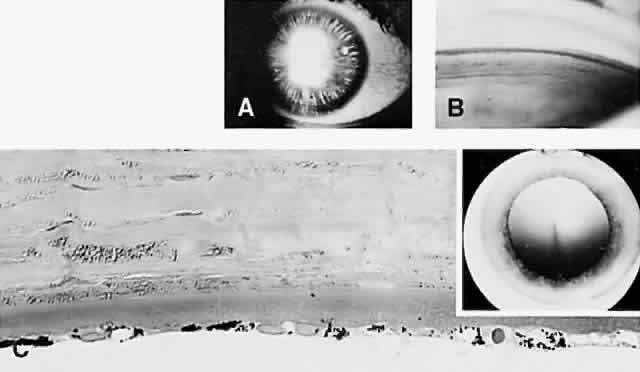
| Fig. 11. Pigment dispersion syndrome. (A) Extensive increased iris transillumination is present mainly in the middle third of the iris. (B) Goniscopic view shows marked deposition of pigment in the anterior chamber angle. (C) Melanin pigment granules present in the corneal endothelial cytoplasm are responsible for the Krukenberg's spindle seen clinically (inset). (A, clinical; B, gonioscopy; C, 1.5-μ section, PD, × 300; inset, clinical) |