Chapter 93
Human Herpes-viruses 6 and 7
HAMIDA B. QAVI
Main Menu   Table Of Contents

Search

HUMAN HERPESVIRUS 6
HUMAN HERPESVIRUS 7
REFERENCES

Herpesviridae are divided among three subfamilies: alphaherpesviruses, betaherpesviruses, and gammaherpesviruses. Human herpesviruses (HHVs) in the alpha subfamily include herpes simplex viruses (HSVs) 1 and 2 and varicella zoster virus (VZV). The beta subfamily consists of cytomegalovirus (HCMV), human herpesvirus-6 (HHV-6), and human herpesvirus-7 (HHV-7). HHV-6 and HHV-7 belong to the Roseolavirus genus of the betaherpesvirus subfamily. The members of the gammaherpesvirus subfamily are human herpesvirus-8 (HHV-8) and Epstein Barr virus (EBV). These viruses have specific cell tropisms and have the ability to establish latent infections and reactivate occasionally.
Back to Top
HUMAN HERPESVIRUS 6

GROWTH PROPERTIES AND VIRUS STRUCTURE

In 1986, Salahuddin and coworkers1 isolated HHV-6 from peripheral mononuclear cells of patients with various lymphoproliferative diseases and from an individual with human immunodeficiency virus type-1 (HIV-1) infection. More than 10 laboratories worldwide have reported the isolation of closely related viruses.2

Because of its similarity to HCMV, HHV-6 is classified as a betaherpesvirus.2 Immunofluorescent staining of HHV-6-infected cultures using specific monoclonal antibodies to other known HHVs—HSV-1, HSV-2, VZV, EBV, and HCMV clearly indicate that HHV-6 is a previously undetected virus. In situ hybridization using cloned viral deoxyribonucleic acid (DNA) probes has further confirmed these findings.3 The HHV-6 isolates from several laboratories exhibit differences in in vitro cell tropism, effects on expression of T-cell markers, the major cell type that supports their growth, the restriction endonuclease patterns of their DNA, differential reactivity with various monoclonal antibodies, seroepidemiology, and disease associations.4,5 Based on these observations, HHV-6 is divided into two groups: A and B. The two variants of HHV-6 are genetically closely related.2–6 A clear etiologic role of HHV-6A has not been identified, although HHV-6B has been shown to be the major cause of exanthem subitum or roseola.7

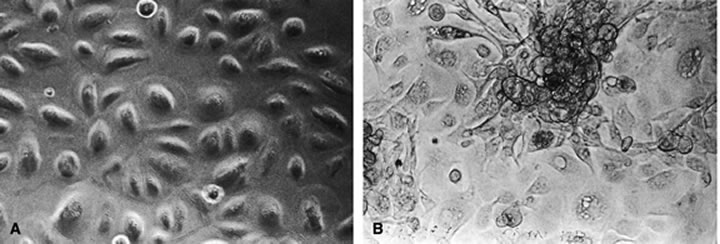
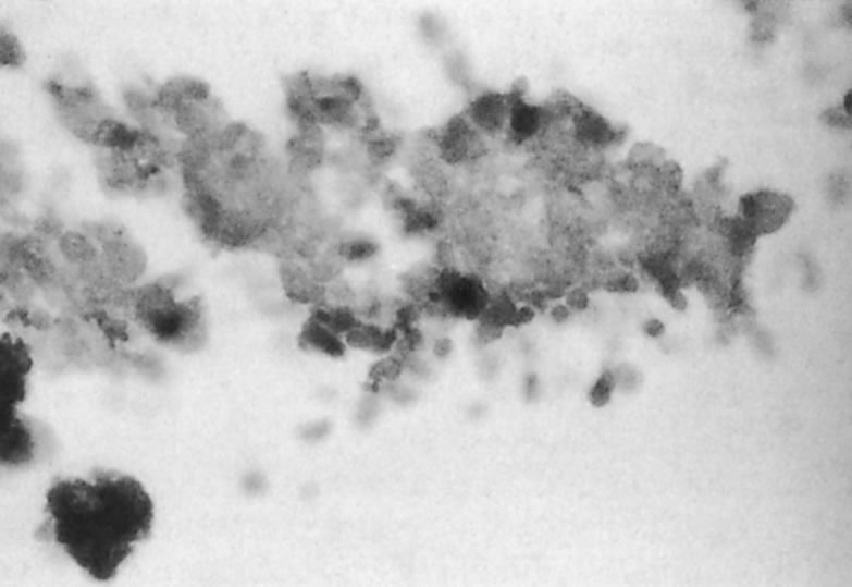
The predominant cell type permissive for replication of both HHV-6A and HHV-6B is CD4+ T cells. Although individual isolates of both variants A and B differ in their host range and ease of adaptability to culture, isolates of both kinds have been adapted by serial passages in continuous cell lines to yield higher titers. An often noted variant differentiating characteristic is the relative ease of adapting HHV-6A isolates for growth in T-cell lines, CCRF HSB2 cells and HHV-6B isolates in Molt 3 cells.6 A range of other cell types (including some EBV transformed B-cell lines, transformed cervical epithelial cells, megakaryocytes, glial cell lines, neurons, astrocytes, primary monocytes/macrophages, natural killer cells, bone marrow, tonsil, thymus, and spleen mononuclear leukocytes) have been infected with HHV-6. HHV-6B isolates have a more restricted cell tropism than the HHV-6A variant.2 HHV-6 has been recovered by cocultivation of corneas with peripheral blood monocytes, and the virus is capable of infecting primary and transformed corneal epithelial cells. HHV-6 can infect corneal cells, which then express viral antigens and DNA sequences.8 HHV-6 induces cytopathic effects in infected cells, including corneal cells (Fig. 1A and B). These changes are characterized by the presence of large balloon-shaped refractile cells, with occasional syncytia formation in the infected cultures. The cytopathic effects are visible 4 to 5 days after infection, and virus is usually cell associated.2,6,8 HHV-6 has also been isolated from and detected in retinas of acquired immunodeficiency syndrome (AIDS) patients9–11 (Fig. 2).

Fig. 1. Phase-contrast micrographs of uninfected (A) and HHV-6 infected (B) corneal epithelial cells at day 7 after infection (× 200). (Qavi HB, Xu B, Green MT et al: Morphological and ultra structural changes induced in corneal epithelial cells by HIV-1 and HHV-6. Curr Eye Res 15:597, 1996)

Fig. 2. Immunoperoxidase staining, showing HHV-6 antigens in a retinal section from an acquired immunodeficiency syndrome (AIDS) patient (× 400). (Qavi HB, Green MT, SeGall, GK et al: Transcriptional activity of HIV-1 and HHV-6 in retinal lesions from AIDS patients. Invest Ophthalmol Vis Sci 33:275, 1992)

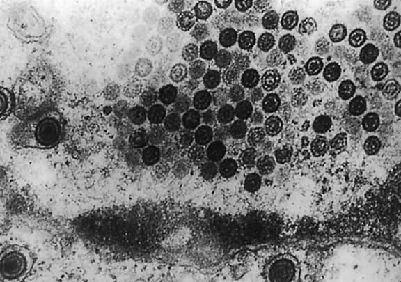
Electron microscopy shows that HHV-6 has features typical of members of the herpesvirus family. Its DNA is present in an icosahedral capsid made up of 162 capsomeres. The enveloped virions are 160 to 200 nm in diameter; naked capsid is 90 to 110 nm, and enclosed cores are 60 nm in diameter. Empty and DNA-filled nucleocapsids are observed in the nucleus. In the cytoplasm, capsids are coated with a fibrous material called tegument, which is 20 to 40 nm in diameter (Fig. 3). The tegument is the most striking morphologic feature of HHV-6; this component of the virion is more clearly demarcated in HHV-6 than in HCMV virions. Mature enveloped virions with prominent tegument accumulate in cytoplasmic vacuoles and on the surface of infected cells. Large inclusion bodies are also present in the cytoplasm of some infected cells.12,13

Fig. 3. Electron micrograph of peripheral blood mononuclear cells from an HHV-6 positive corneal culture, showing particles with teguments and envelopes.

The double-stranded linear DNA genome of the virus has been estimated to be 163 to 170 kilobase pairs (kbp), with an overall G + C content of about 43%. The HHV-6 DNA consists of a 143-kbp unique long segment flanked on both sides by single copies of direct repeated (DR) sequences (i.e., in the same orientation). The size of the DR sequences is 13 kbp in HHV-6A strain and 10 kbp in HHV-6B strains.14 Less than 10% of the genome has been shown to be circularized, directly linking the left DR with the right DR.

Restriction fragment polymorphism has been noted when comparing different HHV-6 isolates, indicating variation in DNA structure among isolates.15 Restriction-enzyme analysis of purified HHV-6 DNA shows a pattern markedly different from other HHVs. Under stringent conditions, however, HHV-6 DNA cross-hybridizes to certain regions of HCMV DNA.16

In the HHV-6 genome, seven blocks in the central region are core genes, which are common to all herpesviruses. Another block spanning open reading frames (ORFs) U2 to U14 contains specific genes for the betaherpesvirus subfamily. The ORFs U15 to U25 are genes specific to roseolavirus (HHV-6 and HHV-7). However, three genes U22, U83, and U95 are specific to HHV-6 and are not found in HHV-7. HHV-6 also closely resembles HHV-7 in genome homology and organization. Amino acid similarity to HHV-7 is 46.6% to 84.9% and to HCMV 41.0% to 75.8%.15–19

REPLICATION, LATENCY, AND REACTIVATION

HHV-6 appears to enter cells by endocytosis. The nature of the receptor is unknown; however, monoclonal antibody to virion glycoprotein complex seems to inhibit the penetration of HHV-6 into the cells.19 The virus has been observed to enter uncoated pits, and the viral membrane fuses with endocytic vesicles, resulting in the release of nucleocapsids into the cytoplasm. The nucleocapsid enters the nucleus, where gene expression begins and viral DNA replication takes place. The nucleocapsid is assembled, acquires the tegument in unique nuclear structures called tegusomes, and is then released into the cytoplasm by fusion of the tegusome with the nuclear membrane. The resulting particles are enveloped by budding into cytoplasmic vacuoles, which then fuse with the cell membranes to release mature virions.13,20

The virus readily infects and replicates in some T-cell lines and fibroblasts in vitro. In other cell lines, however, virus replication is suppressed or the virus may become latent. Latent viral genome has been detected in T- and B-cell lines and fibroblasts that survive viral infection. The only data available to support latency is the failure to recover HHV-6 from peripheral blood lymphocytes of all healthy adults, indicating that the virus may be latent in these cells. During primary infection, HHV-6 DNA is detectable in nonadherent peripheral blood lymphocytes, whereas during convalescence it is only detectable in adherent cells by polymerase chain reaction (PCR). Several investigators have been able to reactivate HHV-6 from latently infected macrophages, from T- and B-cell lines and from fibroblasts using 12-0-tetradecanoylphorbol 13-acetate and hydrocortisone.2,21

VIRAL DNA REPLICATION AND GENE EXPRESSION

The temporal relation of DNA replication and viral gene expression is unknown. Little is known about the regulatory properties of HHV-6 promoters or the role of specific viral genes in HHV-6 gene regulation. HHV-6 codes for a variety of enzymes and regulatory proteins involved in nucleotide metabolism. Some of these have been identified as homologues of genes present in HSV-1, VZV, and HCMV, and others have been biochemically characterized.

These genes encode the major DNA-binding protein, DNA polymerase, polymerase-associated protein, a helicase-primase complex, DNA origin binding protein, and the small subunit of ribonucleotide reductase. Similar to HCMV, however, a virus-specific thymidine kinase activity has not been demonstrated in cells infected with HHV-6. The absence of a gene for thymidine kinase has also been confirmed by sequence homology analysis.15

Because of the low abundance of viral transcripts, many genes expected to be expressed during productive replication have not been identified by Northern blot analysis, in which the only transcript detectable was a 3.5-kb transcript that was identified as a product of the immediate early regulatory gene or genes. The C-terminal portion of this immediate early protein has the ability to transactivate heterologous promoters.22

With an estimated 170-kbp DNA, HHV-6 is capable of coding for more than 70 proteins. Using purified virions, more than 25 HHV-6-specific proteins, ranging from 30 to 200 kilodaltons (kDa), have been identified by immunoprecipitation. Among these, nine are glycoproteins, eight are associated with infected cell membranes, and six are associated with viral envelope. In addition to the previously mentioned proteins necessary for DNA synthesis, HHV-6 encodes genes for a large structural phosphoprotein, a large tegument protein, a major capsid protein, and an HHV-6-induced nuclear antigen.23 In addition, HHV-6 but not HHV-7 carries a homologue of adeno-associated type 2 parvovirus rep gene that is transcribed in latently infected cells.24,25 Recently, the viral protein from the U12 gene has been recognized as a beta-chemokine receptor.25 HHV-6 may play a role in tumor progression through these molecules.

EPIDEMIOLOGY AND PATHOGENESIS

Worldwide, more than 95% of individuals older than the age of 2 years are seropositive for HHV-6 antibody. The level of maternal antibody decreases during the first 5 months of life; antibody levels sharply increase between 6 months and 5 years of age, indicating initial infection. The seroprevalence decreases with age; however, it rises again at age 62 years. The mode of transmission is believed to be saliva. HHV-6 DNA has been detected in saliva of 90% of the United States population.2,27

HHV-6-associated exanthem subitum (a childhood disease) is characterized by high fever and a rash for 2 to 5 days. The incubation period is usually 5 to 15 days, and the disease normally resolves without complications. In rare cases, complications such as recurrent convulsions, meningitis, hepatosplenomegaly, mononucleosis, and fatal fulminant hepatitis have been reported.7,27–29 In the adult population, primary HHV-6 infection is rare. If infection is symptomatic, the symptoms can include lymphadenopathy, mononucleosis, and hepatitis.27

Virus-neutralizing IgM has been reported in patients 4 to 8 days after the onset of exanthem subitum, which usually peaks after 2 to 3 weeks and disappears in 7 to 8 weeks. In most cases, IgG is detectable after the eighth week. Cellular immunity may have a role in controlling HHV-6 infection in organ transplantation patients. The simple tests employed for the detection of HHV-6 antibodies (specifically, IgG levels) are indirect immunofluorescence and anticomplement immunofluorescence. The most widely used and most sensitive test is the enzyme-linked immunosorbent assay. The most sensitive and specific markers for HHV-6 infection are 135-kDa and 100-kDa antigens present in the serum.2

One intriguing characteristic of HHV-6 is its interaction with other viruses. It has been shown that HHV-6 grows with EBV or HCMV simultaneously within the same cells and may interact with each of these viruses. HHV-7 has been demonstrated to activate latent HHV-6. An enhanced expression of human papilloma virus (HPV) oncoproteins, E6 and E7, and transactivation of an HPV-18 promoter in cervical epithelial cells in the presence of HHV-6 infection has been documented, indicating that the virus may be associated with induction of cervical carcinoma caused by HPV.27 There are strong indications that HHV-6 may not transactivate human T-cell leukemia virus type I long terminal repeat.30 It has been shown that HHV-6 accelerates the destruction of CD4+ T lymphocytes in cultures coinfected with HHV-6 and HIV-1.8,31

HHV-6 infection induces nuclear factors that specifically bind to the enhancer region of HIV-1 LTR.31 When cloned into T- cells, HHV-6 DNA fragments containing an immediate early gene encode gene products that are able to activate the HIV-1 LTR.32 Because of its positive interaction with HIV-1, a catalytic role for HHV-6 during progression of AIDS has been proposed. The activation of HIV-1 by HHV-6 in retinal cells in vitro and the activation of HSV-1 in rabbit corneas in vivo have been demonstrated.8,33 These findings suggest that HHV-6 may play an important role in the pathogenesis of retinitis and keratitis.

The oncogenic potential of HHV-6 has been tested in mouse fibroblast cell lines and human epidermal keratinocytes in vitro using three viral DNA fragments.19 One codes DR7 and the other two encompasses the regions spanning U2 to U20 and U31 to U37. DR7 and the two other loci also contain genes with transactivating activity on HIV-1 LTR.34,35

HHV-6 is one of the most neurotropic viruses known in children and adults. A possible correlation between active HHV-6 infection and multiple sclerosis (MS) has also been the focus of much attention in the past few years. MS is a severe central nervous system (CNS) disease of young adults characterized by the progressive demyelination of nerves that lead to paralysis and eventually to death. This is an autoimmune disease of myelin. HHV-6B DNA sequences were found with higher frequency in MS brain specimens using representational difference PCR.36 Oligodendrocyte nuclei were found positive for HHV-6 antigens in MS samples (80%), whereas the controls were negative (0%).The titer of anti-HHV-6 IgM was also higher in samples from MS patients than control (73% vs. 8%).These findings are interpreted as a hallmark of the association between active or reactivable HHV-6 infection and the disease.37–39 The serologic analysis are difficult to interpret, because this disease is caused by an immunologic dysregulation and, therefore, the rise in antibody titer may be the sign of the disease rather than the cause. However, these results were not confirmed by other investigators.40 Therefore, the association between an active infection and MS is still a controversial issue.

Foscarnet, a potent inhibitor of DNA polymerases of other herpesviruses, has been shown to successfully inhibit HCMV and HHV-6 infections in AIDS patients with HCMV retinitis.41 Ganciclovir and foscarnet have been used to successfully treat HHV-6 infections in several organs transplant patients.42 Sophocarpines have been shown to be effective against HHV-6 replication (Qavi and coworkers, manuscript accepted by Phytotherapy for March 2002 issue [in press]).

Back to Top
HUMAN HERPESVIRUS 7

GROWTH PROPERTIES AND VIRUS STRUCTURE

HHV-7 was first isolated from CD4+ T cells of a healthy person.43 It has since been shown to undergo productive replication in CD4+ T cells; it appears to use the CD4 molecules as its receptor and may also infect other hematopoietic progenitor cells that express surface CD4 molecules.44–46 Infection of CD4+ T cells, monocytes, and macrophages results in downregulation of expression of CD4 molecules and restricts subsequent infection of those cells by HIV-1.47,48

The 145-kb genome of HHV-7 has been completely sequenced and is genetically related to HHV-6, sharing 60% nucleotide similarity and more than 55% amino acid homology.15–19,49–52 The origins of DNA replication of HHV-6 and HHV-7 that are used during lytic replication show extensive homology.53 The genome of HHV-7 is bounded by terminal DRs and it is these regions where the greatest differences from HHV-6 exist.50 In addition, divergence occurs in regions encoding immediate early genes thought to be involved in transcriptional regulation.50 As a member of the betaherpesvirus subfamily, HHV-7 shows some genomic relation to HCMV and to lesser extent, human alphaherpesviruses and gammaherpesviruses, generally in genes defined as core genes common to most members of the herpesvirus family.53,54 Even genomes of HHV-6 and HHV-7 are collinear; notable structural difference are apparent especially in the DR area and immediate early regions of the HHV-7 viral sequence, as well as in the absence of an adeno-associated virus type 2 gene homologue.15–19

LATENCY AND REACTIVATION

HHV-7 appears to be able to establish latency, but the exact nature of the latent state of HHV-7 is unknown. The virus appears to be able to become latent in T cells and can be reactivated from peripheral blood of healthy individuals by T-cell activation.55 In addition, virus and viral DNA can be detected in salivary glands of seropositive people.56–59 The frequency of detection of DNA varies in individual salivary glands, but DNA has been detected in 100% of submandibular gland samples of seropositive individuals.58 These observations indicated that HHV-7 may be in latent form in salivary gland cells and may reactivate occasionally.

EPIDEMIOLOGY AND PATHOGENESIS

HHV-7 appears to be ubiquitous in humans. Infection with HHV-7 occurs at high frequency early in life, although somewhat later than HHV-6 infection.60–62 Seropositivity rates among adult populations are generally 85% or more.

Two proteins, a 85-kDa phosphoprotein complex and a 121-kDa protein have been identified as immunodominant proteins, distinct from HHV-6, but cross reactivity of sera between HHV-6 and HHV-7 occurs.61–63 In Britain, virus DNA was detected in saliva of 96% of healthy adult volunteers at an estimated 1.1 × 10⁶ genome copies/ml and in 83% of peripheral blood samples at 267 copies/106 cells.64 A German study reported similar rates of DNA positivity.65 The amount of virus that a person has in saliva or blood appears to remain relatively stable over time. Although both HHV-6 and HHV-7 DNA can be detected in salivary glands, infectious HHV-7 is detected in saliva at a higher rate than HHV-6, suggesting that shedding of HHV-7 is more common. The rate of shedding is further increased in immunocompromised individuals.60,66

Although HHV-6 has been demonstrated to be an important etiologic agent of exanthem subitum in children, HHV-7 infection has been associated with a similar clinical presentation. In one study of 22 cases of primary HHV-7 infection, 47.1% of the children showed clinical signs consistent with exanthem subitum. These children already had HHV-6 antibody at the time of infection.67 In other studies with fewer patients, a similar association was shown.68,69

Reactivation of HHV-7 is detected by increased levels of viral DNA in the peripheral blood. When such reactivation occurs in renal-allograft patients concurrent with increased levels of HCMV DNA, there may be an increased risk of the patient developing clinically apparent HCMV disease.70 HHV-7 has been associated with a variety of other disease manifestations, but these remain to be confirmed. No specific ocular manifestations of HHV-7 infection have been reported.

HHV-7 is susceptible to inhibition by nucleoside analogues that are effective against HCMV and HHV-6 including ganciclovir, foscarnet, and cidofovir but not those antiherpes agents requiring viral thymidine kinase for phosphorylation.71

Back to Top
REFERENCES

1. Salahuddin SZ, Ablashi DV, Markham PD et al: Isolation of a new virus, HBLV, in patients with lymphoproliferative disorders. Science 234:596, 1986

2. Pellet E, Black JB: Human herpesvirus-6. In Fields BW, Knipe DM, Howley PM et al. (eds): Virology. 3rd ed. Philadelphia, Lippincott-Raven, 1996, p 2587

3. Joseph SF, Salahuddin SA, Ablashi DV et al: Genomic analysis of the human B-lympho-tropic virus (HBLV). Science 234:601, 1986

4. Dewhurst S, Melntyre K, Schuabel K et al: Human herpesvirus-6 (HHV-6) variant B accounts for the majority of symptomatic primary HHV-6 infections in a population of U.S. infants. J Clin Microbiol 31:416, 1993

5. Schirmer EC, Wyatt LS, Yamanishi K et al: Differentiation between two distinct classes of viruses now classified as human herpesvirus-6. Proc Natl Acad Sci U S A 88:5922, 1991

6. Black JB, Sanderlin KC, Goldsmith CS et al: Growth properties of human herpesvirus-6 strain 29, J Virol Methods 26:133, 1989

7. Yamanishi, K Okuno T, Shiraki K et al: Identification of human herpesvirus-6 as a causal for exanthem subitum. Lancet 1:1065, 1988

8. Qavi HB, Xu B, Green MT et al: Morphological and ultra structural changes induced in corneal epithelial cells by HIV-1 and HHV-6. Curr Eye Res 15:597, 1996

9. Reux I, Fillet Am, Agut H et al: In situ detection of human herpesvirus-6 in retinitis associated with acquired immunodeficiency syndrome. Am J Ophthalmol 114:375, 1992

10. Qavi HB, Green MT, SeGall GK et al: Transcriptional activity of HIV-1 and HHV-6 in retinal lesions from AIDS patients. Invest Ophthalmol Vis Sci 33:275, 1992

11. Qavi HB, Green MT, Lewis DE et al: HIV-1 and HHV-6 antigens and transcripts in retinas of patients with AIDS in the absence of human cytomegalovirus. Invest Ophthalmol Vis Sci 36:2040, 1995

12. Qavi HB, Green MT, SeGall GK: HIV-1 and HHV-6 infections of human retina and cornea, In Ablashi DV, Krueger GF (eds): Human Herpesvirus 6: Epidemiology, Molecular Biology and Clinical Pathology. Vol 4. Amsterdam, Elsevier Science, 1992, p 263

13. Kramarsky B, Sander C: Electron microscopy of human herpesvirus-6 (HHV-6). In Ablashi DV, Drueger GF (eds): Human Herpesvirusv 6: Epidemiology, Molecular Biology and Clinical Pathology. Vol 4. Amsterdam, Elsevier Science, 1992, p 59

14. Linquester GJ, Pellet PF: Properties of human herpesvirus strain Z29 genome: G-C content, length and presence of variable-length directly repeated terminal sequence elements. Virology 182:102, 1991

15. Gompels UA, Nichols J, Lawrence G et al: The DNA sequence of human herpesvirus-6: Structure, coding content, and genome evolution. Virology 51:209, 1995

16. Neipel F, Ellinger K, Fleckenstein B: The unique region of the human herpesvirus 6 genome is essentially collinear with the UL segment of human cytomegalovirus. J Gen Virol 72:2293, 1991

17. Nicholas J: Determination and analysis of the complete nucleotide sequence of human herpesvirus 7. J Virol 68:597, 1996

18. Megaw AG, Rapaport D, Avidor B et al. The DNA sequence of the RK strain of human herpesvirus 7. Virology 244:119, 1998

19. Campadelli-Fiume G, Mirandola P, Menotti I: Human herpesvirus 6: An emerging pathogen. Emerging Infect Disord 5:353, 1999

20. Foa-Tomasi L, Boscaro A, Gaeta S et al: Monoclonal antibodies to gp100 inhibit penetration of human herpesvirus-6 and polykaryocyte formation in susceptible cells. J Virol 65:4124, 1991

21. Kondo K, Kondo T, Okuno T et al: Latent human herpesvirus-6 infection of monocytes/macrophages. J Gen Virol 72:1401, 1991

22. Schoive U, Neipel F, Schreiner D et al: Structure and transcription of an immediate-early region in the human herpesvirus-6 genome. J Virol 68:2978, 1994

23. Balachandran N, Tirawatnopong S, Pfeiffer B et al: Electrophoretic analysis of human herpesvirus-6 polypeptides immunoprecipitated from infected cells with human sera. J Infect Dis 163:29, 1991

24. Thompson BJ, Weindler FW, Gray D et al: Human herpesvirus 6 (HHV-6) is a helper virus for adeno-associated virus type 2 (AAV-2) and the AAV-2 rep gene homologue in HHV-6 can mediate AAV-2 DNA replication and regulate gene expression. Virology 204:304, 1994

25. Rotola A, Ravaioli T, Gonelli A et al: U94 of human herpesvirus 6 is expressed in latently infected peripheral blood mononuclear cells and blocks of viral gene expression in transformed lymphocytes in culture. Proc Natl Acad Sciences U S A 95:13911, 1998

26. Isegawa Y, Ping Z, Nakano K et al: Human herpesvirus open reading frame U12 encodes a functional beta-chemokine receptor. J Virol 72:6104, 1998

27. Levin PH: A review of human herpesvirus-6 infections. Infect Med 12:395, 1995

28. Suga S, Yoshikawa T, Asano Y et al: Clinical and virological analyses of 21 infants with exanthem subitum (roseola infantum) and central nervous system complications. Ann Neurol 33:597, 1992

29. Stammers TG: Roseola infantum—The neglected exanthem. Practitioner 232:541, 1988

30. Cao W, Sullivan DC: Human herpesvirus 6 infection does not enhance human T-cell lymphotropic virus type (HTLV-1) expression in the HTLV-1 transformed cell line MT4. J Infect Dis 166:446, 1992

31. Ensoli B, Lusso P, Schachter F et al: Human herpesvirus-6 increases HIV-1 expression in co-infected T-cells via nuclear factors binding to HIV-1 enhancer. EMBO J 8:3019, 1990

32. Horvat RT, Wood C, Joseph SF et al: Transactivation of the human immunodeficiency virus promoter by human herpesvirus 6 (HHV-6) strains GS and Z29 in primary human T lymphocytes and identification of transactivating HHV-6 (GS) gene fragments. J Virol 65:2895, 1991

33. Qavi HB: Possible Role of HHV-6 in the enhanced severity of HSV-1 Keratitis. In vivo 13:427, 1999

34. Thompson J, Choudhury S, Kashanchi F et al: A transforming fragment within direct repeat region of human herpesvirus type 6 that transactivates HIV-1. Oncogene 9:1167, 1994

35. Kashanchi F, Araujod, Doniger J et al: Human herpesvirus 6 (HHV-6) ORF-1 transactivating gene exhibits malignant transforming activity and its protein binds to p53. Oncogene 14:359, 1997

36. Challoner PB, Smith KT, Parker JD et al: Plaque associated expression of human herpesvirus 6 in multiple sclerosis. Proc Natl Acad Sci U S A 92:7440, 1995

37. Soldan SS, Berti R, Salem N et al: Association of human herpesvirus 6 (HHV-6) with multiple sclerosis: Increased IgM response to HHV-6 early antigen and detection of serum HHV-6 DNA. Nat Med 3:1394, 1997

38. Ablashi DV, Lapps W, Kaplan M et al: Human herpesvirus 6 (HHV-6) infection in multiple sclerosis: A preliminary report. Multiple Sclerosis 4:490, 1998

39. Friedman JE, Lyons MJ, Cu G et al: The association of the human herpesvirus 6 and MS. Multiple Sclerosis 5:355, 1999

40. Coats AR, Bell J: HHV-6 and multiple sclerosis. Nat Med 4:537, 1998

41. Streicher HZ, Hung CL, Ablashi DV et al: In vitro inhibition of human herpesvirus 6 by phosphonoformate. J Virol Methods 21:301, 1988

42. Williams MV: HHV-6: Response to antiviral agents. In Ablashi DV, Krueger GF (eds): Human Herpesvirus 6: Epidemiology, Molecular Biology and Clinical and Pathology. Vol 4. Amsterdam, Elsevier Science, 1992, p 317

43. Frenkel N, Schirmer EC, Wyatt LS: Isolation of a new herpesvirus from human CD4+ T cells. Proc Natl Acad Sci U S A 87:748, 1990

44. Yasukawa M, Hatta N, Sada E et al: Human herpesvirus 7 infection of CD4+ T cells does not require expression of the OKT4 epitope. J Gen Virol 77:3103, 1996

45. Yasukawa M, Inoue Y, Ohminami H et al: Human herpesvirus 7 infection of lymphoid and myeloid cell lines transduced with an adenovirus vector containing the CD4 gene. J Virol 71:1708, 1997

46. Lusso P, Secchiero P, Crowley RW et al: CD4 is a critical component of the receptor for human herpesvirus 7: Interference with human immunodeficiency virus. Proc Natl Acad Sci U S A 91:3872, 1994

47. Crowley RW, Secchiero P, Zella D et al: Interference between human herpesvirus 7 and HIV-1 in mononuclear phagocytes. J Immunol 156:2004, 1996

48. Furukawa M, Yasukawa M, Yakushijin Y et al: Distinct effects of human herpesvirus 6 and human herpesvirus 7 on surface molecule expression and function of CD4+ T cells. J Immunol 152:5768, 1994

49. Singer O, Frenkel N: Human herpesvirus 7 (HHV-7) DNA-analyses of clones spanning the entire genome. Arch Virol 142:287, 1997

50. Nicholas J: Determination and analysis of the complete nucleotide sequence of human herpesvirus 7. J Virol 70:5975, 1996

51. Ruvolo VR, Berneman Z, Secchiero P et al: Cloning, restriction endonuclease mapping and partial sequence analysis of the genome of human herpesvirus 7 strain JI. J Gen Virol 77:1901, 1996

52. Dominguez G, Black JB, Stamey FF et al: Physical and genetic maps of the human herpesvirus 7 strain SD genome. Arch Virol 141:2387, 1996

53. Bernman ZN, Ablashi DV, Li G et al: Human herpesvirus 7 is a T-lymphotropic virus and is related to, but significantly different from human herpesvirus 6 and human cytomegalovirus. Proc Natl Acad Sci U S A 89:10552, 1992

54. Vanloon N, Dykes C, Deng HY et al: Identification and analysis of a lytic-phase origin of DNA replication in human herpesvirus 7. J Virol 71:3279, 1997

55. Katsafanas GC, Schirmer EC, Wyatt LS et al: In vitro activation of human herpesvirus 6 and 7 from latency. Proc Natl Acad Sci U S A 93:9788, 1996

56. Wyatt LS, Frenkel N: Human herpesvirus 7 is a constitutive inhabitant of adult human saliva. J Virol 66:3206, 1992

57. Black JB, Inoue N, Kite-Powell K et al: Frequent isolation of human herpesvirus 7 from saliva. Virus Res 29:91, 1993

58. Sada E, Yasukawa M, Ito C et al: Detection of human herpesvirus 6 and human herpesvirus 7 in the submandibular gland, parotid gland, and lip salivary gland by PCR. J Clin Microbiol 34:2320, 1996

59. Hikada Y, Liu Y, Yamamoto M et al: Frequent isolation of human herpesvirus 7 from saliva samples. J Med Virol 40:343, 1993

60. Levine PH, Ablashi DV: An etiologic perspective of the new herpesviruses: HHV-7 and HHV-8. Infect Med 16:24,29, 1999

61. Wyatt LS, Rodriguez WJ, Balachandran N et al: Human herpesvirus 7: Antigenic properties and prevalence in children and adults. J Virol 65:6260, 1991

62. Tanaka-Taya K, Kondo T, Mukai T et al: Seroepidemiological study of herpesvirus-6 and -7 in children of different ages and detection of these two viruses in throat swabs by polymerase chain reaction. J Med Virol 348:88, 1996

63. Foa-Tomasi L, Avitabile E, Campadelli-Fiume G: Polyvalent and monoclonal antibodies identify major immunogenic proteins specific for human herpesvirus 7-infected cells and have weak cross-reactivity with human herpesvirus 6. J Gen Virol 75:2719, 1994

64. Kidd IM, Clark DA, Ait-Kaled M et al: Measurement of human herpesvirus 7 load in peripheral blood and saliva of healthy subjects by quantitative polymerase chain reaction. J Infect Dis 174:396, 1996

65. Wilborn F, Schimidt CA, Lorenz F et al: Human herpesvirus type 7 in blood donors: Detection by the polymerase chain reaction. J Med Virol 47:65, 1995

66. DiLuca D, Mirandola P, Ravaioli T et al: Human herpesviruses 6 and 7 in salivary glands and shedding in saliva of healthy and human immunodeficiency virus positive individuals. J Med Virol 45:462, 1995

67. Torigoe S, Kumamoto T, Koide W et al: Clinical manifestations associated with human herpesvirus 7 infection. Arch Dis Childhood 72:518, 1995

68. Tanaka K, Kondo T, Torigoe S et al: Human herpesvirus 7: Another causal agent for roseola (exanthem subitum). J Pediatr 125:1, 1994

69. Asano Y, Suga S, Yoshikawa et al: Clinical features and viral excretion in an infant with primary human herpesvirus 7 infection. Pediatrics 95:187, 1995

70. Osama HK, Peiris JS, Taylor CE et al: “Cytomegalovirus disease” in renal allograft recipients: Is human herpesvirus 7 a co-factor for disease progression? J Med Virol 48:295, 1995

71. Takahashi K, Suzuki M, Iwata Y et al: Selective activity of various nucleoside and nucleotide analogues against human herpesvirus 6 and 7. Antiviral Chem Chemother 8:24, 1997

Back to Top