|
|
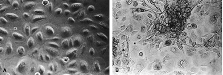
| Fig. 1. Phase-contrast micrographs of uninfected (A) and HHV-6 infected (B) corneal epithelial cells at day 7 after infection (× 200). (Qavi HB, Xu B, Green MT et al: Morphological and ultra structural changes induced in corneal epithelial cells by HIV-1 and HHV-6. Curr Eye Res 15:597, 1996) |