Chapter 45
Host Defense Against Ocular Infection
BRADLEY D. JETT
Main Menu   Table Of Contents

Search

ANATOMIC AND MECHANICAL DEFENSES
DEFENSES OF THE OCULAR SURFACE AND SURROUNDING TISSUES
DEFENSES OF THE OCULAR INTERIOR
SUMMARY
REFERENCES

The eye possesses a diverse array of defense mechanisms that protect the visual field from the damaging consequences of infection. The relatively large number of nonspecific and specific defenses (Table 1), localized within this relatively small organ system, provides a formidable barrier to bacteria, viruses, fungi, and parasites. In this chapter, current information on the host defenses of the ocular surface and its surrounding tissues, as well as the defense mechanisms that exist in the interior of the eye, is discussed.


Table 1. Ocular Host Defense Mechanisms


Nonspecific Ocular DefensesSpecific Ocular Defenses
EyelidsEye-associated lymphoid tissue
TearsAntigen-presenting Langerhans' cells
Ocular epithelium 
Normal ocular bacterial floraImmunoglobulins
MucinClassical complement cascade
Antibacterial factorsT lymphocytes
Alternative complement cascadeB lymphocytes
Natural killer cells 
Macrophages 


The immunologic defenses of the eye, although impressive, present a puzzling enigma: The eye is extremely vulnerable to inflammation. Therefore, the immunologic defense mechanisms described in this chapter can lead to potentially deleterious consequences for the optical clarity and bioelectrical functionality of the visual apparatus. It has been long recognized that the eye represents an immunologically privileged site. Decades of research by Streilein and others has revealed that the eye, like other immune privileged sites, possesses complex mechanisms capable of regulating inflammatory responses. Although ocular immune privilege is a dynamic, rather than passive, state mediated by soluble and cellular factors within the ocular microenvironment, a detailed discussion of this phenomenon is beyond the scope of this chapter.1,2,3 Immune privilege is mentioned in this introduction simply to remind the reader that the immunologic defense mechanisms described herein are also dynamic processes and are susceptible to both activation and suppression.

Back to Top
ANATOMIC AND MECHANICAL DEFENSES

EYELIDS AND EYELASHES

The first line of defense against ocular infection is provided by the eyelids. This mechanical barrier, when closed, protects the ocular surface and its surrounding tissues from exposure to pathogens, allergens, and other foreign bodies. The ocular surface is highly innervated and exquisitely sensitive to mechanical disturbance. Even the slightest contact with the eyelids, eyelashes, or corneal surface elicits the blink reflex. This blinking action serves to wash pathogens and other foreign particles from the ocular surface and to renew the tear film.

Conditions that interfere with normal lid function, including conjunctivitis and keratitis, may render the eye more susceptible to infections.4

TEARS AND LACRIMAL DRAINAGE

The tear film (Table 2) serves multiple functions in the nonspecific defense against ocular infection. It bathes the ocular surface and provides a mechanical barrier to adhesion by pathogenic organisms. It lubricates the corneal surface and facilitates the washing action of the eyelids. Bacteria, fungi, foreign bodies, and desquamated epithelial cells are flushed nasally toward the two puncta by this highly efficient washing mechanism.


Table 2. The Tear Film


ComponentFunction
Flushing actionMechanical removal of pathogens
MucinPrevents pathogen binding to ocular surface, traps microbes for removal via lacrimal drainage
LactoferrinIron-binding protein, interferes with pathogen metabolism
CeruloplasminCopper-binding protein
β-lysinAttacks bacterial membranes
LysozymeHydrolyzes bacterial cell wall peptidoglycan
CytokinesRegulation of immune responses, recruitment and activation of phagocytic cells
ComplementOpsonization of pathogens, attacks pathogen membranes, recruitment of immune cells
ImmunoglobulinsOpsonization of pathogens, blocks pathogen binding to ocular surface, neutralization of toxins
Phospholipase A2Attacks pathogen membrane phospholipids
DefensinsInhibits pathogen growth


The 7-μm thick tear film is comprised of three layers: lipid, aqueous, and mucoid. These layers are derived from the meibomian glands, the lacrimal glands, and conjunctival goblet cells, respectively.5 Specific antimicrobial components of the aqueous and mucoid layers are discussed later. Lipids confer surfactant properties to the tear film, enhancing its ability to trap microbes and efficiently flush them into the lacrimal excretory system. It is, therefore, not surprising that patients suffering from aqueous tear film deficiencies (i.e., dry eye) are at increased risk for corneal infection.

OCULAR EPITHELIUM

The ocular epithelial surface prevents intraocular penetration by most microorganisms. This nonkeratinized squamous epithelium, consisting of five to six cell layers, is supported by a columnar basal layer. The basement membrane, with its numerous junctional complexes, presents a nearly impermeable anatomic barrier.6 The outermost layer of the corneal epithelium is covered by a layer of glycocalyx over which lies a layer of mucus. This glycocalyx serves as a scaffold that binds mucin, antibodies, and other tear film components. Specific components of the mucoid tear film are discussed later.

Relatively few microorganisms are capable of penetrating an intact corneal epithelium; these include Neisseria gonorrhoeae and Haemophilus aegyptius.4 Even those organisms commonly associated with bacterial keratitis, including Pseudomonas aeruginosa and Staphylococcus aureus, have difficulty establishing infection on an uncompromised ocular surface. Laboratory animal models of bacterial keratitis often require some form of pre-inoculation corneal injury or abrasion (Fig. 1), and/or prolonged contact with the pathogen.7,8 Keratitis caused by yeasts, filamentous fungi, and parasites are almost always associated with corneal abnormalities, injury, or surgery.5 Contact lens wear has also been implicated as a predisposing factor to fungal (predominantly Candida spp.) and parasitic (predominantly Acanthamoeba spp.) keratitis, presumably resulting from microabrasions of the corneal epithelium following prolonged contact with the lens polymers.5 Although little is currently known regarding the molecular mechanism for adhesion and invasion of the cornea epithelium by pathogenic organisms, a reasonable hypothesis may involve the unmasking of previously sequestered host molecules for which pathogens possess specific receptors.9

Fig. 1. Scanning electron micrograph of scarified rabbit cornea infected with Staphylococcus aureus. Bacteria were observed to bind relatively poorly to uninjured corneal epithelium compared with areas of injury and exposure of deeper layers. (Courtesy of Dr. M Gilmore)

Back to Top
DEFENSES OF THE OCULAR SURFACE AND SURROUNDING TISSUES

OVERVIEW OF OCULAR MUCOSAL IMMUNITY

The hypothetical involvement of extraocular tissues (i.e., conjunctiva, lacrimal system, etc.) in ocular immunologic processes is not a new concept. It has been long recognized that mucosa-associated lymphoid tissue (MALT) comprises an important component of an organism's innate and adaptive defense against invasion of mucosal surfaces by pathogens. Specific examples of MALT include gut-associated lymphoid tissue (GALT) found in intestinal Peyer's patches and bronchus-associated lymphoid tissue (BALT) of the respiratory tract. MALT, by definition, consists of clusters (follicles) of lymphatic cells situated within and beneath the epithelium of a mucosal surface. These follicles detect antigen and are capable of inducing both cellular and humoral immune responses. MALT follicles possess professional antigen-presenting cells (i.e., macrophages) and are the source of many secreted antimicrobial compounds and cytokines. Specialized vessels within MALT permit lymphocyte recirculation and communication with the central immune system. (For review of mucosal immunology, see references 3, 10, 11, 12, and 13.) Mucosal immunity has long been the subject of intense investigation, because these sites represent an important entry point for many pathogens. It is, therefore, somewhat surprising that research into the mucosal immunology of the eye has gained significant momentum only within the past decade.

Several recent histologic, immunohistochemical, and flow cytometric analyses have been published that suggest a role for extraocular tissues in regional mucosal immunity. Studies of conjunctiva-associated lymphoid tissue (CALT) revealed the presence of discreet follicles with germinal centers, close interactions between lymphocytes and surface lymphoepithelium, layers of IgA+ plasma cells in the lamina propria, and high endothelial venules (HEVs).14,15 Similar studies involving lacrimal drainage-associated lymphoid tissue (LDALT) and lacrimal gland-associated lymphoid tissue (LGALT) have revealed the presence of IgA+ plasma cells, T lymphocytes, major histocompatibility complex (MHC) class II-positive cells, and distinct lymphoid follicles in close association with HEVs.16,17 Because of the apparent biochemical and anatomic integration of lacrimal gland, lacrimal drainage, and conjunctival immune functions, it has been recently proposed to collectively refer to these sites as eye-associated lymphoid tissue (EALT).16 The various humoral and cellular components of the ocular mucosa, which serve both protective and regulatory functions, are discussed in greater detail later.

NORMAL OCULAR MICROFLORA

Colonization of the normal ocular surface and surrounding tissues by microorganisms is a dynamic phenomenon in both quantity and identity of microbes. Although samples taken from the conjunctiva of healthy individuals may be negative for microorganisms on culture, it is generally accepted that most humans harbor at least some normal bacteria on their periocular tissues. Among the normal ocular flora, Staphylococcus sp, Corynebacterium sp, and anaerobic Propionibacterium sp are most frequently isolated.13,18,19 Most fungi isolated from the conjunctiva are thought to be airborne contaminants; and protozoans and helminths are not considered to be normal ocular flora.

The diverse spectrum of normal ocular isolates and the intricacies of colonization dynamics are beyond the scope of this chapter. Nevertheless, normal ocular bacteria may serve a protective role in inhibiting colonization by more pathogenic species. This phenomenon of competitive exclusion has been well studied at other anatomic sites, including the nares, skin, and gastrointestinal tract. Presumably normal ocular organisms may play a similar role; however, a direct relationship between ocular commensal colonization and pathogen inhibition has remained elusive. The normal flora described previously are ironically the most frequent causes of many ocular infectious diseases, which demonstrates the delicate balance of host-parasite relationships in the external ocular microenvironment.

MUCINS

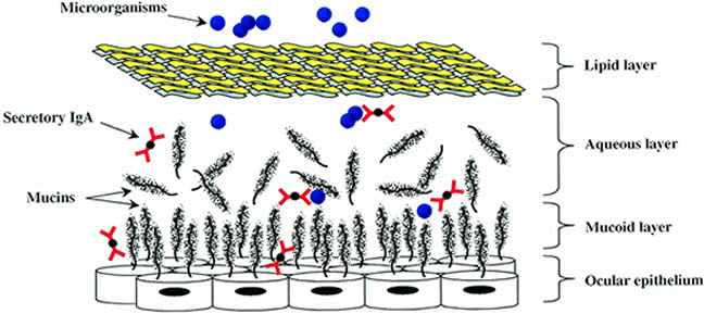
The innermost layer of the preocular tear film predominantly consists of glycoproteins known as mucins. (For review see references 20 and 21.) Ocular mucins are thought to serve a protective role; the mucins trap pathogenic microorganisms until they are swept to the inner canthus by the blinking action of the eyelids. Mucins, by their nature, may also serve as a matrix within which secretory IgA and other antibacterial factors are concentrated (Fig. 2).

Fig. 2. Ocular tear film mucin model. Mucin glycoproteins such as MUC1, MUC4, and MUC5AC are shown in close association with ocular epithelial surface, as well as in the aqueous tear film phase. Secretory IgA expressed in regional lymphoid tissue is shown within the mucin meshwork, as well as in the aqueous phase, in which it is capable of binding pathogenic organisms. Other antimicrobial factors are present but are not shown in the figure.

Mucin glycoconjugates are large (3 × 10⁵ to greater than 4 × 10⁷ kilodaltons [kDa]) and contain many O-linked oligosaccharide side chains, which comprise approximately three fourths of their dry weight.22 Mucins are expressed by most specialized epithelial tissues of mucosal surfaces, and nine mucin genes have been identified.23 Because of their heterogeneous nature and high carbohydrate content, mucins are inherently difficult to purify and analyze. Results from multiple groups using techniques of glycoprotein purification, anti-mucin antibody reactivity, and reverse transcriptase-polymerase chain reaction (RT-PCR) have provided evidence that the preocular tear film consists primarily of MUC1, MUC2, MUC4, and MUC5AC.22–25 Although the exact ocular sites of mucin expression remain unclear, they are generally thought to be produced by stratified epithelial cells of the ocular surface and conjunctiva and by conjunctival goblet cells.

Abnormal expression, distribution, and/or glycosylation of ocular mucins is thought to be a major contributing factor to the pathophysiology of dry eye syndrome.26,27 More unclear, however, is the role of mucins in innate immunity to ocular infectious diseases. The results of a recent study using genetically engineered MUC1 null mice suggested that this mucin plays a protective role against bacteria-induced inflammation.28 However, the exact role of MUC1 in this study or the role of other mucins in protection against ocular infection remains to be determined.

LYSOZYME

Lysozyme is a low-molecular-weight protein (15 kDa) that demonstrates bacteriostatic and bactericidal activity against a wide range of primarily gram-positive bacteria (i.e., Streptococcus, Staphylococcus, etc.). It facilitates the breakdown of bacterial cell wall peptidoglycan by hydrolyzing glycosidic bonds between N-acetylmuramic acid and N-acetylglucosamine.13 Lysozyme is one of the major components of tears, accounting for approximately 30% to 40% of total tear protein, and is most likely synthesized by acinar cells of the lacrimal gland.13,29 Because gram-negative organisms (i.e., Escherichia coli, Pseudomonas, Salmonella, etc.) possess an outer lipid bilayer with high concentrations of lipopolysaccharide, they are generally more resistant to lysozyme compared with gram-positive organisms. Nevertheless, lysozyme may potentiate the antibacterial activity of complement in conjunction with secretory IgA.

PHOSPHOLIPASE A2

Although lysozyme has traditionally been viewed as the principal antibacterial substance of the tear film, recent findings indicate that phospholipase A2 may actually represent the most active component. Phospholipase A2 is found in mucous secretions at other body sites and is known to be produced by leukocytes; its synthesis by cells within the main and accessory lacrimal glands has been suggested.29 The concentration of phospholipase A2 in human tears was determined to be approximately 30 μg/ml, corresponding to 1,000 × and 30,000 × the concentration sufficient to kill S. aureus and Listeria monocytogenes, respectively.30,31

Unlike lysozyme, which attacks the peptidoglycan cell wall, phospholipase A2 directly attacks the phospholipids of bacterial membranes. This low-molecular-weight enzyme is less active against gram-negative bacteria, perhaps because of the nature of their double lipid bilayers. Until recently, it was unclear whether tear film phospholipase A2 was necessary and/or sufficient for bactericidal activity against gram-positive pathogens. Using radiolabeled S. aureus in a rabbit model, Moreau and coworkers32 demonstrated that tear phospholipase A2 cleaved bacterial membranes, releasing arachidonic acid. They also demonstrated that phospholipase A2 could be blocked by polyamine inhibitors (i.e., spermidine). More research is clearly warranted into this potentially important antibacterial factor, including its role and molecular mechanism in the human tear film.

β-LYSIN

The presence of the antibacterial factor β-lysin in tears has been suggested for several decades. Its spectrum of activity is similar to that of lysozyme, but it tends to also be active against organisms that are less susceptible to lysozyme. The target of β-lysin is generally considered to be the bacterial lipid membrane, and β-lysin is known to act synergistically with lysozyme.33 Synthesized primarily by platelets, its presence in the ocular tear film is thought to be due to leakage from the serum. In fact, its presence in the ocular tear film has been questioned.34 Confusion surrounding the expression, spectrum of activity, and identity of ocular β-lysin may stem from its inclusion within a larger family of small (less than 8 kDa) cationic platelet-derived microbicidal proteins, which are a somewhat heterogeneous group of defensin-type molecules with diverse mechanisms of action.35 As protein micropurification techniques become more sophisticated, perhaps the understanding of the expression and/or role of β-lysin in the eye will become more clear.

METAL CHELATORS

Lactoferrin is an 82-kDa iron-binding protein found in the human tear film. It is a major constituent of human tears (approximately 25% of total tear protein), and its expression has been observed in cells lining the nasolacrimal system.36 This multifunctional protein has been shown to enhance natural killer (NK) cell activity and to regulate complement activation.13 Its antimicrobial properties are likely due to its ability to reduce the amount of free iron, which is an essential element for many pathogens.4 Lactoferrin was capable of interfering with herpes simplex virus-1 (HSV-1) infection of murine corneal epithelial cells both in vitro and in vivo. Once infected, however, lactoferrin had little effect on viral replication, suggesting that it may compete for viral binding sites on the host cell.37 Human immunodeficiency virus (HIV)-positive patients demonstrated tear lactoferrin deficiency and concomitant increases in levels of bacterial colonization of periocular tissue.38

Ceruloplasmin, a copper-binding protein of the tear film, may play a similar role in interference with essential element availability for pathogens. Nevertheless, a direct cause and effect relationship between innate ocular mucosal immunity and these metal ion chelators has not been established.

DEFENSINS

Defensins are a highly conserved group of peptide antibiotics found in many animal classes, including mammals, birds, insects, and amphibians.39 Defensins are produced by leukocytes and other cell types within mucous membranes and at other anatomic sites and have been studied primarily in the context of respiratory and intestinal epithelial surfaces. Only recently have defensins been explored as potentially important, innate antimicrobial components of ocular immunity.

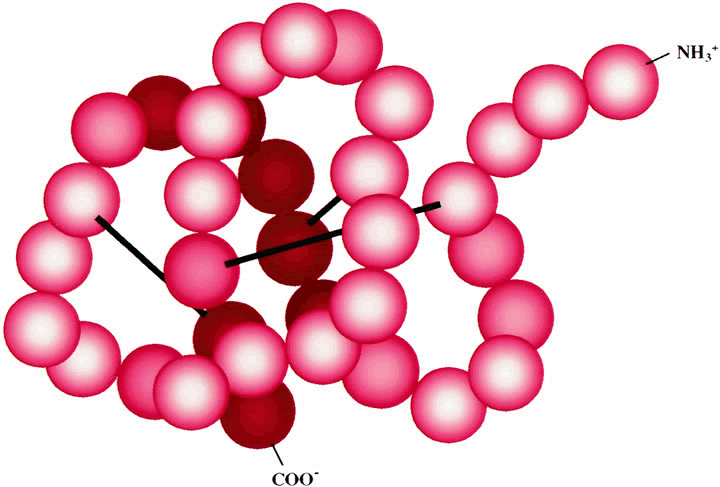
Defensins are short peptide molecules (29 to 35 amino acids) of low molecular weight (3.5 to 4.5 kDa). They are variably arginine-rich and cationic and contain six conserved cysteine residues that participate in forming intramolecular disulfide bonds (Fig. 3). Defensins thus take on a rigid three-dimensional β-sheet structure and function as dimers, forming voltage-sensitive channels (pores) in membranes of target organisms.40 The spectrum of defensin targets is greater than lysozyme or phospholipase A2 and extends to gram-positive and gram-negative bacteria; fungi; and viruses, including HIV and HSV. Two classes of defensins are recognized in humans: neutrophil-derived and Paneth cell-derived α-defensins and epithelial cell-derived β-defensins.

Fig. 3. Human β-defensin model. 3-D model of human β-defensin (HBD-1) showing spatial arrangement of its 36 amino acids. Disulfide bonds between six cysteine residues are shown as black bars. Using standard nomenclature, the amino acid sequence of HBD-1, from N terminus to C terminus is: DHYNCVSSGGQCLYSACPIFTKIQGTCYRGKAKCCK. [GenBank Accession #CAA90650]

Several recent studies have identified ocular sites of defensin expression using immunologic and RT-PCR techniques. The first direct demonstration of ocular defensin production came from Hattenbach and colleagues41 who reported human β-defensin-1 (HBD-1) and human β-defensin-2 (HBD-2) expression in human conjunctival specimens. Haynes and coworkers40 similarly demonstrated expression of HBD-1 and HBD-2 in corneal and conjunctival epithelial cells and human α-defensins 1, 2, and 3 in tears, lacrimal gland, and inflamed conjunctiva. Lehmann and colleagues42 later confirmed these results using semiquantitative real-time PCR, and further suggested that HBD-1 is constitutively produced in extraocular tissues, whereas HBD-2 demonstrated an inducible expression pattern. Others have since extended these studies and have reported nasolacrimal duct expression of HBD-1, HBD-2, and other antimicrobial peptides, including bactericidal-permeability-increasing protein (BPI) and heparin-binding protein (CAP37).43

Together, these findings point to defensins as potential innate antimicrobial factors of the ocular surface and interior. HBD-2 is inducible by gram-positive and gram-negative bacteria, fungi, and inflammatory cytokines, thereby limiting its expression to infectious and/or inflammatory stimuli. Other roles for defensins include epithelial healing; monocytic, dendritic, and T-cell chemotaxis; synergism with lysozyme; and complement activation, which highlight their potential versatility in innate ocular defenses.43

CYTOKINES

Cytokines function as regulators of both specific and nonspecific immunologic responses, and a detailed discussion of their roles is beyond the scope of this chapter. In the context of ocular defense against infectious microorganisms, cytokines likely play an indirect role; induction follows infectious insult, resulting in activation and/or augmentation of humoral and cellular defenses. As discussed previously, the ocular mucosal lymphoid tissues play an active and dynamic role in initiating and regulating innate defenses. These tissues are rich in cells of the specific (T and B lymphocytes) and nonspecific (macrophages) immune systems and likely serve a sentry role in foreign antigen sampling. On contact with a pathogenic organism, these cells may be activated and begin to secrete multiple cytokines. These cytokines, in turn, may further enhance cellular activation, increase vascular permeability leading to deposition of serum proteins in the ocular fluids, initiate maturation of specific T and B lymphocytes, enhance antimicrobial molecule expression (i.e., defensins), and recruit additional immune cells to the site of infection.4

Cytokines thought to be key in the initiation and regulation of immune and inflammatory responses following ocular infection include the chemokines (i.e., interleukin [IL]-8, macrophage-inflammatory protein, monocyte chemoattractant protein), IL-1, IL-6, IL-10, IL-12, tissue necrosis factor (TNF)-α, and interferon (IFN)-γ. Multiple types of intraocular and extraocular cells are capable of producing inflammatory cytokines, including lymphoid cells, fibroblasts, vascular endothelial cells, Langerhans' cells, and epithelial cells.44 Furthermore, a wide variety of infectious agents are capable of inducing ocular cytokine expression, including bacteria,45 viruses,46 and parasitic helminths.47

COMPLEMENT

Complement is a collection of more than 20 serum proteins that, when activated, participate in a cascade of biochemical reactions that lead to lysis of a target cell. (For review, see reference 48.) The end result of target cell destruction can be initiated by two distinct but interrelated pathways. The classical complement pathway is a form of specific immunity and requires immunoglobulins to be bound to a target cell antigen. Once bound, these immunoglobulins serve as a binding site for complement component C1 and catalyze C1 activation (Fig. 4). Activation of subsequent complement components then ensues, ultimately leading to the most crucial step in the complement system—cleavage of C3 into C3a and C3b by complexes known as C3 convertases. The alternative pathway, on the other hand, does not require antigen-antibody interaction and can be activated by bacterial polysaccharides, bacterial cell wall peptidoglycan, bacterial lipopolysaccharide, fungi, viruses, and parasites. It too, converges with the classical pathway at the level of C3 activation but solicits the cooperation of factor B, factor D, and properdin.

Fig. 4. Diagram of classical and alternative pathways of complement activation. The bar over a complement component indicates an active enzyme. B, factor B; D, factor D; P, properdin. C3 represents the pivotal component that is acted on by both the classical and alternative activating pathways.

Activation of complement components generally involves proteolytic cleavage, resulting in release of smaller components, many of which possess potent biologic activities. Activated complement mediates several biologic functions that are integral to innate immune defenses49:

  1. Chemotaxis. Complement component C5a attracts cells of the innate immune system, including neutrophils.
  2. Anaphylatoxin activity. C3a, C4a, and C5a are capable of triggering histamine release from mast cell and basophil granules. The resulting vasodilation and increased vascular permeability enhances deposition of effector molecules and recruitment of immune cells at the site of infection.
  3. Opsonization. C3b is capable of binding to the surface of pathogens, often in conjunction with antigen-antibody complexes. These C3b-coated surfaces are then recognized by macrophages and neutrophils possessing C3b receptors on their surface, resulting in phagocytosis of the pathogen.
  4. Target cell lysis. Activation of the full complement cascade on a target membrane results in the assembly of components C5b, C6, C7, C8, and C9 into a functional membrane attack complex, which forms osmotic pores in the target cell.

The presence of complement components on the ocular surface and within ocular compartments has been somewhat controversial. Some reports indicate the presence of a complete ocular set of complement cascade proteins, whereas others report the presence of only a few components.50 The exact source of complement components in the eye is also not clear. Ocular fibroblasts, macrophages, and certain epithelial cells could express complement, but leakage from the bloodstream also represents a reasonable source. Less debatable is the observation that complement concentrations increase during periods of ocular inflammation and/or infection.51 In these cases, complement is assumed to be the result of infectious/inflammatory ocular insults as well as the cause of inflammatory cascade amplification and perpetuation. These observations suggest that complement formation and deposition within ocular tissues are tightly regulated processes.

The presence of complement-regulatory systems within ocular tissues is an attractive hypothesis, because unregulated complement activation could potentially lead to homologous attack and destruction of sensitive ocular tissues. Recent studies have revealed the presence of several complement-regulatory proteins in corneas, lacrimal glands, tears, and in vitreous and aqueous humor.52–55 These ocular complement regulators include decay accelerating factor (DAF or CD55), a cell surface inhibitor of autologous C3 activation; membrane cofactor protein (MCP or CD46), an additional regulator of C3 activation; and the membrane inhibitor of reactive lysis (MIRL or CD59), a cell surface regulator of membrane attack complex formation.

Although an earlier experiment in mice suggested a role for complement in reducing the severity of bacterial keratitis,56 more research is required to elucidate the exact role of complement and complement-regulatory factors in infectious diseases of the ocular surface.

IMMUNOGLOBULINS

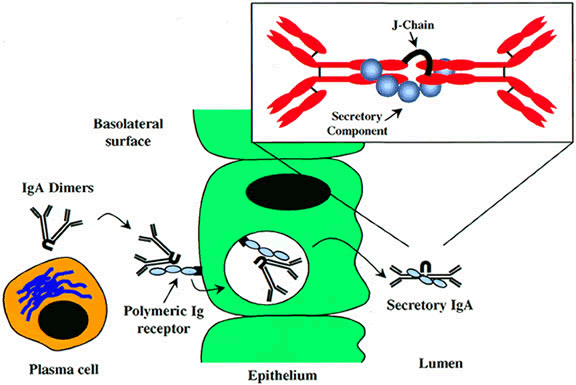
Lacrimal glands and conjunctiva contain large numbers of T and B lymphocytes, with antibody-secreting plasma cells accounting for approximately half of the lymphocytes in these tissues. Immunoglobulin secretion is a powerful, but specific, defense against ocular infectious diseases. IgA is the predominant immunoglobulin in external ocular secretions, and IgG represents the major antibody class found in extraocular tissues.48 IgA is normally present as a dimer, consisting of two IgA molecules coupled to one another at their Fc end via a polypeptide J-chain (Fig. 5). It is generally thought that the lacrimal gland possesses the highest concentration of IgA-secreting B lymphocytes. IgA that is synthesized in local ocular tissues binds to secretory component on the basolateral side of the epithelial cells through which it is to be secreted via the epithelial polymeric-Ig receptor.57 The J-chain is thought to protect IgA from premature proteolytic degradation. Receptor-mediated endocytosis facilitates transmigration of the IgA through the epithelial cell cytoplasm. A cleavage releases the secretory IgA from the polymeric-Ig receptor just before its arrival at the apical membrane, where it is released into the extracellular milieu. Although local IgA antibody expression is likely, leakage from the bloodstream as a result of infection and/or inflammation may also contribute to IgA's relatively high levels in extraocular compartments.

Fig. 5. Model for production and structure of secretory IgA. Homing and maturation of plasma cells within mucosal lymphoid tissue results in formation of dimeric IgA, with monomers linked via J-chain polypeptide. Dimeric IgA is bound by polymeric Ig receptor on basolateral surface of lymphoid epithelial cells. Receptor-mediated endocytosis facilitates transport of dimeric IgA through epithelial cell cytoplasm. Polymeric Ig receptor-IgA complex is proteolytically released from vesicles, with polymeric Ig receptor remaining associated with IgA. On release of IgA onto the luminal surface, polymeric Ig receptor takes on the new name “secretory component” and dimeric IgA becomes known as “secretory IgA” or sIgA.

Repeated ocular injection with antigen may result in a classic secondary response, with higher levels of antigen-specific antibodies being produced. However, the exact site for this memory response remains unknown. Local secretion by ocular resident memory B lymphocytes may account for much of the IgA response; however, B lymphocytes may also be recruited to the eye from the GALT and BALT. Secretory IgA in ocular fluids and tissues likely functions to (1) neutralize pathogens, (2) block host-pathogen interactions at the level of host receptor blockage, and (3) fix complement via the classical complement cascade.13 Regardless of supposed importance of IgA in pathogenicity, patients with low levels or absence of IgA alone are generally not predisposed to keratoconjunctivitis.6

Although secretory IgA on the ocular surface is important in preventing infection by bacteria such as Chlamydia and N. gonorrhoeae, it has recently gained attention from its role in preventing ocular infection with parasites.58,59 Leher and coworkers demonstrated that oral immunization induces production of parasite-specific IgA in mucosal secretions and prevents corneal infection in an animal model. These results point to deposition of IgA or IgA-producing plasma cells in the eye via the circulation. Not only is it feasible to invoke mucosal immunity via immunization strategies but Meek and colleagues59 recently demonstrated local mucosal immunization via naturally acquired exposure. They found that greater than 80% of normal individuals had an anti-Toxoplasma gondii IgA response in their tears, but only 23% of these had evidence of systemic immunity. These findings suggest that either local ocular mucosal IgA production results from common exposure to this pathogen or that humans naturally possess anti-parasitic IgA as part of their antibody repertoire.

IgG and IgM are also found in the eye and, like IgA, can be derived either from localized mucosal plasma cells or from the circulation. IgG is the predominant immunoglobulin in corneal tissue, and it likely diffuses there from the limbal vessels. There is little to no IgM in the corneal stroma, likely because of its extremely large size and poor diffusion characteristics.48 IgG and IgM are much more efficient in complement fixation than IgA, although complement fixation is not the primary means by which IgG and IgM function in protection against ocular viruses. IgG and IgM function primarily at the level of viral neutralization and have a significantly longer primary immune response than IgA, which leads to longer lived protection against repeated viral infection.60

Although IgE is not normally discussed in the context of ocular antimicrobial defenses, it can be found in association with ocular mast cells from normal human conjunctiva.61 Ocular disorders involving IgE are usually associated with an allergic or atopic phenomenon, rather than an infectious disease. Typically, when mast cell-bound IgE encounters antigen, cellular degranulation occurs resulting in release of large amounts of vasoactive substances, including histamine, leukotrienes, and serotonin. Aside from the immediate biologic consequences of hypersensitivity invoked by mast cell degranulation, such responses are important for defending mucous membranes against invasion by parasitic pathogens at other anatomic sites. However, the role of IgE and mast cells in ocular defense against infection remains relatively unexplored.

CELLULAR DEFENSES

Cornea

The central cornea is considered to be relatively devoid of immune cells because of the absence of lymphatic vessels. The peripheral limbal cornea is primarily populated by Langerhans' cells, a type of dendritic cells that plays a role in antigen processing.6 Following corneal injury, surgery, or infection, migration of immune cells into the central cornea can occur; this is presumably the result of production of chemotactic substances at the site of infection or injury, including pathogen-derived components as well as proinflammatory mediators secreted from corneal epithelial cells and fibroblasts. For example, cytokine-induced Langerhans' cell migration into the central cornea was recently demonstrated using gene-targeted knockout mice lacking TNF receptors and/or IL-1 receptors.62 Moreover, the recruitment of neutrophils and eosinophils to the central cornea in a murine model of parasitic infection with Onchocerca volvulus (river blindness) has been demonstrated.63

Eye-Associated Lymphoid Tissue

As discussed earlier in this chapter, recent studies have demonstrated that extraocular tissues, including the conjunctiva and lacrimal drainage system, may constitute an important arm of the common mucosal immune system and are capable of participating in both afferent and efferent immunologic responses.

NK cells are important in the initial nonspecific response to most virus infections. NK cells are large, granular lymphocytes, and following activation they are capable of secreting antiviral cytokines including IFN-γ and TNF-α. NK cells have been demonstrated in the bulbar conjunctiva of normal eyes but in relatively low numbers and only in the substantia propria layer.64 A recent study in which mice were depleted of NK cells demonstrated the importance of NK cells in protection against lethal ocular challenge and corneal scarring caused by HSV-1; however, the effects appeared to be mouse strain-specific. These findings indicate that the conjunctival distribution of NK cells and their role in protection against human ocular infection deserves further investigation.

Macrophages, on the other hand, appear to be the major nonspecific cellular constituent of ocular mucosal lymphoid tissue.64 Macrophages provide first-line defense against bacteria, fungi, and parasites by (1) phagocytosis of complement and/or antibody-opsonized pathogens, (2) oxidative killing of ingested pathogens, and (3) recruitment of additional immune cells via secretion of chemotactic and proinflammatory cytokines. The role of ocular mucosal macrophages in protection against viral infection of the cornea may stem from their role in professional antigen-presentation and subsequent induction of specific immune responses.65

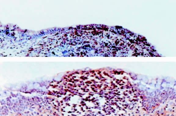
Conjunctival and lacrimal tissues are rich in T and B lymphocytes. As discussed previously in this chapter, these sites have been shown to harbor lymphocytic follicles and may, therefore, constitute MALT (Fig. 6). Hingorani and colleagues64 found CD3+ T cells to be the predominant lymphocyte population in normal human conjunctiva. Differential analysis by this group revealed that the helper to cytotoxic T-cell ratio (CD4:CD8) varied, depending on tissue location (tarsal vs. bulbar) and cellular layer (epithelium vs. substantia propria). Knop and Knop16 demonstrated substantial numbers of IgA+ plasma cells in both the acinar serous glands of the lacrimal sac and the lamina propria of the nasolacrimal duct, suggesting that these tissues comprise an important component of the ocular secretory immune system.

Fig. 6. Baboon conjunctiva-associated lymphoid tissue. Immunohistochemistry demonstrates follicular accumulations rich in T cells (CD3+) and IgA (IgA+).Original magnification × 200. (Courtesy of Dr. J Chodosh)

Despite numerous histologic studies demonstrating the presence of T and B lymphocytes in extraocular tissues, the exact mechanism for induction and/or recruitment of these cells remains unclear. Furthermore, the observation of well-organized lymphoid follicles in normal human extraocular tissue specimens is far from universal, suggesting that the presence of ocular lymphoid tissue may be the result of specific efferent immune responses, as opposed to its presence as a dedicated site for antigen sampling and/or presentation. An extensive review of the literature on ocular mucosal immunity13 suggests a model in which antigen gains access to the GALT via the nasolacrimal drainage system. Antigen-stimulated T and B cells then travel to the lacrimal gland and/or conjunctival tissue via the circulation. On homing to the lacrimal gland, B cells may undergo clonal expansion into IgA-secreting plasma cells. As previously discussed in this chapter, IgA is thought to pass through the acinar cells of lacrimal ductules via poly-Ig receptor and secretory component-mediated processes, leading to its deposition in the tear film. With IgA+ plasma cells, thus, providing specific humoral immunity, clonal expansion of T cells would provide the extraocular tissues with specific cell-mediated protection. This model is supported by the observation that, with some antigens, immunization via direct subconjunctival injection is generally less effective in generating tear IgA responses than either oral immunization (which presumably involves GALT) or topical ocular application (which presumably involves nasolacrimal drainage).60,66,67

In summary, although supported by immunohistochemical and histopathologic analyses, the presence and/or role of EALT continues to be debated. The extraocular tissues are most likely linked to the common mucosal immune system. The exact mechanisms, however, by which this system participates in ocular surface immunology and its potential importance in vaccine development for ocular infectious diseases will likely be the subject of intense study for years to come.

Back to Top
DEFENSES OF THE OCULAR INTERIOR

HUMORAL INTRAOCULAR DEFENSES

The blood-ocular barrier prevents the free passage of most large molecules from the bloodstream into the aqueous and vitreous humor. As a result, levels of soluble immunologic components within the fluid-filled spaces of the eye are relatively low, except in cases of intraocular infection or inflammation.

For more than two decades, Mondino and coworkers have conducted experiments aimed at characterizing intraocular humoral components, especially in the context of intraocular infection. In one study they observed IgG and IgA within aqueous and vitreous humor following intraocular infection with S. aureus.68 The parallel increase of both serum and intraocular immunoglobulin early in infection suggested leakage of antibody from the bloodstream as a result of inflammatory disruption of the blood-ocular barriers. However, IgA was never detected in serum, suggesting local synthesis. Furthermore, as IgG levels declined in the circulation, they simultaneously increased in the vitreous, which also suggests local synthesis. Although it has been difficult to pinpoint antibody-secreting B-lymphocytes within intraocular tissues, local antibody production by these tissues has long been assumed, and quantitation of antibody levels in aqueous and vitreous humor compared with serum levels has been used as a diagnostic tool for intraocular infectious and inflammatory diseases.48,69

Defensins, although likely playing a more prominent role in mucosal immunity, have been recently detected in intraocular tissues and fluids, including the iris, lens capsule, and aqueous and vitreous humor.42,70 It remains to be determined what, if any, role defensins play in clearance of intraocular pathogens.

Complement components have also been detected in the intraocular compartments of normal eyes, although at low levels. Aqueous and vitreous humor complement components, like immunoglobulins, rise following infectious or inflammatory insult. Studies have evaluated the role of complement in the defense of the vitreous against endophthalmitis.71,72 Taking advantage of the ability to render experimental animals (i.e., guinea pigs) complement deficient by injecting them with cobra venom factor, investigators demonstrated that staphylococci grew more rapidly in the eye following intravitreal injection in decomplemented animals than in controls.72 Furthermore, as complement levels were restored, bacterial proliferation was diminished. These results suggested that complement components, and perhaps other humoral factors, leak into the interior of the eye following inflammatory perturbation of the blood-ocular barrier and perform their natural antimicrobial function. Unfortunately, unchecked intraocular complement activation has the potential to damage sensitive intraocular tissues such as the retina. It is, therefore, somewhat ironic that the complement-inhibitory components MCP, DAF, and CD59 (see previous discussion in this chapter) are found in normal aqueous and vitreous humor,52,54 and yet the activity of complement may be an important defense against intraocular infection! These apparently contradictory findings simply highlight the molecular balancing act continually performed by the eye to limit the proliferation of ocular pathogens and the ocular damage resulting from the immune response.

CELLULAR INTRAOCULAR DEFENSES

The aqueous and vitreous humor are not normally populated by immune cells. In cases of intraocular infection, injury, or other similar diseases, these fluid-filled cavities can quickly become packed with inflammatory cells, leading to bystander damage of normal ocular cells and compromising the visual field. As such, most analyses of intraocular immune cell populations focus on the tissues immediately surrounding the aqueous and vitreous humor, including the iris, ciliary body, and the choroid.

The most extensive studies of “resident” ocular immune cells have been performed by McMenamin and coworkers, who have highly refined the technique of intraocular tissue whole-mount histopathology. By preparing whole tissues (i.e., iris, choroid, etc.), rather than thin sections, resident cell densities and distribution patterns are better appreciated. A previously published comprehensive review of such studies presents image data derived from standard histologic, immunohistochemical, and dual-color immunofluorescence laser scanning confocal microscopy.73

McMenamin74 has observed rich networks of intraocular resident tissue macrophages and dendritic cells. Distributed in the stroma of the iris, stroma of the ciliary body, and perivascularly within the choroid, resident macrophages are thought to be involved in phagocytosis of tissue debris, tumor cells, and microorganisms. Situated primarily near intraocular fenestrated vascular beds, a sentinel role for macrophages at the blood-ocular barrier has been implied. Dendritic cells form a resident population distinct from macrophages and are generally more pleomorphic. Their abundance between layers of the ciliary epithelium near the blood-aqueous barrier suggest that they may be involved in antigen sampling. Certain populations of tissue-restricted dendritic cells are believed to be important in the induction of anterior chamber-associated immune deviation (ACAID), a form of ocular immune privilege that results in suppression of damaging intraocular immune responses. This phenomenon again presents an apparent contradiction of function; mechanisms generally thought to provoke an immune response and clearance of an infectious agent may actually be involved in prevention of the response. Nevertheless, the roles of intraocular macrophages and dendritic cells in typical innate immunity and/or presentation of microbial antigens remain poorly defined.

Mast cells appear to be relatively rare in most species examined to date.73 Their characteristic periarteriolar distribution within the choroid suggests that they may be involved in the inductive phase of intraocular inflammation, presumably via secretion of biologically active components stored in their granules. However, the role of mast cells in any intraocular infectious process remains unexplored.

Eosinophils are circulating granulocytes, in contrast to their tissue-resident mast cell cousins. Although eosinophils are known to be vital in protection of the host from mucosal invasion by parasites, virtually nothing is known regarding their intraocular distribution or role in intraocular infectious diseases.

Lymphocytes, including T cells, B cells, and NK cells, are thought to be relatively rare within intraocular tissues. As discussed previously, however, the observance of local antibody production within intraocular tissues and fluids suggests the presence of at least some resident lymphocytes. Cytotoxic T cells and NK cells are thought to be important in limiting the spread of HSV-1 ocular infection.60,75 However, it remains unclear whether intraocular T cells, NK cells, and antibodies originate from local intraocular populations or are the result of lymphatic-derived cells that have homed to the blood-ocular barrier. The rarity of T lymphocytes in intraocular tissues is somewhat puzzling. It has been speculated that apoptosis of lymphocytes via the interaction of Fas+ T cells with Fas-Ligand expressed on normal intraocular cells may represent a mechanism by which the visual field is kept clear of unwanted T-cell infiltration.76

Back to Top
SUMMARY
The eye is well equipped with innate and specific defense systems. Mechanical, soluble, and cellular components work in exquisite synchronization to prevent loss of vision from infectious organisms and from the damaging effects of the host's own immune response. A major thrust of ocular immunologic research is the development of new strategies aimed at preventing establishment and spread of ocular pathogens and/or augmenting existing natural defense mechanisms. For example, recent research into the mechanism of IgA secretion across an epithelial membrane via the polymeric-Ig receptor57 may provide a means for delivering virtually any systemically delivered therapeutic agent to the ocular mucosa, thereby bypassing the need for repeated topical administration of eye drops and ointments. Moreover, if the ocular mucosa is actually an active component of the common mucosal immune system, it may in some cases serve as a potential vaccination site. Novel or naturally occurring proteins and peptides (i.e., defensins, phospholipases, etc.) or yet-to-be-discovered ocular factors may one day take their place among therapeutically useful ocular antimicrobial agents. Lastly, a detailed understanding of the inductive mechanisms and dynamics of the ocular immune response may result in development of antimicrobial agents that target a pathogen while limiting the exacerbation of inflammation. Much has been learned in recent years regarding the humoral and cellular ocular defenses against bacteria, viruses, fungi, and parasites, but ocular microbiology and immunology represents a field rich in unanswered questions and is deserving of continued aggressive research.
Back to Top
REFERENCES

1. Whitcup S: The double-edged ocular immune response: The Cogan lecture. Invest Ophthalmol Vis Sci 41:3243, 2000

2. Streilein J, Stein-Streilein J: Does innate immune privilege exist? J Leukoc Biol 67:479, 2000

3. Streilein J: Regional immunity and ocular immune privilege. Chem Immunol 73:11, 1999

4. Pleyer U, Baatz H: Antibacterial protection of the ocular surface. Ophthalmologica 211(Suppl 1):2, 1997

5. Klotz S, Penn C, Negvesky G et al: Fungal and parasitic infections of the eye. Clin Microbiol Rev 13:662, 2000

6. Nassif K: Ocular surface defense mechanisms. In Tabbara K, Hyndiuk R (eds). Infections of the Eye. New York: Little, Brown and Company, 1996, p 35

7. Kernacki K, Barret R, Hazlett L: Evidence for TIMP-1 protection against P. aeruginosa-induced corneal ulceration and perforation. Invest Ophthalmol Vis Sci 40:3168, 1999

8. Thakur A, Kyd J, Xue M et al: Effector mechanisms of protection against Pseudomonas aeruginosa keratitis in immunized rats. Infect Immun 69:3295, 2001

9. Rhem M, Lech E, Patti J et al: The collagen-binding adhesin is a virulence factor in Staphylococcus aureus keratitis. Infect Immun 68:3776, 2000

10. Hein W: Organization of mucosal lymphoid tissue. In Kraehenbuhl J, Neutra M (eds). Defense of Mucosal Surfaces: Pathogenesis, Immunity, and Vaccines. Berlin: Springer-Verlag, 1999, p 1

11. Kraehenbuhl J, Neutra M: Molecular and cellular basis of immune protection of mucosal surfaces. Physiol Rev 72:853, 1992

12. Streilein J: Regional immunology of the eye. In Pepose J, Holland G, Wilhelmus K (eds). Ocular Infection and Immunity. St Louis: Mosby, 1996, p 19

13. McClellan K: Mucosal defense of the outer eye. Surv Ophthalmol 42:233, 1997

14. Knop N, Knop E: Conjunctiva-associated lymphoid tissue in the human eye. Invest Ophthalmol Vis Sci 41:1270, 2000

15. Chodosh J, Nordquist R, Kennedy R: Comparative anatomy of mammalian conjunctival lymphoid tissue: A putative mucosal immune site. Dev Comp Immunol 22:621, 1998

16. Knop E, Knop N: Lacrimal drainage-associated lymphoid tissue (LDALT): A part of the human mucosal immune system. Invest Ophthalmol Vis Sci 42:566, 2001

17. Saitoh-Inagawa W, Hiroi T, Yanagita M et al: Unique characteristics of lacrimal glands as a part of mucosal immune network: High frequency of IgA-committed B-1 cells and NK1.1+ αβ T cells. Invest Ophthalmol Vis Sci 41:138, 2000

18. Brinser J, Burd E: Principles of diagnostic ocular microbiology. In Tabbara K, Hyndiuk R (eds). Infections of the Eye. New York: Little, Brown and Company, 1996, p 69

19. Armstrong R: The microbiology of the eye. Ophthalmic Physiol Opt 20:429, 2000

20. Argueso P, Gipson I: Epithelial mucins of the ocular surface: Structure, biosynthesis and function. Exp Eye Res 73:281, 2001

21. Strous G, Dekker J: Mucin-type glycoproteins. Crit Rev Biochem Mol Biol 27:57, 1992

22. Pflugfelder S, Liu Z, Monroy D et al: Detection of sialomucin complex (MUC4) in human ocular surface epithelium and tear fluid. Invest Ophthalmol Vis Sci 41:1316, 2000

23. McKenzie R, Jumblatt J, Jumblatt M: Quantification of MUC2 and MUC5AC transcripts in human conjunctiva. Invest Ophthalmol Vis Sci 41:703, 2000

24. Zhao H, Jumblatt J, Wood T et al: Quantification of MUC5AC protein in human tears. Cornea 20:873, 2001

25. Berry M, Ellingham R, Corfield A: Membrane-associated mucins in normal human conjunctiva. Invest Ophthalmol Vis Sci 41:398, 2000

26. Danjo Y, Watanabe H, Tisdale A et al: Alteration of mucin in human conjunctival epithelia in dry eye. Invest Ophthalmol Vis Sci 39:2602, 1998

27. Rolando M, Zierhut M: The ocular surface and tear film and their dysfunction in dry eye disease. Surv Ophthalmol 45(Suppl 2):S203, 2001

28. Kardon R, Price R, Julian J et al: Bacterial conjunctivitis in Muc1 null mice. Invest Ophthalmol Vis Sci 40:1328, 1999

29. Aho H, Saari K, Kallajoki M et al: Synthesis of group II phospholipase A2 and lysozyme in lacrimal glands. Invest Ophthalmol Vis Sci 37:1826, 1996

30. Qu X, Lehrer R: Secretory phospholipase A2 is the principal bactericide for staphylococci and other gram-positive bacteria in human tears. Infect Immun 66:2791, 1998

31. Saari KM, Aho V, Paavilainen V et al: Group II PLA(2) content of tears in normal subjects. Invest Ophthalmol Vis Sci 42:318, 2001

32. Moreau J, Girgis D, Hume E et al: Phospholipase A2 in rabbit tears: A host defense against Staphylococcus aureus. Invest Ophthalmol Vis Sci 42:2347, 2001

33. Asensi V, Fierer J: Synergistic effect of human lysozyme plus ampicillin or beta-lysin on the killing of Listeria monocytogenes. J Infect Dis 163:574, 1991

34. Janssen P, Muytjens H, Van Bijsterveld O: Non lysozyme antibacterial factor in human tears. Fact or fiction? Invest Ophthalmol Vis Sci 25:1156, 1984

35. Xiong Y, Yeaman M, Bayer A: In vitro antibacterial activities of platelet microbicidal protein and neutrophil defensin against Staphylococcus aureus are influenced by antibiotics differing in mechanism of action. Antimicrob Agents Chemother 43:1111, 1999

36. Paulsen F, Pufe T, Schaudig U et al: Detection of natural peptide antibiotics in human nasolacrimal ducts. Invest Ophthalmol Vis Sci 42:2157, 2001

37. Fujihara T, Hayashi K: Lactoferrin inhibits herpes simplex virus type-1 (HSV-1) infection to mouse cornea. Arch Virol 140:1469, 1995

38. Comerie-Smith S, Nunez J, Hosmer M et al: Tear lactoferrin levels and ocular bacterial flora in HIV positive patients. Adv Exp Med Biol 350:339, 1994

39. Martin E, Ganz T, Lehrer R: Defensins and other endogenous peptide antibiotics of vertebrates. J Leukoc Biol 58:128, 1995

40. Haynes R, Tighe P, Dua H: Antimicrobial defensin peptides of the human ocular surface. Br J Ophthalmol 83:737, 1999

41. Hattenbach L, Gumbel H, Kippenberger S: Identification of beta-defensins in human conjunctiva. Antimicrob Agents Chemother 42:3332, 1998

42. Lehmann O, Hussain I, Watt P: Investigation of β-defensin gene expression in the ocular anterior segment by semiquantitative RT-PCR. Br J Ophthalmol 84:523, 2000

43. Paulsen F, Pufe T, Schaudig U et al: Detection of natural peptide antibiotics in human nasolacrimal ducts. Invest Ophthalmol Vis Sci 42:2157, 2001

44. Chandler J: Ocular surface immunology. In Pepose J, Holland G, Wilhelmus K (eds).Ocular Infection and Immunity. St Louis: Mosby, 1996, p 104

45. Rudner X, Kernacki K, Barrett R et al: Prolonged elevation of IL-1 in Pseudomonas aeruginosa ocular infection regulates macrophage-inflammatory protein-2 production, polymorphonuclear neutrophil persistence, and corneal perforation. J Immunol 164:6576, 2000

46. Stumpf T, Shimeld C, Easty D et al: Cytokine production in a murine model of recurrent herpetic stromal keratitis. Invest Ophthalmol Vis Sci 42:372, 2001

47. Hall L, Diaconu E, Patel R et al: CXC chemokine receptor 2 but not C-C chemokine receptor 1 expression is essential for neutrophil recruitment to the cornea in helminth-mediated keratitis (river blindness). J Immunol 166:4035, 2001

48. Waldrep J, Mondino B: Humoral immunity and the eye. In Pepose J, Holland G, Wilhelmus K (eds). Ocular Infection and Immunity. St Louis: Mosby, 1996, p 33

49. Mondino B, Pleyer U: Host defense against bacterial and fungal disease. In Tasman W, Jaeger E (eds.). Foundations of Clinical Ophthalmology, vol 2. Baltimore: Lippincott, Williams, & Wilkins, 1995, p 1

50. Willcox M, Morris C, Thakur A et al: Complement and complement regulatory proteins in human tears. Invest Ophthalmol Vis Sci 38:1, 1997

51. Mondino B, Chou H, Sumner H: Generation of complement membrane attack complex in normal human corneas. Invest Ophthalmol Vis Sci 37:1576, 1996

52. Sohn J, Kaplan H, Suk H et al: Complement regulatory activity of normal human intraocular fluid is mediated by MCP, DAF, and CD59. Invest Ophthalmol Vis Sci 41:4195, 2000

53. Cocuzzi E, Szczotka L, Brodbeck W et al: Tears contain the complement regulator CD59 as well as decay-accelerating factor (DAF). Clin Exp Immunol 123:188, 2001

54. Sohn J, Kaplan H, Suk H et al: Chronic low level complement activation within the eye is controlled by intraocular complement regulatory proteins. Invest Ophthalmol Vis Sci 41:3492, 2000

55. Bora N, Gobleman C, Atkinson J et al: Differential expression of the complement regulatory proteins in the human eye. Invest Ophthalmol Vis Sci 34:3597, 1993

56. Cleveland R, Hazlett L, Leon M et al: Role of complement in murine corneal infection caused by Pseudomonas aeruginosa. Invest Ophthalmol Vis Sci 24:237, 1983

57. Hexham J, White K, Carayannopoulos L et al: A human immunoglobulin (Ig)A c-alpha-3 domain motif directs polymeric Ig receptor-mediated secretion. J Exp Med 189:747, 1999

58. Leher H, Alizadeh H, Taylor W et al: Role of mucosal IgA in the resistance to Acanthamoeba keratitis. Invest Ophthalmol Vis Sci 39:2666, 1998

59. Meek B, Klaren V, Van Haeringen N et al: IgA antibodies to Toxoplasma gondii in human tears. Invest Ophthalmol Vis Sci 41:2584, 2000

60. Hayashi K: Local immune responses in ocular virus infection and their implications for future immunotherapy. Ophthalmologica 211(Suppl 1):45, 1997

61. Stahl J, Cook E, Graziano F et al: Human conjunctival mast cells: Expression of Fc epsilon RI, c-kit, ICAM-1, and IgE. Arch Ophthalmol 117:493, 1999

62. Dekaris I, Zhu S, Dana M: TNF-alpha regulates corneal Langerhans cell migration. J Immunol 162:4235, 1999

63. Kaifi J, Hall L, Diaz C et al: Impaired eosinophil recruitment to the cornea in P-selectin-deficient mice in Onchocerca volvulus keratitis (river blindness). Invest Ophthalmol Vis Sci 41:3856, 2000

64. Hingorani M, Metz D, Lightman S: Characterization of the normal conjunctival leukocyte population. Exp Eye Res 64:905, 1997

65. Cheng H, Tumpey T, Staats H et al: Role of macrophages in restricting herpes simplex virus type 1 growth after ocular infection. Invest Ophthalmol Vis Sci 41:1402, 2000

66. Davidson H, Byrnes S, Montgomery P: The effect of immunization on rat serum and tear antibody responses to Chlamydia trachomatis. Reg Immunol 5:114, 1993

67. Pu Z, Silverstein A, Prendergast R: Conjunctival immunity: Compared effects of ocular or intestinal immunization in rats. Invest Ophthalmol Vis Sci 24:1411, 1983

68. Engstrom R, Mondino B, Glasgow B et al: Immune response to Staphylococcus aureus endophthalmitis in a rabbit model. Invest Ophthalmol Vis Sci 32:1523, 1991

69. Liekfeld A, Schweig F, Jaeckel C et al: Intraocular antibody production in intraocular inflammation. Graefe's Arch Clin Exp Ophthalmol 238:222, 2000

70. Haynes R, McElveen J, Dua H et al: Expression of human beta-defensins in intraocular tissues. Invest Ophthalmol Vis Sci 41:3026, 2000

71. Aizuss D, Mondino B, Sumner H et al: The complement system and host defense against Pseudomonas endophthalmitis. Invest Ophthalmol Vis Sci 26:1262, 1985

72. Giese M, Mondino B, Glasgow B et al: Complement system and host defense against staphylococcal endophthalmitis. Invest Ophthalmol Vis Sci 35:1026, 1994

73. McMenamin P: The distribution of immune cells in the uveal tract of the normal eye. Eye 11:183, 1997

74. McMenamin P: Immunocompetent cells in the anterior segment. Prog Ret Eye Res 13:555, 1994

75. Tanigawa M, Bigger J, Kanter M et al: Natural killer cells prevent direct anterior-to-posterior spread of herpes simplex virus type 1 in the eye. Invest Ophthalmol Vis Sci 41:132, 2000

76. Griffith T, Brunner T, Fletcher S et al: Fas ligand-induced apoptosis as a mechanism of immune privilege. Science 270:1189, 1995

Back to Top