
|
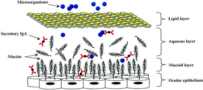
| Fig. 2. Ocular tear film mucin model. Mucin glycoproteins such as MUC1, MUC4, and MUC5AC are shown in close association with ocular epithelial surface, as well as in the aqueous tear film phase. Secretory IgA expressed in regional lymphoid tissue is shown within the mucin meshwork, as well as in the aqueous phase, in which it is capable of binding pathogenic organisms. Other antimicrobial factors are present but are not shown in the figure. |