Chapter 42
The Pathogenesis of Bacterial Infections of the Cornea
GEORGE A. STERN and GREGORY S. SCHULTZ
Main Menu   Table Of Contents

Search


Preparation of this chapter was supported in part by an unrestricted departmental grant (Department of Ophthalmology) from Research to Prevent Blindness, Inc, New York, New York, and from USPHS research grant EY05587 (GS).

LOCALIZATION: THE SOURCE OF BACTERIA
ADHERENCE, COLONIZATION, AND EARLY INVASION: ROLE OF THE EPITHELIAL DEFECT
SPECIAL ROLE OF CONTACT LENSES
FURTHER INVASION AND THE INFLAMMATORY RESPONSE
TISSUE DESTRUCTION: EFFECT OF PROTEOLYTIC ENZYMES AND OTHER VIRULENCE FACTORS
REFERENCES

The tissues lined by the mucosal surface of the eye, the cornea and conjunctiva, are susceptible to a variety of bacterial infections. Infections of the cornea, especially those caused by Pseudomonas aeruginosa, have been the subject of intense research interest. The purpose of this chapter is to review the mechanisms involved in the development of bacterial infections of the cornea, especially Pseudomonas corneal ulceration.

In any infection, pathogenic organisms must first localize at the target organ or tissue. Unless the organisms are isolated in a closed space from which they cannot escape (e.g., bacteria introduced into the eye during ocular surgery, causing endophthalmitis), they must adhere to the tissue and then colonize the infected mucosal surface. The pathogenicity of a bacterium, its ability to cause infection of a given tissue, is largely related to its ability to adhere to and colonize that tissue. Once sufficient numbers of organisms accumulate on the tissue surface and invade deeper structures, an inflammatory response is elicited in response to the presence of the bacteria. Characteristics of inflammation include vascular dilatation and increased vascular permeability, edema, and infiltration of polymorphonuclear leukocytes (PMNs). If unchecked, the infection causes tissue necrosis, ultimately leading to corneal perforation. The destruction of tissue is due to the liberation of proteolytic enzymes by both the bacteria and the PMNs. An organism's virulence (i.e., ability to cause serious disease rapidly) is related to its ability to induce inflammation and complement of destructive enzymes. If the infection is arrested by antibacterial therapy or the host inflammatory response, the cornea then undergoes a process of repair, which includes neovascularization, collagen resynthesis (scar formation), and re-epithelialization.

Back to Top
LOCALIZATION: THE SOURCE OF BACTERIA
Bacteria causing corneal ulceration arise from a number of sources. The most common causes of bacterial keratitis are Staphylococcus aureus, Streptococcus pneumoniae, and gram-negative bacilli, especially P. aeruginosa.1 P. aeruginosa are most commonly found in infections related to the wear of contact lenses.

How do these bacteria reach the eye? The margins of the eyelid are normally populated by coagulase-negative staphylococci and both aerobic and anaerobic diphtheroids (e.g., Propionibacterium acnes). These organisms can often be isolated from the conjunctiva, although it is likely that they do not become adherent to the conjunctival epithelium and therefore represent spillover from the lid margin. Although these species are not common causes of keratitis, they can cause infection in patients that have chronic corneal epithelial disease, leading to persistent punctate or macroscopic epithelial defects. S. aureus, a more virulent corneal pathogen, may be found as part of the eyelid flora in as many as 30% of presumably normal patients.2 In addition, staphylococci and other ocular pathogens may reach the eye when they cause periocular infection. S. aureus is the most common cause of ulcerative blepharitis,3 and both staphylococci and streptococci are frequent causes of chronic dacryocystitis.

P. aeruginosa and other gram-negative bacteria are neither part of the normal lid flora nor causes of chronic periocular infection. They are ubiquitous, found in feces, soil, and most sources of water. Therefore they can cause post-traumatic corneal infection when the cause of the trauma has been contaminated by soil or water. In addition, gram-negative bacilli such as P. aeruginosa, are common contaminants of paraphernalia used in the care of contact lenses.4–6 Pseudomonas, a common contaminant of aqueous solutions, is frequently present in nonsterile sources of water, including commercially available distilled water. In addition, P. aeruginosa has been found in mascara and other cosmetics used around the eye.7,8

Of patients who develop bacterial keratitis associated with the use of contact lenses, a very high percentage care for their lenses improperly.9 The most frequently contaminated item used in the care of contact lenses is the sterilization and storage case.4–6 In more than 50% of asymptomatic contact lens wearers, lens cases are contaminated, and more than 50% of contaminated cases contain gram-negative bacilli. Cleaning and sterilization of the contact lens case is a frequently neglected aspect of contact lens care. Pseudomonas readily adheres to the plastic surfaces of cases, where the adherent organisms secrete a glycocalyx, or slime, that enables them to survive exposure to disinfecting solutions.5 Contact lenses stored in such cases readily become contaminated.

Back to Top
ADHERENCE, COLONIZATION, AND EARLY INVASION: ROLE OF THE EPITHELIAL DEFECT
A longstanding clinical observation is that bacterial corneal infections do not occur in patients with intact healthy epithelium. Bacterial keratitis develops in one of three types of patients: (1) those with traumatic corneal abrasions; (2) those with epithelial defects due to intrinsic disease (e.g., dry eye, exposure keratitis, neurotrophic keratitis, postinfectious persistent epithelial defects); and (3) those who wear contact lenses, especially extended-wear hydrophilic lenses. The common feature linking these three patient groups is a defect in the corneal epithelium, to which the bacteria must adhere in order to initiate the infection.

The mechanisms behind the development of epithelial defects in the first two groups is self-evident. In the third group, contact lenses may lead to epithelial injury in a number of ways.10 The cornea can be injured during the process of insertion or removal of the lenses, by trauma from defects in the lenses or deposits on the lenses, by lens-induced hypoxia, or by chemical toxicity from contact lens disinfectants. It is not necessary for defects in the epithelium to be full thickness. Corneas with partial-thickness epithelial defects are more susceptible to adherence by Pseudomonas than are corneas with a fully intact epithelium,11 and extended wear of contact lenses modifies superficial epithelial cells so that these cells are more susceptible to attachment by Pseudomonas.12 Overnight wear of currently available soft lenses, which do not have adequate oxygen transmissibility to prevent hypoxia, causes superficial desquamation of epithelium and a greater propensity for adherence by Pseudomonas.13 Most important, however, is corneal swelling induced by overnight wear of contact lenses. The cornea normally swells 2% to 4% during sleep.14 In the presence of a contact lens, however, overnight swelling is increased to an average of 15%, and gross stromal edema can be present on awakening.14–16 In some patients, induced corneal swelling can be sufficient to cause the development of bullae; these can rupture, leading to the development of epithelial defects. In experimental models of contact lens-associated Pseudomonas keratitis in which there was no corneal trauma, the degree of induced corneal swelling under closed-eye conditions was the most important variable associated with the development of corneal infection.17,18 In fact, in the absence of corneal trauma, prolonged lid closure is required for infection to develop.18 This is the most likely mechanism responsible for the proven association between the overnight wear of contact lenses and the increased risk of bacterial keratitis.19,20

For bacteria to infect a mucosal surface, they must adhere to that surface to resist the natural mechanisms that inhibit bacterial colonization. In the eye, these include the protective effects of the cilia, the mechanical washing effect of the tear film and blinking, the coating of the cornea with ocular mucous,21,22 and the presence of antibacterial substances in the tears, including lysozyme, lactoferrin, betalysin, and IgA antibodies. Bacteria do not adhere well to intact corneal epithelium or to stroma completely denuded of epithelium; they readily adhere to injured or diseased epithelium at the edge of an epithelial defect.23 For this reason, a defect in the epithelium that exposes epithelial receptors is a prerequisite for the development of a bacterial corneal infection.

It is logical, therefore, to assume that the pathogenicity of a bacterial species for the cornea should be related to its ability to adhere to injured corneal epithelium. The most common causes of bacterial corneal ulceration are P. aeruginosa, S. aureus, and S. pneumoniae.1 Reichert and Stern24 demonstrated that these three organisms exhibit a greater degree of in vitro adherence to human corneal epithelium than do other bacteria that are not common corneal pathogens. Panjwani and colleagues25 reached similar conclusions regarding the adherence of S. aureus and P. aeruginosa to rabbit corneal epithelium.

The process of bacterial adherence generally involves a chemical or structural interaction between a component or appendage of the bacterial cell wall, called an adhesin, and a receptor on the epithelial cell or other structure to which the bacteria adhere. For example, teichoic acids, which are essential components of the cell wall of gram-positive cocci, are responsible for adherence of staphylococci and streptococci to epithelial cells.26,27 In the case of gram-negative organisms, filamentous cell wall appendages called fimbriae (commonly called pili, although this term is best reserved for structures involved in the transfer of genetic material) are often the adhesins.

Although adherence of Pseudomonas to injured epithelium is known to be the initial step in the pathogenesis of corneal ulceration, the exact nature of the interaction between bacterial and epithelial cells is complex and incompletely understood. It is likely that fimbriae are important Pseudomonas adhesins, especially when adhering to traumatized corneas.28 Cell surface glycoproteins appear to be the major receptors for Pseudomonas fimbriae, especially those containing moieties of mannose,29–33 N-acetylglucosamine,29 galactose β(1,3) N-acetylgalactosamine,29 and most importantly, sialic (N-acetylneuraminic) acid.29,33–35 Adherence is not completely correlated, however, with the presence of fimbriae: Mutant hyperfimbriated strains have diminished adherence and virulence in comparison to normal strains of Pseudomonas.36 It is possible that adherence is also related to other structures expressed by the rpoN gene, which regulates transcription of mRNA responsible for the synthesis of fimbrial proteins.37 Cell membrane lipopolysaccharides are also Pseudomonas adhesins,28,38 and it is likely that their epithelial receptors are neutral glycosphingolipids, especially asialo-GM1.38,39

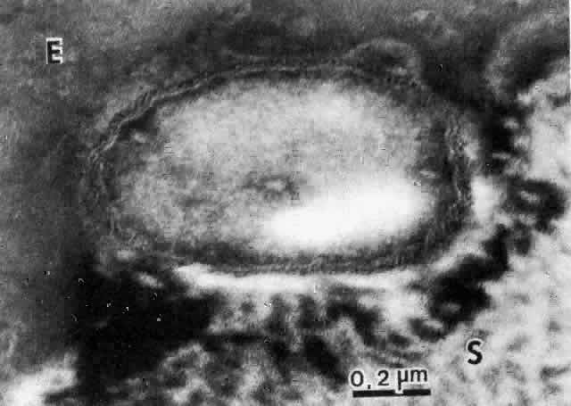
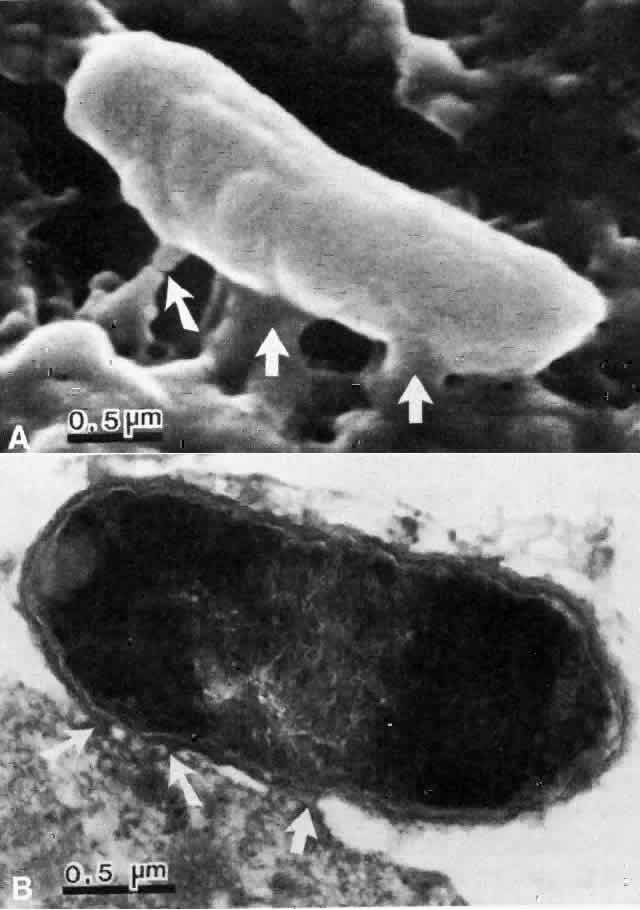
The physical events involved in the adhesion and early penetration of P. aeruginosa to the rabbit cornea have been studied by Stern and co-workers40 using scanning and transmission electron microscopy. Bacteria inoculated onto the surface of a cornea with an epithelial defect adhere to the injured epithelium within 15 minutes. Adherence is the result of an interaction between the bacterial and epithelial cell walls, with fusion of the two structures (Fig. 1). In some cases, fimbriae or other filamentous bacterial appendages are the adhesive structures. Within the subsequent 15 minutes, excavations in the epithelial cells appear to form around the bacteria. It is unknown whether this is the result of bacterial enzymes or an active process in which bacteria are engulfed into the epithelial cell. Forty-five minutes after inoculation, these excavations begin to fill in with the bacteria disappearing into the substance of the epithelial cell. One hour after inoculation, bacteria are no longer found on the epithelial surface. Instead, they are seen within epithelial cells or migrating through epithelial cells into the anterior stroma, and they are replicating at this stage (Fig. 2). Pseudomonas, therefore, appears to reach the anterior stroma by a process of adhesion and transepithelial migration. The invasion of epithelial cells by Pseudomonas renders the bacteria transiently resistant to host defenses and the effects of topical antibiotics.41 Entry of Pseudomonas into the epithelial cell involves active metabolic processes (e.g., tyrosine kinase and cellular actin microfilaments) and may be mediated by binding to integrins.42 The entire process of adherence and early stromal penetration in the experimental rabbit eye occurs in only 1 hour.

Fig. 1. A. Scanning electron micrograph of the Pseudomonas organism adhering to the injured corneal epithelial cell 15 minutes after inoculation. Focal areas of adherence are recognizable (arrows). B. Transmission electron micrograph of same cornea. Focal areas of adhesion between the epithelial cell and the bacterium are seen (arrows), which are consistent with adhesin-receptor interaction.

Fig. 2. Transmission electron micrograph of the cornea 1 hour after inoculation. The organism has migrated through the epithelial cell and is located between the epithelial cell (E) and the stroma (S).

Back to Top
SPECIAL ROLE OF CONTACT LENSES
Contact lenses, especially when worn overnight, are a significant risk factor for the development of bacterial keratitis.19,20 Contact lenses contribute to the pathogenesis of corneal infection in a number of ways:
  1. Contact lenses can cause the epithelial defect necessary for infection to develop. The mechanisms by which this occurs have been discussed previously. In addition, soft contact lens wear increases the expression of glycoprotein bacterial receptors on corneal epithelial cells.33 Contact lenses also modify the mucin layer of tear film, which normally inhibits bacterial attachment to the cornea.21,22
  2. Contact lenses are a site for bacteria to adhere. Many bacteria, particularly gram-negative bacilli, have evolved mechanisms by which they adhere to solid surfaces. These bacteria are capable of adhering to contact lenses. Although bacteria can adhere to unworn lenses,43 adherence, especially of P. aeruginosa, is significantly more pronounced in lenses that have become coated with tear film components such as mucin and proteins.44–48 Such coatings begin to form within minutes after insertion of a lens, and the surface of the lens is completely coated after only 24 hours of wear.49 Sialic acid, an important receptor for Pseudomonas, is a significant component of mucin and is likely responsible for the enhanced adherence of Pseudomonas to worn lenses.48 In addition, other glycoprotein and glycolipid deposits on lenses contribute to the adherence of Pseudomonas.50 Once bacteria adhere to lenses, the production of organic biofilm (glycocalyx) further strengthens the attachment.51,52 The average worn contact lens harbors more than 2000 bacteria, and may carry as many as 150,000 organisms.53
  3. Once the organisms migrate from the lens and become adherent to the cornea, the contact lens protects the bacteria from the defensive mechanisms of the eye by shielding the cornea from the mechanical cleansing effects of blinking, and by reducing tear flow over the cornea. The PMNs that infiltrate the cornea in response to the infection primarily arise from the precorneal tear film. By reducing tear flow, the contact lens delays the inflammatory response, allowing for unhindered replication of infecting organisms.54

The early pathogenesis of bacterial keratitis in the contact lens wearer is described in Figure 3. When a contact lens is placed on the eye, it is rapidly coated with mucoproteinaceous deposits. With increased wear, the lens becomes increasingly susceptible to adherence by bacteria to which it becomes exposed, either while stored in contaminated lens-care solutions or when exposed to periocular bacterial flora. In a non—contact lens wearer, the odds of infection are low because two infrequent events must occur simultaneously: (1) the localization of pathogenic organisms on the eye; and (2) the development of an epithelial defect. In a patient wearing a contaminated contact lens, pathogenic organisms are always present and infection can occur whenever an epithelial defect develops. Thus, the contaminated contact lens is a “time bomb” with the potential to cause a case of ulcerative keratitis. Once epithelial receptors become exposed, bacteria from the contact lens reach the cornea and become adherent to the injured epithelium. The bacteria then penetrate the deeper layers of the cornea, replicate, and incite an inflammatory response, which is seen clinically as ulcerative keratitis.

Fig. 3. Schematic representation of early events in the pathogenesis of Pseudomonas corneal ulceration, including the localization of bacteria, development of an epithelial defect, adherence of bacteria, and initial penetration of bacteria into the cornea.

Back to Top
FURTHER INVASION AND THE INFLAMMATORY RESPONSE
The sequence of events following adherence and initial invasion of the stroma by Pseudomonas has been elegantly described by Hyndiuk.55 Once Pseudomonas organisms reach the stroma, they replicate and spread both radially and deeply, migrating between stromal collagen lamellae. The intrastromal spread of the bacteria is likely facilitated by the production of two matrix metalloproteinases (MMPs), elastase and alkaline protease. Both of these enzymes are potent proteoglycanases that rapidly destroy stromal ground substance.

Invasion of the cornea is rapidly followed by infiltration of the corneal stroma by PMNs. PMNs are chemotactically attracted to the cornea, and they attempt to reach the site of infection by three routes: (1) migration from the limbal blood vessels through the corneal periphery; (2) exudation from inflamed iris blood vessels and migration across the anterior chamber; and (3) surface exudation from inflamed limbal blood vessels and migration through the precorneal tear film. In experimental models in which bacteria are externally applied to a surface wound (superficial inoculation), most closely mimicking the pathophysiology of clinical infections, the third route of PMN migration is most important early in the course of the infection. In models in which bacteria are injected into the corneal stroma (intrastromal inoculation), migration of PMNs through the peripheral cornea appears to be the predominant route,56,57 most likely because the injection disseminates organisms into the corneal periphery and separates peripheral corneal lamellae. The intrastromal inoculation model, however, is not often reproduced in the clinical setting. In models of contact lens overwear in which organisms reach the cornea by contamination of the lens, the sequence of events is very similar to the superficial inoculation model, but the time course is significantly delayed.54 This is probably due to differences in the degree of epithelial trauma and to inhibition of the migration of PMNs to the cornea because of reduced tear flow.

In the superficial inoculation model, PMNs begin to reach the superficial wound as early as 2 hours after inoculation; they fill the wound within 4 hours. PMNs then spread centrifugally from the origin of the infection and within 24 hours reach the midstroma and midperiphery of the cornea. At this time, PMNs are also found in the anterior chamber and are layered on the endothelium. Although PMNs attempt to reach the cornea by migration across the anterior chamber, they do not effectively cross Descemet's membrane; this process does, however, lead to the common clinical findings of hypopyon and inflammatory endothelial plaque. Between 48 and 72 hours after inoculation, PMNs migrate further into the corneal periphery. At this point, there appears to be an increasing component of PMNs that reach the cornea by migration through the peripheral stroma from limbal blood vessels.

Soon after PMNs enter the cornea, a destructive process ensues. The earliest findings are necrosis of stromal keratocytes and phagocytosis of these dead cells by PMNs. This is most likely an effect of the liberation of exotoxin A from the Pseudomonas organisms.58 As PMNs migrate peripherally in the anterior stroma, the overlying epithelial basement membrane is destroyed, leading to sloughing of the overlying epithelium. The enlarged epithelial defect further enhances the migration of PMNs into the corneal stroma. Actual degradation of collagen begins 8 to 16 hours after the onset of infection, but does not become significant for 24 hours. The loss of collagen fibrils and resulting accumulation of electron-dense granules appears to correlate with degranulation of the PMNs, and the area of destruction increases with the peripheral migration of PMNs. Although stromal destruction may be partly caused by the liberation of proteolytic enzymes by the bacteria, it is more likely due to the secretion of a true collagenase by the PMNs. If the replication and spread of bacteria is not halted by the host response or the instillation of antibiotics, the process of stromal degradation ultimately leads to total loss of stromal tissue and corneal perforation.

Back to Top
TISSUE DESTRUCTION: EFFECT OF PROTEOLYTIC ENZYMES AND OTHER VIRULENCE FACTORS
Pseudomonas produces a variety of substances (e.g., exotoxin A, proteolytic enzymes, hemolysins, endotoxins) responsible for the destruction of corneal tissue during infection.

Once reaching the stroma, Pseudomonas bacteria invade the cornea by migrating between stromal lamellae. This process requires the destruction of stromal proteoglycans (ground substance), and since inflammatory cells have not reached the cornea at this stage, dissolution of proteoglycans must be the result of bacteria-derived proteolytic enzymes. Early studies have demonstrated that the intrastromal injection or topical application of either filtrates of Pseudomonas broth cultures or purified Pseudomonas proteases to wounded corneas cause extensive stromal destruction, primarily through the degradation of proteoglycan ground substance, rather than loss of collagen.59–62 These enzymes have been found to be inhibited by Na2EDTA, which is characteristic of MMPs. Pseudomonas produces two MMPs, elastase and alkaline protease, and both of these enzymes have been found to contribute to its virulence. In one study,63 bacteria producing these enzymes were found to be highly virulent, whereas strains of Pseudomonas that were deficient in their production could not cause corneal infection in mice. In another study,64 intrastromal injection of purified elastase into rabbit corneas caused rapid and extensive stromal liquefaction.

Other enzymes must also be responsible for proteoglycanolytic activity, however, because elastase-deficient P. aeruginosa can cause disease identical to the enzymatically intact parent strains from which they are derived.65 One research group66,67 has also demonstrated the importance of Pseudomonas alkaline protease. Mutant strains that could not produce alkaline protease were not able to cause infection, whereas intact parent strains caused severe keratitis; virulence was restored to the enzyme-deficient strains by instillation of small amounts of alkaline protease into the eye. The ability of Pseudomonas to cause descemetoceles in experimental models of keratitis has been correlated with the production of elastase and alkaline protease in culture; the correlation was stronger, however, with levels of alkaline protease.68 In addition, both active and passive immunization against elastase,69,70 alkaline protease,70 and exotoxin A70 have been shown to reduce the virulence of Pseudomonas in experimental models of keratitis.

Hyndiuk55 observed that the earliest signs of tissue destruction were necrosis of keratocytes and phagocytosis of cellular fragments by PMNs. This destruction is most likely caused by the release of Pseudomonas exotoxin A. Either intrastromal injection of purified exotoxin A or topical application to wounded corneas has been found to cause rapid death of keratocytes, epithelial cells, and endothelial cells.58,71 After intrastromal injection, the effect was greatest closest to the injection site; the death of cells more remote from the injection was dependent on the concentration of the exotoxin; and chemotaxis of PMNs, which phagocytized the necrotic cellular debris, followed soon thereafter.55,58,71

Exotoxin A has been proved important in the pathogenesis of experimental keratitis caused by live bacteria. Genetically modified bacteria that were deficient in exotoxin A were shown by one research group65,66 to be less virulent than parent strains, and another group70 found that immunization against exotoxin A reduced the virulence of Pseudomonas early in the course of keratitis. Other toxins produced by Pseudomonas may also contribute to its virulence. In one study,72 strains isolated from human corneal ulcers were found to produce hemolysin concentrations that were higher than those of strains of undetermined virulence, and intrastromal injection of Pseudomonas hemolysin caused cell death and chemotaxis of PMNs, similar to that caused by exotoxin A.

Total corneal ulceration ultimately requires the degradation of collagen, which forms the framework of the corneal stroma. Most evidence indicates that true collagenolysis is the result of enzymes produced by PMNs. PMNs represent the primary host defense against bacterial infection. In one study,73 guinea pig corneas inoculated with either S. aureus or P. aeruginosa showed far greater bacterial replication in neutropenic animals, where there is a significant decrease in PMN infiltration compared to normal animals.

PMNs are chemotactically attracted to the cornea in Pseudomonas infection by the following mechanisms. First, tissue necrosis caused by toxins such as exotoxin A and hemolysin attracts PMNs to the cornea.58,72 It is unclear whether the PMNs are attracted to products released by the necrotic cells or to the toxins themselves. Second, bacterial endotoxins (cell wall lipopolysaccharides) are potent chemotaxins that attract PMNs by the alternative pathway of complement activation.74–76 The role of the classic pathway of complement activation, which also exists in the cornea, is unclear. Mondino and associates77 suggested that it was not important early in the course of the infection because of the time required to develop a true immune response. In rats, however, prior immune recognition is required for optimal chemotaxis of PMNs to the site of infection, phagocytosis of bacteria, and liberation of proteolytic enzymes.78 The presence of the C3 component of complement, which is activated by either pathway and is important for chemotaxis, phagocytosis, and enzyme activation, is necessary for the protective effects of PMNs against Pseudomonas.79,80 The presence of activated PMNs in the stroma amplifies the inflammatory process by further attracting more PMNs to the site of infection.76

The importance of PMNs as the principal cause of collagen degradation has been demonstrated as follows:

  1. Morphologic evidence of collagen degradation is closely correlated to the presence of degranulated PMNs at the site of infection,55–57 and intrastromal injection of concentrated lysosomal preparations from rabbit PMNs have been shown to be collagenolytic.81 The intrastromal injection of heat-killed Pseudomonas bacteria, which do not secrete proteolytic enzymes, causes PMN infiltration and progressive corneal ulceration. Corticosteroids block the infiltration of PMNs and prevent stromal ulceration in this model.82
  2. Bacterially derived proteases probably cannot degrade intact collagen fibrils, and Brown and colleagues60 suggested that the enzyme responsible for collagen degradation had the features of a mammalian (presumably PMN-derived) collagenase. Bacterial enzymes can, however, contribute to collagenolysis once initial collagen breakdown is initiated by a true collagenase.
  3. Inhibition of bacterial MMPs by phosphoramidon,64 Garladin,83 a thiol-based inhibitor,84 or immunization against these enzymes70 substantially delays ulceration early in the course of experimental infection, but ultimately does not prevent corneal melting and perforation. This suggests that factors that are present later in the course of the infection, such as the infiltration of PMNs, play a more important role. Rabbits vaccinated against endotoxins, which are chemotactic for PMNs, are better protected against corneal ulceration in experimental keratitis than animals vaccinated against bacterial proteases.69
  4. Twining and co-workers78 showed that stromal degradation occurred only when PMNs from rats that were immunocompetent against Pseudomonas infiltrated the cornea, and these PMNs secreted a fourfold greater amount of acid protease than those from immunodeficient rats. PMNs are known to produce several other proteases (e.g., two MMPs, gelatinase and interstitial collagenase; a serine protease, neutral elastase).85

Corneal pathogens other than Pseudomonas produce a number of toxins and proteases that contribute to their pathogenicity. S. pneumoniae produces a hemolysin that has similar effects to that produced by Pseudomonas.86 Serratia marcescens produces a neutral MMP that causes rapid corneal destruction when injected intrastromally.87 Similar to proteases produced by P. aeruginosa, this enzyme appears to degrade proteoglycans rather than intact collagen.88 S. aureus produces a serine protease, a thiol protease, and an MMP, whereas S. epidermidis produces an MMP with significant elastase activity.89

The pathogenesis of Pseudomonas corneal ulceration after initial penetration into the stroma is summarized in Figure 4. This pathogenic action is facilitated by the release of proteoglycanolytic MMPs, elastase and alkaline protease. Subsequent release of exotoxin A, hemolysin, and possibly other toxins causes death of keratocytes and overlying epithelial cells. Chemotaxis of PMNs is the result of complement activation via the alternative pathway caused by bacterial lipopolysaccharide (endotoxin) and cell necrosis caused by bacterial toxins. PMNs attempt to reach to the cornea by several mechanisms, but exudation from limbal blood vessels and migration through the tear film is the most important route early in the infection. The enlarging epithelial defect caused by epithelial necrosis facilitates the entry of PMNs into the corneal stroma. PMNs attempt to control the infection by limiting bacterial replication and phagocytosis of dead cells and bacteria. They are also chemotactic for the additional migration of PMNs, amplifying the inflammatory process. PMNs secrete a number of proteolytic enzymes, including a true collagenase. Once this collagenase begins to degrade collagen fibrils, it and other enzymes act synergistically to degrade stromal tissue even further. If this process proceeds unabated, total corneal destruction and perforation ensues. If the PMNs, usually in combination with antibiotic treatment, succeed in arresting the infectious process, a process of repair begins. Neovascularization and disorderly collagen resynthesis restore corneal integrity, but at the expense of a loss of corneal clarity and reduced vision.

Fig. 4. Schematic representation of later events in the pathogenesis of Pseudomonas corneal ulceration, including bacterial invasion of the stroma, role of toxins and enzymes, and elicitation of the inflammatory response.

Back to Top
REFERENCES

1. Hyndiuk RA, Snyder RW: Bacterial keratitis. In Smolin G, Thoft RA (eds): The Cornea: Scientific Foundations and Clinical Practice, Chap 5, pp 193–225. Boston, Little, Brown & Co, 1987

2. Hyndiuk RA, Skorich DN, Burd EM: Bacterial keratitis. In Tabbara KF, Hyndiuk RA (eds): Infections of the Eye, Chap 18, pp 303–330. Boston, Little, Brown & Co, 1986

3. McCulley JP, Dougherty JM, Deneau DG: Classification of chronic blepharitis. Ophthalmology 89:1173, 1982

4. Donzis PB, Mondino BJ, Weissman BA, Bruckner DA: Microbial contamination of contact lens care systems. Am J Ophthalmol 104:325, 1987

5. Wilson LA, Sawant AD, Simmons RB, Ahearn DG: Microbial contamination of contact lens storage cases and solutions. Am J Ophthalmol 110:193, 1990

6. Stern GA: Corneal infections in contact lens wearers. Int Ophthalmol Clin 31:147, 1991

7. Wilson LA, Ahearn DG: Pseudomonas-induced corneal ulcers associated with contaminated eye mascaras. Am J Ophthalmol 84:112, 1977

8. Wilson LA, Julian AJ, Ahearn DG: The survival and growth of microorganisms in mascara during use. Am J Ophthalmol 79:596, 1975

9. Bowden FW, Cohen EJ, Arentsen JJ, Laibson PR: Patterns of lens care practices and lens product contamination in contact lens associated microbial keratitis. CLAO J 15:49, 1989

10. Stern GA: Pseudomonas keratitis and contact lens wear: The lens/eye is at fault. Cornea 9:S36, 1990

11. Klotz SA, Au YK, Misra RP: A partial thickness epithelial defect increases the adherence of Pseudomonas aeruginosa to the cornea. Invest Ophthalmol Vis Sci 30:1069, 1989

12. Fleiszig SMJ, Efron N, Pier GB: Extended contact lens wear enhances Pseudomonas aeruginosa adherence to human corneal epithelium. Invest Ophthalmol Vis Sci 33:2908, 1992

13. Imayasu M, Petroll WM, Jester JV et al: The relation between contact lens oxygen transmissibility and binding of Pseudomonas aeruginosa to the cornea after overnight wear. Ophthalmology 101:371, 1994

14. Holden BA, Mertz GW, McNally JJ: Corneal swelling response to contact lenses worn under extended wear conditions. Invest Ophthalmol Vis Sci 24:218, 1983

15. Bruce AS, Brennan NA: Epithelial, stromal, and endothelial responses to hydrogel extended wear. CLAO J 19:211, 1993

16. O'Neal MR, Polse KA, Sarver MD: Corneal response to rigid and hydrogel lenses during eye closure. Invest Ophthalmol Vis Sci 25:837, 1984

17. Solomon OD, Loff H, Perla B et al: Testing hypotheses for risk factors for contact lens-associated infectious keratitis in an animal model. CLAO J 20:109, 1994

18. Aswad MI, Barza M, Baum J: Effect of lid-closure on contact lens-associated Pseudomonas keratitis. Arch Ophthalmol 107:1667, 1989

19. Poggio EC, Glynn RJ, Schein OD et al: The incidence of ulcerative keratitis among users of daily-wear and extended-wear soft contact lenses. N Engl J Med 321:779, 1994

20. Schein OD, Glynn RJ, Poggio EC et al: Microbial keratitis study group: The relative risk of ulcerative keratitis among users of daily-wear and extended-wear soft contact lenses: A case-control study. N Engl J Med 321:773, 1989

21. Fleiszig SMJ, Fletcher EL, Pier GB: Evidence that precellular factors may be involved in the non-specific inhibition of bacterial adherence to intact cornea. Invest Ophthalmol Vis Sci 33(suppl):844, 1992

22. Zaidi TS, Fleiszig SMJ, Pier GB: Ocular mucus modulates Pseudomonas aeruginosa adherence to both intact and injured cornea. Invest Ophthalmol Vis Sci 34(suppl):847, 1993

23. Stern GA, Weitzenkorn D, Valenti J: Adherence of Pseudomonas aeruginosa to the mouse cornea: Epithelial vs stromal adherence. Arch Ophthalmol 100:1956, 1983

24. Reichert R, Stern G: Quantitation of adherence of bacteria to human corneal epithelial cells. Arch Ophthalmol 102:1394, 1984

25. Panjwani N, Clark B, Cohen M et al: Differential binding of P. aeruginosa and S. aureus to corneal epithelium in culture. Invest Ophthalmol Vis Sci 31:696, 1990

26. Ofek I, Beachey EH, Jefferson W et al: Cell membrane-binding properties of group A streptococcal lipoteichoic acid. J Exp Med 141:990, 1975

27. Aly R, Litz C, Shinefeld et al: Binding of Staphylococcus aureus to human epithelial cells: Role of teichoic acid. Clin Res 27:40A, 1979

28. Fletcher EL, Fleiszig SMJ, Brennan NA: The role of pili and lipopolysaccharide in adherence of Pseudomonas aeruginosa to the rat cornea. Invest Ophthalmol Vis Sci 32(suppl):1167, 1991

29. Rudner XL, Zheng Z, Berk RS et al: Corneal epithelial glycoproteins exhibit Pseudomonas aeruginosa pilus binding activity. Invest Ophthalmol Vis Sci 33:2185, 1992

30. Hazlett LD, Moon MM, Strejc M, Berk RS: Evidence for N-acetylmannosamine as an ocular receptor for P. aeruginosa adherence to scarified cornea. Invest Ophthalmol Vis Sci 28:1978, 1987

31. Spurr-Michaud SJ, Barza M, Gipson IK: An organ culture system for study of adherence of Pseudomonas aeruginosa to normal and wounded corneas. Invest Ophthalmol Vis Sci 29:379, 1988

32. Blaylock WK, Yue BYJT, Robin JB: The use of concanavalin A to competitively inhibit Pseudomonas aeruginosa adherence to rabbit cornea epithelium. CLAO J 16:223, 1990

33. Klotz SA, Misra RP, Butrus SI: Contact lens wear enhances adherence of Pseudomonas aeruginosa and binding of lectins to the cornea. Cornea 9:266, 1990

34. Barrett R, Rudner XL, Berk RS, Hazlett LD: Further characterization of Pseudomonas aeruginosa pilus binding to corneal glycoproteins. Invest Ophthalmol Vis Sci 35(suppl):1339, 1994

35. Hazlett LD, Moon M, Berk RS: In vivo identification of sialic acid as the ocular receptor for Pseudomonas aeruginosa. Infect Immun 51:687, 1986

36. Hazlett LD, Moon MM, Berk RS, Rudner XL: Analysis of adhesion, piliation, protease production, and ocular infectivity of several P. aeruginosa strains. Curr Eye Res 10:351, 1991

37. Fletcher EL, Fleiszig SMJ, Brennan NA, Pier GB: The rpoN gene product of Pseudomonas aeruginosa is required for adherence to the cornea. Invest Ophthalmol Vis Sci 33(suppl):844, 1992

38. Gupta SK, Masinick S, Hazlett LD: LPS and pili mediate Pseudomonas binding to asialo GM1 glycolipid. Invest Ophthalmol Vis Sci 35(suppl):1339, 1994

39. Panjwani N, Zaidi TS, Gigstad JE et al: Binding of Pseudomonas aeruginosa to neutral glycosphingolipids of rabbit corneal epithelium. Infect Immun 58:114, 1990

40. Stern GA, Lubniewski A, Allen C: The interaction between Pseudomonas aeruginosa and the corneal epithelium: An electron microscopic study. Arch Ophthalmol 103:1221, 1985

41. Fleiszig SMJ, Zaidi TS, Fletcher EL et al: Pseudomonas aeruginosa invades into and survives inside cells during corneal infection. Invest Ophthalmol Vis Sci 34(suppl):901, 1993

42. Fleiszig SMJ, Zaidi TS, Pier GB: Pseudomonas aeruginosa may exploit cell motility mechanisms to invade corneal epithelial cells. Invest Ophthalmol Vis Sci 35(suppl):1338, 1994

43. Duran JA, Refojo MF, Gipson IK, Kenyon KR: Pseudomonas attachment to new hydrogel contact lenses. Arch Ophthalmol 105:106, 1987

44. Aswad MI, John T, Barza M et al: Bacterial adherence to extended wear soft contact lenses. Ophthalmology 97:296, 1990

45. Fowler SA, Allansmith MR: The surface of the continuously worn contact lens. Arch Ophthalmol 98:1233, 1980

46. Butrus SI, Klotz SA: Contact lens surface deposits increase the adhesion of Pseudomonas aeruginosa. Curr Eye Res 9:717, 1990

47. Stern GA, Zam ZS: The pathogenesis of contact lens-associated Pseudomonas aeruginosa corneal ulceration. Cornea 5:41, 1986

48. Stern GA, Zam ZS: The effect of enzymatic contact lens cleaning on adherence of Pseudomonas aeruginosa to soft contact lenses. Ophthalmology 94:115, 1987

49. Fowler SA, Allansmith MR: Evolution of soft contact lens coatings. Arch Ophthalmol 98:95, 1980

50. Klotz SA, Misra RP, Butrus SI: Carbohydrate deposits on the surfaces of worn extended-wear soft contact lenses. Arch Ophthalmol 105:974, 1987

51. Miller MJ, Ahearn DG: Adherence of Pseudomonas aeruginosa to hydrophilic contact lenses and other substrata. J Clin Microbiol 25:1392, 1987

52. Slusher MM, Myrvik QN, Lewis JC, Gristina AG: Extended-wear lenses, biofilm, and bacterial adhesion. Arch Ophthalmol 105:110, 1987

53. Mowrey-McKee MF, Monnat K, Sampson HJ et al: Microbial contamination of hydrophilic contact lenses: Part I. Quantitation of microbes on patient worn-and-handled lenses. CLAO J 18:87, 1992

54. Lawin-Brussel CA, Refojo MF, Leong FL et al: Time course of experimental Pseudomonas aeruginosa keratitis in contact lens overwear. Arch Ophthalmol 108:1012, 1990

55. Hyndiuk RA: Experimental Pseudomonas keratitis. Trans Am Ophthalmol Soc 79:541, 1981

56. Van Horn DL, Davis SD, Hyndiuk RA, Pederson HJ: Experimental Pseudomonas keratitis in the rabbit: Bacteriological, clinical, and microscopic observations. Invest Ophthalmol Vis Sci 20:213, 1981

57. Van Horn DL, Davis SD, Hyndiuk RA, Alpren TVP: Pathogenesis of experimental Pseudomonas keratitis in the guinea pig: Bacteriologic, clinical, and microscopic observations. Invest Ophthalmol Vis Sci 17:1076, 1978

58. Iglewski BH, Burns RP, Gipson IK: Pathogenesis of corneal damage from Pseudomonas exotoxin A. Invest Ophthalmol Vis Sci 16:73, 1977

59. Kreger AS, Gray LD: Purification of Pseudomonas aeruginosa proteases and microscopic characterization of pseudomonal protease-induced corneal damage. Infect Immun 19:630, 1978

60. Brown SI, Bloomfield SE, Tam WI: The cornea-destroying enzyme of Pseudomonas aeruginosa. Invest Ophthalmol Vis Sci 13:174, 1974

61. Kawaharajo K, Abe C, Homma JY et al: Corneal ulcers caused by protease and elastase from Pseudomonas aeruginosa. Jpn J Exp Med 44:435, 1974

62. Gray LD, Kreger AS: Rabbit corneal damage produced by Pseudomonas aeruginosa infection. Infect Immun 12:419, 1975

63. Kawaharajo K, Homma JY: Pathogenesis of the mouse keratitis produced with Pseudomonas aeruginosa. Jpn J Exp Med 45:515, 1975

64. Kessler E, Spierer A: Inhibition by phosphoramidon of Pseudomonas elastase injected intracorneally in rabbit eyes. Curr Eye Res 3:1075, 1984

65. Ohman DE, Burns RP, Iglewski BH: Corneal infections in mice with toxin A and elastase mutants of Pseudomonas aeruginosa. J Infect Dis 142:547, 1980

66. Nicas TI, Iglewski BH: The contributions of exoproducts to virulence of Pseudomonas aeruginosa. Can J Microbiol 31:387, 1985

67. Howe TR, Iglewski BH: Isolation and characterization of alkaline protease-deficient mutants of Pseudomonas aeruginosa in vitro and in a mouse eye model. Infect Immun 43:1058, 1985

68. Twining SS, David SD, Hyndiuk RA: Relationship between proteases and descemetocele formation in experimental Pseudomonas keratitis. Curr Eye Res 5:503, 1986

69. Kreger AS, Lyerly DM, Hazlett LD, Berk RS: Immunization against experimental Pseudomonas aeruginosa and Serratia marcescens keratitis: Vaccination with lipopolysaccharide endotoxins and proteases. Invest Ophthalmol Vis Sci 27:932, 1986

70. Steuhl KP, Doring G, Henni A et al: Relevance of host-derived and bacterial factors in Pseudomonas aeruginosa corneal infections. Invest Ophthalmol Vis Sci 28:1559, 1987

71. Hazlett LD, Berk RS, Iglewski BH: Microscopic characterization of ocular damage produced by Pseudomonas aeruginosa toxin A. Infect Immun 34:1025, 1981

72. Johnson MK, Allen JH: The role of hemolysin in corneal infections with Pseudomonas aeruginosa. Invest Ophthalmol Vis Sci 17:480, 1978

73. Chusid MJ, Davis SD: Experimental bacterial keratitis in neutropenic guinea pigs: Polymorphonuclear leukocytes in corneal host defense. Infect Immun 24:948, 1979

74. Mondino BJ, Rabin BS, Kessler E et al: Corneal rings with gram-negative bacteria. Arch Ophthalmol 95:2222, 1977

75. Mondino BJ, Brown SI, Rabin BS, Bruno J: Alternate pathway activation of complement in a Proteus mirabilis ulceration of the cornea. Arch Ophthalmol 96:1659, 1978

76. Chusid MJ, Davis SD: Polymorphonuclear leukocyte kinetics in experimental induced keratitis. Arch Ophthalmol 103:270, 1985

77. Mondino BJ, Brown SI, Rabin BS: Role of complement in corneal inflammation. Trans Ophthalmol Soc UK 98:363, 1978

78. Twining SS, Lohr KM, Moulder JE: The immune system in experimental Pseudomonas keratitis: Model and early effects. Invest Ophthalmol Vis Sci 27:507, 1986

79. Cleveland RP, Hazlett LD, Leon MA, Berk RS: Role of complement in murine corneal infection caused by Pseudomonas aeruginosa. Invest Ophthalmol Vis Sci 24:237, 1983

80. Hazlett LD, Berk RS: Effect of C3 depletion on experimental Pseudomonas aeruginosa ocular infection: Histopathological analysis. Infect Immun 43:783, 1984

81. Rowsey JJ, Nisbet RM, Swedo JL, Katona L: Corneal collagenolytic activity in rabbit polymorphonuclear leukocytes. J Ultrastruct Res 57:10, 1976

82. Kessler E, Mondino BJ, Brown SI: The corneal response to Pseudomonas aeruginosa: Histopathologic and enzymatic characterization. Invest Ophthalmol Vis Sci 16:116, 1977

83. Barletta JP, Balch KC, Dimova HG et al: Inhibition of Pseudomonas corneal ulcers by a synthetic matrix metalloprotease inhibitor GalardinTM (GM[6001]). Invest Ophthalmol Vis Sci 34:1058, 1993

84. Burns FR, Paterson CA, Gray RD, Wells JT: Inhibition of Pseudomonas aeruginosa elastase and Pseudomonas keratitis using a thiol-based peptide. Antimicrob Agents Chemother 34:2065, 1990

85. Hibbs MS, Hasty KA, Seyer JM et al: Biochemical and immunological characterization of the secreted forms of human neutrophil gelatinase. J Biol Chem 260:2493, 1985

86. Johnson MK, Allen JH: The role of cytolysin in pneumococcal ocular infection. Am J Ophthalmol 80:518, 1975

87. Lyerly D, Kreger A: Purification and characterization of a Serratia marcescens metalloprotease. Infect Immun 24:411, 1979

88. Lyerly D, Gray L, Kreger A: Characterization of rabbit corneal damage produced by Serratia keratitis and a Serratia protease. Infect Immun 33:927, 1981

89. Teufel P, Gotz F: Characterization of an extracellular metalloprotease with elastase activity from Staphylococcus epidermidis. J Bacteriol 175:4218, 1993

Back to Top