|
|
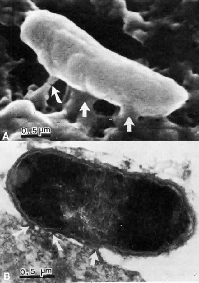
| Fig. 1. A. Scanning electron micrograph of the Pseudomonas organism adhering to the injured corneal epithelial cell 15 minutes after inoculation. Focal areas of adherence are recognizable (arrows). B. Transmission electron micrograph of same cornea. Focal areas of adhesion between the epithelial cell and the bacterium are seen (arrows), which are consistent with adhesin-receptor interaction. |