Chapter 40b
Molecular Diagnostics of Ocular Infectious Disease: Polymerase Chain Reaction Diagnostics of Ophthalmic Disease
ELMA KIM and RUSSELL N. VAN GELDER
Main Menu   Table Of Contents

Search

HISTORY AND BIOCHEMISTRY OF THE POLYMERASE CHAIN REACTION
SPECIMEN ACQUISITION FOR DIAGNOSTIC POLYMERASE CHAIN REACTION
VARIANTS OF POLYMERASE CHAIN REACTION
ADVANTAGES AND DISADVANTAGES OF POLYMERASE CHAIN REACTION COMPARED TO TRADITIONAL DETECTION TECHNIQUES
APPLICATION OF POLYMERSE CHAIN REACTION TO CLINICAL PROBLEMS
CONCLUSION
REFERENCES

In this chapter, we review the biochemistry, indications, applications, and limitations of the polymerase chain reaction (PCR) for the diagnosis of ocular disease. Although PCR is also used in the genetics laboratory for identifying specific mutations, we do not consider that application but restrict our discussion to the use of PCR in the diagnosis of the infectious diseases of the eye.
Back to Top
HISTORY AND BIOCHEMISTRY OF THE POLYMERASE CHAIN REACTION
The modern PCR was first described in 1985.1 In the 20 years since the first publication, the paper describing the modern version of PCR2 has been cited more than 13,000 times, and more than 190,000 papers have been published using this technique.

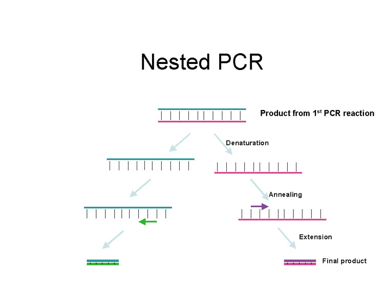
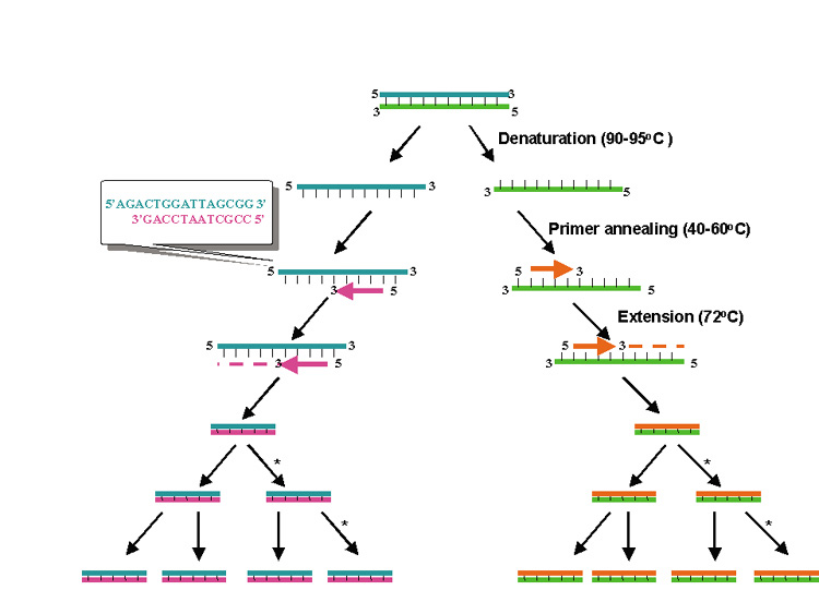
PCR is essentially an in vitro replication of bacterial DNA replication, stripped of all inessential components (Fig. 1). The necessary ingredients are the DNA template (i.e., from a pathogen residing in a biopsy specimen), a pair of 20 to 30 nucleotide oligonucleotide primers specific for the targeted DNA sequence (i.e., a sequence from a viral genome), nucleotide triphosphates (the building blocks for DNA), a thermostable DNA polymerase to catalyze replication, appropriate salts and buffers, and a thermal cycling machine. Once combined, the thermal cycler creates conditions for the three steps of PCR. Each step usually takes between 30 seconds and 2 minutes. The process begins with the denaturation step (typically at approximately 94°C), which separates the double-stranded template DNA into single strands. The temperature is then lowered to the annealing temperature (specific to each pair of primers, typically 45°C to 65°C), which allows the primers to optimally bind to their target sequences. After annealing, the temperature is elevated to an optimal temperature for thermostable DNA polymerase activity (typically between 72°C and 74°C). DNA polymerases capable of functioning at such high temperatures are isolated from thermophilic bacteria (such as those living in geothermal pools). At this temperature, the polymerase catalyzes synthesis of DNA complementary to the template at a rate of approximately 1000 bases per minute; thus a 500 base pair (bp) target may require 30 seconds of extension time. After extension, the temperature is again raised to denature the template DNA from the newly formed complementary strand of DNA. Thus, at the end of the first cycle, the number of target DNA strands has doubled. Both the original template and the new complement strand can then become templates for another round of amplification. PCR is an exponential process; theoretically, after 35 cycles (standard for many protocols), more than 35 billion copies of the original template DNA will have been synthesized. Starting with a single virion (whose DNA weighs 1.6 × 10⁻¹⁶ g), after 35 cycles nearly 20 ng (2.0 × 10⁻⁸ g) of 500 bp product will be produced. This quantity of DNA is adequate for further analysis.

Fig. 1. Schematic of the polymerase chain reaction. Each cycle of the polymerase chain reaction (PCR) includes heat denaturation of the double-stranded DNA template into single strands, annealing of an oligonucleotide primer complementary to sequence being amplified, and extension of the annealed primer using a thermostable DNA polymerase and nucleotide triphosphates. Repeated cycles results in exponential, sequence specific amplification of the original target.

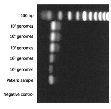
The most commonly used method for detection of the PCR product is electrophoresis in an ethidium bromide-stained agarose gel (Fig. 2). The limit of detection in such a gel is approximately 10 ng of double-stranded DNA. Given the calculations above, PCR should theoretically be able to detect a single genome in a given sample. However, because of imperfect amplification efficiency, maximal sensitivity is generally on the order of 10 genomes. PCR products may also be detected in other ways, using more sensitive dyes such as fluorescent SYBR Green®. This dye may be used in a special variant of PCR (real-time PCR), which allows quantification of product (see later). Sensitivity for detection of PCR products can be improved by use of radioactivity and autoradiography, but these techniques are seldom applied in clinical practice.

Fig. 2. Typical ethidium bromide-stained gel electrophoresis after polymerase chain reaction (PCR) from ocular specimen. First lane shows molecular weight marker. Positive controls are a dilution series of purified herpes simplex DNA, ranging from 105 genomes to 101 genomes. Patient sample is positive and appears to contain between 102 and 103 genomes. Negative control includes all reaction components but substitutes water for patient sample.

Although detection of a band of the correct size on electrophoresis is generally considered indicative of a positive result, PCR products may be more specifically identified in a number of ways. Restriction endonucleases can digest the PCR product into sequence-specific fragments, the pattern of which, on electrophoresis, forms a distinct fingerprint. Alternatively, hybridization methods allow identification by binding the PCR product to a nylon or nitrocellulose filter paper and then incubating the nylon with a labeled DNA or RNA probe that binds to a specific DNA sequence. The gold standard for demonstration of a specific identity for a PCR product is DNA sequencing, which can be performed either directly from the PCR product or after cloning of that fragment into a plasmid vector.

Back to Top
SPECIMEN ACQUISITION FOR DIAGNOSTIC POLYMERASE CHAIN REACTION
Ophthalmic samples for PCR are typically collected in one of three ways: swab samplings of the cornea, conjunctiva, or adnexa; anterior chamber paracentesis; or vitreous biopsy. The type of biopsy taken is guided by disease suspicion, media opacity, structural parameters of the eye, coexistence of pathologic conditions, and the experience of the ophthalmologist. In general, the sample should be taken at the site closest to pathology. For example, vitreous sampling has higher yield than aqueous sampling for ocular toxoplasmosis (although for viral retinitis, both anterior chamber and vitreous biopsy have high yield, likely because of higher infectious burden and smaller and more diffusible pathogen). All samples should be collected aseptically and placed into a sterile container. For a given PCR reaction, 5 to 10 μL of sample is sufficient. However, to allow retesting or testing for multiple pathogens, larger volumes are necessary. Approximately 50 μL from an anterior chamber paracentesis is generally sufficient and should be immediately capped in a 1-mL sterile syringe. For vitrectomy samples, approximately 50 to 100 μL of a dry (i.e., infusion-off) aspirate is collected in a sterile tube and capped upon collection. While PCR can theoretically be performed from vitrectomy cassette material, the extreme dilution of sample by this technique leads to a high incidence of false-negative results.

For external disease, placement of the swab depends on disease presentation. For conjunctivitis, the swab is placed on the conjunctiva without touching the patient's external skin. For a corneal ulcer, the swab should be placed along the periphery of the ulcer, in an active-appearing region. Once the sample is collected, the swab should be placed in a sterile tube containing 0.1 mL of balanced salt solution. In the laboratory, the swab is “milked” and subsequently prepared for PCR.

Once obtained, samples should be placed on ice and, at the earliest opportunity, quick-frozen on dry ice or in liquid nitrogen. The sample should remain frozen because freeze-thaw cycles are potentially deleterious to the sample, potentially degrading nucleic acids (primarily RNA). Swab samples should be stored at −80°C until laboratory processing.

All samples, regardless of source, must be processed to eliminate potential PCR inhibitors.3 Boiling the sample for 10 to 15 minutes is an effective way to remove most inhibitors and to release nucleic acids from relatively acellular sources (such as aqueous or vitreous). Alternatively, inhibitors can be removed by a commercially available DNA purification kit. Such purification is important when analyzing highly cellular samples such as corneal or scleral biopsies. Importantly, if the sample is to be tested for intraocular antibodies (i.e., Goldmann-Witmer testing 4), the laboratory must be notified because boiling irreversibly denatures antibodies from the sample. In these cases, the sample must be aliquoted for separate PCR and antibody testing.

It is essential that both negative controls (water or balanced saline in lieu of patient sample) and positive controls (a quantified sample of known pathogen DNA) be analyzed simultaneously with each PCR sample. Laboratory contamination is a potential cause of false-positive results in PCR; because PCR products are also potential targets for amplification, great care must be taken to avoid contamination of reagents with previous products. The use of consumable supplies and reagents, careful techniques, physical separation of reagents and controls, and routine inclusion of negative controls are necessary. Deoxyuracil may be substituted in PCR for the base thymidine; this base can be deglycosylated with uracil DNA glycosylase prior to the PCR reaction to degrade any product that could potentially serve as template.5

Back to Top
VARIANTS OF POLYMERASE CHAIN REACTION

NESTED POLYMERASE CHAIN REACTION

To increase sensitivity further, nested PCR may be used. Nested PCR is a second round of PCR that uses first-round amplification PCR products as the template DNA. The set of primers used in a nested PCR are internal to the primer pair used in the original PCR (Fig. 3). Nested PCR can have single genome sensitivity; it can also be utilized to improve specificity for detection of a specific pathogen. However, the large number of amplification cycles used in nested PCR (sometimes more than 60) can markedly increase the probability of false positive results. We generally avoid use of nested PCR for routine diagnostic applications.

Fig. 3. Principle of nested polymerase chain reaction (PCR). Small amounts of material amplified in a first (i.e., standard) amplification reaction is used as template for a second round of PCR. Primers for nested PCR are internal to the primers used in the initial amplification. Sensitivity and specificity are generally enhanced by nested PCR, but the high sensitivity can lead to detection of contaminating or commensal DNA.

MULTIPLEX POLYMERASE CHAIN REACTION

At present, clinical testing for multiple organisms in a differential diagnosis requires running individual PCR reactions for each suspected pathogen. In multiplex PCR, more than one set of primer pairs is used simultaneously in a given PCR reaction (Fig. 4). Elnifro et al6 showed that primer sets for both the herpes virus and adenovirus can be used successfully in accurately diagnosing patients with conjunctivitis. There was no difference in PCR sensitivity when comparing uniplex and multiplex PCR reactions for these two organisms. However, when a third set of primers was used, sensitivity decreased dramatically. For posterior segment disease, the STAMP (short tandem amplification of multiple pathogens) technique can be used to simultaneously screen vitreous and aqueous samples for varicella zoster, herpes simplex, cytomegalovirus (CMV), and Toxoplasma gondii.7 Each STAMP primer set, when used individually had a sensitivity on the order of 10 genomes. The sensitivity of the STAMP primers was approximately 1 log lower when used together than when used individually; however, this level of detection was still adequate to diagnose most patients with infectious posterior uveitis.

Fig. 4. Concept for multiplex polymerase chain reaction (PCR). Rather than running multiple separate PCR reactions for organisms on a differential diagnosis (such as herpes simplex virus [HSV], varicella zoster virus [VZV], toxoplasmosis [TOXO], and cytomegalovirus [CMV] as shown here), a single reaction is performed with primers for all organisms present. After amplification, a second detection method—either nested PCR or hybridization—is used to perform definitive detection. While sensitivity is marginally reduced in multiplex reactions, numerous organisms can be screened from a single small sample.

UNIVERSAL PRIMER POLYMERASE CHAIN REACTION

While most PCR primers are specific for a single organism, if multiple organisms share high sequence homology in a given gene, primers can be synthesized that will detect any of these organisms simultaneously. For bacteria and fungi, the DNA sequence encoding ribosomal RNA is highly repeated and conserved. 16S ribosomal primers are used for bacterial species and 18S and 28S ribosomal primers are used for fungal species. With rare exception, universal primers are not available for viruses. Although a positive PCR product demonstrates the presence of bacteria or fungi, respectively, this does not provide final identification. While primer sequences are highly conserved, the intervening sequencing within the PCR amplicon is species-specific. Typically DNA sequencing of the final product is used to achieve definitive identification. Because universal primers can detect all organisms sharing sequence, the technique can be used to amplify DNA from organisms that have not been previously identified.8 Relman et al9 used this technique, for example, to amplify the DNA of the organism responsible for Whipple's disease (Tropheryma whipplei). As a result of this discovery, ocular Whipple's disease can now be diagnosed by vitreous biopsy rather than jejunal biopsy.10

REAL-TIME QUANTITATIVE POLYMERASE CHAIN REACTION

Standard PCR yields a “yes–no” answer to the question of whether a particular organism's DNA is present in a sample. No quantification (similar to a colony count) is provided in standard PCR. In real-time, quantitative PCR, pathogen DNA levels are quantified by using a special thermocycler that can detect fluorescence from each well. Fluorescence is emitted by a dye, such as SYBR Green® dye, which binds to every double-stranded DNA molecule. Alternatively, a third probe (sometimes called the Taqman® probe) that emits fluorescence upon specifically binding PCR product, can be utilized. The quantity of fluorescence generated is compared to a standard curve generated from pure pathogen DNA. Via this technique, viral loads can be calculated. This technique has been commercialized for the quantification of human immunodeficiency virus (HIV) viral loads.11 It has successfully been applied to the quantitation of viral loads in posterior uveitis.12 This technique can potentially be used to differentiate between active and latent infections; for some potential pathogens such as Epstein Barr virus, low copy numbers may indicate commensal or latent DNA, whereas high counts may be associated with active infection.

REVERSE TRANSCRIPTION-POLYMERASE CHAIN REACTION

Standard PCR cannot directly amplify ribonucleic acid (RNA). Reverse transcriptase is a viral enzyme that converts RNA into DNA. Applying reverse transcriptase to convert RNA to DNA before running PCR is known as reverse transcriptase-polymerase chain reaction, or RT-PCR. (This terminology is somewhat confusing because early studies used the same abbreviation for real-time quantitative PCR; that technique is now more commonly called qPCR). RT-PCR is occasionally of value to determine the presence of specific RNAs in a tissue sample. This is essential for directly detecting RNA viruses such as hepatitis C and HIV. This technique can also discriminate between active and inactive viral infections if combined with real-time techniques. The RNA transcripts made by latent virus herpes simplex virus, for example, are different than those made by actively replicating virus. By detection of the latter mRNA transcripts, the presence of active infection can be confirmed.

Back to Top
ADVANTAGES AND DISADVANTAGES OF POLYMERASE CHAIN REACTION COMPARED TO TRADITIONAL DETECTION TECHNIQUES
The advantages of PCR compared to more traditional microbial detection techniques can be summarized with the three S's: speed, specificity, and sensitivity. While culture results can take anywhere from overnight to several weeks, PCR results are generally available within several hours of sample acquisition. PCR is also a nearly perfectly specific test. Even single base mismatches between primer and target sequence can eliminate product formation. Finally, PCR approaches near-perfect sensitivity for detection of pathogen DNA. For some organisms, PCR is becoming the gold standard for sensitivity. In a recent study of delayed onset bacterial endophthalmitis, culture positivity rates from vitreous biopsy for Propionibacterium acnes and other causative organisms were approximately 24%, compared to a 92% detection rate by PCR.13

The two primary disadvantages of PCR can also be traced to its high sensitivity and specificity. Because PCR can potentially detect the presence of a single genome, laboratory contamination is a substantial hazard and can undermine the usefulness of PCR. False-positive results can result from carryover of product DNA via pipettes or even viral shedding by the technician. The importance of testing negative controls with each sample cannot be overstated.

Additionally, PCR does not distinguish between live, latent, or commensal organisms; it detects DNA. For example, latent herpetic viruses present in sample will give positive PCR results. Real-time PCR can be used to distinguish between latent and active viruses.

The high specificity of PCR can also be problematic in two different ways. First, with the exception of the universal primers discussed previously, a different primer set must be used for each organism on the differential diagnosis. Unlike culture techniques, which can detect a wide range of organisms, each organism must be assayed individually in PCR. Thus, the specificity of PCR makes assessment of a large differential diagnosis problematic. Second, if an organism's target DNA is polymorphic (i.e., shows variance between strains of the same organism), false negative results may occur. Certain genes (such as the UL97 polymerase gene of CMV14) are mutational hot spots; such genes should be avoided as targets for PCR. Negative PCR results are more meaningful if generated by several independent primer sets for the organism in question. Additionally, all primer sets should be validated on patient samples (not just on reference strain DNA).

Back to Top
APPLICATION OF POLYMERSE CHAIN REACTION TO CLINICAL PROBLEMS
A good ocular examination including ophthalmoscopy is generally sufficient for a reliable diagnosis of most known infectious ocular inflammatory conditions, making routine PCR unnecessary. PCR is typically used in one of three scenarios: (i) the patient presents with signs and symptoms consistent with an infectious process, but a definitive diagnosis cannot be made because of media opacity or other examination difficulty; (ii) a clinical diagnosis is made but the patient does not respond appropriately to therapy or shows atypical natural history, or (iii) the patient is undergoing a surgery because of a complicating factor and intraocular fluid can be obtained for confirmation of underlying diagnosis. While PCR has been used in single cases to identify a large variety of ocular infections, we limit our discussion to more common clinical applications of PCR.

VIRAL RETINITIS

Historically, posterior segment uveitis was the first condition for which the clinical utility of PCR in ocular diagnostics was established.15 Both ocular toxoplasmosis and viral retinitis can produce a dense vitritis, occasionally making these conditions nearly impossible to diagnose on clinical exam alone. PCR can also be useful for complicated scenarios, such as a patient with AIDS with immune recovery and a concurrent CMV retinitis, atypical presentations of ocular toxoplasmosis, or unexpected negative culture results.

For viral retinitides, culture is a relatively slow method for diagnosis. In contrast, PCR is rapid and has a sensitivity that exceeds 90% for varicella zoster virus (VZV), herpes simplex virus (HSV), and CMV, with specificities over 95%. Ganatra et al16 have shown that viral subtypes can be identified by PCR as well, such as distinguishing between HSV type 1 and 2 in cases of acute retinal necrosis. For CMV retinitis, several ganciclovir resistance-conferring mutations can be rapidly detected by PCR in the UL97 gene followed by restriction endonuclease digestion.17

OCULAR TOXOPLASMOSIS

While classic reactivation toxoplasmosis is a relatively straightforward diagnosis, in the setting of media opacity or atypical primary presentation, the diagnosis can be challenging. PCR sensitivity for T. gondii is approximately 60% to 80% from vitreous biopsy in cases of active ocular toxoplasmosis. This relatively lower sensitivity is thought to be the result of the small number of T. gondii tachyzoites present in the vitreous. Higher sensitivity for diagnosis of ocular toxoplasmosis can be achieved when both PCR and intraocular antibody titers are tested from the same biopsy.18 Unlike serologic testing, PCR can also distinguish between strain subtypes of T. gondii.19 Because the three known subtypes of this organism have different susceptibilities to sulfa medications and extents of virulence in animal models, such information may eventually help guide therapy.

DELAYED-ONSET POSTOPERATIVE ENDOPHTHALMITIS

The most common organisms associated with delayed onset endophthalmitis are Propionibacterium acnes, Staphylococcus epidermidis, Actinomyces israelli, and fungi. Because of low pathogen loads and sometimes fastidious organisms, definitive diagnosis can be challenging. Lohmann et al13 used universal primers for both fungi and bacteria to diagnose delayed-onset endophthalmitis. While culture yields were 0% for aqueous cultures and 24% for vitreous cultures, PCR had sensitivity of 84% for aqueous samples and 92% for vitreous samples.

ANTERIOR SEGMENT AND EXTERNAL DISEASE

Definitive diagnosis of viral conjunctivitis is problematic, because viral cultures tend to be slow and are difficult to perform. Multiplex PCR has been developed for both adenovirus and herpes simplex virus.20 Adenoviral subtypes can be readily identified. Such information may be of clinical utility in rapidly identifying possible cases of epidemic keratoconjunctivitis.

Acanthamoeba is an uncommon but devastating cause of keratitis. Diagnosis is difficult because this organism is especially difficult to culture and has false-negative rates of approximately 50%. Lehmann et al21 have demonstrated PCR sensitivity of 84% for epithelial scrapings and 66% for tear samples, but with a specificity of 95%. Subsequently, Mathers et al22 demonstrated that the use of a combination of primers to detect different polymorphisms of Acanthamoeba yields a sensitivity of 72%. Routine use of PCR in these cases may result in increased rates of diagnosis.

Back to Top
CONCLUSION
PCR is a powerful molecular biology tool that is useful in the diagnosis of infectious eye disease. Clinical application of this technique is presently in its infancy; with further technical improvements and standardization of techniques, PCR will likely become a standard method for the diagnosis of infectious ocular disease. However, proper use of PCR requires an appreciation for the biochemical basis of the technique and an understanding of its strengths and limitations.
Back to Top
REFERENCES

1. Saiki R, Scharf S, Faloona F, et al: Enzymatic amplification of B globin genomic sequences and restriction site analysis for the diagnosis of sickle cell anemia. Science 230:1350, 1985

2. Saiki RK, Gelfand DH, Stoffel S, et al: Primer-directed enzymatic amplification of DNA with a thermostable DNA polymerase. Science 239:487, 1988

3. Wiedbrauk DL, Werner JC, Drevon AM: Inhibition of PCR by aqueous and vitreous fluids. J Clin Microbiol, 33:2643–6,1995

4. Witmer RH: Antibody formation in rabbit eye studied with fluorescein-labeled antibody. AMA Arch Opthalmol 53:811, 1955

5. Longo MC, Berninger MS, Hartley JL: Use of uracil DNA glycosylase to control carry-over contamination in polymerase chain reactions. Gene 93:125, 1990

6. Elnifro EM, Cooper RJ, Klapper PE, et al: Multiplex polymerase chain reaction for diagnosis of viral and chlamydial keratoconjunctivitis. Invest Ophthalmol Vis Sci 41:1818, 2000

7. Dabil H, Boley ML, Schmitz TM, et al: Validation of a diagnostic multiplex polymerase chain reaction assay for infectious posterior uveitis. Arch Ophthalmol 119:1315, 2001

8. Okhravi N, Adamson P, Lightman S: Use of PCR in endophthalmitis. Ocul Immunol Inflamm 8:189, 2000

9. Relman DA, Schmidt TM, MacDermott RP, et al: Identification of the uncultured bacillus of Whipple's disease. N Engl J Med 327:293, 1992

10. Rickman LS, Freeman WR, Green WR, et al: Brief report: Uveitis caused by Tropheryma whippelii (Whipple's bacillus). N Engl J Med 332:363, 1995

11. Berger A, Braner J, Doerr HW, et al: Quantification of viral load: Clinical relevance for human immunodeficiency virus, hepatitis B virus and hepatitis C virus infection. Intervirology 41:24, 1998

12. Dworkin LL, Gibler TM, Van Gelder RN: Real-time quantitative polymerase chain reaction diagnosis of infectious posterior uveitis. Arch Ophthalmol 120:1534, 2002

13. Lohmann CP, Linde HJ, Reischl U: Improved detection of microorganisms by polymerase chain reaction in delayed endophthalmitis after cataract surgery. Ophthalmology 107:1047, 2000; discussion 1051

14. Jabs DA, Martin BK, Forman MS, et al: Mutations conferring ganciclovir resistance in a cohort of patients with acquired immunodeficiency syndrome and cytomegalovirus retinitis. J Infect Dis 183:333, 2001

15. Knox CM, Chandler D, Short GA, et al: Polymerase chain reaction-based assays of vitreous samples for the diagnosis of viral retinitis. Use in diagnostic dilemmas. Ophthalmology 105:37, 1998; discussion 44

16. Ganatra JB, Chandler D, Santos C, et al: Viral causes of the acute retinal necrosis syndrome. Am J Ophthalmol 129:166, 2000

17. Liu W, Kuppermann BD, Martin DF, et al: Mutations in the cytomegalovirus UL97 gene associated with ganciclovir-resistant retinitis. J Infect Dis 177:1176, 1998

18. Fardeau C, Romand S, Rao NA, et al: Diagnosis of toxoplasmic retinochoroiditis with atypical clinical features. Am J Ophthalmol 134:196, 2002

19. Grigg ME, Ganatra J, Boothroyd JC, et al: Unusual abundance of atypical strains associated with human ocular toxoplasmosis. J Infect Dis 184:633, 2001

20. Jackson R, Morris DJ, Cooper RJ, et al: Multiplex polymerase chain reaction for adenovirus and herpes simplex virus in eye swabs. J Virol Methods 56:41, 1996

21. Lehmann OJ, Green SM, Morlet N, et al: Polymerase chain reaction analysis of corneal epithelial and tear samples in the diagnosis of Acanthamoeba keratitis. Invest Ophthalmol Vis Sci 39:1261, 1998

22. Mathers W, Nelson S, Lane J, et al: Confirmation of confocal microscopy diagnosis of Acanthamoeba keratitis using polymerase chain reaction analysis. Arch Ophthalmol 118:178, 2000

Back to Top