Chapter 12
Metabolism and Photochemistry of the Retina
MUAYYAD R. AL-UBAIDI and ROBERT E. ANDERSON
Main Menu   Table Of Contents

Search

CHEMISTRY OF PHOTORECEPTOR OUTER SEGMENTS
PHOTOCHEMISTRY OF VISUAL PIGMENTS
BIOCHEMICAL BASIS OF RETINAL DEGENERATION
MACULAR DEGENERATION
SUGGESTED SITES FOR RETINAL DISEASES
SUGGESTED SITES FOR RETINAL FUNCTION
ACKNOWLEDGMENTS
REFERENCES

The retina is a thin, delicate, transparent tissue that lines the posterior of the eye and is situated between the vitreous humor and the retinal pigment epithelium (RPE) (Fig. 1). The inner limiting membrane, which separates the retina from the vitreous humor, is composed of the foot processes of the glial cell, the Müller cell. This is the only cell that traverses the entire retinal expanse; the distal ends of the Müller cell form the outer limiting membrane between the cell bodies of the photoreceptor cells. Three distinct cell layers are evident in Figure 1. The most distal one contains the photoreceptor cells, the rods and cones. Rods are used for night vision and are most abundant in the peripheral retina. Cones are located in the central region of the retina, the macula, and are responsible for color perception and visual acuity. The nuclei of rod and cone cells make up the outer nuclear layer. Both rods and cones make synaptic contact in the outer plexiform layer with horizontal cells and bipolar cells. The nuclei of the horizontal and bipolar cells make up the inner nuclear layer. Bipolar cells are the second-order neurons of the retina and transfer the visual message generated in the photoreceptors to the ganglion cells, the third-order neurons. Amacrine and interphotoreceptor cells also make synaptic contact in the outer plexiform layer with ganglion and bipolar cells, as well as with each other. The visual message exits the eye by way of the ganglion cell axons, which collectively form the optic nerve.

Fig. 1. Histologic cross section (A) and schematic diagram (B) of the retina. (ILM, inner limiting membrane; NFL, nerve fiber layer; GCL, ganglion cell layer; IPL, interplexiform layer; INL, inner nuclear layer; OPL, outer plexiform layer; ONL, outer nuclear layer; OLM, outer limiting membrane; POS, photoreceptor outer segments; RPE, retinal pigment epithelium; BM, Bruch's membrane; a, amacrine cell; b, bipolar cell; c, cone; g, ganglion cell; h, horizontal cell; i, interplexiform cell; m, Müller cell; r, rod; Cap, a retinal capillary). (Fliesler SJ, Anderson RE: Chemistry and metabolism of lipids in the vertebrate retina. In Holman RT [ed]. Progress in Lipid Research. Vol 22. Elmsford, New York: Pergamon Press, 1983:81)

The function of the retina, to transduce light into an electrical signal that can be transmitted to the brain, is accomplished through the photolysis of light-sensitive visual pigments that are present in the outer segments of photoreceptor cells. A schematic of a photoreceptor cell, presented in Figure 2, illustrates the high degree of polarization of this structure. The proximal end of the photoreceptor cell is a nerve terminal, which makes chemical synapses with horizontal and bipolar cells. The inner segment contains the organelles, which are responsible for the metabolic activity of the cell. The biosynthesis of proteins and lipids occurs on the endoplasmic reticulum in the myoid region of the inner segment, and energy (e.g., adenosine triphosphate [ATP], guanosine triphosphate [GTP]) is generated in the mitochondria in the ellipsoid region of the inner segment. The inner segment is connected to the distal end of the photoreceptor cell, the outer segment, by a narrow connecting cilium. Outer segments occupy the subretinal space and are made up of stacks of membrane discs enclosed within a plasma membrane. The subretinal space is an organized network of fibers made up of complex proteoglycan macromolecules that surround the outer segments of both rod and cone cells. This fibrous network is called the interphotoreceptor matrix. The distal tips of the outer segments are in intimate contact with epithelial processes of the RPE. This contact is essential for the survival of photoreceptor cells. Disruption of the interphotoreceptor matrix or separation of the outer segments from the pigment epithelium leads to a retinal detachment.

Fig. 2. Schematic diagram of a rod cell and its spatial relationship to the retinal pigment epithelium (RPE). (bi, basal enfoldings; c, connecting cilium; d, discs; E, ellipsoid region of the inner segment; g, Golgi apparatus; m, mitochondria; M, myoid region of the inner segment; N, nucleus; pm, plasma membrane of the outer segment; RIS, rod inner segment; ROS, rod outer segment; SP, synaptic pedicle). (Fliesler SJ, Anderson RE: Chemistry and metabolism of lipids in the vertebrate retina. In Holman RT [ed]: Progress in Lipid Research. Vol. 22. Elmsford, New York: Pergamon Press, 1983:1–52)

Four types of photoreceptor cells are found in the human retina, and these are determined by the types of light-sensitive protein photopigments that are contained in their outer segment membranes. Each photopigment has a distinctive absorption spectrum. Rhodopsin is the photopigment of the rod outer segment (ROS) and absorbs light maximally at 500 nm. There are three types of cone photoreceptor cells; each contains only one type of photopigment. The cone pigments are named for the color of light they absorb: blue (440 nm), green (540 nm), and red (570 nm). Cone photoreceptor cells are arranged in a mosaic pattern throughout the macula.

The outer segments are made up of stacks of membranous discs surrounded by a plasma membrane (see Fig. 2). The plasma membrane is continuous with the inner segment through the narrow connecting cilium. In ROS, these discs are free-floating, except for a few at the base, and are not continuous with each other or with the plasma membrane. Cone outer segment discs are not free-floating, so the plasma and disc membranes are continuous.

Back to Top
CHEMISTRY OF PHOTORECEPTOR OUTER SEGMENTS
The bulk of the chemical analyses on photoreceptor cell outer segments have been done on ROS, because rods are by far the most abundant of the photoreceptors. Large quantities of relatively pure ROS are easy to prepare. Isolated ROS contain both disc and plasma membranes; however, because the plasma membrane is only 1% to 2% of the total ROS, an analysis of ROS is essentially an analysis of disc membranes. Only in recent years have relatively pure plasma membranes been prepared in quantities large enough for analysis.

ROD DISC MEMBRANES

The chemical makeup of the disc membranes is relatively simple. The membrane structure is a bilayer; about half of the molecules are integral proteins, and the other half are lipids. Phospholipids make up about 90% of the disc membrane lipids. Phosphatidylcholine (40% to 45%) and phosphatidylethanolamine (35% to 40%) are the major phospholipids, but relatively large amounts of phosphatidylserine (10% to 15%) are also found. Minor phospholipids (about 5%) include phosphatidylinositol (PI), phosphatidylinositol 4-phosphate (PIP), phosphatidylinositol 4,5-bisphosphate, and sphingomyelin. The cholesterol content of the discs is less than 10%, which is lower than what is usually found in plasma membranes. Small amounts of free fatty acids and diacylglycerol are also found in disc membranes. An interesting feature of cholesterol in the discs is that the most basal discs have higher levels of cholesterol than those near the apical tips.1,2 There does not appear to be a similar gradation in phospholipid or fatty acid distribution. The fatty acids of ROS lipids are somewhat unique in that the major component is docosahexaenoic acid (22:6n-3), a long-chain polyunsaturated fatty acid with six double bonds.3 This fatty acid also occurs in large amounts in synaptic terminals in the central nervous system but is not found to any great extent in nonneural tissues.

ROS contain integral (part of the membrane), peripheral (associated with the membrane), and soluble proteins. The major integral protein in rod disc membranes is the visual pigment rhodopsin (approximately 90%), a glycolipoprotein of about 40,000 molecular weight. The interaction of rhodopsin in the disc membrane is shown schematically in Figure 3. Rhodopsin has seven α-helical loops that traverse the lipid bilayer.4 These are important in some of the interactions of rhodopsin with other ROS proteins. Its C terminus is oriented toward the cytoplasmic surface of the disc, and its N terminus, which contains two carbohydrate side chains, is oriented toward the intradisc space. The C terminus contains six residues of serine and threonine, three of which are phosphorylation sites5,6 (serines 334, 338, and 343; see Fig. 3). 11-cis-Retinal is the chromophore of rhodopsin and is linked chemically by a Schiff base to lysine-296. The loops of rhodopsin outside of the lipid bilayer are important in the binding of peripheral and soluble proteins following photobleaching. Two adjacent cysteine molecules near the C terminus contain thioesters of palmitic acid, whose hydrophobic tails are embedded in the lipid bilayer7 anchoring the C terminus to the disk membrane and creating a specialized surface region—a fourth cytoplasmic loop (amino acids 310 to 321).8,9 This anchoring of the C terminus of rhodopsin is expected to cause conformational constraints.10 It was suggested that the fourth cytoplasmic domain created by the insertion of the palmitoyl groups into the membrane play a role in the binding and activation of transducin.

Fig. 3. Schematic diagram of the interaction of rhodopsin with the disc membrane. The C terminus (-COOH) is on the cytoplasmic side of the disc and contains the three serines (serines 334, 338, and 343) that are phosphorylated upon activation of rhodopsin by a photon. The N terminus is in the intradiscal space and contains two asparagines (Asn) that are glycosylated. Also shown is the interaction of the two palmitoyl moieties attached to cysteines 322 and 323 with the plasma membrane (yellow solid box). Open boxes represent the hydrophobic domains of rhodopsin that are imbedded in the membrane.

Another integral protein present in ROS in small amounts (1% to 2%) is peripherin/rds, which appears to be located at the edges of the discs near the plasma membrane.11 Nonintegral proteins of ROS include a cyclic guanosine monophosphate (cGMP)-specific phosphodiesterase, guanylate cyclase, arrestin (S antigen, 48K protein), rhodopsin kinase, protein kinase C, other protein kineses, phosphoprotein phosphatase, phosducin, transducin, phospholipase C, alcohol dehydrogenase, glucose-6-phosphate dehydrogenase, glutathione peroxidase, glutathione reductase, diacylglycerol kinase, PI kinase, PIP kinase, recoverin, and probably others yet to be discovered. Several of the ROS proteins are directly involved in visual transduction and are discussed later in greater detail.

ROD PLASMA MEMBRANES

Because the discs are derived from the plasma membrane, one would think that the chemistry of the disc and plasma membranes would be the same. However, this is not the case. Studies have now shown that the ROS plasma membrane has a different lipid and protein composition.12,13 The major differences in the lipids are a larger amount of cholesterol and lower level of 22:6n-3 in the plasma membrane. Rhodopsin is present in the plasma membrane, although at a much lower levels than in the discs. The plasma membrane contains the sodium channel, an integral protein that controls the passive entry of sodium (and, to a lesser extent, calcium) into the outer segment in the dark. Calcium is extruded from the outer segment by a calcium-sodium exchanger in the plasma membrane, and sodium is extruded from the photoreceptor cell by ouabain-sensitive pumps located in the ellipsoid region of the inner segment. This passive influx of sodium in the dark is the dark current described by electrophysiologists.

CONE VISUAL PIGMENTS

All three cone pigments have been identified and their amino acid sequence is known. All three share sequence homologies with rhodopsin, suggesting that all four visual pigments are derived from a common ancestral gene. Point mutations involving single amino acid substitutions, as well as cone pigment genes rearrangements have been described in color-blind individuals.14,15 Thus, color blindness can result from a single point mutation in the DNA encoding one of the color pigments, leading to the synthesis of a nonfunctional visual protein. Alternatively, recombinational rearrangements in the red/green pigments genes can also lead to color blindness.15,16 These proteins either do not form a photolabile molecule with 11-cis retinal or do not activate transducin and, therefore, fail to activate the phosphodiesterase when photobleached. In some cases of color pigments gene rearrangements a functional pigment that absorbs photons and initiates visual excitation can be generated. This is the so-called anomalous color blindness, which is identified psychophysically when the test subject does not match the “normal” spectral sensitivity patterns of human visual pigments. Recently, it was shown that patients with a mutation (Pro50Leu) in the gene encoding guanylate cyclase activator protein-1 (GCAP-1) exhibit decreased visual acuity and loss of color vision that occurred after the age of 20 years. Electrophysiologic testing revealed generalized loss of cone function, with preservation of rod function.17

BIOSYNTHESIS AND TURNOVER OF ROD OUTER SEGMENT MEMBRANES

All of the components of cone and rod outer segments are constantly being renewed by molecules synthesized in the inner segment. The reason for renewal is not known, but it seems reasonable for the cell to have a mechanism to replace old and dysfunctional molecules. In 1976, Young described two mechanisms of outer segment renewal: molecular replacement and membrane replacements. In molecular replacement, individual components of the outer segments are replaced molecule for molecule. An example is the soluble proteins of ROS, which diffuse into the outer segment after synthesis in the inner segment. Another example is the exchange of single lipid molecules catalyzed by lipid transfer proteins.18 In membrane replacement, vesicles containing newly synthesized lipids and proteins are transported to the base of the outer segment, where they fuse with the plasma membranes near the connecting cilium.19 The increase in plasma membrane surface area causes the basal membranes of the outer segment to evaginate and fold over (see Fig. 2). In ROS, these basal unfoldings eventually pinch off to form a free-floating disc.20 In cones, the plasma and disc membranes remain continuous.

Let us follow the synthesis of rhodopsin in the rod photoreceptor cell. The gene that codes for rhodopsin directs the synthesis of a messenger RNA (transcription) specific for rhodopsin. The message is translated on membrane-bound ribosomes, where opsin is synthesized, acetylated, glycosylated at its N terminus, and palmitoylated at its C terminus; disulfide bonds are established and the molecule is incorporated into the membrane of the endoplasmic reticulum. This membrane buds and fuses with the Golgi cisternae where the two sugar residues are modified. Finally, vesicles containing newly synthesized rhodopsin are released and make their way to the base of the outer segment. This transport process to the base of the outer segments is an active process that is facilitated by several members of a small GTP-binding proteins family known as Rab proteins.21

As new discs are added at the base of the outer segment, membranes at the apical tip are shed and phagocytized by the RPE. It takes about 10 days in mammals for a newly incorporated disc at the base to reach the tip of a ROS. Integral proteins such as rhodopsin, once incorporated into a disc, remain with that disc until it is shed.

Lipids are renewed by both membrane and molecular replacement. The vesicles that contain newly synthesized opsin also contain lipid. When fused with the plasma membrane, these lipids become part of the basal unfoldings that eventually give rise to the discs. However, the retina has soluble lipid transfer proteins that affect a one-for-one exchange of lipids between intracellular membranes.18 Also, studies have shown that lipid incorporation into ROS can proceed in the absence of protein synthesis.22 Thus, both membrane and molecular replacement are important in the renewal of outer segment lipids.

Several years ago, it was discovered that rods and cones shed their tips at precise times of the day.23 Rods shed in the morning at about the time of subjective sunrise, and cones shed in the evening about the time of subjective sunset. In mammals, these are circadian events whose periodicities are set, but not driven, by light.24 In amphibians, the rod-shedding event can be driven by light.25 Invertebrates also exhibit a similar phenomenon in which light provokes the disruption of their microvilli, followed by a rapid reorganization of their characteristic structure.26 The balance between renewal and shedding maintains a functional outer segment of uniform length throughout the lifetime of the cell. Anything that disrupts this delicate balance can lead to functional anomaly and may cause death of the photoreceptor cell. The best example of this is Royal College of Surgeons (RCS) rats, whose photoreceptors renew properly but whose RPE cannot phagocytize shed ROS tips.27 As a result, RCS photoreceptors degenerate within 2 months after birth.

Back to Top
PHOTOCHEMISTRY OF VISUAL PIGMENTS

PHOTOBLEACHING OF RHODOPSIN

The 11-cis-retinal chromophore of rhodopsin is oriented perpendicular to the fatty acid chains in the lipid bilayer of the ROS discs, a position that enhances photon capture. A single photon can isomerize the cis double bond, producing all-trans-retinal (Fig. 4). The conversion of 11-cis-retinal to all-trans-retinal is the only known action of light in the retina and leads to dramatic changes in the three-dimensional structure of retinal, which ultimately provokes conformational changes in rhodopsin. These conformational changes have been captured at low temperatures by a number of investigators who have studied their spectroscopic and kinetic properties. The so-called bleaching cascade of rhodopsin is presented in Figure 5. The first stable intermediate at physiologic temperatures is metarhodopsin II, and its formation from metarhodopsin I within milliseconds of photon capture leads to visual excitation. Each of these intermediates is a different conformational state of photoactivated rhodopsin (opsin) that results from the cis to trans isomerization of retinal.

Fig. 4. Chemical structure of 11-cis-retinal and all-trans-retinal. The only known action of light in the retina is to photoisomerize the cis double bond at position 11 to a trans double bond. (Anderson RE [ed]. Biochemistry of the Eye. San Francisco: American Academy of Ophthalmology, 1983:190)

Fig. 5. Bleaching sequence of rhodopsin. Each intermediate was stabilized at low temperatures and characterized by its unique absorption spectrum. (Anderson RE [ed]. Biochemistry of the Eye. San Francisco: American Academy of Ophthalmology, 1983:191)

THE VISUAL CYCLE

All-trans-retinal produced by the photoactivation of rhodopsin must be isomerized enzymatically to 11-cis-retinal, the substrate for regeneration of rhodopsin. The isomerase enzymes are located in the RPE28; no isomerase activity has been found in the retina. Therefore, the transport of all-trans-retinal to and from the RPE is an important part of the visual cycle, which is depicted in Figure 6. All-trans-retinal released from photolyzed rhodopsin is reduced rapidly to all-trans-retinol by the enzyme retinol dehydrogenase. The cofactor nicotinamide adenine dinucleotide phosphate in its reduced form (NADPH) is generated by glucose-6-phosphate dehydrogenase, an enzyme of the hexose monophosphate shunt. In low concentrations, all-trans-retinol can disrupt membranes. Therefore, it must be removed rapidly from the outer segments to the RPE to prevent damage to the discs. The mechanism of transport of all-trans-retinol to the RPE is not understood. The interphotoreceptor matrix contains high levels of interphotoreceptor retinoid binding protein (IRPB), which is thought by some to move retinoids between the ROS and the RPE.29 However, other evidence has shown that retinol can move between the two cells by simple diffusion.30 Both mechanisms most likely are used. Once in the RPE, all-trans-retinol is converted to all-trans-retinol esters, which are isomerized to 11-cis-retinol by the enzyme retinoid isomerase.31 After the isomerization event, 11-cis-retinol is returned to the ROS, where it is oxidized to 11-cis-retinal and used for the regeneration of rhodopsin.

Fig. 6. The visual cycle. All-trans-retinol generated in the ROS goes to the RPE, where it is isomerized to 11-cis-retinol and returned to the ROS. There it reacts with opsin to regenerate a photolabile molecule of rhodopsin. (Courtesy of Richard A. Alvarez)

RETINOL STORAGE AND TRANSPORT

The pigment epithelium can store both 11-cis-retinol and all-trans-retinol as esters of palmitic, stearic, and oleic acids. This storage ensures the retina of a steady supply of 11-cis-retinal, even at the brightest light levels. However, the main organ for retinol storage is the liver. Retinol obtained from the diet or synthesized in the intestine from dietary carotenoids is transported to the liver in chylomicrons, where it is esterified to fatty acids and stored in small lipid droplets. For transport to other tissues, one molecule of retinol binds to a specific protein, serum retinol binding protein (SRBP), which complexes with another serum protein, prealbumin. This complex reacts with specific receptors on cell surfaces to deliver retinol to target cells. The plasma membrane of the basal surface of RPE cells has receptors that recognize and bind SRBP.27

VISUAL TRANSDUCTION

For vision to occur, the message that a photon was absorbed in the outer segment must be transmitted to the synaptic terminal of the photoreceptor cell. This is accomplished through rapid communication between the disc and plasma membrane of the outer segment, mediated by the small water-soluble molecule cGMP.32 In the dark, the level of cGMP in the outer segments is high compared with other retinal cells (Fig. 7). At high concentrations, cGMP interacts with the sodium channel in the plasma membrane and keeps it open to the passive influx of sodium from the interphotoreceptor space. This depolarizes the cell, allowing neurotransmitter to be released from the synaptic terminal in the dark. In the light, the photobleaching of rhodopsin is followed rapidly (in milliseconds) by a precipitous decline in cGMP, leading to closure of the sodium channel and hyperpolarization of the photoreceptor cell. Neurotransmitter release is inhibited, causing hyperpolarization of horizontal cells and either hyperpolarization or depolarization of bipolar cells, depending on the cell type. Photoreceptor, horizontal, and bipolar cells respond to light with sustained, graded potentials rather than action potentials. The retina is, thus, allowed to make discriminations in intensity and duration of visual stimuli that would not be possible if the cells fired on an all-or-none basis. The control of cGMP levels in ROS is achieved through a series of biochemical reactions, depicted in Figure 8, and involves several proteins. The conformational changes in photoactivated rhodopsin (R*) lead to the binding of transducin (Tαβγ-GDP) to R* and the exchange of GTP for guanosine diphosphate (GDP). This complex dissociates into Tα-GTP, R*, and Tβγy. Tα-GTP reacts with a cGMP-specific phosphodiesterase (PDEαβγ) and removes the inhibitory subunit (PDEγ). Relieved of its inhibitor, the activated cGMP phosphodiesterase (PDEαβ hydrolyzes cGMP in the ROS cytoplasm. Two amplification steps are essential to achieve the rapid reduction in cGMP levels. The first occurs in the activation of transducin, when one molecule of R* can generate more than a hundred molecules of Tα-GTP. The second is at the enzyme level, where one PDEαβ molecule can hydrolyze thousands of molecules of cGMP. These two amplification steps translate the photolysis of one molecule of rhodopsin into the hydrolysis of hundreds of thousands of molecules of cGMP. This is an amazing biochemical feat, considering that visual transduction occurs over a time frame of a few milliseconds.

Fig. 7. Schematic of light- and dark-adapted photoreceptor cell. Levels of cGMP are high in the dark and the sodium channels are open, depolarizing the cell. In the light, the cGMP levels decrease rapidly, causing the cell to hyperpolarize. (Courtesy of Richard A. Alvarez). (Basic and Clinical Science Course, Section 1: Fundamentals and Principles of Ophthalmology. San Francisco: American Academy of Ophthalmology, 1990:188)

Fig. 8. Phototransduction cascade. Schematic representation of events involved in the control of cGMP levels in the photoreceptor cell. Activation of rhodopsin by light sets in motion a cascade of events that ultimately lead to the hydrolysis of cGMP. These biochemical events occur within few milliseconds of photon capture. (P, phosphate; PP2A, protein phosphatase 2A; M II, metarhodopsin II; RGS, regulator of G-protein signaling; PDE, phosphodiesterase; GCAP, guanylate cyclase activation protein). (Courtesy of Michael W. Bell, PhD)

Left unchecked, R* will continue to bind transducin and activate the phosphodiesterase. This is not a desired effect, because it would lead to permanent hyperpolarization of the photoreceptor cell and loss of sensitivity to changes in the visual stimulus. Efficient single-photon detection by retinal rod photoreceptors requires timely and reproducible deactivation of rhodopsin (rhodopsin cycle, see Fig. 8). Rhodopsin contains multiple sites for phosphorylation at its C-terminal domain.

Outer segments contain a soluble protein kinase, rhodopsin kinase, that phosphorylates three serines on R*.5,6 The enzyme requires no soluble cofactors, and its activity is regulated by another protein called recoverin in a calcium-dependent manner.33–40 Thus, once R* begins to activate transducin, a competing phosphorylation reaction also begins, deactivating R*. After phosphorylation, the soluble protein arrestin (also called S antigen or 48K protein) binds to R* to prevent further activation of transducin. The second site of control of phosphodiesterase activity is the deactivation of Tα-GTP. Tα is a guanosine triphosphatase and slowly hydrolyzes its bound GTP to GDP. A newly identified protein called regulators of G protein signaling (RGS) enhances the GTPase activity of Tα.41–44 When this happens, the affinity of Tα-GDP for the inhibitory subunit of phosphodiesterase is reduced. PDEγ is released from Tα-GDP and binds PDEαβ, resulting in deactivation of the enzyme.

The patency of the sodium channels in the plasma membrane is controlled by the concentration of soluble cGMP in the ROS. There are two sources of cGMP. One is cGMP bound by discs that can be released when the cytosolic cGMP levels are decreased. The other source is the enzymatic synthesis of cGMP by guanylate cyclase, which is present in the ROS. The activity of this enzyme is regulated by guanylate cyclase activator protein (GCAP) in a calcium-dependent manner.45–48 In the resting state (dark), when calcium levels in the ROS are highest (because of passive influx through the sodium channel), the activity of guanylate cyclase is reduced. However, following closure of the sodium channels, the concentration of free calcium in the ROS is at its basal level regulated by the action of the calcium-sodium exchanger in the plasma membrane, which continues to pump calcium out of the cell. The reduction in free calcium results in the liberation of GCAP that in turn activates guanylate cyclase, resulting in increased synthesis of cGMP. As the level of cGMP increases in the ROS, the sodium channels reopen, the calcium level rises, and guanylate cyclase activity is inhibited. The steady-state level of cGMP is maintained by these interactions.

Back to Top
BIOCHEMICAL BASIS OF RETINAL DEGENERATION
Degenerative diseases of the retina are a major cause of blindness in the world. Many laboratories have searched for biochemical abnormalities, and impressive important discoveries have now been made. These relate to inherited degenerations, in which a defective gene and (consequently) gene product may be identified. Because of the overwhelming progress made in the knowledge of retinal degenerative disorders, this section is limited to discussion of diseases caused by mutations in the rhodopsin gene and macular degeneration. For further information please refer to the Internet sites listed later.

In 1993, Dryja and associates49 were the first to report that a substitution of histidine for proline at position 23 (see Fig. 3) in the rhodopsin gene is associated with retinitis pigmentosa. Later another mutation where arginine substituted for threonine at position 58 and leucine or serine for proline at position 347 were reported.50 Shortly thereafter, Ingelhearn and colleagues51 reported a deletion of isoleucine at position 255 or 256 of rhodopsin in a large Irish family with autosomal dominant retinitis pigmentosa (ADRP). To date, there are over 100 different mutations in the rhodopsin gene that have been identified in patients with different forms of retinal degenerative disorders (for a list see http://mol.ophth.uiowa.edu/MOL_WWW/Rhotab.html). Mutations in rhodopsin contribute to the clinical phenotype in 30% of patients with ADRP. Expression of some of these mutations in experimental (transgenic) animals leads to retinal degenerative changes that mimicked those observed in human patients.52–58

One of the major challenges in understanding the effects of mutations in the rhodopsin gene on the function of the photoreceptor is that there does not appear to be a direct correlation between the mutation and the disease phenotype. As an example, rhodopsins with mutations at codons 344 and 347, both in the cytoplasmic domains, cause two distinct phenotypes. Mutations in codon 347 cause early onset night blindness and visual field loss,59 whereas codon 347 mutations cause late onset night blindness and mild disease.60 Meanwhile, a deletion at codon 341 to 343 leads to complete loss of rod function with preserved central vision and late onset of night blindness.61 Furthermore, the substitution of leucine for proline at codon 347 causes more damage to the retina than when the proline in substituted for by an arginine.

In conclusion, mutations in the rhodopsin gene can cause clinical phenotypes as severe as congenital retinal dysfunction with loss of both rod and cone function or as mild as late onset night blindness with well-preserved cone function.

Back to Top
MACULAR DEGENERATION
Age-related macular degeneration (AMD) is the most common cause of legal blindness in people older than the age of 55 in developed countries. It affects about 6% of Americans between the ages of 65 and 75, and over 20% of those older than 75 years. AMD has been associated with environmental and genetic factors. However, the clinical heterogeneity, late age at onset, and complex etiology have confounded genetic studies of the disorder. Methods applicable to the study of single-gene and some complex disorders (i.e., linkage analysis, sib-pair analysis, transmission disequilibrium test, etc.) have had limited utility in elucidating the genetic components of the complex AMD trait. Recently, disease-associated variants were reported in the ATP-binding cassette transporter (ABCR, also called ABCA4) gene in a subset of patients affected with AMD.62–64 It was shown that the risk of AMD is elevated approximately threefold in carriers of one mutation in the ABCR gene (D2177N) and approximately fivefold in G1961E carriers.64 However, another study showed that ABCR is not a major genetic risk factor for AMD.65–68 Interestingly, alterations in the ABCR gene can also cause Stargardt disease (STGD), a common autosomal recessive maculopathy of early and young-adult onset.69,70 Additional genetic studies are needed to more fully evaluate the role of ABCR in AMD.

Recently, the vitelliform macular dystrophy (VMD2) gene has been identified as the causative gene in juvenile-onset vitelliform macular dystrophy (Best disease), a central retinopathy primarily characterized by an impaired function of the RPE.71–73 Although AMD shares some phenotypic features with Best disease such as abnormal subretinal accumulation of lipofuscin material, progressive geographic atrophy, and choroidal neovascularization and may be the consequence of a common pathogenic mechanism, no disease causing mutations in the VMD2 gene were identified in AMD patients.71 These findings exclude a direct role for VMD2 in the predisposition to AMD. In another study, it was concluded that patients with the clinical diagnosis of Best disease are significantly more likely to have a mutation in the VMD2 gene if they also have a positive family history.74

Back to Top
SUGGESTED SITES FOR RETINAL DISEASES
http://www.sph.uth.tmc.edu/RetNet/

http://mol.ophth.uiowa.edu/

http://www.lib.uiowa.edu/hardin/md/ophth.html

http://www.netserv.net.au/doonbank/rd.html

Back to Top
SUGGESTED SITES FOR RETINAL FUNCTION
http://www.webvision.med.utah.edu/

http://eye.med.uth.tmc.edu/Vision/Carter-Dawson/lecture1/index.htm

http://www.lfhk.cuni.cz/marekova/fototransmise/

http://www.biol.tsukuba.ac.jp/~nakatani/scheme1.htm

Back to Top
ACKNOWLEDGMENTS
Muayyad R. Al-Ubaidi's research is supported by a grant from the National Eye Institute (EY 11376) and the Foundation Fighting Blindness. Dr. Anderson's research is supported by grants from the National Eye Institute (EY00871, EY04149, and EY12190) and the Foundation Fighting Blindness.
Back to Top
REFERENCES

1. Andrews LD, Cohen AI. Freeze-fracture evidence for the presence of cholesterol in particle-free patches of basal disks and the plasma membrane of retinal rod outer segments of mice and frogs. J Biol Chem 1979;81:215

2. Boesze-Battaglia K, Fliesler SJ, Albert AD. Relationship of cholesterol content to spatial distribution and age of disc membranes in retinal rod outer segments. J Biol Chem 1990;265:18867

3. Fliesler SJ, Anderson RE. Chemistry and metabolism of lipids in the vertebrate retina. In Holman RT (ed). Progress in Lipid Research. Elmsford, New York: Pergamon Press, 1983:79–131

4. Findlay JBC, Pappin DJC, Eliopoulos EE. The primary structure, chemistry and molecular modeling of rhodopsin. In Osbourne NN, Chader GJ, (eds). Progress in Retinal Research. Elmsford, New York: Pergamon Press, 1988:63–87

5. Ohguro H, Johnson RS, Ericsson LH et al. Control of rhodopsin multiple phosphorylation. Biochemistry 1994;33:1023

6. Ohguro H, Van Hooser JP, Milam AH et al. Rhodopsin phosphorylation and dephosphorylation in vivo. J Biol Chem 1995;270:14259

7. St. Jules RS, Smith SB, O'Brien PJ. The localization and timing of post-translational modifications of rat rhodopsin. Exp Eye Res 1990;51:427

8. Hargrave PA, McDowell JH. Rhodopsin and phototransduction: A model system for G protein-linked receptors. FASEB J 1992;6:2323

9. Moench SJ, Moreland J, Stewart DH et al. Fluorescence studies of the location and membrane accessibility of the palmitoylation sites of rhodopsin. Biochemistry 1994;33:5791

10. O'Dowd BF, Hnatowich M, Caron MG et al. Palmitoylation of the human β2-adrenergic receptor. J Biol Chem 1989;264:7564

11. Connell GJ, Molday RS. Molecular cloning, primary structure, and orientation of the vertebrate photoreceptor cell protein peripherin in the rod outer segment disk membrane. Biochemistry 1990;29:4691

12. Molday RS, Molday LL. Differences in the protein composition of bovine retinal rod outer segment disk and plasma membranes isolated by a ricin-gold-dextran density perturbation method. J Cell Biol 1987;105:2589

13. Boesze-Battaglia K, Albert AD. Fatty acid composition of bovine rod outer segment plasma membrane. Exp Eye Res 1989;49:699

14. Nathans J, Thomas D, Hogness DS. Molecular genetics of human color vision: The genes encoding blue, green, and red pigments. Science 1986;232:193

15. Nathans J, Piantanida TP, Eddy RL et al. Molecular genetics of inherited variations in human color vision. Science 1986;232:203

16. Nathans J. The evolution and physiology of human color vision: Insights from molecular genetic studies of visual pigments. Neuron 1999;24:299

17. Downes SM, Holder GE, Fitzke FW et al. Autosomal dominant cone and cone-rod dystrophy with mutations in the guanylate cyclase activator 1A gene-encoding guanylate cyclase activating protein-1. Arch Ophthalmol 2001;119:96

18. Dudley PA, Anderson RE. Phospholipids transfer protein from bovine retina with high activity towards retinal rod disc membranes. FEBS Letters 1978;95:57

19. Besharse JC, Pfenninger KH. Membrane assembly in retinal photoreceptors, part I. Freeze-fracture analysis in cytoplasmic vesicles in relationship to disc assembly. J Cell Biol 1980;87:451

20. Steinberg RH, Fisher SK, Anderson DH. Disc morphogenesis in vertebrate photoreceptors. J Comp Neurol 1980;190:501

21. Deretic D. Rab proteins and post-Golgi trafficking of rhodopsin in photoreceptor cells. Electrophoresis 1997;18:2537

22. Fliesler SJ, Basinger SF. Monensin stimulates glycerolipid incorporation into rod outer segment membranes. J Biol Chem 1987;262:17516

23. Young RW. The daily rhythm of daily shedding and degradation of rod and cone outer segment membranes in the chick retina. Invest Ophthalmol Vis Sci 1978;17:105

24. Tierstein PS, Goldman AI, O'Brien PJ. Evidence for both local and central regulation of rat rod outer segment disc shedding. Invest Ophthalmol Vis Sci 1980;19:1268

25. Hollyfield JG, Basinger SF. Photoreceptor shedding can be initiated within the eye. Nature 1978;274:794

26. Blest AD. The rapid synthesis and destruction of photoreceptor membrane by a dinopid spider: A daily cycle. Proc R Soc Lond 1978;200:463

27. Bok D, Hall MO. The role of the pigment epithelium in the etiology of inherited 
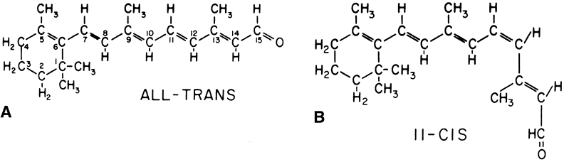
retinal dystrophy in the rat. J Cell Biol 1971;49:664

28. Bernstein PS, Law WC, Rando RR. Isomerization of all-trans-retinoids to 11-cis-retinoids in vitro. Proc Natl Acad Sci U S A 1987;84:1849

29. Chader GJ. Interphotoreceptor retinoid-binding protein (IRBP): A model protein for molecular biological and clinically relevant studies. Friedenwald lecture. Invest Ophthalmol Vis Sci 1989;30:7

30. Ho MT, Massey JB, Pownall HJ et al. Mechanism of vitamin A movement between rod outer segments, interphotoreceptor retinoid-binding protein, and liposomes. J Biol Chem 1989;264:928

31. Rando RR. Membrane phospholipids as an energy source in the operation of the visual cycle. Biochemistry 1991;30:595

32. Fung BK-K. Transducin: Structure, function and role in phototransduction. In Osborne NN, Chader GJ (eds). Progress in Retinal Research. Elmsford, New York: Pergamon Press, 1987:151–177

33. Sears S, Erickson A, Hendrickson A. The spatial and temporal expression of outer segment proteins during development of Macaca monkey cones. Invest Ophthalmol Vis Sci 2000;41:971

34. Hargrave PA. Rhodopsin structure, function, and topography the Friedenwald lecture. Invest Ophthalmol Vis Sci 2001;42:3

35. Sallese M, Iacovelli L, Cumashi A et al. Regulation of G protein-coupled receptor kinase subtypes by calcium sensor proteins. Biochim Biophys Acta 2000;1498:112

36. Mendez A, Burns ME, Roca A et al. Rapid and reproducible deactivation of rhodopsin requires multiple phosphorylation sites. Neuron 2000;28:153

37. Bruel C, Cha K, Niu L et al. Rhodopsin kinase: Two mAbs binding near the carboxyl terminus cause time-dependent inactivation. Proc Natl Acad Sci U S A 2000;97:3010

38. Bruel C, Cha K, Reeves PJ et al. Rhodopsin kinase: Expression in mammalian cells and a two-step purification. Proc Natl Acad Sci U S A 2000;97:3004

39. Chen CK, Hurley JB. Purification of rhodopsin kinase by recoverin affinity chromatography. Methods Enzymol 2000;315:404

40. Johnson WC, Palczewski K, Gorczyca WA et al. Calcium binding to recoverin: Implications for secondary structure and membrane association. Biochim Biophys Acta 1997;1342:164

41. Zhou J, Moroi K, Nishiyama M et al. Characterization of RGS5 in regulation of G protein-coupled receptor signaling. Life Sci 2001;68:1457

42. Lyubarsky AL, Naarendorp F, Zhang X et al. RGS9-1 is required for normal inactivation of mouse cone phototransduction. Mol Vis 2001;7:71

43. He W, Lu L, Zhang X et al. Modules in the photoreceptor RGS9-1.Gbeta 5L GTPase-accelerating protein complex control effector coupling, GTPase acceleration, protein folding, and stability. J Biol Chem 2000;275:37093

44. Chen CK, Burns ME, He W et al. Slowed recovery of rod photoresponse in mice lacking the GTPase accelerating protein RGS9-1. Nature 2000;403:557

45. Koch KW, Stryer L. Highly cooperative feedback control of retinal rod guanylate cyclase by calcium ions. Nature 1988;334:64

46. Gorczyca WA. Role of calcium ions in vertebrate phototransduction. Pol J Pharmacol 1999;51:167

47. Duda T, Goraczniak R, Surgucheva I et al. Calcium modulation of bovine photoreceptor guanylate cyclase. Biochemistry 1996;35:8478

48. Gorczyca WA, Polans AS, Surgucheva IG et al. Guanylyl cyclase activating protein. A calcium-sensitive regulator of phototransduction. J Biol Chem 1995;270:22029

49. Dryja TP, McGee TL, Reichel E et al. A point mutation of the rhodopsin gene in one form of retinitis pigmentosa. Nature 1990;343:364

50. Dryja TP, McGee TL, Hahn LB et al. Mutations within the rhodopsin gene in patients with autosomal dominant retinitis pigmentosa. N Engl J Med 1990;323:1302

51. Inglehearn CF, Bashir R, Lester DH et al. A 3-bp deletion in the rhodopsin gene in a family with autosomal dominant retinitis pigmentosa. Am J Hum Genet 1991;48:26

52. Chen J, Makino CL, Peachey NS et al. Mechanism of rhodopsin inactivation in vivo as revealed by COOH-terminal truncation mutant. Science 1995;267:374

53. Gryczan C, Kusvak JR, Novak L et al. A transgenic mouse model for autosomal dominant retinitis pigmentosa caused by a three base pair deletion in codon 255/256 of the opsin gene. Invest Ophthalmol Vis Sci 1995;36:S423

54. Naash MI, Hollyfield JG, Al-Ubaidi MR et al. Simulation of human autosomal dominant retinitis pigmentosa in transgenic mice expressing a mutated murine opsin gene. Proc Natl Acad Sci U S A 1993;90:5499

55. Naash MI, Al-Ubaidi MR, Hollyfield JG et al. Simulation of autosomal dominant retinitis pigmentosa in transgenic mice. In Hollyfield JG, Anderson RE, LaVail MM (eds). Retinal Degeneration: Clinical and Laboratory Applications. New York: Plenum, 1993:201–210

56. Olsson JE, Gordon JW, Pawlyk BS et al. Transgenic mice with a rhodopsin mutation (Pro23His): A mouse model of autosomal dominant retinitis pigmentosa. Neuron 1992;9:815

57. Sung C-H, Makino C, Baylor D et al. A rhodopsin gene mutation responsible for autosomal dominant retinitis pigmentosa results in a protein that is defective in localization to the photoreceptor outer segment. J Neurosci 1994;14:5818

58. Zozulya SA, Gurevich VV, Zvyaga TA et al. Functional expression in vitro of bovine visual rhodopsin. Protein Eng 1990;3:453

59. Berson EL, Rosner B, Sandberg MA et al. Ocular findings in patients with autosomal dominant retinitis pigmentosa and rhodopsin, proline-347-leucine. Am J Ophthalmol 1991;111:614

60. Jacobson SG, Kemp CM, Sung CH et al. Retinal function and rhodopsin levels in autosomal dominant retinitis pigmentosa with rhodopsin mutations. Am J Ophthalmol 1991;112:256

61. Apfelstedt-Sylla E, Kunisch M, Horn M et al. Ocular findings in a family with autosomal dominant retinitis pigmentosa and a frameshift mutation altering the carboxyl terminal sequence of rhodopsin. Br J Ophthalmol 1993;77:495

62. Allikmets R, Shroyer NF, Singh N et al. Mutation of the Stargardt disease gene (ABCR) in age-related macular degeneration. Science 1997;277:1805

63. Sun H, Smallwood PM, Nathans J. Biochemical defects in ABCR protein variants associated with human retinopathies. Nat Genet 2000;26:242

64. Allikmets R. Further evidence for an association of ABCR alleles with age-related macular degeneration. The International ABCR Screening Consortium. Am J Hum Genet 2000;67:487

65. Rivera A, White K, Stohr H et al. A comprehensive survey of sequence variation in the ABCA4 (ABCR) gene in Stargardt disease and age-related macular degeneration. Am J Hum Genet 2000;67:800

66. Fuse N, Suzuki T, Wada Y et al. Molecular genetic analysis of ABCR gene in Japanese dry form age- related macular degeneration. Jpn J Ophthalmol 2000;44:245

67. Souied EH, Ducroq D, Rozet JM et al. ABCR gene analysis in familial exudative age-related macular degeneration. Invest Ophthalmol Vis Sci 2000;41:244

68. De La Paz MA, Guy VK, Abou-Donia S et al. Analysis of the Stargardt disease gene (ABCR) in age-related macular degeneration. Ophthalmology 1999;106:1531

69. Simonelli F, Testa F, de Crecchio G et al. New ABCR mutations and clinical phenotype in Italian patients with Stargardt disease. Invest Ophthalmol Vis Sci 2000;41:892

70. Lewis RA, Shroyer NF, Singh N et al. Genotype/phenotype analysis of a photoreceptor-specific ATP-binding cassette transporter gene, ABCR, in Stargardt disease. Am J Hum Genet 1999;64:422

71. Kramer F, White K, Pauleikhoff D et al. Mutations in the VMD2 gene are associated with juvenile-onset vitelliform macular dystrophy (Best disease) and adult vitelliform macular dystrophy but not age-related macular degeneration. Eur J Hum Genet 2000;8:286

72. Caldwell GM, Kakuk LE, Griesinger IB et al. Bestrophin gene mutations in patients with Best vitelliform macular dystrophy. Genomics 1999;58:98

73. Marquardt A, Stohr H, Passmore LA et al. Mutations in a novel gene, VMD2, encoding a protein of unknown properties cause juvenile-onset vitelliform macular dystrophy (Best's disease). Hum Mol Genet 1998;7:1517

74. Lotery AJ, Munier FL, Fishman GA et al. Allelic variation in the VMD2 gene in best disease and age-related macular degeneration. Invest Ophthalmol Vis Sci 2000;41:1291

Back to Top