|
|
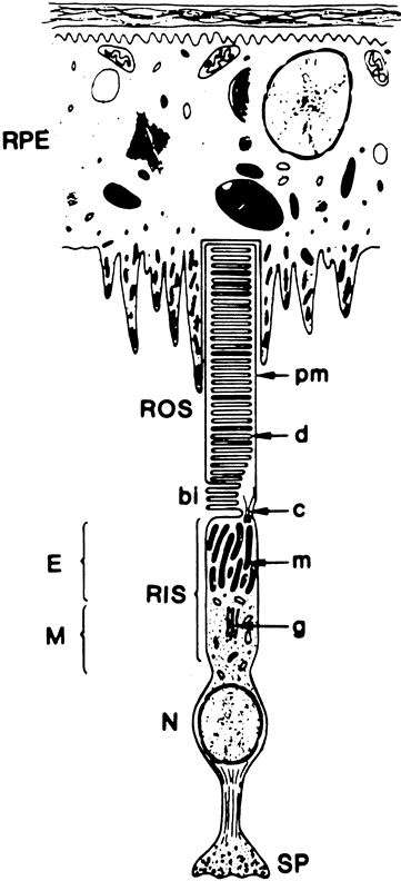
| Fig. 2. Schematic diagram of a rod cell and its spatial relationship to the retinal pigment epithelium (RPE). (bi, basal enfoldings; c, connecting cilium; d, discs; E, ellipsoid region of the inner segment; g, Golgi apparatus; m, mitochondria; M, myoid region of the inner segment; N, nucleus; pm, plasma membrane of the outer segment; RIS, rod inner segment; ROS, rod outer segment; SP, synaptic pedicle). (Fliesler SJ, Anderson RE: Chemistry and metabolism of lipids in the vertebrate retina. In Holman RT [ed]: Progress in Lipid Research. Vol. 22. Elmsford, New York: Pergamon Press, 1983:1–52) |