Chapter 2a
Physiology of the Lacrimal System
CAT NGUYEN BURKAT, ROBIN R. HODGES, MARK J. LUCARELLI and DARLENE A. DARTT
Main Menu   Table Of Contents

Search

SECRETORY SYSTEM
EXCRETORY SYSTEM
REFERENCES

SECRETORY SYSTEM

LACRIMAL GLAND AND ACCESSORY GLANDS

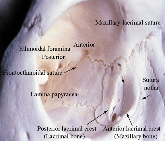
The exocrine main lacrimal gland is located in the superotemporal orbit in a shallow lacrimal fossa of the frontal bone. It is surrounded by fibrous tissue that is attached superiorly to the frontal bone periosteum. In the adult, the lacrimal gland measures 20 mm by 12 mm by 5 mm and weighs approximately 0.78 g. It is incompletely divided by the lateral horn of the levator aponeurosis into a larger orbital lobe, which comprises 60% to 70% of its total mass, and a smaller palpebral lobe below.1,2 The two lobes are connected by vessels, nerves, and the lacrimal ducts. The orbital lobe lies posterior to the orbital septum and preaponeurotic fat, and anterior to the levator aponeurosis.1 With increasing age, the gland may herniate through a weakened orbital septum, resulting in fullness of the lateral upper eyelid. The palpebral lobe is located posterior to the levator aponeurosis in the subaponeurotic space and directly anterior to the conjunctiva, which is adherent to the posterior aspect of the gland. Medially, Müller's muscle may intervene between the posterior gland and the conjunctiva. Anteriorly, the palpebral lobe may extend beyond the orbital margin to lie in the superolateral fornix, where it may be visible through the conjunctiva with eyelid eversion.

The lacrimal gland is divided into numerous lobules, separated by interlobular fibrovascular connective tissue. Each lobule is structurally composed of the acinar unit and the ductal system. Two to six secretory ducts from the orbital lobe of the lacrimal gland pass through the palpebral lobe or along its fibrous capsule, joining with ducts from the palpebral lobe to form 6 to 12 tubules that empty into the superolateral conjunctival fornix 4 to 5 mm above the tarsus.3,4 Excision of the palpebral lobe may therefore interrupt drainage from the orbital lobe. The ducts, lined by pseudostratified, nonkeratinizing squamous epithelium, form a branching pattern within the lacrimal gland. Secretion from the acinar units first drains into the smaller intralobular ducts, proceeds through the larger interlobular ducts, and eventually passes into the main excretory ducts before draining into the superolateral conjunctival fornix. The surface of the ductal lumina contains microvilli, similar to the acinar secretory cells.5,6 The epithelial walls of the larger ducts contain three to four layers of columnar or cuboidal cells, compared with a single layer in the smaller intralobular ducts.

Each acinar unit, or secretory unit, of the lacrimal gland is composed of an inner layer of columnar or pyramidal-shaped secretory cells around a central lumen, and a surrounding basal layer of myoepithelial cells. The nucleus is located at the base of the acinar secretory cell, and numerous membrane-bound zymogenic secretory granules are present in the apical cytoplasm.7 The number and size of granules, which contain predominantly serous proteins for secretion, vary from cell to cell. Typically, larger granules are located in the peripheral lobules.6,8 The zymogenic secretory granules fuse with the apical cell membrane and release their granular contents into the acinar lumen through a mechanism known as emiocytosis.5,6,9 Myoepithelial cells are spindle- or basket-shaped cells, composed of cytoplasmic myofilaments, located between the base of the secretory cell and the basement membrane of the acinar unit.7 Contraction of the myoepithelial cells helps drive secretions into the drainage tubules and ducts.

Accessory lacrimal glands are located in the conjunctival fornices and along the superior tarsal border. There are approximately 20 to 40 accessory glands of Krause in the superior conjunctival fornix. There are approximately half the number of glands of Krause present in the lower eyelid as in the superior conjunctiva. Accessory glands of Wolfring, which are slightly larger, are located along the superior tarsal border in the upper eyelid, with 4 to 20 located in the upper eyelid, and 2 to 4 in the lower eyelid.1,3,10,11 The accessory lacrimal gland lobules empty onto the conjunctiva surface through small branching ducts that drain into a single excretory duct that is lined by one or two layers of cells.11

Like the main lacrimal gland, the accessory lacrimal glands are also innervated. Parasympathetic and sympathetic nerves have been identified in accessory lacrimal glands of humans.11–14 The presence of nerves suggests that secretion from accessory lacrimal glands is neurally regulated. Therefore, the accessory lacrimal glands can also contribute to stimulated tear secretion.

INNERVATION OF THE SECRETORY SYSTEM

The lacrimal gland receives innervation from cranial nerves V and VII, as well as from the sympathetics of the superior cervical ganglion.15 The lacrimal branch of the ophthalmic division of the trigeminal nerve carries sensory stimuli from the lacrimal gland, with the cell bodies located within the Gasserian ganglion. Afferent sensory fibers exit the posterior aspect of the lacrimal gland, travel along the superior border of the lateral rectus muscle, and exit the orbit through the superior orbital fissure.

Parasympathetic secretomotor fibers originate in the lacrimal nucleus of the caudal pons and exit the pontomedullary junction between cranial nerves VI and VIII. The fibers enter the internal auditory canal and travel a long course within the nervus intermedius, the greater superficial petrosal nerve, the deep petrosal nerve, and the vidian nerve to finally synapse in the pterygopalatine ganglion.4 The greater superficial petrosal nerve arises from the preganglionic parasympathetic fibers of the geniculate ganglion. The deep petrosal nerve carries sympathetic fibers from the internal carotid plexus. Postganglionic parasympathetic fibers leave the pterygopalatine ganglion through the pterygopalatine nerves to innervate the lacrimal gland.16,17 Some fibers may join the zygomatic nerve as it branches from the maxillary division of the trigeminal nerve and enters the orbit through the inferior orbital fissure. Branches of the zygomatic nerve may ascend and enter the posterior surface of the lacrimal gland either alone or in combination with the lacrimal nerve.1 Traditionally, it was thought that secretomotor nerves pass to the gland through the zygomatic and lacrimal nerves. More recently, Ruskell18 found that parasympathetic fibers traveled along a branch of the middle meningeal artery through the superior orbital fissure before joining the ophthalmic or lacrimal artery to supply the lacrimal gland. After parasympathetic denervation, the lacrimal gland has been reported to be supersensitive to parasympathomimetic agents such as topical or systemic pilocarpine.19,20

Postganglionic sympathetic nerves, arising from the superior cervical ganglion and internal carotid artery plexus, arrive with the lacrimal artery branch of the ophthalmic artery. Sympathetic nerve fibers also arrive along with parasympathetics in the zygomatic nerve. The zygomatic branch of the maxillary trigeminal nerve gives off the lacrimal branch before dividing into zygomaticotemporal and zygomaticofacial branches. This lacrimal branch anastomoses with the lacrimal nerve of the ophthalmic trigeminal nerve or travels along the periorbita to independently enter the gland at its posterolateral aspect.

The lacrimal gland receives arterial supply from the lacrimal artery of the ophthalmic artery, with contributions from the recurrent meningeal artery and a branch of the infraorbital artery. The arterial supply enters the posterior aspect of the gland and eventually continues anteriorly as the arterial arcades of the eyelids. The venous drainage follows approximately the same intraorbital course of the artery and drains into the superior ophthalmic vein and cavernous sinus. Lymphatic channels in the interstitial tissue between the gland lobules join the lymphatic drainage from the lateral eyelids and conjunctiva to empty into the preauricular lymph nodes.

REGULATION OF SECRETION

The lacrimal gland secretes water, electrolytes, and protein in response to neural and hormonal stimulation and is the major contributor to the aqueous portion of the tear film with additional contributions from the accessory lacrimal glands, the cornea, and the conjunctiva.21 Thus, the lacrimal gland is extensively innervated with parasympathetic, sympathetic, and sensory nerves. Parasympathetic nerves, containing acetylcholine (ACh) and vasoactive intestinal peptide (VIP), and sympathetic nerves containing norepinephrine, are potent stimuli of lacrimal gland secretion.21 In addition, many hormones from the hypothalamic-pituitary-gonadal axis have significant influences on the lacrimal gland, including stimulating secretion of secretory immunoglobulin A (SIgA).22–24 Sensory nerves are the least prevalent and release substance P and calcitonin gene-related peptide. In the lacrimal gland, each of these agonists bind to specific receptors on the acinar cells and initiate a different cascade of intracellular events, or cellular signal transduction pathway. Stimulation of sensory nerves in the cornea or conjunctiva carries the afferent limb of the tear reflex arc, and the efferent parasympathetic and sympathetic nerves subsequently stimulate lacrimal gland secretion.

The lacrimal gland consists of three main types of cells: acinar, ductal, and myoepithelial cells. Plasma cells, lymphocytes, macrophages, and mast cells are also present. Myoepithelial cells are basket-shaped cells that surround the acinar cells. Because of the abundance of α-smooth muscle actin present in these cells, it is believed that they are involved in contraction to expel the secretory product. In support of this hypothesis, these cells have been shown to express several neurotransmitter receptors.25,26 Ductal epithelial cells form the lacrimal ducts and modify the secretory product as it moves through the ducts. The acinar cells are the predominant cell type in the lacrimal gland and make up approximately 80% of the cells in the gland. These cells form the secretory unit of the gland termed an acinus. In cross-section, acini are seen as a ring of pyramidal-shaped cells joined by tight junctions on the lateral side.21 The presence of tight junctions results in the formation of highly polarized cells and division of the plasma membrane into apical membranes (membranes bordering the ducts), lateral membranes (membranes between adjacent cells), and basal membranes (membranes that are exposed to extracellular matrix and blood supply). This formation ensures unidirectional secretion of the secretory product toward the ductal lumen. Receptors for hormones and neurotransmitters are located on the basolateral membranes. Once activated, the signal from these receptors is transduced through the cell leading to fusion of secretory vesicles with the apical membrane to release their contents into the ducts. Ductal cells modify the secretory product before it is released onto the ocular surface. Electrolyte concentration of the secreted lacrimal gland fluid varies with flow rate, with hypertonic fluid produced during low flow rates and isotonic fluid at faster flow rates.27 The secretion of electrolytes and water into the lumen is dependent on the activation of ion transport proteins and ion channels also located in the apical and basolateral membranes.

The lacrimal gland is able to secrete proteins by two different processes, each of which is regulated differently. In the first process, known as constitutive secretion, the proteins are synthesized in the endoplasmic reticulum, modified in the Golgi apparatus, and sorted into secretory vesicles. These vesicles immediately fuse with the apical membrane.22–24 These vesicles are not stored; thus, regulation of constitutive secretion occurs at the level of protein synthesis. An example of a constitutively secreted protein is SIgA. The second type of secretion is called regulated protein secretion. In this type of secretion, the proteins are synthesized in the endoplasmic reticulum, modified in the Golgi apparatus, and packaged into secretory vesicles. These vesicles are stored until the appropriate stimulus, such as the release of neurotransmitters from nerves, occurs, at which time they fuse with the apical membrane. The lacrimal gland secretes in a merocrine fashion, that is, only a small portion of granules fuse on stimulation.

Clinically, tear secretion from the main lacrimal gland may be stimulated by reflex tearing from stimulation of the conjunctiva and cornea by irritating phenomena, psychogenic tearing, or aberrant regeneration. Reflex tearing may also occur by stimulation of the retina and optic nerve by bright light, and by spicy foods because of a reflex arc between the gustatory and the lacrimal nucleus. In psychogenic tearing, strong emotions and stress cause afferent pathways from the frontal lobe, basal ganglia, thalamus, and hypothalamus to interact with the lacrimal nucleus.28 Regeneration of injured parasympathetic secretomotor fibers destined for the salivary glands may aberrantly innervate the lacrimal gland, resulting in crocodile tears, or frank epiphora with gustatory stimulation. These stimuli affect the reflex tear production from the main lacrimal gland, with the continuous basal tear secretion from the accessory lacrimal glands, conjunctival goblet cells, and sebaceous glands all left unaffected.

Excision of the main lacrimal gland may result in lacrimal insufficiency, with profound changes in water and electrolyte secretion, loss of the normal tear film, and corneal sequelae.29,30 However, total removal of the main lacrimal gland does not in itself lead to keratoconjunctivitis sicca, which suggests that tear production from the accessory lacrimal glands may be sufficient to maintain a stable tear layer on the cornea.31

STIMULATION OF PROTEIN SECRETION BY PARASYMPATHETIC NERVES

Neurotransmitters released from parasympathetic nerves are potent stimuli of lacrimal gland secretion. In the lacrimal gland, these nerves contain ACh and VIP that activate two distinct intracellular signaling pathways. ACh binds to M3 muscarinic receptors present on basal and lateral membranes of acinar cells (Fig. 1).32 The activated M3 muscarinic receptor is coupled to the G protein, Gαq/11.33–36 The G protein, in turns, activates phospholipase C (PLC). PLC hydrolyzes phosphatidylinositol bisphosphate (PIP2), a membrane phospholipid, into two second messengers, 1,4,5-inositol trisphosphate (1,4,5-IP3) and diacylglycerol (DAG).32,37–40 1,4,5-IP3 binds to IP3 receptors on the endoplasmic reticulum to release its Ca2+ stores, increasing the intracellular concentration of calcium, or [Ca2+]i.41 In the lacrimal gland, cholinergic agonists increase 1,4,5-IP3 within seconds resulting in a rapid increase in [Ca2+]i.41 When the intracellular Ca2+ stores are depleted, a feedback mechanism is activated whereby Ca2+ enters the cells through Ca2+ channels in the plasma membrane of the acinar cell.42,43 This Ca2+ influx is termed “stored operated Ca2+ entry” or “capacitative Ca2+ influx” and is responsible for the slower, sustained phase of increased [Ca2+]i. Therefore, the cholinergic agonist-stimulated Ca2+ response is a biphasic response of a rapid burst released from intracellular stores followed by movement of Ca2+ into the cell that is sustained until the stimulus is removed or decreases slowly over time.41,44 In conjunction with calmodulin, the Ca2+ phosphorylates and therefore activates proteins, as of yet unidentified, that lead to protein secretion (Fig. 1). The increase in intracellular calcium is necessary for both muscarinic and adrenergic agonist-evoked exocytosis from acinar cells, and may even be sufficient to induce exocytosis in the absence of agonist stimulation.45

Fig. 1. Signal transduction pathways activated by cholinergic agonists in the lacrimal gland. ACh, acetylcholine; M3, muscarinic receptor subtype 3; Gαq, alpha subunit of Gq G protein; PLC, phospholipase C; Pyk2 and Src, nonreceptor tyrosine kinases; raf, mitogen-activated protein kinase kinase kinase; MEK, mitogen-activated protein kinase kinase; MAPK, p44/p42 mitogen-activated protein kinase; IP3, inositol trisphosphate; PKC, protein kinase C; ER, endoplasmic reticulum. (Modified from Dartt DA:Regulation of lacrimal gland secretion by neurotransmitters and the EGF family of growth factors. Exp Eye Res 73:741, 2001, with permission.)

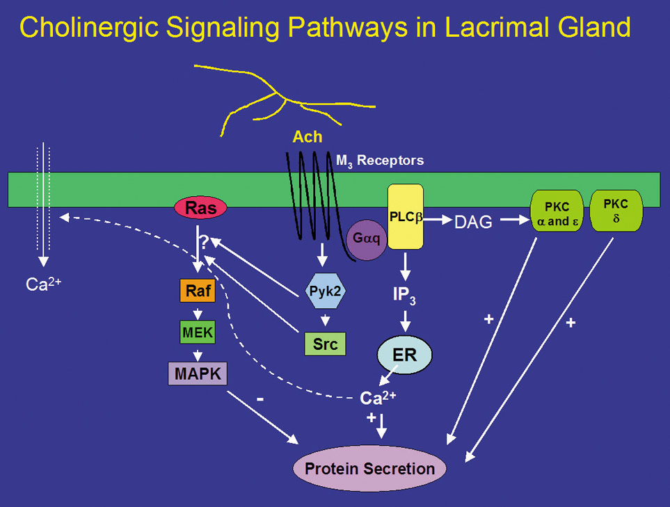
DAG, which is also produced on hydrolysis of PIP2, binds to and activates several members of the protein kinase C (PKC) family.46 The PKC family includes 10 different isoforms and has been divided into three groups depending on cofactor requirements.47–50 Classic PKCs include PKCα, -βI, -βII, and -γ and require calcium and phospholipids for activation. Novel PKC isoforms include PKCδ, ε, η, and θ, and are calcium independent but phospholipid dependent. Atypical PKC isoforms are PKCλ and ζ. These isoforms are calcium and phospholipid independent. The lacrimal gland contains at least four PKC isoforms, PKCα, δ, ε, and λ. PKD, also known as PKCμ, is also present in the lacrimal gland.51 In general, PKC isoforms have cell-specific localizations and functions, and the lacrimal gland is no exception.51 In the lacrimal gland, PKCα was found to be located in the cytoplasm and plasma membranes of acinar cells. PKCδ was found in the cytoplasm of acinar and myoepithelial cells. PKCε was located in the cytoplasm, basal, and lateral membranes, and on an apical membrane system that is similar to actin networks of acinar cells. PKCε was also seen occasionally on myoepithelial cells. PKCλ was observed in acinar cells on an endomembrane system, potentially the endoplasmic reticulum or Golgi apparatus.51

Translocation of PKC from the cytosol to membrane fractions can be an indication of activation. Stimulation of lacrimal gland acini with cholinergic agonists for less than 1 minute translocates PKCα from the cytosol to the basal and lateral membranes.52 Cholinergic agonists also translocate PKCδ and ε, albeit with a longer time course.53 With specific inhibitors of the α, δ, and ε isoforms of PKC, cholinergic agonists were shown to use all three isoforms to varying degrees to stimulate secretion. Inhibition of PKCα reduced cholinergic agonist-induced protein secretion approximately 75%, whereas inhibition of PKCε reduced cholinergic agonist-induced secretion by approximately 50%. Inhibition of PKCδ reduced cholinergic agonist-induced secretion by approximately 20%.53 These findings were confirmed by down-regulating PKC using phorbol esters. Down-regulation with phorbol esters decreased the amount of PKCα by 79%, PKCδ by 53%, and PKCε by 10%.54 This treatment also decreased cholinergic agonist-stimulated protein secretion more than 90%.54 Thus PKCα, δ, and ε play differential, but overlapping, roles in cholinergic agonist-stimulated protein secretion (Fig. 1).51,55,56

In summary, cholinergic agonists bind to M3 muscarinic receptors to activate PLC. PLC hydrolyses PIP2 to produce 1,4,5-IP3 and DAG. 1,4,5-IP3 increases [Ca2+]i, whereas DAG activates PKCα, δ, and ε. Thus, activation of protein secretion stimulated by cholinergic agonists uses at least three PKC isoforms along with Ca2+ to stimulate protein secretion (Fig. 1).

In addition to ACh, parasympathetic nerves also release the neuropeptide VIP on stimulation. VIP is a 28-amino acid regulatory peptide that is found in parasympathetic nerves of most exocrine glands. VIP binds to VIP receptors (VIPR) of which two types have been identified. These receptors are called VIPR1 and VIPR2.26 In the lacrimal gland, VIPR1 is present on the basolateral membranes of acinar cells, whereas VIPR2 is found on myoepithelial cells.26 VIPRs are coupled to the G protein, Gαs, to activate adenylyl cyclase (AC) (Fig. 2).26,35 The lacrimal gland contains at least three of the six known isoforms (I-VI) of AC, namely, ACII, ACIII, and ACIV. The presence of the other AC isoforms has not been detected in the lacrimal gland.26 Interestingly, none of the isoforms were localized to the plasma membrane as would be expected for interaction with Gαs. ACII was present almost exclusively on myoepithelial cells, whereas AC III was found only on the occasional duct, blood vessel, and myoepithelial cell. ACIV was found to be present in acinar cells but with an intracellular localization that appeared to be either endoplasmic reticulum or Golgi apparatus. It is possible that another AC isoform is responsible for the coupling of Gαs to its downstream effects or that ACIII is recruited to the plasma membrane on stimulation. This has been shown to be the case for muscarinic receptors in the lacrimal gland.57 Activation of AC leads to the formation of cyclic-adenosine monophosphate (cAMP) from AMP. cAMP, in turn, binds to and activates a cAMP-dependent protein kinase, known as protein kinase A (PKA) (Fig. 2). PKA is responsible for phosphorylating proteins that are components of the protein secretory machinery or ion transport channels necessary for secretion to occur, although none of the substrates have been identified in the lacrimal gland. Inhibition of PKA in the lacrimal gland with a specific inhibitor decreased VIP-stimulated protein secretion by 70%.26

Fig. 2. Signal transduction pathways activated by vasoactive intestinal peptide (VIP) in the lacrimal gland. VIPR, VIP receptor; Gαs, alpha subunit of Gs G protein; AC, adenylate cyclase; cAMP, cyclic adenosine monophosphate; ATP, adenosine triphosphate; AMP, adenosine monophosphate; PKA, protein kinase A; cAMP PDE, cAMP phosphodiesterase. (Modified from Dartt DA: Regulation of lacrimal gland secretion by neurotransmitters and the EGF family of growth factors.Exp Eye Res 73:741, 2001, with permission.)

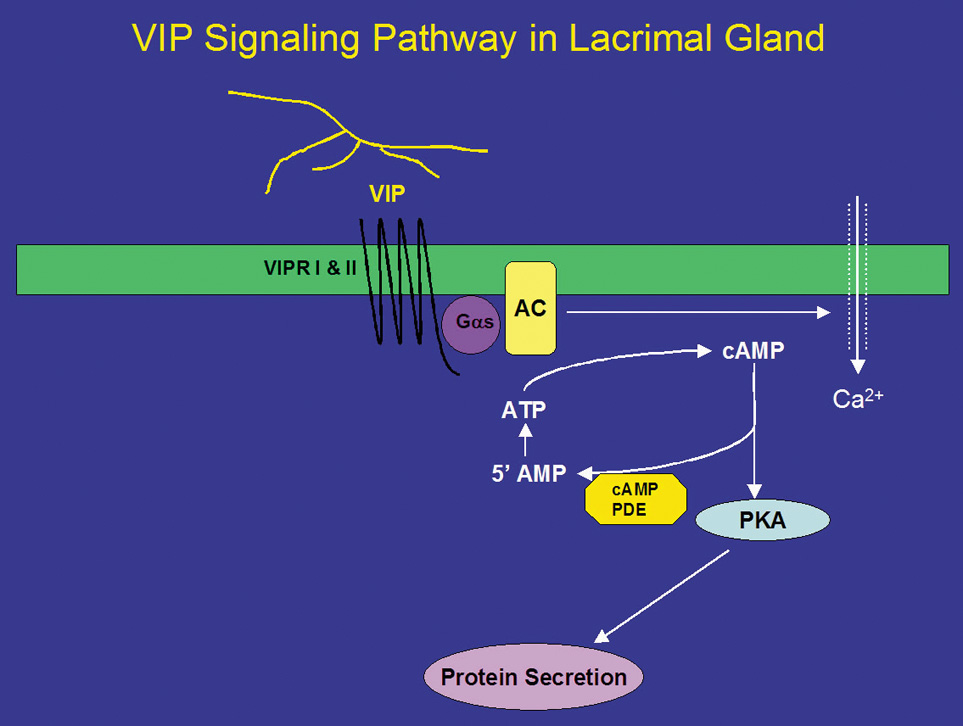
The signal is terminated when cAMP is degraded by cAMP-dependent phosphodiesterases, which are present in the cell. Eleven different families of phosphodiesterases have been identified, each with varying specificities to cAMP and cyclic guanosine monophosphate.58 However, the types of phosphodiesterases present in the lacrimal gland remain unidentified. It is known that the nonspecific phosphodiesterase inhibitor 3-isobutyl-1-methylxanthine (IBMX) stimulates secretion in the absence of agonists implying that the basal rate of secretion from the lacrimal gland is partially dependent on cyclic nucleotides. The effects of IBMX are also dependent on Ca2+ because removal of Ca2+ partially reduces IBMX-stimulated protein secretion.59

In addition to activation of the cAMP-dependent pathway, addition of VIP to lacrimal gland acinar cells also increases [Ca2+]i.41 The increase in [Ca2+]i obtained in the presence of VIP is 25% of the increase obtained when the cells were stimulated with cholinergic agonists.26,41 Unlike cholinergic agonists, VIP does not increase levels of IP3 but increases [Ca2+]i by modulating Ca2+ influx.60 Increased cAMP leads to increased intracytoplasmic concentrations of Ca2+ through plasma membrane channels and activates Ca2+-dependent K+ channels that play an essential role in fluid secretion.61

In addition to VIP, two other peptide hormones, α-melanocyte-stimulating hormone (α-MSH) and adrenocorticotropic hormone (ACTH) stimulate lacrimal gland protein secretion.62 These peptides are not released from nerves, but are delivered by the blood supply to the lacrimal gland. α-MSH and ACTH stimulate protein secretion by activation of the cAMP-dependent pathway. Only the melanocortin 5 receptor, which can bind both α-MSH and ACTH, has been identified in the lacrimal gland.63,64 This receptor could mediate the effects of both hormones in the lacrimal gland.

STIMULATION OF PROTEIN SECRETION BY SYMPATHETIC NERVES

Norepinephrine, the neurotransmitter released from sympathetic nerves, stimulates lacrimal gland protein secretion.65,66 Norepinephrine can bind to both α- and β-adrenergic receptors present in the gland. Activation of β-adrenergic receptors causes an increase in protein secretion, which appears to occur through the cAMP-dependent pathway67 similar to the VIP pathway shown in Figure 2.

Norepinephrine, when bound to α1-adrenergic receptors in the lacrimal gland, is a potent stimulator of protein secretion. However, the signal transduction pathways used by α-adrenergic receptors are not completely understood. α1-Adrenergic receptors, similar to muscarinic receptors, activate the G-protein Gαq/11 (Fig 3).68 In contrast with muscarinic receptors, activation of Gαq/11 by α1-adrenergic receptors only accounts for a small portion (∼30%) of adrenergic agonist-induced secretion.68 The Gαq/11 presumably activates an unknown effector phospholipase, although it is known that the phospholipase is neither PLC nor phospholipase D (PLD).41,69 α1-Adrenergic agonists do not increase IP3 or IP4.50,70 However, they do cause a small increase in intracellular Ca2+, contributing only 20% of that caused by cholinergic agonists.70,71 The increased Ca2+ and activated PKC then result in protein secretion. In addition, α1-adrenergic agonist-induced protein secretion was not inhibited by chelation of extracellular Ca2+, indicating that the increase in [Ca2+]i does not play a major role in this secretion.70

Fig. 3. Signal transduction pathways activated by α1-adrenergic agonists in the lacrimal gland. Norepi, norephinephrine; Gαq, alpha subunit of Gq G protein; DAG, diacylglycerol; PKC, protein kinase C; ErbB1, the EGF receptor; ErbB, the EGF family of growth factor receptor; P, phosphorylated tyrosine residues on the ErbBs; SOS, Grb2 and Shc, adaptor proteins, raf, mitogen-activated protein kinase kinase kinase; MEK, mitogen-activated protein kinase kinase; MAPK, p44/p42 mitogen-activated protein kinase; ER, endoplasmic reticulum. (Modified from Dartt DA: Regulation of lacrimal gland secretion by neurotransmitters and the EGF family of growth factors. Exp Eye Res 73:741, 2001, with permission.)

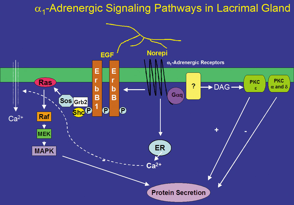
Like cholinergic agonists, α1-adrenergic agonists have also been shown to influence protein secretion by PKC. Down-regulation of PKC with phorbol esters, however, increased α1-adrenergic agonist-induced protein secretion.72 By using inhibitors specific to PKCα, δ, and ε, Zoukhri et al showed that inhibition of PKCα and δ increased α1-adrenergic agonist-induced protein secretion, and that inhibition of PKCε decreased this secretion.53 These results suggest that α1-adrenergic agonists activate PKCα, δ, and ε, but only PKCε stimulates α1-adrenergic agonist-induced protein secretion whereas PKCα and δ inhibit it (Fig. 3). Thus, muscarinic and α1-adrenergic receptors used the same PKC isoforms but in different ways to control protein secretion. This suggests that cellular localization of PKC, through anchoring proteins, is vital for agonist-induced protein secretion.

STIMULATION OF PROTEIN SECRETION BY GROWTH FACTORS

Growth factors, such as the epidermal growth factor (EGF) family of growth factors, have been identified as stimuli of protein secretion from the lacrimal gland.73 This family includes EGF, transforming growth factor (TGF)-α, heparin-binding EGF (HB-EGF), heregulin, and other members of its growth factor family. These molecules are synthesized as transmembrane precursor molecules that are proteolytically cleaved to the soluble pro-form of the growth factor. For EGF, it is further cleaved to the mature form. The transmembrane precursor molecule and the pro- and mature forms are catalytically active and capable of binding to the EGF receptor subtypes.

There are four types of the EGF receptors, referred to as erbB1-4. These receptors are tyrosine kinases. On ligand binding, the erbB receptors form homo- and heterodimers with each other and phosphorylate one another on multiple tyrosine residues. Depending on the tyrosine residues phosphorylated, different adaptor proteins are recruited resulting in the activation of a physiologic process. The dimers can bind different ligands, recruit different adaptor molecules, activate different processes, and have varying potencies.74 The lacrimal gland contains all four types of erbB receptors, although their locations differ.73,75 ErbB1, also known as the EGF receptor, is present on the apical, basal, and lateral membranes of acinar and ductal cells, whereas erbB2 (known as HER2) is located on the basal and lateral membranes of acinar and ductal cells. The location of erbB3 has been reported to be on ductal cells,75 and erbB4 (known as Neu) is found on the apical, basal, and lateral membranes of ductal cells.73

After EGF binds to an erbB receptor and forms a dimer with another erbB receptor, several adaptor proteins can be recruited, each linking the receptor to a different signaling pathway. To induce lacrimal gland protein secretion, EGF binds to the erbB1 receptor and recruits PLCγ, which, in turn, produces 1,4,5-IP3 and DAG. 1,4,5-IP3 increases [Ca2+]i, whereas the DAG activates PKC (Fig. 4).54 EGF therefore stimulates protein secretion through Ca2+ and PKC signaling pathways. Studies using chelation of Ca2+ and down-regulation of PKC were found to inhibit EGF-stimulated protein secretion.54 Heregulin, HB-EGF, and TGFα also stimulate protein secretion, although the precise cellular mechanisms have not been determined.73

Fig. 4. Signal transduction pathways activated by EGF in the lacrimal gland. ErbB1, the EGF receptor; ErbB, the EGF family of growth factor receptors; PLC, phospholipase C; PI3K, phosphatidylinositol 3 kinase; P, phosphorylated tyrosine residue on the EGF receptor; raf, mitogen-activated protein kinase kinase kinase; MEK, mitogen-activated protein kinase kinase; MAPK, p44/p42 mitogen-activated protein kinase; PKC, protein kinase C. (Reprinted from Hodges RR, Dartt DA: Regulatory pathways in lacrimal gland epithelium. Int Rev Cytol 231:129, 2003, with permission.)

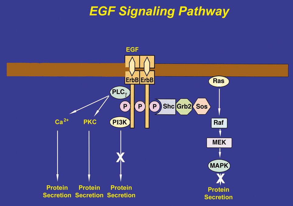
If the adaptor proteins, Shc and Grb2, are recruited and tyrosine-phosphorylated by the erbB receptor, the protein SOS binds to Grb2. SOS catalyzes the exchange of GDP for GTP on Ras to activate it. Ras initiates the activation of a series of serine-threonine kinases including Raf (a mitogen-activated protein kinase [MAPK]), which in turn activates MEK (another MAPK), ultimately leading to activation of p42/p44 MAPK. Activated p42/p44 MAPK can then translocate to the nucleus to influence gene expression to initiate cell proliferation, migration, and apoptosis. Activated p42/p44 MAPK may also remain in the cytosol to influence secretion and other short-term processes.76–78 In the lacrimal gland, EGF causes the tyrosine-phosphorylation of Shc and Grb2, indicating that this pathway is activated.78 However, EGF activation of p42/p44 MAPK does not cause protein secretion, although it does modify cholinergic- and α1-adrenergic agonist-induced protein secretion. Heregulin, HB-EGF, and TGFα also activate p42/p44 MAPK in the lacrimal gland.73

The third protein that can be recruited to the erbB receptors is phosphatidylinositol 3-kinase (PI-3K). PI-3K phosphorylates several lipids including phosphatidylinositol 4,5-bisphosphate to produce phosphatidylinositol 3,4,5-trisphosphate. This stimulates processes such as cell growth, survival, and adhesion. Inhibitors of PI-3K do not have any effect on EGF-stimulated protein secretion in the lacrimal gland, which implies that either PI-3K is not recruited by erbB receptors in the lacrimal gland or PI-3K is recruited for another function other than secretion.54

In many different types of tissues, including the lacrimal gland, the signal transduction pathways of EGF and G-protein coupled receptors interact with one another. Indeed, in the lacrimal gland, inhibition of MEK increased cholinergic and α1-adrenergic agonist-induced protein secretion suggesting that activation of p42/p44 MAPK negatively modulates agonist-stimulated secretion.78 Cholinergic agonists have been shown to activate p42/p44 MAPK through activation of the nonreceptor tyrosine kinases, Pyk2 and Src (Fig. 1).78 In contrast, α1-adrenergic agonists use a different mechanism to activate p42/p44 MAPK. These agonists activate EGF to recruit Shc and Grb2, which activate the cascade of kinases that result in activation of p42/p44 MAPK (Fig. 2).78

Neuropeptides, such as substance P and calcitonin gene-related peptide, may indirectly affect lacrimal gland secretion. Substance P may stimulate protein secretion indirectly by causing mast cell degranulation, with the released mediator, serotonin, stimulating the lacrimal acinar cells. In contrast, calcitonin gene-related peptide inhibits degranulation of mast cells, resulting in decreased serotonin release and thus decreased protein release from the acini.79

REGULATION OF CONSTITUTIVE PROTEIN SECRETION

Proteins secreted through the constitutive pathway are secreted shortly after synthesis rather than being stored in secretory vesicles. The regulation of this secretion lies at the level of gene transcription and translation. SIgA and secretory component (SC) are examples of proteins secreted by this pathway. SIgA is composed of IgA and a J chain coupled to SC. SC is synthesized in the acinar and ductal cells, whereas IgA and the J chain are synthesized by plasma cells present in the lacrimal gland.22 SC is synthesized as a precursor molecule, known as the polymeric immunoglobulin receptor, which inserts into the basal and lateral membranes. IgA binds to this receptor and is endocytosed into distinct vesicles that are transported directly to the apical membrane. These vesicles fuse with the apical membrane releasing SIgA into the lumen.

Androgens play a significant role in the maintenance and function of the lacrimal gland.23 Dihydrotestosterone has been shown to increase both the synthesis and secretion of SC.80 Androgens appear to regulate this pathway by diffusing to the nucleus of the acinar cell where they bind to specific receptors.23 The receptors are members of a family of ligand-activated transcription factors, and when activated by androgens from the bloodstream, they regulate SIgA by controlling the rate of synthesis of the SC.23

SIgA antibodies act to inhibit viral, bacterial, and parasitic invasion, and damage to the lacrimal gland and ocular surface. Fluid secretion of the lacrimal gland is dependent on the presence of androgens; ovariectomy of female rabbits has been demonstrated to cause decreased tear secretion from the lacrimal gland.81 Likewise, the predominance of dry eyes in postmenopausal or pregnant women, or those taking oral contraceptives, suggests that the decrease in circulating androgens because of decreased function of the ovaries affects lacrimal tear production to some degree.82 Circulating androgens also exert an anti-inflammatory effect on the lacrimal gland by androgen-dependent production of anti-inflammatory cytokines (e.g., TGFβ) that suppress lymphocytic inflammation.23,82

MECHANISM OF WATER AND ELECTROLYTE SECRETION

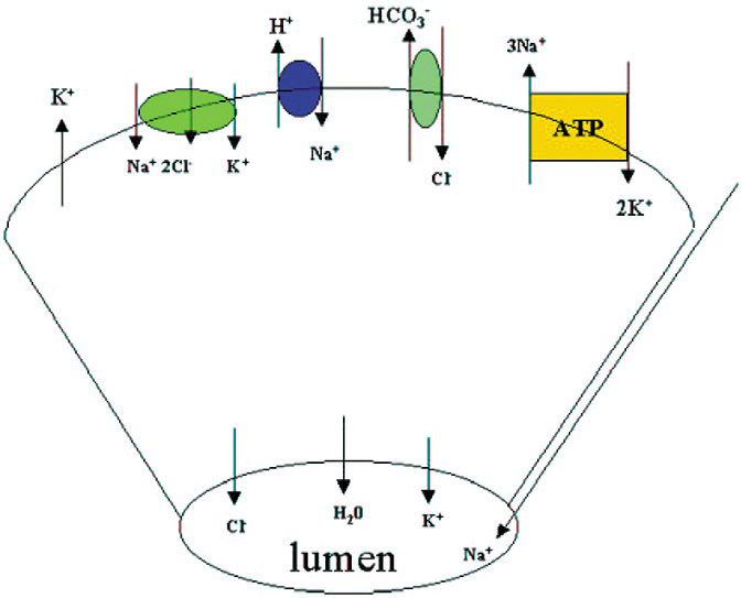
Along with protein, the lacrimal gland also secretes water and electrolytes. Acinar cells secrete fluid with an electrolyte composition similar to plasma. The increase in [Ca2+]i produced by cholinergic and α1-adrenergic agonists, as well as the increase in cAMP induced by VIP, activates K+ channels in the basal and lateral membranes and Cl channels in the apical membranes of the acinar cells (Fig. 5).83 The result is a net efflux of K+ and Cl into the lumen. Movement of these ions creates a negative potential difference causing Na+ to move into the lumen by a paracellular pathway through channels located between the cells.84 Water is then secreted into the lumen through water channels called aquaporins.85 The Na+/K+/Cl cotransporter, Cl/HCO3 exchangers, and Na+/H+ exchangers replace the Cl that exited the cell through the apical membrane. The increase in Na+ as a result of the Na+/K+/Cl cotransporter and Na+/H+ activates the Na+-K+-ATPase in acinar cell membranes. This latter transporter pumps Na+ out of the cell and K+ into the cell. Cl enters the cell as its transport is coupled to Na+ movement. Cl/HCO3 and Na+/H+ exchangers extrude HCO3 from the cell and are replaced by cellular carbonic anhydrase. As the fluid passes through the ducts, the ductal epithelial cells modify the fluid by secreting a fluid rich in KCl. The final fluid that exits onto the ocular surface is an isotonic solution containing Na+, K+, Cl, and HCO3 (Fig. 5).86

Fig. 5. Fluid secretion by rat lacrimal gland. Receptor activation stimulates K+ channels that extrude K+ across the basolateral membrane and Cl and K+ across the apical membrane into the lumen. Water then follows. Movement of Cl into the lumen drives cations from paracellular spaces into the lumen. Because of the decreased cellular concentrations of K+ and Cl, the Na+/K+/2Cl cotransporter becomes active to replace intracellular K+ and Cl. The Na+/H+ exchanger becomes active to extrude H+. The Cl/HCO3 exchanger drives Cl uptake. Na+-K+ ATPase becomes active to extrude Na+ and cause K+ re-uptake. (Reprinted from Hodges RR, Dartt DA: Regulatory pathways in lacrimal gland epithelium. Int Rev Cytol 231:129, 2003, with permission.)

Back to Top
EXCRETORY SYSTEM

LACRIMAL DRAINAGE SYSTEM

Anatomy

The lacrimal excretory system begins with a 0.3-mm opening on the medial portion of each eyelid, termed the punctum.3,87 Because of more rapid growth of the maxilla compared with the frontal bone during embryologic development, the lateral migration pulls the inferior canaliculus laterally.88 This results in the lower eyelid punctum to be located approximately 1.0 mm lateral to the upper eyelid punctum.89 Both puncta are directed posteriorly to appose the lacrimal tear lake. Causes such as horizontal eyelid laxity, disinsertion of the inferior retractors from the tarsus, cicatricial shortening of the anterior lamella of the medial eyelid, or trauma may interfere with the precise apposition of the puncta to the lacrimal lake, and thus affect normal functioning of the excretory system. The punctal opening widens into the ampulla, which is 2 mm in height and directed perpendicular to the eyelid margin, before making a sharp turn into the canaliculi. The ampulla, with a diameter of 2 to 3 mm, is the widest part of the proximal drainage system, and thus a common site for formation of dacryoliths.

The canaliculi measure 0.5 to 1.0 mm in diameter and run parallel to the eyelid margins. The superior canaliculus is shorter, measuring 8 mm in length, and the inferior canaliculus measures 10 mm (Fig. 6). The canaliculi are lined with 6 to 12 layers of nonkeratinizing, stratified squamous epithelium and surrounded by superficial pretarsal orbicularis oculi muscle as they traverse the medial eyelids and medial canthal region. The deep portion of the pretarsal orbicularis muscle (Horner-Duverney muscle, tensor tarsi, or pars lacrimalis) is important in lacrimal outflow. It passes posterior to the lacrimal sac and the posterior limb of the medial canthal tendon, and inserts onto the upper posterior lacrimal crest.3,87,90 The orbital septum inserts along or just posterior to the inferior posterior lacrimal crest. The deep head of the preseptal orbicularis oculi muscle (Jones' muscle) attaches to the lacrimal sac and lacrimal fascia (Fig. 7).91

Fig. 6. Approximate dimensions of the lacrimal excretory system. BE, bulla ethmoidalis; IT, inferior turbinate; MS, maxillary sinus; MT, middle turbinate.

Fig. 7. Relationship of the right medial canthal tendon to the nasolacrimal sac: A. Diagrammatic illustration. B. Anatomic dissection. The thick anterior limb of the medial canthal tendon wraps along the anterior upper half of the lacrimal sac to insert onto the anterior lacrimal crest, and the thin posterior limb passes behind the sac to insert onto the posterior lacrimal crest. The upper eyelid tarsus is much greater in height than the lower eyelid tarsus. IO, inferior oblique muscle origin; LCT, lateral canthal tendon; MCT-a, medial canthal tendon-anterior limb; MCT-p, medial canthal tendon-posterior limb; NLS, nasolacrimal sac; SN, sutura notha; T, tarsus.

The superior and inferior canaliculi merge to form a common canaliculus before entry into the nasolacrimal sac in more than 90% of individuals.3,92 In a study using digital subtraction dacryocystograms, the common canaliculus was present in 94% of lacrimal drainage systems. The upper and lower canaliculi join at the lacrimal sac wall without a common canaliculus in an additional 4%, with only 2% of systems having completely separate drainage of the upper and lower canaliculi into the lacrimal sac.93 The opening of the common canaliculus into the lacrimal sac is known as the common internal punctum, which should be free of any mucosal membrane or stricture for success of dacryocystorhinostomy surgery.

The functional valve between the common canaliculus and the lacrimal sac has traditionally been attributed to the valve of Rosenmüller, although some studies have been unable to document this structure.94 Tucker demonstrated that the canaliculi consistently angle posteriorly en route to the lacrimal sac.95 The canaliculi then bend at the canaliculus-common canaliculus junction at an angle of 118 degrees, before passing anteriorly to enter the lacrimal sac at an acute angle of 58 degrees. This consistent angulation may contribute to a valve-like effect that prevents retrograde flow of tears from the lacrimal sac.

The nasolacrimal sac and duct are portions of the same continuous structure. The lacrimal sac is lined by nonciliated columnar epithelium. The total sac measures 12 to 15 mm vertically and 4 to 8 mm anteroposteriorly, although it may be slightly larger in males.91 The fundus of the sac extends 3 to 5 mm above the medial canthal tendon, and the body of the sac measures 10 mm in height (Fig. 6).3 The lacrimal sac is ensheathed in the lacrimal fascia, which refers to the periorbital lining that splits at the posterior lacrimal crest into one layer that lines the fossa, and another layer that encases the lateral sac to reach the anterior lacrimal crest. Between the lacrimal fascia and the lacrimal sac there is a narrow space that contains a fine venous plexus draining into the angular or supraorbital vein.96

In addition, the lacrimal sac is wrapped by the thick anterior and thin posterior limbs of the medial canthal tendon. The structure of the medial canthal tendon has been described as having superior and inferior crura that arise from the tarsal plates. The crura combine to form the medial canthal tendon that then divides into a thicker anterior limb that wraps along the anterior upper half of the lacrimal sac before inserting onto the anterior lacrimal crest, and a very thin posterior limb that passes behind the sac to insert onto the posterior lacrimal crest (Fig. 7). The posterior limb helps maintain the position of the medial canthus in instances of traumatic or surgical disinsertion of the anterior limb. Anatomic dissections have also demonstrated a superior supporting branch of the tendon that courses superiorly to insert onto the frontal bone periosteum at the superomedial orbital rim.97 Below the anterior limb, the inferior portion of the lacrimal sac is bound anteriorly only by the orbital septum, which also inserts onto the anterior lacrimal crest. A distended sac may herniate initially through this thinner inferior septal lining.

The sac rests in the lacrimal sac fossa, with its medial aspect tightly adherent to the periosteal lining of the fossa. The lacrimal sac fossa is a depression in the inferomedial orbital rim, formed by the maxillary and lacrimal bones. It is bordered by the anterior lacrimal crest of the maxillary bone, and the posterior lacrimal crest of the lacrimal bone (Fig. 8). The fossa is approximately 16 mm high, 4 to 9 mm wide, and 2 mm deep.87,98 The fossa is widest at its base, where it is confluent with the opening of the nasolacrimal canal. The lower nasolacrimal fossa and the nasolacrimal duct are narrower in females, which may account for the female predominance of nasolacrimal obstruction.99

Fig. 8. Bony anatomy of the lacrimal sac fossa and medial orbital wall, right orbit. The anterior and posterior lacrimal crests are formed by the maxillary and lacrimal bones, respectively.

The nasolacrimal duct, measuring 3 to 4 mm in diameter, travels inferolaterally and slightly posteriorly in its bony course to the inferior turbinate for an interosseous distance of 12 mm (Fig. 9). The anterior, lateral, and posterior walls of the canal are formed by the maxillary bone, and the medial wall is formed by the lateral nasal wall and descending process of the lacrimal bone.100 The long axis of the duct and canal forms an angle of 15 to 25 degrees posterior to the frontal plane, or in a line connecting the medial commissure to the first molar tooth.87 The nasolacrimal duct terminates as a 5-mm extension into the inferior nasal meatus on the lateral wall or roof of the inferior meatus. The ostium is located, on average, 25 to 30 mm posterior to the lateral margin of the anterior nares, or 20 mm in children.87,101

Fig. 9. Anatomic dissection of the lacrimal drainage system within the bony wall between the nasal cavity and maxillary sinus. The nasolacrimal duct drains into the inferior meatus. C-i, inferior canaliculus; C-s, superior canaliculus; IM, inferior meatus; MS, maxillary sinus; NLD, nasolacrimal duct; NLS, nasolacrimal sac; NS, nasal septum.

When the nasolacrimal duct ostium is obstructed, the duct and sac distend with accumulated fluid. Distention of the intraosseous duct is limited to a diameter of 4 mm because of the rigid bony walls of the canal, with necessary compression of the lymphatic and venous plexi surrounding the duct. The lacrimal sac, in contrast, is not restricted by bone, and may expand laterally and anteriorly. Distention of the sac initially occurs inferiorly, because the medial canthal tendon surrounds the superior half of the sac.

The lacrimal drainage system histology transitions from a nonkeratinizing stratified squamous epithelium in the puncta and canaliculi to a dual columnar layer epithelium in the nasolacrimal sac. As the duct approaches the nasal cavity, it gradually shifts to mucoperiosteum with scattered mucus-secreting goblet cells.87 Multiple mucosal folds and sinuses have been reported within the lacrimal drainage system, and they may play a role in regulating tear drainage. The previously mentioned valve of Rosenmüller is located at the junction of the common canaliculus and sac, and the valve of Krause is located between the sac and duct. A mucosal flap, Hasner's valve (or plica lacrimalis), may be present at the opening of the duct into the inferior meatus of the nose.102

VASCULAR SUPPLY AND INNERVATION TO THE EXCRETORY SYSTEM

The vascular supply to the nasolacrimal drainage system is composed of contributions from the ophthalmic artery, angular artery, infraorbital artery, and internal maxillary artery. The ophthalmic artery courses anteromedially within the orbit after passing over the optic nerve to become the dorsal nasal artery. The dorsal nasal artery gives rise to the superior medial palpebral arteries that supply the lacrimal sac. The facial artery crosses the mandible diagonally and runs along the line of the nasojugal skinfold to become the angular artery. The angular artery passes deep to the orbicularis oculi muscle and superficial to the medial canthal tendon, with the angular vein immediately lateral to the artery (Fig. 10). Both vessels are located approximately 5 mm anteromedial to the anterior lacrimal crest, or 8 mm medial to the medial commissure of the eyelids.87 The angular artery then perforates the orbital septum above the medial canthal tendon to supply branches to the nasolacrimal sac and duct. The infraorbital artery mainly supplies the inferior sac and upper nasolacrimal duct. The inferior nasolacrimal duct receives its arterial supply from the nasal branch of the sphenopalatine artery, a branch of the internal maxillary artery.87,103 On the frontal process of the maxilla just anterior to the lacrimal sac fossa, a fine groove termed the sutura notha or sutura longitudinalis imperfecta of Weber, runs parallel to the anterior lacrimal crest (Fig. 8).87,96 Small twigs of the infraorbital artery pass through this vascular groove to supply the bone and nasal mucosa.

Fig. 10. The angular vein (AV) (blue) and artery (red) are located approximately 5 mm medial to the anterior lacrimal crest. FV, frontal vein; MCT, medial canthal tendon.

A fine venous plexus surrounds the nasolacrimal duct, which drains into the angular vein and infraorbital vein superiorly, and into the nasal cavity, the pterygoid plexus through sphenopalatine veins, and the internal maxillary vein inferiorly. Venous drainage continues down into the facial vein, or courses through the superomedial orbit to empty into the superior ophthalmic vein. Lymphatic flow drains into the submaxillary nodes or the deep cervical nodes.

Sensory innervation to the nasolacrimal sac is derived from the infratrochlear nerve, which is a terminal branch of the nasociliary branch of the ophthalmic trigeminal nerve. The inferior nasolacrimal duct is also innervated by the anterior superior alveolar branch of the maxillary trigeminal nerve.

EXCRETORY REGULATION AND THE TEAR PUMP MECHANISM

The amount of tears present on the ocular surface at any given time is a function both of secretion of the components that make up tears and the drainage and evaporation of tears. Although the regulation of tear production has been the subject of extensive studies, the regulation of tear drainage has only recently begun to be understood.

Tears secreted by the main and accessory lacrimal glands pass over the surface of the conjunctiva from lateral to medial to reach the puncta. The proper position of the eyelids against the globe and the position of the puncta to the tear lake are important for capillary attraction to draw the tears into the canaliculi. Malposition of the puncta, eyelids, or globe may interfere with this functional relationship.

Jones and Wobig104 popularized the lacrimal pump theory, which proposed that contraction of the pretarsal orbicularis muscle fibers during eyelid closure compresses and shortens the canaliculi, pumping tears toward the lacrimal sac. Furthermore, contraction of the deep head of the preseptal orbicularis muscle, which is attached to the lacrimal fascia and sac, results in simultaneous lateral movement of the lateral sac wall. This was thought to generate a negative pressure within the lacrimal sac that draws tears into the sac from the canaliculi.

In contrast, other anatomic and physiologic studies90,105–109 have found that a positive-pressure mechanism, rather than a negative-pressure mechanism, is responsible for the lacrimal pump during eyelid closure. Rosengren106 intubated the lacrimal sac through the nasolacrimal duct to measure pressures within the sac, and found that pressure in the lacrimal sac increases with eyelid closure and decreases with eyelid opening. Similarly, Ploman et al105 and Maurice107 measured positive pressures in the lacrimal sac during eyelid closure by intubation through the canalicular system.

Doane108 measured tear fluid movement during blinking by instilling carbon black into the tear film and taking high-speed photographs. He found that a pressure increase occurs in the canaliculi, and to a lesser extent in the nasolacrimal sac during eyelid closure. On eyelid closure, the puncta are occluded by the opposing eyelid, which prevents fluid regurgitation. Contraction of the pretarsal orbicularis oculi muscle compresses the ampulla and shortens the canaliculi, propelling tear fluid within the canaliculi toward the lacrimal sac.101,108 Simultaneously, contraction of the deep head of the preseptal orbicularis oculi muscle exerts lateral traction on the lacrimal sac wall, drawing fluid from the canaliculi into the lacrimal sac. He further theorized that as the tension increases on the lacrimal fascia to open the fundus of the sac, the inferior portion closes more tightly, preventing aspiration of air from the nose. As the eyelids open, the puncta initially remain closed by the opposing lid until the end of the opening movement when they suddenly pop apart. Tone in the superficial and deep heads of the pretarsal orbicularis muscle decreases, allowing the canaliculi to reopen and the lacrimal sac to close. The expansion of the canaliculi draws in a new flow of tears aided by capillary attraction. With relaxation of the deep head of the preseptal orbicularis muscle, elastic recoil of the lacrimal fascia collapses the lacrimal sac, expelling any fluid within the sac down into the now patent nasolacrimal duct.3 The influence of eyelid movement on tear flow ends as the tears move from the expandable lacrimal sac into the bony-surrounded nasolacrimal duct.108

The positive-pressure pumping of tears through the canaliculi appears to be important for successful drainage of tears after dacryocystorhinostomy, in which the positive- or negative-pressure mechanisms within the lacrimal sac are not applicable. Epiphora in the setting of pathologic facial paralysis or pharmacologic paralysis of the medial orbicularis oculi muscle with botulinum toxin also demonstrates the important contribution of the orbicularis oculi muscle to the lacrimal pump mechanism.110

A single blink may transport more tears than produced by basic secretion in 1 minute, suggesting that the lacrimal excretory system functions far below capacity.107 In other words, the lacrimal sac may not empty with each blink, but accumulates tear fluid until the volume is sufficient to bypass the resistance to drainage through the nasolacrimal duct and valve of Hasner. The volume of tears drained per blink has been measured to be 1.8 μL by Rosengren and approximately 2.0 μL by Sahlin and Chen.106,111 Although the majority of tears drain through the excretory system, 10% to 25% of secreted tear volume may be lost to evaporation under normal conditions.112 A small portion of the tear volume may be absorbed by the epithelial lining of the nasolacrimal sac and duct.56,113,114 Goblet cells and intraepithelial mucous glands located within the epithelium produce a mucous layer covering the distal excretory system.115 In a study by Chavis et al,113 no technetium tracer passed into the nose of one-third of subjects under basal secretion rates, suggesting that tear fluid may be absorbed within the excretory system before reaching the nasal cavity. More recently, it has been shown in an animal model that lipophilic substances are absorbed from the tear fluid by the epithelium of the nasolacrimal duct.116 Lacrimal drainage capacity has also been found to correlate to the blink rate when the nasolacrimal duct is in a horizontal position.111 Gravity may also contribute to increased lacrimal drainage.111 Other factors that assist in tear drainage include capillary attraction by the canaliculi, and the Venturi effect and Bernoulli's principle in the nasolacrimal duct, in which an increase in velocity of fluid flow occurs with a concomitant decrease in pressure.

Thale et al56 used histologic, immunohistochemical, and scanning electron microscopic techniques to demonstrate that the wall of the lacrimal sac is composed of collagen, elastic, and reticular fiber bundles arranged in a helical pattern. They proposed that the lacrimal sac distends and is pulled superolaterally because of its medial attachments when the lacrimal orbicularis muscle contracts with blinking. In addition, the helical collagen and elastic fibers encircling the sac may result in the sac being “wrung out” as another mechanism of lacrimal drainage.

A vascular plexus has been demonstrated surrounding and within the walls of the nasolacrimal sac, and filling the space between the nasolacrimal duct and its bony canal. This vascular plexus has specialized blood vessels similar to a cavernous body, and is thought to play a role in tear fluid absorption or regulation of tear outflow.10,56,117–119 In a histoanatomic study of the lacrimal drainage system, Thale et al found this plexus of large capacitance vessels to be continuous with the cavernous body of the inferior turbinate.56 By using anatomic dissection and electron microscopy, Paulsen et al118,119 found that the vascular plexus fills more than two-thirds of the bony canal, and hypothesized that swelling and shrinkage of the cavernous plexus regulates tear outflow by controlling the opening and closing of the lacrimal passage lumen. The cavernous plexus swells when the barrier arteries open and the throttle veins close, resulting in filling of the capacitance veins, swelling of the cavernous plexus, and subsequent closure of the lacrimal passage lumen. In contrast, closing the arteries and opening the throttle veins allow the collected blood in the capacitance veins to drain. This shrinks the cavernous plexus and increases the lumen of the excretory passageway. The blood vessels within the plexus have been found to be innervated, which suggests that arterial vasoconstriction and vasodilation by neural regulation may be a factor in the regulation of tear drainage.119,120 Ayub et al120 found a similar function to the vascular plexus in an experiment using an endoscope to observe the transit time of a fluorescein drop through the excretory system. However, they found that there was an ideal degree of cavernous plexus swelling that permitted optimal tear drainage under normal conditions. Excessive swelling of the vascular body would narrow the lacrimal passage lumen and decrease outflow. Similarly, significant shrinkage of the vascular body would dilate the lumen too much such that the mucosal folds of the lumen were too distant to enable optimal drainage.

The valve of Rosenmüller or the angulation of the canaliculi may prevent retrograde flow of fluid from the lacrimal sac into the canaliculi. The valve of Hasner at the nasal ostium prevents reflux upward from the nasal cavity into the nasolacrimal duct. As long as the pressure within the nose is less than that within the nasolacrimal duct, the valve remains open. When the intranasal pressure increases, such as during nose-blowing, Hasner's valve closes, preventing air or mucus in the nose from entering the nasolacrimal duct.

Incompetence of Hasner's valve is most often congenital or caused by retraction or fibrosis of the valve secondary to chronic inflammation and infection. In approximately 30% of neonates, a delicate membrane persists at the distal nasolacrimal duct, and usually undergoes spontaneous perforation within 6 months after birth. Once the ostium is patent, the residual membrane thickens to become the valve of Hasner. In 2% to 4% of infants, the ostium remains imperforate despite digital massage or nasolacrimal probing. In premature infants, approximately 70% have congenital nasolacrimal obstruction.101

Epiphora results from anatomic obstruction of the nasolacrimal passages, or from nonobstructive factors such as eyelid malposition, lacrimal pump insufficiency, tear film instability, or primary or reflex hypersecretion. Various tests are available that evaluate nasolacrimal patency, such as dacryocystography, computed tomography, nasal endoscopy, and lacrimal probing and irrigation. Dacryocystography, in which a radiopaque dye is injected into the excretory system and radiographs are taken of the transit of dye through the passages, may be helpful to define the presence and location of an anatomic obstruction from a mass, but is not performed under normal physiologic conditions. Physiologic tests for obstruction include the Jones dye test,3,121 fluorescein dye disappearance,122 saccharin taste test,123,124 and dacryoscintigraphy. Physiologic tests have the advantage of not requiring any instrumentation or injection that distends the lacrimal passageways from exogenous pressure. Therefore, functional rather than anatomic obstruction may be determined in the setting of normal physiologic conditions.

The primary dye test was described by Jones in 1962. In this procedure, 2% fluorescein was instilled into the tear film and a cotton pledget placed in the inferior meatus at 5 minutes was evaluated for presence of fluorescein. The fluorescein dye disappearance test described by Zappia and Milder122 measured the disappearance of fluorescein from the tear film as evidence of an intact drainage system. The saccharin test, initially described by Lipsius123 and revised by Hornblass,124 used a 1% saccharin solution in distilled water placed in the inferior cul-de-sac to determine functional patency of the lacrimal system. Ninety percent of patients tasted the sweet solution in the oropharynx by 15 minutes, although the test was ineffective in patients with tasting problems. The fluorescein appearance test125 was another physiologic test that used 2% fluorescein instilled in the cul-de-sac and visualized by ultraviolet light in the oropharynx. The oropharynx was examined every 5 minutes for 1 hour and fluorescein was seen by 30 minutes in 90%.

An elevated tear lake has been recognized in some studies to be a sign of lacrimal outflow obstruction.126,127 Measuring the tear meniscus level on slit-lamp examination, which is on average 0.2 mm in unobstructed systems and 0.6 mm in obstructed systems, or the ratio between two sides have been shown to be useful indicators of nasolacrimal obstruction.127

Back to Top
REFERENCES

1. Dutton JJ: The lacrimal systems. In Dutton J, ed. Atlas of Clinical and Surgical Orbital Anatomy. Philadelphia: WB Saunders, 1994: 140–142

2. Morton AD, Elner VM, Lemke BN, et al: Lateral extensions of the Müller muscle. Arch Ophthalmol 114:1486, 1996

3. Jones LT: An anatomical approach to problems of the eyelids and lacrimal apparatus. Arch Ophthalmol 66:111, 1961

4. Lemke BN, Lucarelli MJ: Anatomy of the ocular adnexa, orbit, and related facial structures. In Nesi FA, Lisman RD, Levine MR, Brazzo BG, Gladstone GJ, eds. Smith's Ophthalmic Plastic and Reconstructive Surgery, 2nd Edition. St. Louis: Mosby, 1998: 3–78

5. Fine BS, Yanoff M: Ocular Histology: A Text and Atlas, 2nd Edition. New York: Harper & Row, 1979

6. Iwamoto T, Jakobiec F: Lacrimal glands. In Duane WD, Racords RE, eds.: Biomedical Foundations of Ophthalmology, Vol 1, Chap 30. Philadelphia: Lippincott Williams & Wilkins, 1982

7. Egeberg J, Jensen OA: The ultrastructure of the acini of the human lacrimal gland. Acta Ophthalmol 47:400, 1969

8. Orzalesi N, Riva A, Testa F: Fine structure of human lacrimal gland: I. The normal gland. J Submicrosc Cytol 3:283, 1971

9. Weingeist TA: The glands of the ocular adnexa. Int Ophthalmol Clin 13:243, 1973

10. Duke-Elder S, Wybar KC: The anatomy of the visual system. In Duke-Elder S, ed. System of Ophthalmology. St. Louis: Mosby, 1961: 563–565

11. Seifert P, Spitznas M, Koch F, et al: Light and electron microscopic morphology of accessory lacrimal glands. Adv Exp Med Biol 350:19, 1994

12. Seifert P, Spitznas M: Demonstration of nerve fibers in human accessory lacrimal glands. Graefes Arch Clin Exp Ophthalmol 232(2):107, 1994

13. Seifert P, Stuppi S, Spitznas M: Distribution pattern of nervous tissue and peptidergic nerve fibers in accessory lacrimal glands. Curr Eye Res 16(4):298, 1997

14. Seifert P, Spitznas M: Vasoactive intestinal polypeptide (VIP) innervation of the human eyelid glands. Exp Eye Res 68(6):685, 1999

15. Walcott B: Anatomy and innervation of the human lacrimal gland. In Albert DM, Jakobiec F, Robinson N, eds.: Principles and Practice of Ophthalmology: Basic Sciences. Philadelphia: WB Saunders Co, 1994: 454–458

16. Ruskell GL: The orbital branches of the pterygopalatine ganglion and their relationship with internal carotid nerve branches in primates. J Anat 106:323, 1970

17. Ruskell GL: The distribution of autonomic post-ganglionic nerve fibers to the lacrimal gland in monkeys. J Anat 109:229, 1971

18. Ruskell GL: Distribution of pterygopalatine ganglion efferents to the lacrimal gland in man. Exp Eye Res 78(3):329, 2004

19. de Haas EBH: The response of the lacrimal gland to parasympathetic denervation. Ophthalmologica 141:46, 1961

20. Sears ML, Selker RG: Denervation supersensitivity of the lacrimal gland. Am J Ophthalmol 63:481, 1967

21. Hodges RR, Dartt DA: Regulatory pathways in lacrimal gland epithelium. Int Rev Cytol 231:129, 2003

22. Sullivan DA: Ocular mucosal immunity. In Ogra P, Metecky J, Lamm ME, eds. Handbook of Mucosal Immunology, 2nd Edition. Orlando: Academic Press, 1998: 1241

23. Sullivan DA, Wickham LA, Rocha EM, et al: Influence of gender, sex steroid hormones, and the hypothalamic-pituitary axis on the structure and function of the lacrimal gland. Adv Exp Med Biol 438:11, 1998

24. Sullivan DA: Immunology of the lacrimal gland and tear film. Dev Ophthalmol 30:39, 1999

25. Lemullois M, Rossignol B, Maudit P: Immunolocalization of myoepithelial cells in isolated acini of rat exorbital lacrimal gland: Cellular distribution of muscarinic receptors. Biol Cell 86:175, 1996

26. Hodges RR, Zoukhri D, Sergheraert , et al: Identification of vasoactive intestinal peptide receptor subtypes in the lacrimal gland and their signal transducing components. Invest Ophthalmol Vis Sci 38:610, 1997

27. Fullard R: Interactions of eyelids and tears in corneal wetting and the dynamics of the normal human eyeblink. Am J Ophthalmol 89:507, 1994

28. Jones LT: The lacrimal secretory system and its treatment. Am J Ophthalmol 62:47, 1996

29. Sjögren H, Kronning E: Keratoconjunctivitis sicca after partial extirpation of the palpebral lacrimal gland. Acta Ophthalmol 29:355, 1951

30. Elliot AJ: Lacrimal hyposecretion. Can Med Assoc J 79:371, 1968

31. Maitchouk DY, Beuerman RW, Ohta T, et al: Tear production after unilateral removal of the main lacrimal gland in squirrel monkeys. Arch Ophthalmol 118:246, 2000

32. Mauduit P, Jammes H, Rossignol B: M3 muscarinic acetylcholine receptor coupling to PLC in rat exorbital lacrimal acinar cells. Am J Physiol 264:C1550, 1993

33. Evans M, Marty A: Potentiation of muscarinic and alpha-adrenergic responses by an analogue of guanosine 5'-triphosphate. Proc Natl Acad Sci U S A 83:4099, 1986

34. Marty A, Evans M, Tan YP, Trautmann A: Muscarinic response in rat lacrimal glands. J Exp Biol 124:15, 1986

35. Meneray MA, Fields TY, Bennett DJ: Gs and Gq/11 couple vasoactive intestinal peptide and cholinergic stimulation to lacrimal secretion. Invest Ophthalmol Vis Sci 38:1261, 1997

36. Meneray MA, Fields TY: G protein coupling of receptor activation to lacrimal secretion. Adv Exp Med Biol 438:133, 1998

37. Godfrey PP, Putney JW Jr: Receptor-mediated metabolism of the phosphoinositides and phosphatidic acid in rat lacrimal acinar cells. J Biochem 218(1):187, 1984

38. Morris AP, Gallacher DV, Irvine RF, Petersen OH: Synergism of inositol trisphosphate and tetrakisphosphate in activating Ca2+-dependent K+ channels. Nature 330(6149):653, 1987

39. Kwan CY, Takemura H, Obie JF, Thastrup O, Putney JW Jr: Effects of MeCh, thapsigargin, and La3+ on plasmalemmal and intracellular Ca2+ transport in lacrimal acinar cells. Am J Physiol 258:C1006, 1990

40. Berridge MJ: Inositol trisphosphate and calcium signaling. Nature 361:315, 1993

41. Dartt DA, Dicker DM, Ronco LV, et al: Lacrimal gland inositol trisphosphate isomer and inositol tetrakisphosphate production. Am J Physiol 259:G274, 1990

42. Putney JW Jr, Huang Y, Bird GS: Calcium signaling in lacrimal acinar cells. Adv Exp Med Biol 438:123, 1998

43. Putney JW Jr, Broad LM, Braun FJ, et al: Mechanisms of capacitative calcium entry. J Cell Sci 114:2223, 2001

44. Bird GS, Rossier MF, Hughes AR, et al: Activation of Ca2+ entry into acinar cells by a non-phosphorylatable inositol trisphosphate. Nature 352(6331):162, 1991

45. Sundermeier T, Matthews G, Brink PR, Walcott B: Calcium dependence of exocytosis in lacrimal gland acinar cells. Am J Physiol Cell Physiol 282:C360, 2002

46. Nishizuka Y: The role of protein kinase C in cell surface signal transduction and tumor promotion. Nature 308:693, 1984

47. Zoukhri D, Hodges RR, Sergheraert C, Dartt DA: Protein kinase C isoforms differentially control lacrimal gland functions. Adv Exp Med Biol 438:181, 1998

48. Nishizuka Y: Intracellular signaling by hydrolysis of phospholipids and activation of protein kinase C. Science 1992;258(5082):607, 1992

49. Newton AC: Protein kinase C: Structure, function, and regulation. J Biol Chem 270(48):28495, 1995

50. Gromada J, Jorgensen TD, Dissing S: Role of protein kinase C in the regulation of inositol phosphate production and Ca2+ mobilization evoked by ATP and acetylcholine in rat lacrimal acini. Pflugers Arch 429:578, 1995

51. Zoukhri DR, Hodges RR, Sergheraert C, Toker A, Dartt DA: Lacrimal gland PKC isoforms are differentially involved in agonist-induced protein secretion. Am J Physiol 272(1 Pt 1):C263, 1997

52. Zoukhri DR, Hodges RR, Dicker DM, Dartt DA: Role of protein kinase C in cholinergic stimulation of lacrimal gland protein secretion. FEBS Lett 351:67, 1994

53. Zoukhri DR, Hodges RR, Willert S, Dartt DA: Immunolocalization of lacrimal gland PKC isoforms. Effect of phorbol esters and cholinergic agonists on their cellular distribution. J Membr Biol 157(2):169, 1997

54. Tepavcevic V, Hodges RR, Zoukhri DR, Dartt DA: Signal transduction pathways used by EGF to stimulate protein secretion in rat lacrimal gland. Invest Ophthalmol Vis Sci 44(3):1075, 2003

55. Zoukhri DR, Hodges RR, Sergheraert C, Dartt DA: Lacrimal gland functions are differentially controlled by protein kinase C isoforms. Ann N Y Acad Sci 842:217, 1998

56. Thale A, Paulsen F, Rochels R, et al: Functional anatomy of the human efferent tear ducts: A new theory of tear outflow mechanism. Graefes Arch Clin Exp Ophthalmol 236:674, 1998

57. Bradley ME, Peters CL, Lambert RW, Yiu SC, Mircheff AK.: Subcellular distribution of muscarinic acetylcholine receptors in rat exorbital lacrimal gland. Invest Ophthalmol Vis Sci 31(5):977, 1990

58. Matsumoto T, Kobayashi T, Kamata K: Phosphodiesterases in the vascular system. J Smooth Muscle Res 39(4):67, 2003

59. Dartt DA, Donowitz M, Joshi VJ, Mathieu RS, Sharp GW: Cyclic nucleotide-dependent enzyme secretion in the rat lacrimal gland. J Physiol 352:375, 1984

60. Mauduit P, Herman G, Rossignol B: Newly synthesized protein secretion in rat lacrimal gland: Post-second messenger synergism. Am J Physiol 253(4 Pt 1):C514, 1987

61. Hodges RR, Zoukhri D, Lightman JP, Dartt DA: Identification and cellular localization of the components of the VIP signaling pathway in the lacrimal gland. Adv Exp Med Biol 38:169, 1998

62. Jahn R, Padel U, Porsch PU, Soling HD: Adrenocorticotropic hormone and alpha-melanocyte-stimulating hormone induce secretion and protein phosphorylation in the rat lacrimal gland by activation of a cAMP-dependent pathway. Eur J Biochem 126(3):623, 1982

63. van der Kraan M, Adan RA, Entwistle ML, et al: Expression of melanocortin-5 receptor in secretory epithelia supports a functional role in exocrine and endocrine glands. Endocrinology 139(5):2348, 1998

64. Akbulut S, Byersdorfer CA, Larsen CP, et al: Expression of the melanocortin 5 receptor on rat lymphocytes. Biochem Biophys Res Commun 281(5):1086, 2001

65. Thorig L, Van Haeringen NJ, Timmermans PB, Van Zwieten PA.: Peroxidase secretion from rat lacrimal gland cells in vitro. I. Alpha-adrenergic stimulation in the absence of alpha-adrenoceptors. Exp Eye Res 37(5):475, 1983

66. Dartt DA: Regulation of inositol phosphates, calcium, and protein kinase C in the lacrimal gland. Prog Retin Eye Res 13:443, 1994

67. Mauduit P, Herman G, Rossignol B: Protein secretion induced by isoproterenol or pentoxifylline in lacrimal gland: Ca2+ effects. Am J Physiol 246(1 Pt 1):C37, 1984

68. Meneray MA, Fields TY: Adrenergic stimulation of lacrimal protein secretion is mediated by G(q/11)alpha and G(s)alpha. Curr Eye Res 21(2):602, 2000

69. Zoukhri D, Dartt DA: Cholinergic activation of phospholipase D in lacrimal gland acini is independent of protein kinase C and calcium. Am J Physiol 268(3 Pt 1):C713, 1995

70. Hodges RR, Dicker DM, Rose PE, et al: Adrenergic and cholinergic agonists use separate signal transduction pathways in lacrimal gland. Am J Physiol 262:G1087, 1992

71. Dartt DA, Hodges RR, Zoukhri D: Signal transduction pathways activated by cholinergic and alpha 1-adrenergic agonists in the lacrimal gland. Adv Exp Med Biol 438:113, 1998

72. Hodges RR, Dicker DM, Dartt DA: Role of protein kinase C in alpha 1-adrenergic and cholinergic agonist stimulated protein secretion. Adv Exp Med Biol 350:147, 1994

73. Chen L, Johansson J, Hodges RR, et al: Differential effect of the EGF family of growth factors on protein secretion, MAPK activation, and intracellular calcium concentration in rat lacrimal gland. Exp Eye Res 80:379, 2005

74. Tzahar E, Yarden Y: The ErbB-2/HER2 oncogenic receptor of adenocarcinomas: From orphanhood to multiple stromal ligands. Biochem Biophys Acta 1377(1):M25, 1998

75. Arango ME, Li P, Komatsu M, et al: Production and localization of Muc4/sialomucin complex and its receptor tyrosine kinase ErbB2 in the rat lacrimal gland. Invest Ophthalmol Vis Sci 42(12):2749, 2001

76. Keely SJ, Calandrella SO, Barrett KE: Carbachol-stimulated transactivation of epidermal growth factor receptor and mitogen-activated protein kinase in T(84) cells is mediated by intracellular ca(2+), PYK-2, and p60(src). J Biol Chem 275(17):12619, 2000

77. Kanno H, Horikawa Y, Hodges RR, et al: Cholinergic agonists transactivate the EGFR and stimulate MAPK to induce goblet cell secretion. Am J Physiol Cell Physiol 284:C988, 2003

78. Ota I, Zoukhri D, Hodges RR, et al: Alpha 1-adrenergic and cholinergic agonists activate MAPK by separate mechanisms to inhibit secretion in lacrimal gland. Am J Physiol Cell Physiol 284(1):C168, 2003

79. Singh J, Sharkey KA, Lea RW, Williams RM: Effects of neuropeptides on serotonin release and protein and peroxidase secretion in the isolated rat lacrimal gland. Adv Exp Med Biol 438:145, 1998

80. Sullivan DA, Kelleher RS, Vaerman JP, Hann LE: Androgen regulation of secretory component synthesis by lacrimal gland acinar cells in vitro. J Immunol 145(12):4238, 1990

81. Walcott B, Moore L, Birzgalis A, et al: A model of fluid secretion by the acinar cells of the mouse lacrimal gland. Adv Exp Med Biol 506(Pt A):191, 2002

82. Stern ME, Gao J, Siemasko KF, et al: The role of the lacrimal functional unit in the pathophysiology of dry eye. Exp Eye Res 78:409, 2004

83. Lechleiter JD, Dartt DA, Brehm P: Vasoactive intestinal peptide activates Ca2(+)-dependent K+ channels through a cAMP pathway in mouse lacrimal cells. Neuron 1988;1(3):227, 1988

84. Nauntofte B: Regulation of electrolyte and fluid secretion in salivary acinar cells. Am J Physiol 263(6 Pt 1):G823, 1992

85. Raina S, Preston GM, Guggino WW, et al: Molecular cloning and characterization of an aquaporin cDNA from salivary, lacrimal, and respiratory tissues. J Biol Chem 270:1908, 1995

86. Mircheff A: Water and electrolyte secretion and fluid modification. In Albert DM, Jakobiec F, Robinson N, eds.: Principles and Practices of Ophthalmology: Basic Sciences. Philadelphia; WB Saunders Co, 1994: 466–472

87. Whitnall SE: The Anatomy of the Human Orbit and Accessory Organs of Vision. New York: Oxford University Press, 1932

88. Singh J, Djali PK, Adeghate E: Immunohistochemistry and secretory effects of leucine enkephalin in the isolated pig lacrimal gland. Adv Exp Med Biol 438:157, 1998

89. Hurwitz JJ: Embryology of the lacrimal drainage system. In Hurwitz JJ, ed.: The Lacrimal System. Philadelphia: Lippincott-Raven, 1996: 9–13

90. Ahl NC, Hill JD: Horner's muscle and the lacrimal system. Arch Ophthalmol 100:488, 1982

91. Lemke BN, Della Rocca RC: Surgery of the Eyelid and Orbit: An Anatomical Approach. Norwalk: Appleton and Lange, 1990: 90–114

92. Lemke BN: Lacrimal anatomy. Adv Ophthalmol Plast Reconstr Surg 3:11, 1984

93. Yazici B, Yazici Z: Frequency of the common canaliculus: A radiological study. Arch Ophthalmol 118(10):1381, 2000

94. Schaeffer JP: The genesis and development of the nasolacrimal passages in man. Am J Anat 13:1, 1912

95. Tucker NA, Tucker SM, Linberg JV: The anatomy of the common canaliculus. Arch Ophthalmol 114:1231, 1996

96. Shinohara H, Taniguchi Y, Kominami R, et al: The lacrimal fascia redefined. Clin Anat 14(6):401, 2001

97. Anderson RL: Medial canthal tendon branches out. Arch Ophthalmol 95:2051, 1977

98. Bailey JH: Surgical anatomy of the lacrimal sac. Am J Ophthalmol 6:665, 1923

99. Groessl SA, Sires BS, Lemke BN: An anatomical basis for primary acquired nasolacrimal duct obstruction. Arch Ophthalmol 115:71, 1997

100. Whitnall SE: The naso-lacrimal canal: The extent to which it is formed by the maxilla, and the influence of this upon its caliber. Ophthalmoscope 10:557, 1912

101. Cassady JV, Cassady JR: Lacrimal drainage problems in infancy and childhood. J Pediatr Ophthalmol 1:27, 1964

102. Aubaret E: The valves of the lacrymo-nasal passages (Les Replis valvulaires des canalicules et du conduit lacrymo-nasal). Arch d'Ophthal 28:211, 1908

103. Jones LT, Wobig JL: Surgery of the Eyelids and Lacrimal System. Birmingham, AL: Aesculapius, 1976

104. Jones LT, Wobig JL: Congenital anomalies of the lacrimal system. In Jones LT, Wobig JL: Surgery of the Eyelids and Lacrimal System. Birmingham, AL: Aesculapius, 1976: 157–73

105. Ploman K, Engel A, Knutsson F: Experimental studies of lacrimal passageways. Acta Ophthalmol 6:55, 1928

106. Rosengren B: On lacrimal drainage. Ophthalmologica 164:409, 1972

107. Maurice DM: The dynamics and drainage of tears. Int Ophthalmol Clin 13:103, 1973

108. Doane MG: Blinking and the mechanics of the lacrimal drainage system. Ophthalmol 88:844, 1981

109. Becker BB: Tricompartment model of the lacrimal pump mechanism. Ophthalmol 99:1139, 1992

110. Sahlin S, Chen E, Kaugesaar T, et al: Effect of eyelid botulinum toxin injection on lacrimal drainage. Am J Ophthalmol 129:481, 2000

111. Sahlin S, Chen E: Gravity, blink rate, and lacrimal drainage capacity. Am J Ophthalmol 124:758, 1997

112. Mishima S: Some physiologic aspects of the precorneal tear film. Arch Ophthalmol 73:233, 1965

113. Chavis RM, Maisey MN, Whelham RA: Quantitative lacrimal scintillography. Arch Ophthalmol 96:2066, 1978

114. Sisler HA: Current concepts in the understanding of lacrimal drainage. Adv Ophthalmic Plast Reconstr Surg 3:25, 1984

115. Paulsen FP: The human nasolacrimal ducts. Adv Anat Embryol Cell Biol 170III-XI:1, 2003

116. Paulsen F, Föge M, Thale A, et al: Absorption of lipophilic substances from tear fluid by the epithelium of the nasolacrimal ducts. Invest Ophthalmol Vis Sci 43:3137, 2002

117. Paulsen F, Thale A, Kohla G, et al: Functional anatomy of human lacrimal duct epithelium. Anal Embryol 198:1, 1998

118. Paulsen FP, Thale AB, Hallmann UJ, et al: The cavernous body of the human efferent tear ducts: Function in tear outflow mechanism. Invest Ophthalmol Vis Sci 41(5):965, 2000

119. Paulsen FP, Hallmann UJ, Paulsen J, Thale A: Innervation of the cavernous body of the human efferent tear ducts and function in tear outflow mechanism. J Anat 197:177, 2000

120. Ayub M, Thale AB, Hedderich J, et al: The cavernous body of the human efferent tear ducts contributes to regulation of tear outflow. Invest Ophthalmol Vis Sci 44(11):4900, 2003

121. Zappia RJ, Milder B: Lacrimal drainage function: 1. The Jones fluorescein test. Am J Ophthalmol 74:154, 1972

122. Zappia RJ, Milder B: Lacrimal drainage function: 2. The fluorescein dye disappearance test. Am J Ophthalmol 74:160, 1972

123. Lipsius EI: Sodium saccharin for testing the patency of the lacrimal passages. Am J Ophthalmol 41:320, 1956

124. Hornblass A: A simple taste test for lacrimal obstruction. Arch Ophthalmol 90:435, 1973

125. Flach A: The fluorescein appearance test for lacrimal obstruction. Ann Ophthalmol 11:237, 1979

126. Vicks V, Holds JB, Hartstein ME, et al: Tarsal strip procedure for the correction of tearing. Ophthal Plast Reconstr Surg 2004;20:37, 2004

127. Burkat CN, Lucarelli MJ: Tear meniscus level as an indicator of nasolacrimal obstruction. Ophthal Plast Reconstr Surg 2005 (in press)

Back to Top