Chapter 31
Functional Anatomy of the Extraocular Muscles
HOWARD M. EGGERS
Main Menu   Table Of Contents

Search

EMBRYOLOGY
GROSS ANATOMY
MICROSCOPIC ANATOMY
FUNCTION
REFERENCES

EMBRYOLOGY
The pattern of human eye muscle development is similar to that in elasmobranchs, reptiles, birds, marsupials, and the cat.1–5 A pattern of six extraocular muscles and motor nuclei is present in all vertebrates.6,7 The similarity in muscular architecture and neuronal organization of the extraocular muscles in all vertebrates reflects a highly conserved developmental pattern.

Functionally, the muscles are organized into three pairs that are closely aligned with the axes of the semicircular canals, allowing congruence of sensory and motor frames of reference.8,9 In mammals below primates, an additional retractor bulbi system translates the eye posteriorly to provide a protective reflex in conjunction with the nictitating membrane. This system is reduced to an accessory lateral rectus muscle in primates and is absent in humans.10

The extraocular muscles are composites of connective and muscle tissue that have embryologic origins in different locations. In the vertebrate head, connective tissue and smooth muscle is formed from the neural crest, an ectodermal structure, rather than from mesoderm, as occurs in the trunk. Neural crest cells migrate from their origin in the neural fold and some invade the paraxial mesoderm, where they appear to play a patterning role and contribute to the development of the condensations of mesoderm that give rise to the extraocular muscles.

Mammalian head paraxial mesoderm contains a series of seven hills, called somitomeres,11 demarcated on the surface by shallow furrows. The eighth somitomere is the first somite. Somitomeres give rise to both muscles and connective tissue for the entire head.12 In human embryos of 14 somites (25 days) a pair of premandibular mesodermal condensations may be seen that give rise to the eye muscles innervated by the oculomotor nerve (superior, medial, and inferior recti, and inferior oblique). Two separate tissue masses in the maxillomandibular mesoderm give rise to the lateral rectus and superior oblique. These three primordial condensations on each side of the head (premandibular and the two maxillomandibular) are homologous to the premandibular, mandibular, and hyoid head cavities of lower vertebrates.1 The eye muscle primordia cannot be assigned to segments or metameres, and there is little evidence that the vertebrate head has a metameric organization.

The connective tissue, and not the myogenic precursors, determines the exact site where the muscles form. The mesoderm that gives rise to the eye muscles must have both proper spatial positioning for recognition by the appropriate motor nerve and correct positioning with respect to the eye. It appears that periocular crest cells acquire patterning information appropriate for the site from the optic vesicle.

The myoblasts that give rise to the extraocular muscles are thought to arise from a population of cells that migrates from the somitomere to the presumptive eye muscle region before muscle fibers are formed. The eye muscles form from superior and inferior mesenchymal condensations.13,14 The superior condensation gives rise to the superior rectus, superior oblique, levator, and upper half of medial and lateral recti; the inferior condensation gives rise to the inferior rectus and oblique muscles and the lower half of the medial and lateral recti.

Primary fibers containing the different types of myosin heavy chain form in the absence of innervation. In chick limb muscle, fibers containing all three types of myosin heavy chain (fast, slow, and mixed fast and slow) exist in the muscle before innervation. The primary fibers are replaced with a new population of secondary myoblasts, containing the myosin type of the final muscle.15,16 There are thus two stages of myogenesis.17 In the first, primary myoblasts in populations containing different types of myosin heavy chain are guided to the muscle site by neural crestderived connective tissue. In the second phase, proper formation of secondary muscle fibers requires interaction between nerve and muscle.

The motor axon terminal induces the postjunctional membrane in the muscle to form aggregates of proteins: acetylcholine receptors and acetylcholinesterase. A protein, agrin, is synthesized in the cell body of motor neurons and is transported to the nerve terminals, where it is found in an active form bound to the basal lamina of the nerve terminal. Agrin is thought to mediate the motor neuron–induced aggregation of synaptic proteins at the neuromuscular junction.18

It has traditionally been said that the muscles differentiate from the apex of the orbit forward, whereas the sclera simultaneously differentiates in the opposite direction.3 An alternative view has been put forward that the individual muscles develop concurrently along their entire length.13 The differentiation of myoblasts to mature fibers is dependent on innervation by motor nerves.19 Motor neuron cytogenesis in the brain stem extraocular muscle nuclei undergoes a caudal to rostral gradient that is concurrent with, but independent of, muscle differentiation.20-22 At about 1 month, the nerves to the extraocular muscles reach their respective destinations in the sequence oculomotor, abducens, and trochlear. There is initially an overproduction of axons in the motor nerve, followed by a thinning out. This pattern may correlate with loss of polyneuronal innervation or death of excess motor neurons.23,24 An excess of nerve fibers in a small area containing primordia of several extraocular muscles may also relate to occasional anomalous innervation of muscles.25

The trochlea begins to form at 6 weeks of age. The levator palpebrae arises by delamination from the superior rectus during the seventh week, but separation is not complete until the end of the fourth month. Muscle cell striations are recognizable early in the second month; at the end of the second month, strands of long cells have formed.

At about 3 months, the muscle cells have become enveloped by collagen fibers, representing the earliest fascia, and condensations of vascular and perineural adventitia are seen. Connective tissue septa develop a little later; they are formed by the end of the fourth month and continue to enlarge through the second trimester along with the muscle fascia.

The orbital mesenchyma differentiates last.26 Islands of capillary beds develop in the areas of loose mesenchyme between connective tissue septa, and in the fifth month adipose cells arise about these beds, thus completing the overall structural plan of the orbit. By 6 months the muscles are in their final positions. During the last trimester, all the existing tissues and structures enlarge further.

The differentiation of the orbital connective tissue starts later than that of the orbital walls or eye muscles. It is linked with the development of mesenchymal condensations at about 3 months and with the formation of capillary beds at about 4 months, around which the orbital fat develops. The orbital connective tissue attains its adult configuration by 6 months.

Prenatal studies in monkeys have shown that the global mutiplyinnervated, tonically contracting muscle fiber type develops first.27 The chief proprioceptive sensory structure of the eye muscle (the palisade endings) involves this fiber type. Thus, early maturation would allow sensory feedback to be used in muscle development. The last fiber type to mature is the orbital, singly innervated twitch fiber. This fiber contains a rich supply of mitochondria and depends on aerobic metabolism. It is chronically active in its role of positioning the orbital pulley (vide infra).

The growth of the muscle tendons in width and location is linear with time during fetal development.28 The tendon insertions initially extend from the limbus to the equator. With time, the tissue underlying the tendon degenerates, freeing it from the sclera and more clearly defining the exact location of the insertion. The insertion moves farther posterior to the limbus and the anterior segment of the globe enlarges. The muscle insertions continue to change until 18 months to 2 years after birth.13 Infants with esotropia show no anomalies of the medial and lateral rectus tendons, and no correlation exists between the angle of strabismus and the locations of muscle insertion.13

Postnatally, there is further development and refinement in extraocular muscle anatomy as well as changes in the motor nerve (e.g., myelination and distribution of fiber diameters).29 In the rat, postnatal differentiation of fiber types in the orbital layer of the muscle precedes that in the global layer.30 In the cat, the order is reversed.31,32 The changes that occur involve fiber size, maturation of nerve terminals, vascularity, ultrastructural features, histochemistry, and the composition of myosin.

In the primate, the orbital layer twitch fiber shows continued postnatal development of mitochondrial aggregates for up to 6 months.30 This continued development parallels improvements in eye rotation range and fixation stability.

Postnatal muscle development can be disturbed by abnormal visual experience. In cats, lid-sutured eyes have shown decreases in muscle fiber cross-sectional area, oxidative enzyme activity, and capillary density compared with the normal side.34 The concomitant functional alterations are reduced speed of contraction and reduced fatigue resistance.35 In lid-sutured rats, a 15% reduction in the mean fiber size of the trochlear nerve has been found, suggesting an effect on the postnatal development of the extraocular motor neurons.36

In one study, expression of 13 known genes was examined using cDNA microarray in rats raised in the dark to postnatal age of 45 days.37 There was a higher expression level of genes involving tissue remodeling and lower for a gene marker for cellular de-differentiation. Isometric contractile properties were affected. Twitch and tetanic tension were maintained, but twitches were slowed and the muscles fatigued more easily. Reponse at higher stimulation frequencies (over 200 Hz) was significantly lower. Visual experience is thus necessary for development of the normal phenotype.

Extraocular muscle continues to express molecules that are normally expressed in development.38 Studies of rabbit extraocular muscle have shown that there is a population of activated satellite cells (muscle stem cells) that divide and provide daughter myoblasts that fuse with existing adult extraocular muscle myofibers. Labeled nuclei are randomly distributed along the muscle length and are located at the sarcolemmal periphery (characteristic of growth and repair). This may allow regulation of the total number of sarcomeres. Satellite cells in skeletal muscle are activated by injury or exercise. Extraocular muscle appears to have a constant level of satellite cell activation. Local control of satellite cell activation, movement, and turnover is likely.

Back to Top
GROSS ANATOMY

Each eye lies within a bony, roughly cone-shaped cavity in the head, called the orbit. The medial walls of the orbit are approximately parallel to the midsagittal plane, and the lateral walls form an angle of approximately 90 degrees with each other. The medial and lateral orbital walls thus form an angle of approximately 45 degrees with each other. Each orbital axis therefore diverges from the midline by about 22.5 degrees and from the axis of the other orbit by about 45 degrees (Fig. 1)39

Fig. 1. Geometry of the orbits. (Modified from Duke-Elder S, Wybar KC: System of Ophthalmology. St. Louis, Mosby, 1961.)

MUSCLES

There are six extrinsic extraocular muscles—four rectus and two oblique—that attach to and rotate the eye. They are arranged anatomically and functionally in three pairs.40–45 Each of the six muscles attaches at one end to the sclera and at the other end to the bony orbit. Five of these muscles have their origin at the orbital apex; the sixth (inferior oblique) originates at the front of the orbit. A seventh muscle, the levator palpebrae superioris, elevates the upper lid but plays no role in positioning the eye; it will not be directly discussed further. The four rectus muscles—superior, inferior, medial, and lateral—are approximately 40 mm in length and have a width six times their thickness. They arise from a fibrous ring at the orbital apex, the anulus of Zinn, and course forward, unconstrained by connective tissue, as progressively flat strips in a cone-shaped configuration, attaching to the sclera several millimeters posterior to the limbus.46–59 Exact insertion points have been measured in a number of studies; some of the results are excerpted in Table 1. More recently, results of a large and complete series were similar to those of the older studies for the parameters that were measured (see Fig. 3). It shows that the old series measured from the anterior limbal edge to the middle of the insertion. The tendons vary in length: medial 4 mm, inferior and superior 6 mm, and lateral 9 mm. The insertions vary somewhat in shape and location (Table 1, Fig. 2). The distance of the insertions behind the limbus increases in the sequence medial, inferior, lateral, and superior rectus. This progression is known as the spiral of Tillaux (Fig. 3)60

Table 1. Mensuration of Human Extraocular Muscles*
Click here to view Table 1


Fig. 2. Variability of the extraocular muscle insertions. C, corneal limbus; E, equator; Int, medial rectus; VS, superior rectus; Ex, lateral rectus; V Inf, inferior rectus. (Howe L: The Muscles of the Eye. New York, GP Putnam, 1907.)

Fig. 3. Location of average muscle insertions with respect to the corneal limbus, based on measurements in 100 autopsy eyes. Dimensions are in millimeters. IR, superior rectus; MR, medial rectus; IR, inferior rectus; LR, lateral rectus. (Apt L: An anatomical reevaluation of rectus muscle insertions. Tr Am Ophth Soc 78:365, 1980.)

The anulus of Zinn is oval in cross section and overlies the optic foramen and the medial portion of the superior orbital fissure (Fig. 4). The lower portion of the ring (tendon of Zinn) is attached to the inferior root of the lesser wing of the sphenoid below the optic foramen and gives rise to the inferior rectus and part of the medial and lateral recti. The attachment site may be developed into a small tubercle, the infraoptic tubercle. The upper portion of the ring (tendon of Lockwood) attaches to the body of the sphenoid and bridges over the superior orbital fissure; where it attaches to the greater wing of the sphenoid, a bony spur may occur (spina recti lateralis). Through the anulus pass the optic nerve, ophthalmic artery, abducens nerve, upper and lower divisions of the oculomotor nerve, and nasociliary branch of the ophthalmic nerve.

Fig. 4. Structures at the orbital apex, showing muscle insertions into the anulus of Zinn and the location of various vessels and nerves. (Housepian EM: Intraorbital tumors. In Schmidek HH, Sweet WH (eds): Current Techniques in Operative Neurosurgery. New York, Grune & Stratton, 1977.)

The two oblique muscles approach the eye from the front.61,62 The superior oblique muscle arises from the frontoethmoidal suture, superomedial to the anulus of Zinn and the origin of the medial rectus. It courses forward along the junction of the medial orbital wall and roof; after passing through the pulley, or trochlea (Fig. 5), it is redirected posteriorly and inserts in the lateral, posterior, superior octant of the globe (Fig. 6), passing between the superior rectus muscle and the globe. The superior oblique muscle is more rounded or fusiform than the other extrinsic muscles. The inferior oblique muscle arises behind the lower orbital margin on the orbital plate of the maxilla and proceeds in a direct path to its insertion in the lateral, posterior, inferior octant of the globe. It passes below the inferior rectus. The neurovascular bundle to the inferior oblique muscle courses forward along the lateral border of the inferior rectus and enters the oblique muscle at its midportion. Associated with it is strong connective tissue such that when the muscle is disinserted and transposed forward to insert next to the interior rectus, the neurovascular bundle appears to form a new point of origin for contraction of the inferior oblique, inducing a depressing action.63Mensuration data for the six oculorotary human muscles are shown in Table 1. Table 2 lists the locations of the origins and insertions in a three-dimensional coordinate system. The x-axis is positive laterally, the y-axis positive anteriorly, and the z-axis positive superiorly. The origin is at the center of the eye. These measurements were made usingvarying techniques, and the differences among the sets of measurement are of interest. These coordinates are useful in quantitative calculations of the directions of muscle pull and the three-dimensional cooperation of the extraocular muscles. Figure 7 shows the positions of the extraocular muscles when plotted from such data. Orbital pulleys (vide infra) determine the functional origins of the muscles, and similar coordinates can be provided for each pulley. These coordinates can be determined by analysis of orbital magnetic resonance images obtained with the eye in specified directions. The pulley is at a point that is relatively invariant in position as the eye assumes various directions of gaze.


Table 2. Coordinates of the Points of Origin and Insertion of the Extraocular Muscles for Primary Position of the Eye


  Insertion
Muscle Coordinates* Origin Fick49 Ruete54 Volkntann50 Fick49 Ruete54 Volkmann56
Medial rectus X -18 -14.67 -17 -9.1 -9.9 -9.65
  Y -30 -32 -31.29 +7.9 +6 +7.55
  Z +4 0 +0.6 0 0 0
Inferior rectus X -17 -10.8 -16 0 +2.2 0
  Y -30 -32 -33.05 +7.9 +5.77 +6.73
  Z +2 -4 -2.4 -9.1 -10 -10.24
Lateral rectus X -15 -5.4 -13 +9.1 +10.8 +10.08
  Y -31 -32 -35.29 +7.9 +5.67 +6.34
  Z +2 0 +0.6 0 0 0
Superior rectus X -16 -10.67 -16 0 +2 0
  Y -31 -32 -33.05 +7.9 +5 +5.21
  Z +6.5 +4 +3.6 +9.1 +10 +10.48
Superior oblique X -19.6 -14.1 -15.27 +4.6 +2 +2.9
  Y +10.9 +10 +6.95 -2.7 -3 -5.70
  Z +12.8 +12 +12.25 +9.9 +11 +11.05
Inferior oblique X -18 -8.1 -11.1 +10.4 +8 +8.71
  Y   +6 +10.05 -6 -9 -8.47
  Z   -15 -15.46 0 0 0

*Coordinates are in millimeters from the left of eye in the rectilinear Cartesian coordinate system. X is positive laterally, Y is positive to the front, and Z is positive upward.
von Helmholtz H: Handbuch der Physiologischen Optik. 3rd ed. Hamburg, 1910.


Fig. 5. Scanning electron micrograph of human trochlea, left orbit from the front. The tendon has been sectioned (arrowheads). The tendon is surrounded by fibrovascular tissue and supported by a crescent-shaped plate of cartilage underneath. The tendon is about 3 mm in diameter, and the plate of cartilage is about 6 mm broad and 4 mm long. The cartilage is attached to the fovea trochlearis of the frontal bone by fibrous tissue to form a tube through which the tendon slides. The tendon from here to its insertion is about 20 mm in length. (Courtesy of Dr. Eugene Helveston.)

Fig. 6. Posterior surface of a right eye showing average insertions of the oblique muscles. Dimensions are given in millimeters. SO, superior oblique; IO, inferior oblique; LR, lateral rectus; EQ, equator. (Modified from Fink WH: Surgery of the Oblique Muscles of the Eye. St. Louis, Mosby, 1951.)

Fig. 7. Projection of the positions of the extraocular muscles onto a horizontal plane. Dimensions, to scale, are from measurements in rectilinear three-dimensional coordinates (see Table 2, Ruete's figures). The oblique muscles have nearly the same plane of action. (Modified from Hering E: The Theory of Binocular Vision. New York, Plenum Press, 1977.)

The eye muscles grow with the eye and orbit in early childhood.65–67 The lengths in infancy and at age 1 year are listed in Table 3. Lengths in adults are the same within measurement error. Note that in Figure 8, which shows an infant eye and an adult eye superimposed, the angular proportions stay the same. Eye muscle lengths increase by about the same proportion (40% to 50%). Thus the percent contraction of each muscle stays the same for a given size eye movement, and it seems probable that the central innervation patterns need only minor adjustments to compensate for growth.


Table 3. Growth in Length of the Rectus Muscles with Age (mean ± standard deviation)


  Age
Rectus Muscle Premature Infant 1 year 20–30 years 40–50 years 60–83 years
Medial 19 22.6 ± 1.3 27.0 ± 1.4 36.2 ± 5.3 35.8 ± 5.6 38.4 ± 5.1
Inferior 16 17.8 ± 2.6 28.0 ± 8.0 36.0 ± 5.8 37.2 ± 7.3 34.8 ± 3.1
Lateral 22 24.8 ± 1.7 30.5 ± 1.9 39.6 ± 4.1 40.2 ± 4.8 41.2 ± 6.7
Superior 20 23.5 ± 1.7 33.3 ± 3.5 44.2 ± 4.4 43.8 ± 5.4 41.8 ± 5.1

Mühlendyck H: Wachstum und Lange der ausseren Augenmuskeln. Ber Dtsch Ophthalmol Ges 75:449, 1978.


Fig. 8. Growth of the eye and extraocular muscles based on measurements of the adult (Volkmann)55 and infant (Schneller66) Dimensions are given in millimeters. Note how angular relationships are preserved even though lengths change. (Modified from Mühlendyck H: Wachstum und Lange der ausseren Augenmuskeln. Ber Dtsch Ophthalmol Ges 75:449, 1978.)

The monkey is commonly used as a model for human anatomy and physiology.68 The orbit in the monkey is somewhat different in shape from that in humans; consequently the muscles have qualitatively different primary position actions. The monkey orbital axis is 2 degrees more lateral and points upward by 15 degrees, compared to 1 degree downward in humans. Monkey horizontal recti have a significant torsional action, unlike those in humans Monkey vertical recti ack the adducting action present in humans. In the primary position, the oblique muscles of the monkey have a more vertical than torsional action.

BLOOD SUPPLY

The extraocular muscles are supplied by the muscular branches of the ophthalmic artery, which usually occur as two main stems and a varying number of smaller twigs.44,45 Further blood supply may arise from the lacrimal and infraorbital arteries. The venous drainage of the extraocular muscles is into the superior and inferior orbital veins.

The lateral (or upper) muscular branch of the ophthalmic artery supplies the lateral and superior rectus, superior oblique, and levator muscles. The levator may also receive twigs from the supraorbital artery. The medial (or inferior) muscular branch, the larger of the two, supplies the medial and inferior recti and the inferior oblique. The infraorbital artery sends a branch to the inferior oblique. The lacrimal artery sends branches to the lateral and superior recti.

The extraocular muscles have fibers in two layers: orbital and global. Each contains different types of muscle fibers (vide infra). The orbital layer has continually contracting, mitochondria-rich fibers that use aerobic metabolism; therefore, the blood supply to the orbital layer is greater than that to the global layer. The orbital layer thus has more vessels per unit of area, more vessels per muscle fiber, and more vascular lumenal area than the global layer.69

The anterior ciliary arteries, which supply blood to the anterior segment of the eye, pass through the rectus muscles and penetrate the sclera at the muscle insertions. There are two anterior ciliary arteries in each rectus except the lateral, which has only one. The anterior ciliary arteries merely pass through the rectus muscles and do not supply blood to these muscles. Anterior segment ischemia may result from interruption of the anterior ciliary arteries; a clinical rule of thumb is to avoid surgery on more than two rectus muscles at a time to minimize this risk.

INNERVATION

The third cranial nerve (oculomotor) divides within the orbit into superior and inferior branches. The superior division innervates the superior rectus and levator palpebrae. The inferior division innervates the inferior and medial recti and the inferior oblique. The fourth (trochlear) nerve innervates the superior oblique, and the sixth (abducens) cranial nerve innervates the lateral rectus.44,45

The nerves enter the rectus muscles from within the muscle cone at approximately the junction of the middle and posterior thirds of the muscle (for the superior, medial, and inferior recti) and just posterior to the middle (for the lateral rectus). The superior oblique is supplied by its nerve in several branches that enter the anterior half of the posterior third of the muscle, on its orbital surface. The inferior oblique is supplied at about the middle of its posterior border.

ORBITAL TISSUES

The orbital tissues of importance for the extraocular muscles are Tenon's capsule70 and the system of connective tissue septa that span the orbit. The literature contains numerous descriptions of the orbital connective tissue and Tenon's capsule based on gross dissection or injection of preserved specimens. These techniques tend to produce nonexistent fascial planes and ligaments. Whitnall's study of Tenon's capsule in fresh postmortem preparations has given us one of the most accurate descriptions of this structure.44

Tenon's Capsule

Tenon's capsule, or the fascia bulbi, is a layer of delicate connective tissue that completely envelops the globe from the limbus to the optic nerve, forming a potential space free of any tissue attachments that might restrict globe rotation. Posteriorly, it is loosely attached by extremely fine connective tissue filaments to the sclera. This episcleral space contains no fluid or endothelium and is not a full-fledged ball-and-socket joint in function. The globe can rotate some within the capsule; however, beyond small rotations, the globe and capsule move together, deflecting the orbital fat attached behind. This is to be expected, since the capsule is attached firmly to the eye at the limbus and to the muscles at their points of entrance.

The anterior one third of the capsule lies anterior to the rectus muscle insertions and is thinner and more adherent to the globe, fusing to the sclera at the corneoscleral limbus. On its outer surfaces, Tenon's capsule in front is loosely adherent to the conjunctiva and can be readily separated from it. In contrast, the posterior capsule is firmly attached to the orbital fat and connective tissue meshwork.

Tenon's capsule is penetrated by the optic nerve, the extraocular muscles, and the blood vessels and nerves serving the globe. About the optic nerve the capsule is penetrated and broken up by multiple posterior ciliary arteries and nerves, and some have considered it to cease a few millimeters away from the optic nerve. There is no sheath continuing backward around the optic nerve.

The rectus muscles pierce Tenon's capsule anterior to the equator of the globe and pick up sleevelike extensions of the capsule. The intracapsular portion of the rectus muscles is 7 to 10 mm in length. From the point of capsular penetration to the insertion, each muscle has fascial expansions from its edges that attach to the overlying Tenon's capsule, forming falciform folds,72 and insert into the sclera lateral and posterior to the muscle insertion. Small attachments between the undersurface of the muscle and the sclera just behind the muscle insertion are called footplates. Outside Tenon's capsule, in their posterior one third, the muscles have only a very thin sheath. Closer to Tenon's capsule the muscle sheaths become thicker and more opaque and, for a distance of 10 to 12 mm, give off lateral expansions that connect between adjacent muscles to form an intermuscular membrane. The intermuscular membrane has only a limited extent and is not found more than a short distance behind the globe.

The medial and lateral rectus muscle sheaths give off attachments from their external surfaces that are known as ailerons or check ligaments. These arise from the muscle sheath anterior to the point of muscle passage through the capsule and insert on the zygomatic tubercle (for the lateral rectus) and on the lacrimal bone behind the lacrimal crest (for the medial rectus). The muscles do not move within their sheaths so that these attachments limit extreme rotations of the eye; their severance or fibrosis can free the rotation even more or can restrict it. In addition to “checking” rotation, these attachments help to anchor the whole fascial apparatus in position.

The orbital fat is prevented from attaching to the globe by Tenon's capsule. Sometimes Tenon's capsule is perforated during surgery and the orbital fat has been sufficiently traumatized that adhesions can form between the fat and the globe. Such an adhesive syndrome significantly impairs eye rotations. Penetration of Tenon's capsule can release fat as forward as 10 mm behind the limbus.

The sheath of the superior rectus blends with that of the overlying levator palpebrae superioris and assists synergic action between the two muscles. This explains the apparent or “pseudo” ptosis that accompanies a hypotropia. The forward expansion of the inferior rectus passes from the muscle undersurface above the inferior oblique and inserts between the tarsus and orbicularis oculi muscle. This mechanical linkage helps to lower the lid on downgaze. It also explains a change in lid fissure width after inferior rectus surgery. Resections of this muscle tend to raise the lower lid and narrow the lid fissure, whereas recessions tend to widen the lid fissure by lowering the lid. Each rectus muscle sends connective tissue expansions that attach to and retract the conjunctival cul-de-sac. When the eye moves, this attachment prevents the conjunctiva from dragging over the cornea.72

Pulleys

The structure and organization of the orbital connective tissue have long been neglected. Koornneef, using a thick section histologic technique,73 has demonstrated the consistent architecture of this system.74–76 Periocular and retro-ocular orbital spaces are filled with fat pads organized as adipose tissue space, surrounded by collagenous strands and septa. The septa are 0.3 mm or more in thickness and subdivide the orbital volume. The strands consist of large, meshed, collagenous tissue containing fibroblasts and collagen fibers. Anteriorly, the septa are radial around the globe and connect the periorbita to Tenon's capsule and the intermuscular membrane (Figs. 9 and 10).

Fig. 9. Connective tissue septa of the anterior orbit about the globe. 1, periorbita; 2, connective tissue strands; 3, intermuscular membrane; 4, Tenon's capsule. Asterisks indicate areas where smooth muscle cells were found. (Koornneef L: Details of the orbital connective tissue system in the adult. Acta Morphol Neerl Scand 15:1, 1977.)

Fig. 10. Orbital connective tissue septa. The diagrams represent cross sections perpendicular to the orbital axis. A. Near orbital apex. B. Just posterior to globe. C. Slightly anterior to the posterior surface of the globe. D. Approximately at the ocular equator, slplsr, superior levator palpebra–superior rectus complex; Irm, lateral rectus muscle; mrm,medial rectus muscle; irm, inferior rectus muscle; som, superior oblique muscle; iom, inferior oblique muscle; mm, Müller's muscle of the orbit; on, optic nerve. (Koornneef L: The architecture of the musculo-fibrous apparatus in the human orbit. Acta Morphol Neerl Scand 15:35, 1977.)

Sleeves or pulleys anchor the positions of the rectus muscles near and posterior to the equator of the eye.77,78 The pulleys lie within Tenon's fascia and are composed of dense collagen bands woven at right angles and supported from the orbital walls by elastin and smooth muscle bundles.79 These bands determine the functional origin of the muscles and have consistent locations across all subjects. Coronal plane coordinates have a 95% confidence interval of less than 1 mm.80 The muscles telescope within the pulley, sliding on thin collagenous sheets (Figs. 11 and 12).

Fig. 11. Extraocular tissue architecture, anterior view. Serial sections of human orbit, stacked and reassembled by computer. View from front. Green, sclera; Red, muscle; Pink, smooth muscle; Blue, collagen; Orange, elastin. Note heavy elastin and smooth muscle concentration along the inferior half of the globe, constituting part of Lockwood's ligament. (Miller JM, Demer JL, Poukens V et al:Extraocular Tissue Architecture H7, privately published Quicktime VR scene. © 2002 Joel M. Miller.)

Fig. 12. Extraocular tissue architecture, medial view. Serial sections of human orbit, stacked and reassembled by computer. Green, sclera; Red, muscle; Pink, smooth muscle; Blue, collagen; Orange, elastin. Note broad band of elastin and smooth muscle stretching from medial orbit to cross-over of the inferior oblique and the inferior rectus, lying within the region of Lockwood's ligament. Equatorially distributed smooth muscle probably is involved in stabilizing the pulleys. (Miller JM, Demer JL, Poukens V et al: Extraocular Tissue Architecture H7, privately published Quicktime VR scene. © 2002 Joel M. Miller.)

The smooth muscle is innervated by projections from the superior cervical ganglion using norepinephrine, from the pterygopalatine ganglion using nitroxidergic innervation, and from the pterygopalatine or ciliary ganglion using acetylcholine. 83 Innervation of the smooth muscle anchoring the pulleys may have a functional role of maintaining or correcting pulley location. Magnetic resonance imaging (MRI) studies have shown that the pulleys have stable locations with varying directions of gaze, generally shifting less than a millimeter transversely in a coronal plane with eye rotations. Excessive shifts in pulley location result in incomitant strabismus.83 The inferior oblique and inferior rectus pulleys are mechanically coupled via Lockwood's ligament. Smooth muscle in the pulley suspension runs from the medial to the inferior rectus pulley, constituting Muller's peribulbar muscle.

Each extraocular muscle has two layers: orbital and global (vide infra). The orbital layer inserts on the pulleys so that muscle contraction will translate the pulley posteriorly. The orbital layer of the inferior rectus inserts on the lateral rectus pulley and the inferior oblique pulley. Partial coupling of the inferior rectus with the inferior oblique pulley moves its axis with vertical gaze. The orbital layer of the inferior oblique inserts on the inferior rectus pulley, translating it nasally with contraction of the inferior oblique. The orbital layer of the superior oblique inserts on a dense collagenous sheath posterior to the trochlea. The sheath goes through the trochlea and then inserts on the nasal border of the superior rectus pulley, pulling it nasally when the superior rectus contracts.84

The so-called check ligaments and the suspensory ligament of Lockwood are part of this system. Postoperative fibrotic changes in these septa are important in restricting ocular motility. Lockwood's ligament is an expansion of the inferior rectus sheath to the surrounding septa to form a suspensory hammock for the globe.

The orbital connective tissue constrains the position of the eye muscles. The rigidity of the resulting musculo-orbital coupling determines how far the muscles shift with eye movements and consequently plays a role in determining the exact eye movement resulting from a change in muscle innervation. Thus, when the eye looks up, the horizontal rectus muscles move upward only in the portion distal to an anchoring point near the ocular equator. This preserves the purely horizontal movement produced by the horizontal rectus muscles and limits cross-coupling of the action of one muscle to another.

Back to Top
MICROSCOPIC ANATOMY

CONNECTIVE TISSUE

The connective tissue surrounding the extraocular muscles is more delicate than that around ordinary skeletal muscles. The muscle fibers are distributed in a capillary-rich, loose connective tissue. The capillaries in extraocular muscle are of the closed type, with the bidirectional transfer of macromolecules achieved by transendothelial vesicular transport.85

Adjacent to the surface of each muscle fiber (cell) is a layer about 50-nm thick that stains as a mucopolysaccharide. This, in turn, is surrounded by a delicate reticulum of argyrophilic (collagenous) fibers (Fig. 13) and elastin fibers (Fig. 14).86,87 The fibers are uniformly distributed, and the muscle is penetrated by a few intramuscular septa (the internal perimysium) that consist of collagenous fibers and contain many nerves and blood vessels. Argyrophilic fibers occur at the muscle fiber–connective tissue junction. The internal perimysium is continuous with the external muscle sheath or capsule (external perimysium or epimysium). Between the muscle fibers are numerous nerve fibers. Capillary vessels are most numerous on the outer, orbital surface of the muscle, which contains the smaller muscle fibers that are constantly active. Blood flow measurements in the cat show that the average flow in the six extraocular muscles is greater than that in any skeletal muscles and exceeded only by that in the myocardium.

Fig. 13. Cross section of human extraocular muscle showing argyrophilic reticulin fibers ensheathing each muscle fiber. (Valu L: Über die normale Struktur und Alternenderungen der Bindegewebs fasern der äussern Augenmuskeln. Albrecht von Graefes Arch Klin Exp Ophthalmol 169:272, 1966.)

Fig. 14. Cross section (A) and longitudinal section (B) of human extraocular muscle showing elastin fibers. (Valu L: Über die normale Struktur und Alternen-derungen der Bindegewebs fasern der aussern Augenmuskeln. Albrecht von Graefes Arch Klin Exp Ophthalmol 169:272, 1966)

The muscle sheath is very thin for 2 cm or so from the muscle origin.44,45 On approaching the back of the globe, a thicker, opaque sheath that contains inner longitudinal fibers and outer circumferential fibers develops. The outer layer fuses with Tenon's capsule.

MUSCLE FIBERS

Eye muscle histology is a subject vexed by confusion and controversy. This is partly due to interspecies variation in the eye muscles but also to difficulty in making correlations between structure and function. The basis for understanding the extraocular muscle fiber types is the work that has been done on skeletal muscle. With the use of a variety of techniques—physiological,88 histochemical,89,90 biochemical,91,92 immunocytochemical,93–95 and ultrastructural 93,96–98—three or four fiber types have been delineated in skeletal muscle. When studied in detail, the extraocular muscle fibers are not exactly like those of any skeletal muscle.

In broad outline, extraocular muscle shares with skeletal muscle a spectrum of twitch fibers, differing in rapidity of contraction and in fatiguability (“white” versus “red”). In addition, there are tonically contracting fibers, which do not occur at all in skeletal muscle. Skeletal muscles differ one from another in the proportion of fiber types. In contrast, except for one fiber type, all extraocular muscles are similar in fiber type composition. Like skeletal muscle, all fibers innervated by one axon are of the same type.

Light Microscopy

A gross muscle contains clusters of adjacent parallel muscle cells that are delimited by connective tissue septa. Such a cluster is a fasciculus. The muscle cells, which are also called fibers, are roughly cylindrical, multinucleated cells bounded by a plasma membrane. The ends fuse with the inelastic tissue of the muscle tendon.

The extrinsic eye muscles have structural features not found in limb muscle, and the microscopic appearance is therefore somewhat different from that of ordinary skeletal muscle. This is not surprising in view of the unique functional abilities of the extraocular muscles—constancy of activity, rapidity, and fine gradation of contraction. Extraocular muscles have a lower innervation ratio, greater vascularity,99 and a looser connective tissue envelope, rich in elastic fibers,86,87 than are found in skeletal muscle.

Normal extraocular muscle has features that in skeletal muscle are associated with myopathy. The fiber shape and size are variable, and there are more small fibers.100 There is usually a mild mononuclear inflammatory cell infiltration. Compared with skeletal muscle, there are more fibrous connective tissue and internal nuclei. Thus, ordinary myopathy and neuropathy cannot be distinguished in extraocular muscles using conventional morphologic criteria.101 Furthermore, individual fibers may show changes, observable by light or electron microscopy, which in skeletal muscle are considered pathologic. Such changes are whorled fibers, ringbinden fibers, vacuoles within myofilament bundles, subsarcolemmal inclusions, sarcomere disruption, nemaline rods, smearing of Z lines, mitochondrial clumping, mitochondrial abnormalities, zebra bodies, and Hirano bodies.102 These findings occur in children as well as in adults.

The early histologic literature contains evidence of more than one fiber type in extraocular muscle. Fibers were distinguished by diameter, content of sarcoplasm, size and location of nuclei, fibrils, and presence of hypolemmal rings. Thus, in 1910, Cilimbaris103 noted clear fibers with few nuclei and little sarcoplasm, dark fibers with many nuclei and plentiful sarcoplasm, and intermediate types. Thulin,104 in 1914, described three muscle types in human and monkey extraocular muscle. The first contained irregular fibrils of varying size (i.e., Felderstruktur). Furthermore, he mentioned that this type had nerve terminals of a special structure (which he did not describe). A second type showed abundant cytoplasm with a regular arrangement of fibrils (i.e., Fibrillenstruktur). The third type had a fibrillar, cross-striated appearance but also had hypolemmal, circularly arranged fibrils. These are now known as ringbinden fibers and probably represent a degenerative change.

Kato, in 1938,51 distinguished two zones within extraocular muscle, differing in cell size and staining reactions: an inner layer next to the eye itself (the global layer) and an outer, orbital layer. Sometimes an additional intermediate zone can be distinguished. In rectus muscles the orbital layer is C-shaped. The oblique muscles are more circular in cross section and have a concentric arrangement of orbital and global layers. The intermediate zone is conceived of as having an admixture of cell types from the orbital and global layers. It is now known that in the rectus muscles each layer has a different insertion.105 The orbital layer inserts on a connective tissue pulley that constrains the muscle path in the orbit, and the global layer inserts on the globe itself. The global layer extends the entire length of the muscle and through the pulley without inserting. The orbital layer does not extend as far because it inserts on the pulley, located in the equatorial region of the eye.106

Siebeck and Kruger, in 1955,107 clearly identified two types of muscle fiber in extraocular muscle on the basis of light microscopic appearance and introduced the names Fibrillenstruktur and Felderstruktur. Functionally, Fibrillenstruktur fibers are similar to the twitch fibers of ordinary skeletal muscle and Felderstruktur fibers to tonically contracting muscle, most studied in amphibians. The muscle fibers (cells) are arranged longitudinally, many fibers probably running most of the length of the muscle,108 although there have been reports of fibers terminating or arising along the course of the muscle.108-110 Most muscles have fibers that are shorter than the muscle length.111 Cross sections of the muscle at various points along its length reveal a varying proportion of different fiber sizes.110,112–114 In humans, the global layer contains cells with a mean diameter about 50% greater than that of the orbital layer.

Serial sections show fibers to be most numerous in mid orbit.106 Mean numbers across four human specimens, ranging in age from 17 months to 93 years, were approximately 5,900 for the medial, 5,600 for the lateral, and about 4,000 for the vertical recti. In an older study of human inferior rectus muscles, a predominance of small fibers was found proximally95; in rabbits, a predominance was found in the distal part of the muscle.109

The fiber numbers also differ by layer. The human rectus muscle global layers all have approximately 12,000 fibers. The orbital layers vary from 37% to 49% of the total fiber count. Thus the orbital layer has a little less than half the fibers. The global layer is expected to be more uniform because the globe has the same inertia and viscosity in all directions. The orbital layers insert on the orbital connective tissue (pulleys) which couples to the orbital wall, equatorial Tenon's capsule, and adjacent extraocular muscles with elastin, collagen, and smooth muscle. The fiber counts vary in proportion to the connective tissue load constituted by each pulley.

Serial sections of rabbit rectus muscles have been performed114 and show marked, systematic variation of fiber structure and, in some fibers, of diameter, with position along the fiber; this has not been shown in the mouse.113 Relatively little is known about short fibers, which may occur at either end of the muscle, although they may be muscle stem cells (satellite cells).38,113–117 Such variation could mean that all the large fibers do not travel the full length of the muscle, that the fibers taper, or that the fibers split into several smaller branches—as observed by Hines109 in rabbits at either end of the muscle. Lockhart and Brandt108 be lieved the fibers extended the entire length of the muscle in human eyes, as did Cooper and Daniel.115 This is significant because if some fibers do not run full length, a cross section of the muscle will not show every fiber, and the number and proportion of fiber types determined at one point may be invalid elsewhere in the muscle.

By using plastic resins for embedding, it is possible to section the tissue thin enough to obtain adequate resolution to count the small muscle fibers accurately. In cat inferior oblique muscle, Alvarado and van Horn110 found the greatest number of fibers in the middle third of the muscle, the count dropping toward either end of the muscle. Some fibers terminated several millimeters short of the ends. The longest fibers ran the entire length of the muscle. A significant number of small fibers terminate within the muscle. Other departures from a simple parallel arrangement have been found in animals—branching of muscle cells near their ends in the rabbit109 and sheep117 and myomyous junctions between short cells in the cat,118,119 rat,120,121 and sheep.117 A similar arrangement of fibers exists in skeletal muscle.

In human material, Voss112 found only small fibers at the muscle end but larger fibers as well in the typical layered arrangement toward the middle. However, he did not do quantitative fiber counts along the length of the muscle. In gross serial sections of the orbit, Nakagawa53 found that the rectus muscle area was maximum at the level of the posterior surface of the globe (5 mm behind the posterior surface of the globe for the lateral rectus), and the muscles tapered uniformly anterior and posterior to the zone of maximum area. Muscle cell branching and myomyous junctions have not been reported in human material.

In cross section, fibers in the eye muscle are not as tightly packed together as those in skeletal muscle.100 In skeletal muscle, the fibers are arranged in compact bundles and each fiber is polygonal in cross section. There is a scant amount of connective tissue between fibers, but thick septums penetrate between bundles. In the extraocular muscles each cell is separated enough from its neighbors to be approximately round in shape and to have its own fine connective tissue sheath. There are few large septa between fibers, and the interfiber spaces contain a multitude of nerve fibers.

Light and electron micrographs of muscle show a typical cross-striated pattern formed by a registration of sarcomeres in adjacent myofibrils (Fig. 15). The transverse striations are due to the arrangement of filaments into alternating isotropic (I) and anisotropic (A) (i.e., doubly refractile) bands. The I band is transversely bisected by a dark Z line. The sarcomere varies in length from about 1.5 to 3.5 μm depending on the amount of shortening of the muscle.

Fig. 15. Electron micrograph of a twitch fiber in human extraocular muscle. This fiber shows less development of the sarcoplasmic reticulum and T system than the cell in Figure 17. Mitochondria occur in rows between the myofibrils. This cell would be classified as a “red” twitch fiber. The diagram below the electron micrograph shows how the filaments produce the banding pattern. (Courtesy of Dr. T. Iwamoto.)

The I band contains only thin filaments 5 to 6 nm thick and approximately 1 μm in length composed of three proteins: actin, troponin, and topomyosin. The A bands contain thicker (1.1 nm) filaments,, which are about 1.5 μm in length and are composed of the protein myosin. The thick and thin filaments overlap within the A band, where they are linked by crossbridges; a central zone free of thin filaments in the center of the A band is called the H zone (Hensen's stripe). An inconsistent feature is a line centered within the H zone, the M line (Mittelscheibe); its presence depends on the species.

Fiber Types

The variety of fiber types is bewildering, all the more so considering the difficulty in drawing correspondences among various species. There are two basic classes of muscle cells in extraocular muscle: singly innervated (twitch, conducted action potential) and multiply innervated (tonic, graded contraction, no conducted action potential). The different types of muscle fiber can be classified by their location within the muscle (orbital versus global layer), innervation (single or multiple), metabolism (oxidative versus glycolytic), and speed of contraction (fast or slow twitch versus sustained, graded contraction). A cross section of extraocular muscle shows a layered organization, in which a layer of smaller diameter cells (5 to 15 μm) is on the orbital surface (orbital layer), and the bulk of the muscle adjacent to the globe (global layer) contains larger cells of 10 to 40 μm diameter 51,100,101,112 (Table 4). The muscle cells of various diameters may have many other differing structural features, and the muscle fibers may be grouped into several classes on the basis of structure and function (Table 5).


Table 4. Average Fiber Diameters in Human Extraocular Muscle*


Age Diameter (μm)
Third trimester 2.5–5.0
Newborn 7.5
  8.6 (2.0)
  7.5 (4–11)
3 months 8.1 (1.9)
1 year 11.8 (2.9)
3 years 11.5 (6–19)
  12.3 (3.8)
Adult 15 (9–23)
  17.5


*Multiple values from various studies. Numbers in parentheses are either range or standard deviation.
Engel WK, Brooke MH: Muscle biopsy as a clinical diagnostic aid. In Fields WS (ed): Neurological Diagnostic Techniques, pp 90–146. Springfield, IL, Charles C Thomas, 1966.


Table 5. Correspondence of Human Muscle Fiber Type Properties
Click here to view Table 5


The two basic functional types of muscle fiber were first observed in the frog as fast and slow contractile systems by Sommerkamp,122 and the two morphologic patterns were described and named by Kruger.123 Ordinary light microscopy shows two anatomic patterns of the contractile fibrils within the muscle fibers that correspond to the two innervation patterns. The fibrils in singly innervated fibers are arranged in discrete bundles separated by sarcoplasmic reticulum (Fibrillenstruktur). Multiply innervated fibers have a poorly developed sarcoplasmic reticulum and irregular, larger myofibrils that are partly fused (Felderstruktur). In hematoxylin-eosin stained light micrographs, Fibrillenstruktur fibers stain palely and have a finely stippled appearance, regular spacing of fibrils, and mostly peripherally located nuclei. Felderstruktur fibers stain more darkly and have irregularly clumped fibrils that are not clearly separated; their nuclei are central as well as subsarcolemmal.

In addition to light microscopy, electron microscopy and histochemical reactions are used as an aid in classification. Through testing the activity of various metabolic enzymes, it is possible to obtain a better estimate of the functional capabilities of a cell.

The overall plan of organization of fiber types is not as complex as it first seems if one does not too closely compare detailsof morphology across species. Table 6 shows that, in fact, there is considerable agreement among species in the broad outlines of fiber architecture. All the muscles listed have similar fiber arrangements (Table 7). Each muscle has two layers. The outer or orbital surface layer contains small-diameter, singly innervated twitch fibers and small-diameter, multiply innervated tonic fibers, both containing many mitochondria. Morphologically, these fibers are capable of prolonged contractile activity without fatigue. The inner or global layer of muscle contains a spectrum of three types of singly innervated twitch fibers (“red,” “intermediate,” and “white,” according to mitochondrial content, development of sarcoplasmic reticulum, and diameter) and a “white,” multiply innervated tonic fiber. If the global, red twitch fiber is considered to be the same as the similar cell in the orbital layer, there are only five types of fiber. The orbital twitch fiber is distinct, however, in the extreme extent to which its morphology is affected by botulinum toxin chemodenervation.124


Table 6. Fiber Types in Extraocular Muscle
Click here to view Table 6

Table 7. General Schema of Mammalian Extraocular Muscle Fiber Architecture


Layer Morphology
Orbital 1) Small, singly innervated twitch fiber, many mitochondria (red)
  2) Small, multiply innervated tonic fiber, many mitochondria (red)
Global 3) Small, singly innervated twitch fiber, many mitochondria (red)
  4) Medium, singly innervated twitch fiber, moderate mitochondria (intermediate)
  5) Large, singly innervated twitch fiber, few mitochondria (white)
  6) Variable size, multiply innervated tonic fiber, few mitochondria (white)


Within a species, the extraocular muscles may differ from one another in the proportion of the fiber types.125 In the human, the largest difference is in the percentage of twitch fibers in the orbital layer. The medial rectus has the highest percentage of twitch fibers in the orbital layer. The medial rectus has the highest percentage and the lateral rectus the lowest percentage of these fibers.101

The orbital layer twitch fiber shows the most variation in mitochondrial content between species. Mitochondrial content is most highly developed in primates. In lower species, rows of numerous and large mitochondria lie between myofibrils in the center of the cell. In primates, this fiber type has, in addition to the central mitochondria, large subsarcolemmal clusters of mitochondria, correspondingly appropriate oxidative enzyme profile on histochemistry and a rich capillary supply. In the global layer, the most prominent species dif ference is the diameter of the muscle fibers.

Electron Microscopy

Extraocular muscles were first examined with the electron microscope in the guinea pig by Hess in 1961.126 His work clearly showed two morphologic fiber types. Hess and Pilar,127,128 in 1963, extended this work to the cat and combined it with physiologic studies establishing two types of contractile fiber. Initial work on other species confirmed these two broad types of muscle fiber in cats,129 rabbits,130,131 monkeys,132,133 and humans.134–136 Combined with histochemistry, five muscle fiber types were shown in monkeys.137

Beyond fiber size and location in the muscle (orbital or global layer) and features observable by light microscopy, electron microscopy enables observation of many morphologic details by which fibers may be classified. The features used for classification include diameter; position in the muscle; number of mitochondria; development of endoplasmic reticulum; size and development of transverse tubules; arrangement of fibrils; fine structure of fibrils, such as presence of the M line or thickness of the Z line; various biochemical properties, such as the amount of glycogen or lipid droplets, or the activity of various metabolic enzymes; and the innervation pattern, which is single or multiple.

Generally, the grouping of features for any single parameter is loose, specifying, for example, more or less sarcoplasmic reticulum. However, after observation of enough fibers, associations between these morphologic parameters are evident. In transverse electron micrographs, the most reliable criteria to classify fiber types are size and delineation of myofibrils; size, number, and location of mitochondria; and development of sarco plasmic reticulum and T tubules. These subcellular morphologic details relate directly to the functional specialization of the various fiber types. Detailed electron microscopic work has shown five to seven types of muscle fiber based on ultrastructural morphology in each species studied: mouse,113 rat,116,138 rabbit,139 sheep,117 cat,110,140 and monkey.141,142 Spencer and Porter have reviewed the comparative ultrastructural morphology of the various fiber types in detail.143 Table 6 lists some correspondences among several electron microscopic studies of extraocular muscle fibers that have been done in mammals.101,113,114,116,117,137,140,144

The external cell membrane, the sarcolemma, is a bilamellar leaflet 7.5- to 10-nm thick that is universal to all cells. This, in turn, is surrounded by a basement membrane 30- to 50-nm thick and a very delicate connective tissue meshwork. Some authors consider the sarcolemma to contain both the plasma and basement membranes. Each cell contains internal substructures, known as filaments, that are arranged in repeating subassemblies, called sarcomeres, and into parallel structures called fibrils (or myofibrils), which produce the contractile force of the muscle. The bulk of each fiber consists of myofibrils that lie parallel to the length of the cell and run almost end to end. The very ends of the cell taper. The myofibrils are cylinders and have no membrane sheath. In eye muscle they range from 0.1 to 0.5 μm in diameter. In addition to myofibrils the cytoplasm of the muscle cell contains multiple nuclei, Golgi apparatus, mitochondria, glycogen granules, ribosomes, lipid bodies, transverse tubules, and sarcoplasmic reticulum.

The spaces between the fibrils are interlaced with two membranous systems, the transverse tubular system (T system) and the sarcoplasmic reticulum, a specialization of the endoplasmic reticulum of cells in general. The T system is continuous with the sarcolemma, and large molecules added to the bathing medium may be found within the T system. The T system penetrates between the fibrils, in places seeming to encircle them, at a location along the sarcomere that is species specific; in human extraocular as well as skeletal muscle it is at the junction of the A and I bands. The sarcoplasmic reticulum contains irregular tubules and sacs that also lie in the spaces between the fibrils and, at the A–I junction, forms small cisternae that closely abut the transverse tubule.145,146 This close association, however, does not form a tight junction. The cluster of T tubule plus cisternae on either side is called a muscle triad.

The multiple nuclei are elongated in the direction of the long axis of the cell and are up to 11 μm in length. The nuclei are surrounded by an inner and an outer membrane 50 nm apart that are penetrated by pores 50 to 70 nm wide. Near the nucleus lies the Golgi apparatus, consisting of a closely spaced group of flattened smooth tubules.

The mitochondria can occur under the plasma membrane, aggregated around nuclei and under synapses, and between myofilaments. They have a double membrane separated by 8 nm. The inner membrane infolds to form the cristae within the mitochondrion. The mitochondria contain oxidative enzymes.

Lipid bodies, 0.5 to 1 μm in diameter, are sometimes membrane bound. They occur under the plasma membrane, near mitochondria, and around the nuclei. Lipid bodies are commonly accepted as being a type of lysosome.147

Ribosomes, particles 10 to 15 nm in diameter, are difficult to distinguish from glycogen granules. They are somewhat smaller than glycogen granules but are not usually seen in muscle sarcoplasm.148

Human extraocular muscle has been less exhaustively studied by electron microscopy, although three types of muscle fiber can be easily demonstrated in humans (Figs. 15, 16, 17, and 18). Mukuno149 found evidence for up to six fiber types in human extraocular muscle, but these were not exhaustively studied (Table 8). Electron microscopy of Fibrillenstruktur fibers shows several morphologic varieties based on distribution of mitochondria, sarcoplasmic reticulum, glycogen, and M lines. This suggests more than a simple division into red and white fibers. However, electron microscopic studies of human extraocular muscle have focused on the Fibrillenstruktur and Felderstruktur distinction,134,135 and no comprehensive subdivision of Fibrillenstruktur fibers has appeared.

Fig. 16. Low-power electron micrograph showing two types of Fibrillenstruktur (twitch) fibers. Both fibers show discrete and well-aligned myofibrils. The cell to the left contains many mitochondria and some lipid droplets (arrow) (“red” fiber). The cell to the right has sparse mitochondria (“white” fiber). (Courtesy of Dr. T. Iwamoto.)

Fig. 17. Electron micrograph showing a fast-twitch fiber in human extraocular muscle. The myofibrils are separated by a well-developed sarcoplasmic reticulum and T system that form triads at the junction of the A and I bands. Glycogen particles occur between adjacent I bands. The Z line is straight and thin. (Courtesy of Dr. T. Iwamoto.)

Fig. 18. Electron micrograph of a multiply innervated, tonic fiber. The Z line is zigzag and irregular; very little sarcoplasmic reticulum (SR) or T system (T) is evident. Inset shows irregular attachment of thin filaments to the Z line. (Mukuno K: The fine structure of the human extraocular muscle: II. The classification of muscle fibers. Jpn J Ophthalmol 12:111, 1968.)


Table 8. Electron Microscopic Classification of Human Extraocular Muscle Neuromuscular Junctions and Corresponding Fiber Morphology
Click here to view Table 8


Fibrillenstruktur fibers (see Figs. 15, 16, and 17) are 25 to 50 μm in diameter and show fibrils that are distinctly separated from one another by the surrounding large amount of agranular cytoplasm. The Z lines are straight and thin. Mitochondria lie in rows between fibrils or adjacent to the sarcolemma. The sarcoplasmic reticulum and transverse tubular system are well developed, and the motor end plates resemble those of skeletal muscle (en plaque).135 In skeletal muscle, three types of Fibrillenstruktur fibers can be distinguished on the basis of number and size of mitochondria, the development of the sarcoplasmic reticulum, and the presence and distribution of glycogen and lipid droplets.130,131

The Felderstrucktur fibers (see Fig. 18) are 9 to 15 μm in diameter and have less sarcoplasm and fewer mitochondria, which are mostly arranged in small groups in peripheral areas of sarcoplasm devoid of myofibrils.134,135,152,153 The filaments merge into poorly demarcated fibrils. Z lines are thick, appear to extend across several fibrils, and wander up and down the longitudinal length of the cell. The sarcoplasmic reticulum and T system are also less evident but still present. The nerve endings are multiple (en grappe).116

MOLECULAR ANATOMY

The contractile proteins in extraocular muscle occur in a variety of slightly varying molecular forms, referred to as isoforms. There is some correlation between the fiber functional type, the type of contractile proteins in the filaments, and the histochemical profile of the cell. In the fast fiber subtypes of skeletal muscle there is no simple one-to-one relationship between histochemical fiber type and the expression of different thin filament protein isoforms (troponin and tropomyosin).154

In extraocular muscle, the various fiber types differ in isoforms of myosin light chain and myosin heavy chain, as well as in thin filament proteins. Extraocular muscle contains all six myosin light chains common to fast and slow skeletal muscle.155 However, unlike skeletal muscle, extraocular muscle has relatively high activities of both glycolytic and oxidative enzymes and displays a complex pattern of lactate dehydrogenase (LDH) isozymes, which is uncharacteristic either for fast or slow muscle. Although the predominant myosin light chain species are of the fast type, the LDH isozyme and the enzyme activity patterns suggest that extraocular muscles are well suited for both aerobic and anaerobic metabolism.

The multiply innervated fibers of mammalian extraocular muscle contain a type of myosin antigenically similar to that found in amphibian and avian slow muscle and distinct from that present in the slow twitch fibers of mammalian limb muscles.156,157

Mammalian extraocular muscle also contains a distinct myosin heavy chain isoform that is specifically recognized by anti-extraocular muscle antibodies. It is antigenically different from the fast myosin heavy chain isoforms identified in type 2A and type 2B fibers of limb muscles in various mammalian species156 and occurs in the extraocular muscle analog of type 2 fast twitch fibers, which represent the major fiber population in extraocular muscles. It is unique to extraocular muscle.

At least six different myosin heavy chain genes are expressed at the mRNA level in adult extraocular muscle. These types are slow, fast oxidative, fast glycolytic, embryonic, neonatal, and the form unique to extraocular muscle. Protein and immunochemical analysis confirms four different myosin heavy chain types in extraocular muscle. The predominant myosin heavy chain forms in adults are fast. Immunocytochemical results suggest that different myosin isoforms are segregated into different fiber populations, although some fibers appear to contain both neonatal and fast myosin or embryonic and neonatal myosin.156,158 Embryonic and neonatal myosin heavy chains have been found in a subpopulation of fast fibers, predominantly distributed in the orbital layers.156,158

Immunocytochemistry demonstrates that different myosin isoforms tend to segregate into individual myofibers, although some fibers seem to contain more than one myosin heavy chain type.159 There are marked longitudinal differences in the proportion of myofibers expressing a given myosin isoform in the middle of the muscle compared to the ends. This change appears to occur within individual fibers as well as along the series of fibers composing the muscle. The change in isoform along a myofiber may have to be taken into account in fiber type classification schemes, adding another parameter to the otherwise simpler grouping into six types distributed into two groups (orbital and global).

The myosin heavy chain gene family exhibits sequential activation of genes clustered on a single chromosome. Expression of these genes is modulated by hormonal (particularly thyroid)160–162 and neural163 influences. During neonatal development, five different myosin heavy chain mRNAs are expressed in extraocular muscle: embryonic, neonatal, fast oxidative, slow twitch, and “slow twitch-like.” Protein electrophoresis of myosin shows that the predominant form is neonatal. In the adult, the transcripts from these genes are still retained but supplemented with a fast glycolytic and the extraocular muscle–specific myosin heavy chain. There appear to be four types of terminal isoform expression: embryonic, neonatal/adult, adult similar to hind limb, and adult extraocular muscle–specific. These fiber types thus reflect different times of arrest of the myosin heavy chain developmental program. Although this myosin heavy chain transition program could be regulated at a translational level, current evidence demonstrates that myosin heavy chain synthesis is controlled mainly at the pretranslational level.164

The unique features of eye muscle are also reflected in gene expression, and extraocular muscle is defined by a distinct gene expression profile. DNA microarray analysis of extraocular muscle in the rat identified 400 genes as having an expression pattern specific for extraocular muscle compared to masticatory or limb muscle.165 The serial analysis of gene expression technique similarly shows significant differences of extraocular muscle from skeletal muscle.166 Abundant transcript classes in extraocular muscle are those associated with sarcomeric structure, energy metabolism, and ribosomal structural protein expression. Gene expression is consistent with the lower cross-sectional area occupied by contractile filaments in extraocular muscle versus skeletal muscle, and the high mitochondrial content.167

HISTOCHEMISTRY

Muscle cells may be classified in groups on the basis of various histochemical staining reactions (Table 9).168–183 These reactions test for enzyme activities involved in the various aspects of cellular function and make it possible to estimate in some respects the functional capabilities of a muscle fiber.


Table 9. Purpose of Some Histochemical Stains


Purpose Stain
Morphology Hematoxylin and eosin
  Gomori trichrome
  Van Gieson
Myofibrils Myofibrillar adenosine triphosphatase(ATPase) without preincubation at pH 9.4
  ATPase with preincubation at pH 4.6
  ATPase with preincubation at pH 4.2
Oxidative pathway (Krebs' cycle, mitochondria) Succinate dehydrogenase (SDH)
  Nicotinamide adenine nucleotide dehydrogenase–tetrazolium reductase (NADH-TR)
  Menadione-linked α-glycerophos phate dehydrogenase (men-α-GPD)
Glycolytic pathway (sarcoplasm) Periodic acid–Schiff (PAS) (for glycogen)
  Phosphorylase (for glycogen synthesis and breakdown)
Anaerobic glycolysis Lactate dehydrogenase (LDH)
Neutral lipids Sudan black B
Lysosomes Acid phosphatase
Neuromuscular junctions Nonspecific esterase
Acetylcholine receptor α-Bungarotoxin-immunoperoxidase
Arterioles Alkaline phosphatase


The oldest classification of fiber types was based on the color of the gross appearance—red or white.184 Muscle fibers of the type most common in red muscles were called red fibers, and those most common in white muscles were called white fibers. The red fibers are redder aside from their greater vascularity.

Dubowitz and Pearse171,172 found a reciprocal relationship between phosphorylase and oxidative enzyme content in human skeletal muscle. Two types of fiber were evident: (1) a generally smaller-diameter fiber rich in mitochondrial oxidative enzymes and weak in phosphorylase and (2) a generally larger-diameter fiber with converse staining reactions. The first fiber type, thus, would seem to rely on the Krebs' cycle for its energy supply and the second type to synthesize and utilize glycogen. Later work has relied on the myosin adenosine triphosphatase (ATPase) reaction (pH 9.4) to classify fibers unambiguously into two groups: type I, which gives a light reaction, and type II, which gives a dark reaction. Type II can be further divided on the basis of acid lability of the myosin ATPase179,180,182 (IIA inhibited below pH 4.5, IIB inhibited below pH 4.3) or differential response to some other enzyme such as diphosphopyridine nucleotide (DPNH) dehydrogenase (IIB least reaction, IIA intermediate). The myosin heavy chain is responsible for the characteristic pH sensitivity of the histochemical myofibrillar ATPase activity. The various myosins do differ in the speed and force of contraction.

Krebs' cycle activity is indicated by mitochondrial enzymes (e.g., succinate dehydrogenase [SDH] or nicotinamide adenine nucleotide dehydrogenase–tetrazolium reductase [NADH-TR]). Sarcoplasmic oxidative enzyme activity is indicated by LDH. Anaerobic metabolism is indicated by menadione-linked α-glycerophosphate dehydrogenase (men-α-GPD) and sarcoplasmic phosphorylase activity (indicating utilization of glycogen).

The patterns of enzyme staining reactions have been studied in many species,101,117,125,137,155,182,185–193 but they do not form corresponding classes, and interspecies comparisons are difficult and confusing. Table 10 lists some approximate correlations among various systems of nomenclature. The type I and II classification is not directly relevant to extraocular muscle because the staining profile of extraocular muscle is different from that of skeletal muscle. In particular, extraocular muscle shows simultaneous capability for aerobic and anaerobic metabolism. Histochemistry of mammalian extraocular muscle shows six fiber types,189,191 as does human material in some studies.101,143,193,194 Five types have been found in the monkey.137


Table 10. Some Correlations of Mammalian Skeletal Muscle Fiber Nomenclature


Study Animal Fiber Types Basis of classification
Dubowitz and Pearse171,72 (1960) Human I II II Reciprocal relationship between phosphorylase and oxidate enzymes
Stein and Padykula178 (1962) Rat B C A Cytochemical distribution of mitochondria, succinate dehydrogenase stain
Engel173 (1962) Human I II II Myosin ATPase reaction
Romanul177 (1964) Rat III II I Histochemical profile, as many as eight fiber types
Henneman and Olson175 (1965) Cat Slow twitch, intermediate Fast twitch, red Fast twitch, white Mitochondrial ATPase
Padykula and Gauthier181 (1967) Rat Intermediate Red White Morphologic criteria, mitochondrial content
Brooke and Kaiser169 (1970) Human, rat I IIA IIB Myosin ATPase and its pH lability
Yellin and Guth183 (1970) Cat β α,β α Myosin ATPase α-acid labile, base stable β-base labile, acid stable
  Rat β α α,β  
Barnard et al168 (1971) Gunica pig Slow twitch Fast twitch Fast twitch Myosin ATPase and contraction time
Burke et al170 (1971) Cat S, slow contracting FR, fast contracting, fatigue resistant FF, fast contracting, fast fatigue Physiologic parameters
Peter et al176 (1972) Guinea pig and rabbit SO, oxidative FOG, fast twitch, oxidative, glycolytic FG, fast twitch, glycolytic Contraction time, glycolytic and oxidative capacities


The distinction between the orbital and global layers in the rectus and oblique extraocular muscles is easily discerned by histochemistry, particularly in regard to aerobic versus anaerobic metabolism. In general, the orbital layer of muscle stains intensely for SDH, an oxidative exzyme. There is a gradient of activity dropping off from the orbital to the global layer. The opposite pattern is found for men-α-GPD, an anaerobic enzyme. The capillary blood supply is richest in the orbital layer.

Human muscle fiber types are called granular, fine, and coarse on the basis of the appearance of a cross section on trichome stain (Fig. 19). The percentage representation of fiber types is the same in all six oculorotary muscles. None of the correspondences among these sets of staining reactions are exact, neither between human limb and extraocular muscle nor between human and primate extraocular muscle. This seems to reflect a precision in tailoring of the muscle cell's biochemical machinery to its exact task, which frustrates our desire to have tidy classes.

Fig. 19. Histochemistry of normal human superior rectus muscle fibers: serial cross sections. Left-hand column, orbital layer; right-hand column, global layer. A and B. Gomori trichrome stain. C and D. Myosin ATPase, pH 4.6. E and F. Succinate dehydrogenase (SDH). G and H. Nicotinamide adenine nucleotide dehydrogenase-tetrazolium reductase (NADH-TR). I and J. Menadione-linked α-glycerophosphate dehydrogenase (men-α-GDP). (Courtesy of Dr. Robert Spencer.)

The identification of functional type with appearance on trichome stain depends on whether the orbital or global layer is being looked at. In the orbital layer, the singly innervated, fast twitch fiber appears coarse, owing to a high intermyofibrillar content. In the global layer, the singly innervated, fast twitch fibers appear as coarse/granular (“red”), granular (“intermediate”), and granular/fine (“white”). The orbital multiply innervated, tonic fiber is granular in appearance and the global tonic fiber is fine.

In keeping with its great number of mitochondria, the orbital singly innervated muscle fiber (Table 11, fiber type 1) has high activity of mitochondrial and sarcoplasmic oxidative enzymes (NADH-TR, SDH), similar to skeletal muscle type IIA. However, unlike skeletal muscle there is also high activity of anaerobic enzymes (phosphorylase, men-α-GPD) and a high lipid content. Trichrome staining shows a coarse pattern, caused by a high intermyofibrillar content (see Fig. 17). Although being fast-twitch fibers, the neuromuscular junction in primates consists of a zone of end plates that wrap around the muscle (suggestive of a spiral ending).


Table 11. Histochemistry of Human Extraocular Muscle


  Orbital Fiber Type: Global Fiber Type
Characteristic 1 2 3 4 5 6
Trichrome Coarse/granular, Granular/fine Coarse/granular Granular Granular/fine Fine
Innervation Single Multiple Single Single Single Multiple
Myosin ATPase (pH 9.4) +++ +++ +++ +++ +++ ±
Myosin ATPase (pH 4.6) ± +++ ± ± ± ++++
Mean Diameter (μm) 24.8±3.8 19.3±3.2 27.2±4.7 34.5±4.6 46.7±6.2 35.7±4.1
Percentage 80 20 33 25 32 10
SDH +++/++++ ++ ++++ +++ ++ +
NADH-TR +++ ++ ++++ +++ ++ +
LDH ++/+++ ++ ++++ +++ ++ +
Phosphorylase ++/+++ + +++ + + +
Men-α-GDP ++/+++ + ++ ++++ ++++ +

±, very low; +, low; ++, intermediate; +++, high, ++++, very high.
SDH, succinate dehydrogenase; NADH-TR, nicotinamide adenine nucleotide dehydrogenase–tetrazolium reductase; men-α-GPD, menadione-linked α-glycerophosphate dehydrogenase.
Modified from Spencer RF, Porter JD: Structural organization of the extraocular muscles. In Büttner-Ennever [ed]: Neuroanatomy of the Oculomotor System, pp 33–79. Amsterdam, Elsevier, 1988.


The orbital multi-innervated, tonic fiber (see Table 11, fiber type 2) is unique among the six fiber types in extraocular muscle in possessing both alkaline-stable and acid-stable myosin ATPase. This fiber shows systematic variability in histochemistry and ultrastructure along its length.188 The unique ATPase is found in the middle portion of the muscle. At the ends, the ATPase is alkaline labileand acid stable.

The global red, singly innervated fiber (see Table 11, fiber type 3) is most common in an intermediate zone adjacent to the orbital layer. This fiber appears coarse on trichrome stains. Like the orbital twitch fiber, this fiber is highly oxidative; however, in contrast the anaerobic enzymes are not as active. This fiber should be well adapted to repetitive contraction and able to replenish its energy stores quickly after a bout of exercise. It does not rely on glycolysis for rapid energy.

The global intermediate, singly innervated fiber (see Table 11, fiber type 4) is granular in appearance on trichrome stain. It shows moderate activity of both oxidative and anaerobic enzymes.

The global pale, singly innervated fiber (see Table 11, fiber type 5) is granular on trichrome staining. Oxidative enzyme activity is only modest, but anaerobic activity is intense. In contrast to the nearest skeletal muscle analog (type IIB), which shows similar levels of phosphorylase and men-α-GPD, there is less phosphorylase activity in the extraocular muscle fiber. This cell presumably is recruited only when a large muscle force is required. The mitochondria are few because they cannot possibly supply the need for ATP during contraction, so the cell depends on glycolysis for energy during contraction. This fiber therefore fatigues rapidly, and during rest the mitochondria replenish the cell's energy stores.

The global multiply-innervated, tonic fiber (see Table 11, fiber type 6) shows a pattern of low activity of both aerobic and anaerobic enzymes, similar to that in amphibians. Unlike the other extraocular muscle fiber types, this type shows alkaline-labile, acid-stable ATPase (similar to type I skeletal muscle). However, unlike type I skeletal muscle fibers, the oxidative and glycolytic activities are low.

Energetics

Histochemistry gives the best guide as to the fuel the various muscle cells use. Proteins, lipids, and carbohydrates are all usable as energy sources, although in ordinary circumstances only lipids and carbohydrates are used. Glycogen, a storage form of carbohydrate, can be quickly broken down through glycolysis to support bursts of intense activity. Lipids require some time to be mobilized and are best suited for sustained activities. The slow, tonic, en grappe innervated fibers contain lipid droplets and perform continuous sustained contraction. Therefore, these fibers would seem to be suitable for using lipids as well as carbohydrates as an energy source. The high activity of oxidative enzymes in these cells implies usee of the Krebs' cycle oxidative carbohydrate metabolism as well to provide ATP. The breakdown of lipids is an aerobic process, one that would not be suitable for bursts of activity when the tissue oxygen supply may be inadequate. In this regard, it is of interest that the richest capillary plexus surrounds the small fibers of the orbital surface layer, which are predominantly tonic fibers and require a rich oxygen supply for their sustained activity.101 The large fast-twitch fibers that are recruited on maximum gaze effort need to provide only bursts of energy. In this case, the ability to provide ATP rapidly through glycolysis is sufficient, and the glycogen used in such a burst can be replenished at leisure by mitochondrial oxidative metabolism. It appears that the number of mitochondria is tailored to the recruitment frequency.

The energy cost of contraction is incurred when the crossbridges are formed. Slow-twitch fibers have a slower crossbridge cycle. This limits the speed at which the filaments can slide past one another as successive bridges are formed, but it also means that each crossbridge requires less frequent repriming with ATP. Thus, the slow-twitch fibers are more economical of ATP in sustained contractile activity.

The energy cost of contraction is paid for by the hydrolysis of ATP by myosin ATPase. ATP is rapidly rephosphorylated after breakdown by phosphorylcreatine, which actually exceeds ATP in quantity by three to five times and is the main energy store. The consumption of ATP depends on the number of crossbridges formed and broken and can be measured by the heat production during contraction. It has long been known that an isotonic contraction (in which the muscle shortens against a constant load) produces more heat than an isometric contraction—the Fenn effect.195,196 Hill called the extra heat “shortening heat” and the heat produced during an isometric contraction “maintenance heat.”196 The amount of shortening heat depends on the amount of the shortening. The initial phase of an isometric contraction releases heat at a greater rate than later on during the contraction, owing to the energy requirement of contracting against the series elastic element. A possible role of the constant tonus of the extraocular muscles may be to reduce some of this series elastic slack. Collins and co-workers197,198 found that a saccade could be approximated by an initial isometric contraction, terminal isometric relaxation, and intervening movement against a viscous load (which produces a load force proportional to velocity). The energy cost of a saccade is only 0.6 erg/degree. The isometric components of this movement may have some role in minimizing the energy cost of saccades.

NERVES

The motor nerve enters the extraocular muscles in the region of the junction of the middle and posterior thirds. It breaks up into a great multitude of smaller fascicles, which pass between the muscle fibers and run both proximally and distally. Numerous observers have commented on the richness of nerve fibers seen in extraocular muscle.86,100,199 A perineural sheath, of decreasing thickness, envelopes the nerve in its intramuscular portion, terminating just short of the neuromuscular junction.200 Some eye muscle fibers, almost exclusively of Felderstruktur type (tonic fiber), have a unique sheathing of the neuromuscular junction, composed of extensions of perineural epithelial cells. The sheath length varies from a few to 225 μm. The enclosed space is an extension of the perineural space of the nerves,178 but it is probably open-ended and not sealed off from the endomysium. The incidence of sheathing increases from zero in early childhood to 41% of the total number of junctions in the eighth decade of life.

Two basic types of neuromuscular junction (Fig. 20) have been described for extrafusal muscle fibers.129,133,135,202–206 The classic end plate of skeletal muscle is found in the singly innervated fibers (termination en plaque). The second type has been fancied to resemble a bunch of grapes, and it is called a termination en grappe207 (atypical motor ending of Retzius).208 In the older literature, en grappe terminals were thought to be autonomic.209–211 They are found on the multiply innervated fibers.

Fig. 20. Light micrographs showing morphology of en plaque (bottom, labeled A) and en grappe (top) neuromuscular junctions in human extraocular muscle. The en plaque endings are sometimes lobulated. Rarely, two terminals occur on muscle fiber, as illustrated here. En grappe endings contain multiple, small, oval disks spread along a single muscle fiber. (Cheng K: Cholinesterase activity in human extraocular muscles. Jpn J Ophthalmol 7:174, 1963.)

A cholinesterase stain will react with the acetylcholinesterase of the postjunctional muscle membrane and mark the pattern of the end plate.212 The en plaque endings are large, compact, intensely staining, and oval but often lobulated; the nerve is medullated. The en grappe endings are smaller, lighter staining clusters of small beads, round or oval, arranged in chains or loose clusters, sometimes in a spiral around the surface of the muscle cell; the nerve is smaller than that supplying en plaque endings and may be myelinated or nonmyelinated. In the cat213 and rat,214 the cell body of origin for the en grappe endings has been located within the motor nucleus. In the rabbit these fine fibers have been found to exit the brain stem in the ocular nerves.209

Cholinesterase stains of whole muscle show an irregular band of staining lying within the middle third of the muscle just distal to the point of nerve entry, representing en plaque endings and, in addition, a diffuse speckled staining extending along the entire muscle due to the diffuse multiple innervation of tonic fibers.135,202 However, en grappe endings are concentrated in the distal third of the muscle. Teasing the muscle fibers under a dissecting microscope shows that occasionally a single fiber receives two en plaque endings.202,215 Numerous en grappe endings occur on a single muscle cell, although the spacing between clusters of terminals varies from 10 μm to 2 or 3 mm.202 Both terminal types stain with acetyl and butyryl cholinesterase. Table 12 gives data on the innervation of muscle fibers. In the human, roughly 85% of fibers are singly innervated.


Table 12. Innervation of Extraocular Muscle Fibers


Animal Author Zone Single (%) Multiple (%)
Monkey Mayr et al.133   85 15
Baboon Durston191 Orbital 20 80
    Global 88 12
Rat Mayr116   86 14
Sheep Harker117   84 16
Cat Alvarado and Van Horn110   70 30
Human        
Child Mühlendyck233   86 14
Adult Namba et al203   80–90 10–20
Adult Ringel et al101 Orbital 8 92
    Global 67 33


Table 13 gives mensuration data on the two types of nerve terminals. It is clear from these data that the multiply innervated small fibers have a considerably greater synaptic area (12% to 48% larger) than the larger, singly innervated fibers. This difference is even more striking when compared with cell volume.216


Table 13. Nerve Terminals in Human Extraocular Superior Rectus Muscle


Characteristic En plaque,
90% in Middle
Third of Muscle
En grappe,
Uniformly Distributed
Throughout Muscle
Muscle fiber diameter 30–50 μm 10–20 prin
Terminal diameter 27.3±0.44 μ* 9.6±0.14 μm*
Median of all terminals 25 μm 10 μm
Percent of terminals 42% 68%
Percent of muscle fibers having this terminal 80%–90% 10%–20%
Mean area of terminal disc 585 μm2 72 μm2
Number of terminals per muscle fiber 1 (rarely 2) 10–12

*Mean ± standard error of mean.
Namba T, Nakamura T, Grob D: Motor nerve endings in human extraocular muscle. Neurology [Minneap] 18:403, 1968.


Electron microscopy of human extraocular muscle fibers identified as having en plaque or en grappe endings confirms that the en plaque fibers have Fibrillenstruktur and the en grappe fibers Felderstruktur.129,133,135,204 Mukuno217 and Spencer and Porter143 have studied the neuromuscular junctions in human extraocular muscle with electron microscopy and have noted six different types of junction (Fig. 21). As occurs in skeletal muscle, there is a correlation in synapse morphology, particularly subjunctional folding, to fiber type.218 The en plaque endings have highly developed secondary clefts branching off a primary synaptic cleft or gutter that protrudes into the muscle fiber. The en grappe endings have only a few rudimentary synaptic clefts. On light microscopy the en plaque endings are hypolemmal and the en grappe endings are epilemmal. Three of Mukuno's types (1, 2, and 5) have typical junctional folding of the muscle cell membrane and an indentation of the cell surface to form a synaptic gutter. The development of their features varies considerably. Type 1 seems to correspond to the en plaque type of ending typical of skeletal muscle. Three other types of junction (3, 3a, and 4) have hill-like protrusions of muscle cell cytoplasm at the junction site and little development of junctional folds.

Fig. 21. Six types of neuromuscular junction in human extraocular muscle based on electron microscopic findings. N, nucleus; A, axon; S, Schwann cell; M, mitochondrion. (Modified from Mukuno K: Fine structure of the human extraocular muscles: III. Neuromuscular junctions in the normal human extraocular muscles. Acta Soc Ophthalmol Jpn 72:104, 1968.)

Types 3 and 4 show cytoplasmic bridges, and in types 3 and 3a the axon penetrates within the junctional hillock. Type 5 shows two adjacent types of ending, one indenting the cell and having no synaptic folds and the other lying on the surface and having synaptic folds. This type is suggestive of polyneuronal innervation, although this innervation pattern has not yet been demonstrated for mammalian extraocular muscle.219 Unfortunately, Mukuno was unable to determine whether the described neuromuscular junctions were part of a single or multiple innervation patterns.

Electron microscopy of en grappe endings in extraocular muscles of the monkey131,204 and the rat206 has shown postsynaptic folds that make these terminals different from classic (amphibian) en grappe endings. Furthermore, in the rat, Teravainen206 found two types of multiple innervation: type 1, consisting of small end plates with subneural folds and numbering up to five plates per muscle cell, and type 2, consisting of smaller but more numerous endings that do not indent the muscle cell. Type 1 may be the rat analog of the multiply innervated twitch fibers that may exist in the cat.220,221 Mukuno's type 2 terminal is similar to Teravainen's type 1 and appears to be on a (morphologically) tonic fiber. Similarly, Teravainen's type 1 may be analogous to Mukuno's type 3 or 3a.

Extraocular muscle contains spiral nerve endings that simply wrap around extrafusal muscle fibers, classically described by Daniel in the human (Fig. 22), but also found in the cat and the monkey.222–226 Daniel suggested that these endings are sensory, but Hirano thought they were motor endings.211 These spiral endings were considered to be an atypical form of muscle spindle, unique to extraocular muscle, but are now known to be a special type of motor terminal, the dapple motor end plate. They occur in the middle of the muscle in an area devoid of classic spindles. The nerve is myelinated and takes between three and eight complete turns around a single muscle fiber of any size and ends in a single fine process, somewhat resembling a classic en plaque motor end plate. There are also multiple spirals given off by a single axon, which wrap around the fiber in opposite directions. The single spirals are distinctly less numerous than the multiple ones. Further varieties of spiral terminals were described by Sas and Appeltauer.223 These authors do not say how many of these variant endings they observed; however, it seems they are much less common than the spirals described by Daniel.222

Fig. 22. Light micrograph of an “atypical muscle spindle” in hman extraocular muscle. The terminal fiber spirals around a single extrafusal muscle fiber and terminates in a small end bulb. Electron-microscopic examination has produced evidence that these are actually motor endings.226 (silver stain) (Daniel P: Spiral nerve endings in the extrinsic eye muscles of man. J Anat 80:189, 1946.)

Daniel interpreted the spiral shape as being suited for sensing the cross-sectional expansion of the muscle that occurs with contraction. Ruskel225,226 has re-examined the spiral ending and finds that these nerve endings have fine structural features of motor terminals. Spiral terminals differ from other motor end plates in being discontinuous, larger in total area, and spread over a greater length. The spiraling distributes boutons all around the muscle fiber. This architecture would be expected to occur in a fast-twitch fiber.

Most spiral terminals occur on orbital layer twitch fibers and constitute 5% of the motor terminals in a cross section through the end plate zone. Spiral end branches terminate in neuromuscular junctions consisting of a single continuous end plate 10 to 40 μm wide, or in several discrete, small clusters of motor end plate boutons (dapple motor end plates). Each cluster is typically 6 to 12 μm in diameter, but the total area is larger than in conventional end plates. The distance from the first to the last cluster varies with the number of clusters, from 40 μm (two clusters) to 360 μm (nine clusters).

Terminal boutons contain large numbers of agranular vesicles along with mitochondria. A basal lamina occupies the synaptic cleft of the junctions, and postjunctional folds are shallow and irregular. The muscle fiber is thickened at the junctional site by accumulations of subsarcolemmal mitochondria and sole plate nuclei. Two thirds of spiral endings are enveloped by sheaths, formed from extensions of the perineurium of the terminating nerve.

The nerves to the extraocular muscles contain both myelinated and nonmyelinated axons. Older studies failed to observe small, nonmyelinated axons. The sympathetic innervation of the extraocular muscles is through small unmedullated fibers from the superior cervical ganglion and is associated with blood vessels.109,227

Counts of nerve fibers have been performed.228–233 Bors found that the sixth and fourth nerves contained, on entry into the muscle, an 11% greater number of fibers than on exit from the brain stem.230 Torre found a similar increase for the sixth nerve.232 He ascribed the increase in fibers to a division of some of the fibers within the course of the nerve.

A histogram of nerve fiber sizes shows a bimodal distribution, with maxima found at 5 and 11 μm in human trochlear nerve (Fig. 23). Myelinated fibers as small as 1 μm occur. Ruskel's analysis of the nerve to the inferior oblique in the monkey shows peaks at 2.5 to 3.0 μm and 7 to 11 μm.234 Unmyelinated fibers were less than 10% of the total. In the older literature, it was theorized that the large and small fibers served motor units of different function, and the small fibers were assigned to the neuromuscular spindles.235,236 Today, we assign the smaller-diameter fibers to sensory or sympathetic innervation and to the en grappe innervation of the tonic fibers.

Fig. 23. Histogram showing nerve fiber diameters in human trochlear nerve. The small nerve fibers go to tonic and multiply innervated muscle fibers, and the larger nerve fibers go to twitch muscle fibers. (Redrawn from Torre M: Nombre et dimensions des unités matrices dans les muscles extrinsèques de l'oeil et en général dans les muscles squélettiques reliés à des organes de sens. (Schweiz Arch Neurol Psychiatry 72:362, 1953.)

Mühlendyck has recalculated the innervation ratio assuming small nerve fibers innervate Felderstruktur muscle fibers and large nerve fibers innervate Fibrillenstruktur fibers.233 This yields a ratio of 1:3 or 1:4 for the tonic fibers and about 1:10 for the twitch fibers.

The nerves to the extraocular muscles have also been shown to contain sensory fibers from the various receptive end organs in the muscles. The function of the classic spindles and Golgi tendon organs is presumed to be similar to that determined experimentally in animals. The other endings are presumed to be sensory on the basis of morphology without physiologic proof.

SENSORY RECEPTORS

The sensory receptors in extraocular muscle (see Table 14) include neuromuscular spindles and Golgi tendon organs, as found in skeletal muscle. In addition, there are myotendinous cylinders or palisade endings, which may serve a function similar to the Golgi tendon organ. Wolter237 has described a variety of other sensory endings that occur in the interstitial connective tissue (Fig. 24).


Table 14. The Sensory Apparatus of Human Extraocular Muscle


Muscle Receptor Figure
Interstitial sensors Fig. 24
Muscle spindle Figs. 25 through 27
Golgi tendon organ Fig. 28
Myotendinous cylinder Fig. 29


Fig. 24. Sensory endings in interstitial connective tissue; human extraocular muscles. A. Flower bud–like ending. B. Brushlike sensory end structure. C. Sensory spool. D. Arboreal sensory ending. E. Sensory end bulb. (Wolter J: Morphology of the sensory nerve apparatus in striated muscle of the human eye. Arch Ophthalmol 53:201, 1955.)

The afferent nerves from the extraocular muscles have a course that depends on the species. The two chief possibilities are that sensory fibers travel in the ophthalmic division of the trigeminal nerve or in the oculomotor nerve itself. In the sheep, goat, cow, pig, and cat, branches from the three ocular motor nerves join the ophthalmic division of the trigeminal nerve in the cavernous sinus.238–243 The ganglion cell body for these fibers has been shown to be in the semilunar (trigeminal) ganglion. In the sheep, the second-order neuron has been located in the descending trigeminal nucleus.244 In the cat, first-order neurons have been located in both the semilunar ganglion and the mesencephalic nucleus of the trigeminal nerve.245 Also, Tarkhan noted that although sensory afferents may travel for a distance in the oculomotor nerve of the cat, their origin is in the sensory nucleus of the trigeminal nerve.246 Cooper and co-workers concluded that, in the goat, afferents from low-threshold receptors travel in the ophthalmic division of the trigeminal nerve, and afferents from high-threshold receptors travel in the oculomotor nerve roots.247

The peripheral pathway for muscle afferents in the monkey was studied by Tozer and Sherrington.248 Section of the three ocular motor nerves (III, IV, and VI) at their points of exit from the brain stem resulted in virtually complete degeneration of these nerves at the muscles, implying that the afferents run in these nerves. The few remaining small nerves were considered to be autonomic. These authors concluded that any contribution in the orbit from the trigeminal nerve was insignificant. Warwick sectioned the oculomotor and trochlear nerves and found degeneration in the mesencephalic nucleus of the trigeminal nerve, implying that while the afferent fibers may travel in the oculomotor nerve, the cell body of those fibers is in the trigeminal sensory nucleus.249 Taren, after placing a lesion in the nucleus of the mesencephalic tract of the trigeminal nerve, found degeneration in the trochlear and abducens nerves (the lesion was too caudal to involve the oculomotor nerve).250 Thus, in the monkey the first-order neuron is displaced into the brain stem. Work with horseradish peroxidase tracers has shown that the central termination of afferents is in the pars interpolaris of the spinal trigeminal nucleus and in the cuneate nucleus in the caudal medulla,251–253 in an area that also serves neck afferents. In humans, small anastomoses between the ophthalmic and oculomotor nerves are found in the region of the superior orbital fissure or the walls of the cavernous sinus. Similar anastomoses to the trochlear or abducens nerve are less constant.254,255 Thus, an anatomic basis is present for either pattern of afferent anatomy.

Muscle Spindles

The muscle spindles in the extraocular muscles have caused perhaps more confusion than the different fiber types, first because of their variable presence in various species, and second concerning their functional role. Spindles have been described in the human, chimpanzee, macaque, mouse, pig (wild and domestic), sheep, goat, cow, ox, deer, giraffe, and gnu. None have been found in the cat, dog, rabbit, rat, hare, fox, baboon, or birds.232 There is no evident rhyme or reason for this assortment of species—why one species has spindles and another does not. Early reports of spindles in human extraocular muscle were overlooked,86,257–259 and it was widely thought that humans, like common laboratory animals, lacked eye muscle spindles, until they were redescribed by Cooper and Daniel in 1949 (Figs. 25, 26, and 27).112,115,260–262 Data on human extraocular muscle spindles are summarized in Tables 14, 15, and 16.


Table 15. Number of Muscle Spindles in Human Extraocular Muscles


  No. of Spindles    
  Cooper and Daniel115 (1959) Merrillees, et al261 (1950)    
Muscle Proximal Distal Proximal Third Distal Third Voss112 (1957) Inoue265 (1958)
Meidal rectus     37 34 11 47
Inferior rectus 41 6     31 48
Lateral rectus         3 31
Superior rectus     18 33 0 67
Inferior oblique         0 26
Superior oblique     17 54 35 41
      19 40    
      11 40    
       3 19    
       9 26    
      18 32    


Table 16. Morphology of Spindles in Human Extraocular Muscle


  Average (Range)
Length900 μm (100–2600 μm); 30% exceed 1000/μm
DiameterMajority 35–65 μmm (20–100 μm)
Number of interafusal fibers4 μ (1–15 (μm); most 3–7 μm
Diameter of intrafusal fibers10 μm 5–30 μm)

Merrillees NCR, Sunderland S, Haylow W: Neuromuscular spindles in the extraocular muscles in man. Anat Rec 108:23, 1950.


Fig. 25. Light micrograph. Cross section of human extraocular muscle showing several spindles (arrows). The extrafusal fibers are small, well-separated from each other, and approximately round. The connective tissue septa are delicate. (Cooper S, Daniel PM: Muscle spindle in human extrinsic eye muscles. Brain 72:1, 1949.)

Fig. 26. Spindle distribution in an entire human inferior rectus. Spindle lengths are drawn to scale. (Cooper S, Daniel PM: Muscle spindles in human extrinsic eye muscles. Brain 72:1, 1949.)

Fig. 27. Longitudinal sections (A) and cross sections (B) of human extraocular muscle spindles. The torpedo-shaped capsule is delicate and is separated from the intrafusal fibers by a periaxial space. The longitudinal section shows the point of entry of a nerve trunk and at least two intrafusal muscle fibers, which are smaller in diameter than the extrafusal fiber. A capillary runs within the capsule along its left side. The cross section shows three intrafusal fibers and several nerve fibers and capillaries. (Cooper S, Daniel PM: Muscle spindles in human extrinsic eye muscles. Brain 72:1, 1949.)


Almost all the spindles occur in the proximal or distal third of the muscle, away from the motor end plate zone in the middle third (see Fig. 26), and tend to lie at the junction of the orbital and global layers. Cooper and Daniel found more spindles proximally in an inferior rectus115; Merrillees and colleagues found more distally in a superior rectus and about an equal distribution in a medial rectus.261 Whether these distributions are typical of these specific muscles is not known. Merrillees and colleagues studied six superior obliques, and all of them had more spindles in the distal third. The number of spindles counted per muscle varied from 22 to 71.261 Voss's report of no spindles in two human extraocular muscles has been unconfirmed.112 More recent work has produced generally similar findings 262Anatomic abnormalities are common among spindles, and these call into question whether the spindles have any functional ability. Atrophy of intrafusal fibers is common both in adults and children 263,264

Skeletal muscle spindles have been found to have two types of intrafusal muscle fiber (nuclear bag and nuclear chain), two types of motor fiber (γl and γ2), differing neuromuscular junctions, and two types of afferent terminal (annulospiral and flower spray).266,267

Extraocular muscle spindles appear to be simple, containing only one type of muscle fiber and motor nerve terminal. Human extraocular muscle spindles conain one to six intrafusal muscle fibers of the nuclear chain type. The sensory endings are not directly comparable with the annulospiral or flower spray endings.134,135,201,268

The spindle in extraocular muscle is small and more delicate than that in skeletal muscle. The average size is 900 μm long and about 50 μm in diameter. The capsule is thin and contains only two or three laminae of fibrous tissue (see Fig. 27). In places it may be split to include an extrafusal fiber. The intrafusal striated muscle fibers run from pole to pole and are closely invested by the capsule at their poles, but a narrow periaxial space exists about the fibers in the equatorial portion of the spindle. The intrafusal striated muscle fibers often have central nuclei but do not show a nuclear bag region, as is found in skeletal muscle spindles. Delicate motor end plates on the intrafusal fibers occur at the poles of the spindles. At the equator there is also a complex arrangement of nerve fibers and endings. Both myelinated and nonmyelinated fibers may be seen encircling individual intrafusal muscle fibers.

Ultrastructural study of spindles in skeletal muscles of the cat,269,272 rat,271 and human272 shows that the sensory terminals contain mitochondria, neurofilaments, and microvesicles. Correlations of structure and function have not been done on intrafusal muscle fibers. Histochemistry273 shows a high level of ATPase, which is reversed on acid preincubation, and of oxidative enzymes and nonspecific esterases. Intrafusal fibers show low reactivity for glycogen, phosphorylase, lipids, and αα-glycerophosphate dehydrogenase. In human skeletal muscle, both types of intrafusal fiber have histochemical profiles different from that of extrafusal fibers.

The atypical muscle spindles, classically described by Daniel,222 are now known to be motor terminals. The spiraling of the terminal nerve is a way to reach the several bouton clusters of the dapple motor end plate. The further varieties of spiral terminals described by Sas and Appeltauer223 are also presumably all motor in function, reflecting minor anatomic variants.

Myotendinous Endings

Dogiel, in 1906, described two types of encapsulated nerve end organs in the myotendinous region of extraocular muscle.274 These tendon organs comprise the classic Golgi tendon organs and myotendinous cylinders (or palisade endings).275–277

Golgi described the tendon organs that bear his name in skeletal muscle but overlooked them in ocular muscle.278 Their presence in ocular muscle of a variety of species, including humans, was reported by Marchi and illustrated in great detail for the human by Ciaccio (Fig. 28).279,280 Althoughle they have definitely been found in human extraocular muscle, their presence appears to be inconstant, and at times they cannot be found. 276,277

Fig. 28. Drawing of a Golgi tendon organ in a human extraocular muscle. A single afferent fiber has trifurcated (top) and ramifies into a multitude of branches that surround and insinuate themselves between the tendon fibers. The capsule is not shown. (Ciaccio GV: Sur les plaques nerveuses finales dans des vertèbres. Arch Ital Biol 14:31, 1891.)

This organ is of spindle form and is exceptionally small in extraocular muscle.281 The average size in the monkey is 270 × 36 μm, one half to one third the size found in skeletal muscle. Only one or two occur in each extraocular muscle.

Most skeletal muscle tendon organs lie at myotendinous junctions, and numerous muscle fibers attach to the external surface of the capsule. In contrast, in extraocular muscle only one or two muscle fibers attach to each tendon organ and the muscle fiber may enter the capsule. The muscle fiber appears to be Felderstruktur in type.

The capsule is formed from perineurium, supplemented by processes from fibroblasts. It appears to isolate the intracapsular space from the surround. Collagen bundles are spaced apart, with nerve terminals in between. Capillaries are found both inside and outside the spindle. One or several medullated nerves may go to a spindle. The nerve branches and subdivides into numerous ramifications that compose the nerve plaque within the tendon organ. The axis cylinders contain enlargements or plates of various shapes and sizes and run between loose fascicles of tendon. They penetrate the fascicles as they continue to divide, each branch finally surrounding, in a ring or spiral, a small bundle of tendon fibers.

Electron microscopy282–284of a Golgi tendon organ (rat) shows this classic spray of nerve endings to be composed of rounded nerve processes surrounded by Schwann cell cytoplasm and irregular, naked, mitochondrion-filled processes indented by bundles of collagen. The plasma membrane of the nerve terminals is occasionally in direct contact with the tendon bundle, but usually there is a thin intervening layer of basement membrane material or Schwann cell cytoplasm. Crowded mitochondria are seen in the terminal nerve cytoplasm, as are a few vesicles, in keeping with the sensory function of these endings.

In contrast to the Golgi tendon organ, myotendinous cylinders are numerous.275,276 Ruskel found 350 in the horizontal recti of the monkey.275 In the myotendinous cylinder, the capsule is formed of fibroblasts and the collagen compact. As in the Golgi tendon organ, the capsule appears to separate the intracapsular space from the surround. Felderstruktur fibers attach to or enter the cylinder. The myelinated nerve has terminal expan sions that contain clear vesicles and mitochondria. Neuromuscular clefts are 20 to 40 μm, and there is no basal lamina or specialization of the sarcoplasm at the contact. The nerve fiber branch, terminal, muscle fiber, and its tendon are all encapsulated to form a myotendinous cylinder, averaging 125 μm in length (Fig. 29).

Fig. 29. Camera lucida drawing of human palisade endings. The nerve terminals ramify about the tip of a single muscle fiber. (Richmond FJR, Johnston WSW, Baker RS, Steinbach MJ: Palisade endings in human extraocular muscles. Invest Ophthalmol Vis Sci 25:471, 1984.)

The presence of forms of encapsulated nerve end organs intermediate between Golgi tendon organs and myotendinous cylinders imply that in extraocular muscle there is a spectrum of morphologies within a single class of receptor.

The capsule of muscle spindles and Golgi tendon organs and the sheath of spiral motor terminals all derive from extensions of perineural epithelial cells. The capsules are sealed from the surrounding interstitial space; the sheath is open to the surround. The palisade ending has a sealed capsule derived from fibroblasts.

Back to Top
FUNCTION
The unusual contractile properties of the extraocular muscles have been known for several decades. These properties result from the variety of fiber types. Cooper and Eccles showed, in 1930, that the extraocular muscles are the most rapidly contracting mammalian muscle.257 This is due to the fast-twitch muscle fibers. In contrast, Duke-Elder and Duke-Elder, also in 1930, found that extraocular muscles undergo a prolonged contracture when exposed to cholinergic agents, as do many amphibian muscles.286 This is now known to be due to the multiply innervated tonic fibers, which fail to conduct action potentials and produce a slow, graded tension on repetitive stimulation.287,288 These fibers were first examined physiologically in extraocular muscle by Hess and Pilar.127 On morphologic examination, the fibers that showed multiple innervation and a Felder struktur were found to have unique physiologic properties.

The function of the extraocular muscles are discussed here in reverse order to anatomy (i.e., from the microscopic to the gross), beginning with the fibers and building up to the cooperative functioning and interaction of the entire muscles. At each succeeding level of organization there are new degrees of freedom in the arrangement of the muscle fibers; the way in which these possibilities of structural organization are realized has functional implications. Thus, the progressive overlap of thick and thin filaments in the sarcomere determines the linear nature of the length tension curve in the muscle's physiologic range of contraction.

The next level of organization is that of the muscle fibers in the gross muscle. This can be done in several ways. For example, the eye muscles have fibers running in a parallel arrangement most of the length of the muscle. This stacks as many sarcomeres end to end as possible and maximizes the distance through which each fiber can contract and, therefore, the angular rotation range of the eye. Pennate arrangements of fibers can provide greater force but a shorter amplitude of movement. Thus, each muscle has the maximum possible length of linear length–tension curve. In addition, the parallel arrangement of fibers gives the greatest speed of contraction, because the distance of shortening of all the sarcomeres is summated.

The last level of organization is that of the three-dimensional arrangement of the gross muscles. This has obvious implications for the eye movement produced by contraction of a single muscle or of all the muscles in concert, and determines the innervation pattern that must be calculated by the central nervous system (CNS) to point the eye in a particular direction in space.

The command to a muscle to contract is a nerve impulse originating in the CNS. This impulse gives rise to several activation processes that follow each other in rapid succession: transmitter release in the neuromuscular junction, muscle cell membrane depolarization, muscle action potential generation (in twitch fibers), and calcium release from the sarcoplasmic reticulum. Calcium stimulates the contractile proteins to produce a contraction force (i.e., a tendency to shorten). The exact contraction depends on the load against which the muscle pulls. The eye muscles are simpler than most skeletal muscles in that the load (inertia of the globe, stretch of the antagonist and orbital tissues, and viscosity) is approximately constant. A contraction occurring with fixed muscle length is termed isometric. A contraction against a constant load is said to be isotonic. Even when a muscle is held constant in length, the sarcomeres shorten somewhat as the elasticity of the tendon and internal tissues is taken up (series elastic element).289

The mechanical response of the muscle cell to a single nerve action potential is a twitch (i.e., a brief contraction). Because the muscle excitation processes and contraction response take much longer than the nerve impulse, it is possible to restimulate the muscle with a second impulse while it is still responding to a first. This yields progressively overlapping twitches until at a high enough frequency (the fusion frequency), the responses fuse (called a tetanus).

FIBERS

Activation Processes

SYNAPSYNAPSE AND MUSCLE DEPOLARIZATION.

Membrane permeability to a specific ionic species is mediated through channels in the membrane.290,291 Each channel is a large protein molecule containing several subunits that span the cell membrane. Each channel spontaneously fluctuates between states that are open or closed to the passage of a particular ionic species. In electrically excitable tissue, the three-dimensional conformation and, therefore, ionic conductance of this protein is affected by the transmembrane electrical potential.

By using the patch clamp method to record the open and closed times of a single channel, it has been observed that the open and closed time histograms are self-similar; that is, they appear the same no matter what the speed or magnification of the time axis.292,293 A phenomenon is statistically self-similar (fractal) if a property measured at scale x is proportional to the same property measured at scale ax:L(x) = kL(ax). At any level of transmembrane potential, the channel oscillates randomly between open and closed states, but with the property that the longer it is in one state the more likely it is to stay there. Thus, the measured rate constants for the probability of changing to the other conductance state change with the elapsed time in the current state. The value of the rate constant depends on the time period over which it is measured. In reality, the ion channels have a multitude of conformational states separated by small activation energy barriers, which are also changing in time. These many states are not independent but are linked by cooperative physical processes that result in fractal scaling.

Synaptic transmission is implemented through a sequence of steps. Initially, the neurotransmitter is synthesized from precursors within the nerve and stored in small synaptic vesicles in the nerve terminal. The nerve terminal is depolarized by an action potential; voltage-regulated Ca2+ channels open, allowing calcium ions to diffuse down their electrochemical gradient into the nerve terminal.294 The rise in free cytoplasmic Ca2+ must be present during the period of depolarization to lead to discharge of transmitter (acetylcholine) from its storage sites. The flow of Ca2+ links membrane depolarization to transmitter release (depolarization secretion coupling). Synaptosome channels similar to the channels occurring in gap junctions have been found.295 Their properties are large conductance, low cation selectivity, calcium activation, and voltage regulation. It is hypothesized that activated synaptosome channels transiently form a direct communication between the synaptosome and the extracellular space. The transmitter diffuses across the synaptic cleft and binds to receptors, opening receptor-operated ion channels in the postsynaptic membrane. The action of the transmitter is limited by enzymatic inactivation (acetylcholinesterase).

Botulinum toxin has been developed as a therapeutic drug for strabismus and in involuntary movement disorders.296,297 Botulinum toxin disrupts neuromuscular transmission by inhibiting release of acetylcholine from the presynaptic membrane of the neuromuscular junction.298,299 In pure lipid membranes it forms channels very similar to those of diphtheria toxin.300 The toxin enters nerve terminals through receptor-mediated endocytosis. The exact mechanism of action has not been determined. Botulinum toxin has been found to form channels in extraocular muscle plasma membrane.301 The role of any direct effect on muscle is unknown.

Quantal transmitter release occurs through specialized areas of the nerve terminal membrane (“active zones”). Spontaneous miniature end plate potentials (MEPPs) and nerve impulse depolarization evoked end plate potentials (EPPs) are associated with quantal release of acetylcholine from these sites. These release mechanisms are strictly Ca2+ dependent, with transmembrane Ca2+ currents triggering evoked release and MEPP frequency being determined by the intracellular and extracellular Ca2+ concentrations. A second mechanism is a constant leak of acetylcholine from the nerve terminal in a nonquantal fashion. A third mechanism of transmitter release, which is entirely independent of calcium ions, appears to be activated when Ca2+-dependent quantal transmitter release is blocked by botulinum toxin: giant, slow MEPPs.302,303 This spontaneous, chronic, quantal leak of transmitter has been thought to have a trophic physiologic role.

The nerve impulse, on reaching the neuromuscular junction or synapse, causes the release of acetylcholine transmitter,304 which then diffuses across the 10- to 20-nm synaptic cleft to reach receptors on the crests of the postjunctional sarcolemmal folds.305 In vertebrate muscle, the effect is excitatory (depolarizing). Unlike the electrically excited, propagated action potential, the depolarization at the end plate is due to an increase in permeability to both Na+ and K+ (-10 to -20 mV).306

Patch clamp studies of mammalian skeletal muscle show a variety of channels.307 Inwardly rectifying K+ channels conduct when the inside of the cell is negative with respect to the outside. The Ca2+dependent K+ channel is activated by depolarization. An ATP-sensitive K+ channel is blocked by ATP on the inside surface of the mem brane. Channels for Cl- and Na+ are also present. Similar channels are present in extraocular muscle.308,309 Whether channels are the same in fast- and slow-twitch fibers remains to be shown.

Each receptor pore or channel remains open for an average of 1 msec. In singly innervated twitch fibers, the depolarization initiates a propagated action potential similar to that in nerve fibers. The propagation velocity is about ten times slower than in a nerve.

In multi-innervated tonic127,310,311 fibers, each EPP is smaller than in twitch fibers, owing to release of less transmitter and a smaller end plate area, and the membrane regions between terminals are depolarized by passive electrotonic spread of depolarization. The membrane cannot propagate an action potential.127,312 The tonic fibers have a membrane resistance six or more times greater than the twitch fibers and generally a lower resting potential.127,220,139 The resting potential of twitch fibers ranges between –65 and –90 mV, as in other mammalian twitch fibers. The potential of the tonic fibers is generally less negative, but some of this reduction may be due to shunting resistance around the electrode insertion.313

Singly innervated fibers show two types of synaptic activity: MEPPs and EPPs.139 The MEPPs are small (up to 3 mv), transient depolarizations that result from the spontaneous release of a single quantal packet of acetylcholine and lead to a propagated action potential as the threshold potential (-60 to -55 mV) is reached.

Multiply innervated fibers do not conduct action potentials. They show two kinds of spontaneous synaptic potentials: miniature small nerve junction potentials (MSJPs),127,314 physiologically equivalent to MEPPs, and small nerve junction potentials (SJPs),127,315 representing the end plate depolarization. MSJPs are distinguishable from MEPPs. They range in amplitude from baseline noise to 6 mV, have a longer duration (about ten times that of MEPPs),139 and can be recorded anywhere on the muscle. The SJP is 15 mV or less in amplitude and can also be recorded anywhere on the muscle. It results from electrotonic spread of depolarization at terminal sites. As the strength of nerve stimulation increases, the SJP shape may change and become more complex as the potentials of various amplitudes from multiple end plate regions at varying distances are summated.

There have been some reports of multiply innervated fibers that can conduct an action potential. These are open to various methodologic criticisms, especially that the fibers were not directly identified as being multiply innervated. Such a direct identification has been provided for a very small number of fibers,127,315 which would seem by their numbers to contribute little to the overall functioning of the entire muscle.

CALCIUM RELEASE.

Depolarization of the sarcolemma leads to a release of Ca2+ from the sarcoplasmic reticulum, which acts as a store of Ca2+. The sarcoplasmic membrane actively transports Ca2+ into its tubules and sacs and shows a corre sponding ATPase activity. In resting muscle, the Ca2+ concentration in sarcoplasm is very low, on the order of 10-9 M. The link between the sarcolemma and sarcoplasmic reticulum is the T system, which transmits the depolarization of the action potential of the sarcolemma, probably also by its own propagated action potential. The excitation in the T system then leads, through a mechanism not yet understood, to a release of Ca2+, probably by an electrically induced permeability change of the sarcoplasmic reticulum. The muscle twitch follows the pulse of Ca2+ release and reuptake.317 The level of Ca2+ reached determines the actomyosin ATPase activity and thereby the level of developed tension.318 In tonic fibers the T system presumably works through electrotonus. There is some evidence that tonic fibers depend on Ca2+ from the extracellular space instead of the sarcoplasmic reticulum.319

Contractile Processes

FILAMENTS.

Although muscles may vary in the arrangement of myofilaments—regular in striated fibers and more haphazard in smooth muscle—the basic mechanism of contraction is the same (i.e., the sliding of the thick and thin filaments past each other).320,321 The thin filaments in the I band are composed of the proteins actin, tropomyosin, and troponin. Actin is a globular protein that polymerizes into a chain. The I filament is composed of two actin chains helically twined around one another. The tropomyosin protein molecule is long and rod-shaped, and it is believed that two strands of this molecule lie in the grooves between the actin chains. In this arrangement the tropomyosin molecule lies alongside seven actin monomers. Troponin is a globular protein that binds to tropomyosin but not to actin. Troponin and tropomyosin are involved in regulating the formation of crossbridges in response to Ca2+. Actin is not an ATPase.

The thick filaments of the A band are composed of the protein myosin. Each myosin molecule consists of two heavy chain subunits, each having a tail portion of intertwined α-helices (that form the thick filament) and a head region, associated with two different types of light chain subunit. The light chains are globular and are responsible for the formation of crossbridges to actin and carry the enzymatic sites for ATP hydrolysis.

CONTRACTION.

When, in the presence of Mg-ATP and Ca2+, myosin and actin interact, contraction is possible. It is believed that with Ca2+ bound to troponin, the tropomyosin molecule moves away to expose the binding site on the actin. The crossbridge is the light chain globular head of the myosin. The association between actin and myosin occurs at the ATPase site of one of the globular heads of myosin and a receptor on actin. In the association, ATP is split. A cross-bridge cycle that contains four steps has been postulated: (1) attachment, (2) conformation change and force generation, (3) detachment, and (4) recovery.322 It is commonly assumed that one ATP molecule is used for each mechanical cycle. There is some correlation between actomyosin ATPase activity and velocity of shortening.323 The force produced is proportional to the amount of filament overlap (Fig. 30).324 The maximum velocity of shortening is independent of the amount of overlap. Thus, the crossbridges act as independent force generators. Furthermore, their activity appears to be unsynchronized.320

Fig. 30. Diagram showing the dependence of muscle tension on filament overlap. Tension progressively increases with filament overlap until all possible crossbridges have formed. The tension plateaus until the thin filaments begin to slide past each other, producing some reduction in tension. The tension drops rapidly as the thin filaments overlap into the crossbridge-forming portion of the thick filament and presumably interfere with crossbridge formation. (Modified from Gordon AM, Huxley AF, Julian FJ: The variation in isometric tension with sarcomere length in vertebrate muscle fibers. J Physiol (Lond) 184:170, 1966.)

A very low tension output is characteristic of extraocular muscles. Extraocular muscle generates less force for its cross-sectional area than does skeletal muscle and also has a unique form of myosin heavy chain. In the rat, the maximum isometric tetanic tension per unit of cross-sectional area in extraocular muscle is one third to one half that of limb muscle.325 The total force generated by a single crossbridge may be different for the various myosin heavy chain isoforms. Thus, the performance of various myosin isoforms may vary both in the rate of crossbridge cycling (determining the velocity of contraction) and in the force each crossbridge generates (determining the maximum tension developed).

The intensity of contraction of twitch fibers is graded by the frequency of action potentials. In tonic fibers, the extent of contraction depends on the extent of membrane depolarization. Because end-plate potentials have no refractory period, they can be overlapped by rapid stimulation and summated, building the membrane depolarization to any desired level by using the proper frequency. The phenomenon of facilitation—in which the second of two impulses sufficiently close together in time produces a greater increment of depolarization than the first—is also of importance in tonic fibers.

It has long been known that the extraocular muscles have the fastest twitch of any muscle in the body. Cooper and Eccles measured contraction times in the range of 7.5 to 10 msec for the medial rectus compared with 40 msec for the medial gastrocnemius (cat).285 The corresponding fusion frequencies were 350 Hz and 100 Hz, respectively. The tonic fibers have a rise time of contraction of some 25 msec220 and a contraction time of 20 to 50 msec.220,326 It is the tonic fibers that produce the slow decay in tension of the muscle after a single maximal shock to the nerve.

The contraction time of all twitch fibers is not the same. In studies of motor units of the cat inferior oblique muscle by Lennerstrand, contraction times varyied from 3.5 to 20 msec, with a corresponding range of fusion frequencies.221 Fast-twitch and slow-twitch units can also be stimulated separately in the motor nucleus through intracellular current injections of individual neurons.327 These responses correspond to the different morphologic types of twitch fibers. Although faster or slower units may be found, histograms of twitch contraction time, fusion frequency, and twitch tension are broad and unimodal,328 implying a continuum of twitch unit properties.

Another unique functional feature of extraocular muscles is the small ratio between a single twitch response and tetanus (about 0.1).220,221,285 Skeletal muscle has a ratio at least two times greater. As the frequency of stimulation is increased, the twitch fiber response fuses at a rate that also produces the maximum tetanic response (300 to 450 pulses/sec).329 Further increases of frequency (up to 600 to 800 pulses/sec) produce a greater rate of tension rise but no greater contraction force.330,331 In tonic units, the contractions fuse at 200 pulses/sec or lower, but the contraction force increases with greater frequencies of stimulation up to 400 pulses/sec. At still higher frequencies, there is no greater response in either force or rate of tension rise. The tetanic tension of extraocular muscle has been measured at 1 kg/cm2,332 about one third to one half that in skeletal muscle (rat).333

MECHANICAL PROPERTIES OF MUSCLE

The salient mechanical feature of muscle is the contractile force it produces. This depends on the innervation (i.e., the number of nerve action potentials that reach the muscle per unit time) and the muscle length, which determines the degree of filament overlap and the stretch placed on the passive tissues. These relationships are summarized by a family of length-tension curves. Figure 31 shows such a family for human extraocular muscle. The interaction of these length-tension curves with the three-dimensional geometry of the muscle anatomy and the innervation patterns determines the actual direction in which the eye points. A disturbance of any of these factors—mechanics, innervation, or geometry—disturbs the proper functioning of the oculomotor system, which is to point the eye in a desired direction.

Fig. 31. Length-tension curves in a human lateral rectus muscle that was detached from the globe. The innervation is determined by the fixation effort of the opposite eye. At each innervation level, the tension required to stretch the muscle to various lengths is plotted. The dotted lines represent developed tension (i.e., total tension minus curve PM, the passive muscle tension). N, nasal; I, temporal. (Robinson DA, O'Meara DM, Scott AB et al: Mechanical components of human eye movements. J Appl Physiol 26:548, 1969.)

A length-tension curve plots the contractile force of the muscle versus its length at various innervations.334 At any given innervation effort (specified by the direction of gaze), the length-tension curve is linear beyond a nonlinear region at short lengths. At extended lengths, the tension produced by stretch of passive tissue elements becomes important and acts to linearize the curve by adding force in a region where developed muscle force levels off.

Of note is the fact that the muscle length at maximum developed tension (L0) is at the limit of the physiologic range of eye rotation.334 In primary position the length is about 85% of this, and over a range of eye movement of ±50° the muscle functions from about 0.65 to 1.1 times L0. This means that at the functional length of the muscle the myofilaments have passed each other beyond the point of maximum overlap. Thus, for an extraocular muscle the force of pull lessens as the muscle shortens. This makes for a stable mechanical equi librium when a muscle pulls against its antagonist on the opposite side of the globe. For an eye movement, as the level of innervation to the agonist increases, it shifts to a new length-tension curve, which produces more force at the given length. The agonist shortens, reducing its force, and the antagonist stretches, increasing its force. At some point, these opposing forces balance, and the eye is held in a new direction. One extra factor is that the agonist must balance the passive spring forces of the orbital tissues as well as the antago nist pull. (Fig. 32).

Fig. 32. A family of human medial rectus length-tension curves determined from indwelling strain-gauge measurements. The point on each curve at the eye position for that curve's gaze effort is the actual realized force in the muscle. Observe that the force rises beyond 15 degrees of movement out of the muscle's field of action. The loop shows that the force during a saccade is no more than 30 to 40 gram-force beyond that required for fixation. The small rise in force during the return saccade, when the muscle is inhibited as the antagonist, is due to viscosity. (Modified from Collins CC, Scott AB, O'Meara D: Muscle tension during unrestrained human eye movements. J Physiol [Lond] 245:351, 1975.)

If muscle length is kept fixed, a plot of developed force versus innervation is produced. Such a curve is parabolic:


force = m(θ + 50°)2

where θ is the gaze angle of the other eye and m is a constant depending on the subject.198,335 This is an artificial situation, and in a normally moving eye these forces are not reached because the agonist muscle contracts and slides down its length-tension curve to lower tensions (slope approximately 0.8 gram-force/degree). The actual forces required to maintain fixation, shown in Figure 30, also form a parabolic curve. The agonist forces represented on this curve are those required to counteract the orbital spring forces and antagonist stretch. Beyond 15 degrees out of its field of action, stretch increases the actual force in the muscle even though it is innervated less. In fact, in the range of ± 15 degrees the muscle force changes very little. It is misleading to think of innervation as an increase in tension. This is correct only in an isometric situation. The actual muscle force is not a particularly good indicator of how a muscle is participating in an eye movement. Innervation changes move the eye by means of changes in equilibrium position determined by the length-tension curves.

The mechanical properties of muscle are exceedingly complex.336–343 For many purposes, such as modeling the position the eye assumes for given muscle innervation, a specification of muscle elasticity is sufficient.344 For discussing eye dynamics rather than statics, however, other properties, particularly viscosity, are also important.198 Further properties are hysteresis, thixotropy, creep, and stress relaxation.

The tendency of solid physical bodies to restore themselves to their original shape following deformation is known as elasticity. This may be conveniently expressed as the ratio of change in length to the applied force increment, which is the slope of the length-tension graph. This slope is the elastic stiffness or spring constant. Viscosity is a resistance to change in length that is proportional to velocity, expressed as the ratio of the resisting force to the velocity of length change (e.g., gram-force/degree/second). Both elasticity and viscosity are nonlinear functions of muscle length; however, they become linear, and therefore are more easily managed, when expressed as functions of muscle tension.183 An additional complication is the fact that viscosity also depends on innervation and on velocity of shortening.198,342,343 The latter, the property known as thixotropy, becomes significant in the range of saccadic velocities. For muscle lengthening, viscosity is greater than for shortening and is no longer a function of velocity. Contrary to simple expectations, viscous drag causes the actual force produced in the antagonist muscle to rise during a saccade even though the muscle is totally inhibited.198

Creep and stress relaxation are properties of muscle tissue in general and refer to the relaxation of the internal stresses that return the muscle to its original length after it has been stretched.337 Creep is the change in length that results from the deficient internal restoration force. Stress relaxation is linear against the logarithm of time following stretch. The passive viscoelastic orbital tissues also show these properties.334

Cyclic stretching and relaxation of a muscle (at a fixed level of innervation) show that more tension is generated on lengthening than on shortening; thus the length-tension curve does not retrace its tracks but moves along two separate curves. This phenomenon of the output (tension) leading the input (length) is known as antihysteresis (in hysteresis, the output lags the input).198 The rather marked antihysteresis found by this stretching maneuver is not seen at the level of the motor neuron,345 which fires at a rate that is largely a function of eye position, with little influence by the direction of approach to that position. The difference is that in normal physiology the muscle shortens as its force increases, whereas in the test situation the muscle lengthens as the force increases.

By applying forces to the eye, its rotational mechanical properties can be assessed. The mass of the eye is sufficiently small that its inertia can be neglected. On rapid rotation the vitreous body lags behind, further reducing the eye's apparent rotational inertia.346 The predominant forces affecting globe rotation are an elasticity and a viscosity. The elasticity (stiffness) is about 1.25 gram-force/degree, of which 0.5 is due to passive tissues and 0.75 is due to the length-tension relationship of the horizontal rectus muscles.334 The viscosity is quite high. If the eye is forcibly deviated from its direction of gaze and then suddenly released, it returns to its original position in a path showing at least two phases: (1) an initial rapid return lasting some 10 to 20 msec and (2) a slower return lasting up to nearly a second. It is the viscosity or damping that produces this retardation in realignment. Robinson characterized the viscosity of the human eye with time constants of 8, 285, and 500 msec.342,343 Assuming a mean time constant, one can calculate the coefficient of viscosity itself as about 0.06 gram-force/degree/sec.198

MUSCLE COORDINATION

Kinematics

As stated previously, Tenon's capsule follows eye rotations too well to be considered a very good socket; nevertheless, the eye's rotations are a close approximation of the a ball-and-socket concept347,348 The eye translates a slightly vertically during horizontal rotations (0.4 mm for extreme movements). For horizontal movements the instantaneous center of rotation moves with respect to both the orbit and the eye.349 As the eye rotates from about 40 degrees nasalward to 40 degrees temporalward, the center of rotation moves forward about 1.8 mm in the eye and 1.2 mm in space (the eye thus retracts 0.6 mm) and moves horizontally 0.8 mm in the eye (to a maximum nasal displacement from the visual axis of 1.6 mm near primary position) and 0.6 mm in space. Despite all these displacements, the visual axis does pass through a point fixed in space, or nearly fixed.350 It is usually assumed for convenience that the eye rotates about a fixed point 13.5 mm behind the cornea on the visual axis. This point is a little more posterior in myopic eyes and a little more anterior in hyperopic eyes.

During vergence eye movements, there are violations of exact ball-and-socket behavior.351 The eye moves temporally within the orbit by 200 μm during strong convergence. The agonist muscle has been shown by magnetic resonance imaging to follow a straighter course in the orbit than the antagonist, which bows away from the orbital axis, presumably due to pressure transmitted through the fat pad.352,353 This asymmetry in muscle position may be responsible for the translational displacement of the eye. During maintained vergence, the instantaneous axis of rotation for small horizontal saccades is 1 mm anterior to its position compared with a state with relaxed convergence. Under the dynamic conditions of a saccade, the transient increase in force in both the agonist and antagonist muscle results in a transient retraction of the eye (about 100 μm for an 8-degree saccade), which decays with a half-time of 100 msec.354 The rise in force in the inhibited antagonist is due to viscosity. Transient cyclotorsion of up to one degree also may occur, depending on the starting position of the eye. It decays with a half-time of about one second.

The specification of the orientation of the eye in space requires three coordinates, representing rotational positions about three axes. A coordinate system frequently used for this is Fick's (Fig. 33), which is similar to longitude and latitude for horizontal and vertical positions, respectively. The third axis is orientation around the line of sight (torsion or cyclorotation). Primary position is gaze straight ahead. Secondary positions are gaze directly up, down, right, or left. Tertiary positions are in oblique directions away from primary position. Eye movement nomenclature is listed in Table 17. Rotations of one eye considered by itself are known as ductions. Rotations of both eyes in the same direction are known as conjugate movements or versions, and rotations in opposite directions are known as disjunctive movements or vergences.


Table 17. Terminology of Ocular Movements


Uniocular Binocular Conjugate Disjugate
Movement Name Movement Name Movement Name
Up Elevation (sursumduction) Up Elevation (sursumversion) Both eyes adduct Convergence
Down Depression (deorsumduction) Up and right (left) Dextroelevation (levoelevation) Both eyes abduct Divergence
Medial Adduction Down Depression (deorsumversion) Right (left) eye elevates left (right) eye depresses Positive (negative) vertical divergence or right (left) sursumvergence
Lateral Abduction Down and right (left) Dextrodepression (levodepression)    
Medial rotation of 12 o'clock meridian Intorsion (incycloduction) Right Dextroversion    
    Left Levoversion    
Lateral rotation of 12 o'clock meridian Extorsion (excycloduction) Rightward (leftward) movement of 12 o'clock meridian Dextrocycloversion (levocyeloversion) Intorsion (extorsion) of 12 o'clock meridians Incyclovergence (conclination) or excyclovergence (disclination)
Rightward (leftward) rotation of 12 o'clock meridian Dextrocycloduction (levocycloduction)        
Forward out of orbit Protrusion        
Backward into orbit Retraction        


Fig. 33. Three commonly used coordinate systems for specifying eye position. In Fick's coordinate system, the horizontal gaze angle (longitude) is specified first. In Helmholtz's system, vertical gaze angle is specified first. The perimetric system is the logical one to use for Listing's law.

It is a geometric fact that the orientation in space of a solid object is determined by three coordinates. Rotations of a solid body about axes fixedin space do not commute, that is, the final orientation is different for the same rotations performed in a different sequence. A 90-degree rotation about a horizontal axis followed by a 90-degree rotation about a vertical axis will end up with the object in a different orientation than would result if the sequence of rotations were reversed. (Try it.)

Donders, in 1847, discovered that for stationary fixation in the absence of head tilt the orientation of the eye about the fixation axis (torsion), measured in any coordinate system, is determined solely by the horizontal and vertical coordinates.355 Thus, vertical and horizontal gaze directions are the two independent rotational coordinates; the eye is restricted to a two-dimensional subspace of the three-dimensional space of all possible orientations. Put another way, there is one eye position (three coordinates) for every gaze direction (two coordinates). Preceding eye movements have no effect on the torsional posture, which is determined solely by gaze direction. Because the orientation of the eye is the same regardless of the sequence of rotations required to bring it to its current gaze direction, eye orientation is commutative. 356,357

Ocular torsion can be measured as a function of gaze direction. When this is done under certain conditions —a stationary, erect head with fixation at infinity—the result is called Listing's law.358,359 It specifies which torsional posture is actually used by the eye, and hence to which two-dimensional subspace out of all possible three-dimensional positions the eye is restricted. The actual orientation is what it would be if the eye had rotated from primary position to its final position about an axis lying in the frontal plane which passes through the center of rotation of the eye (Listing's plane). Conversely, the eye, from any direction, can be brought back to primary position by rotation about an axis in Listing's plane. When the eye moves between tertiary positions not involving primary, the rotation is about an axis not in Listing's plane.

Listing's law does not apply360 in the presence of head tilt (which elicits countertorsion), optokinetic torsion, or cyclofusional or cyclophoric torsion, or during convergence,361 which has its own associated extorsion. If head tilt is allowed, countertorsion produces a varying torsional position at each direction of gaze, depending on the amount of head tilt. Donders' law is still valid but Listing's law per se does not apply. However, there is still a two-dimensional subset of torsional positions assumed by the eye as a function of gaze direction, but the relationship is something other than Listing's law. This law may not hold exactly during a smooth pursuit movement,362 but the advent of exact measurement of eye position in three dimensions with magnetic induction coils has shown that, to a good approximation, Listing's law holds during smooth pursuit and saccadic eye movements.363–366

Projection of afterimage crosses, imprinted in primary position, on a spherical surface367–369 (to prevent distortion of the shape of the cross) shows almost 10 degrees of tilt with respect to the objective vertical in gaze 45 degrees eccentric and 45 degrees oblique. A formula for the amount of torsion is given by von Helmholtz.359 Hering pointed out that a consequence of Listing's law is that retinal images of oblique lines maintain the same orientation as the eye scans along the line.370 A possible perceptual benefit of this is that the same set of orientationally tuned cortical neurons will then be continually stimulated.

Depending on the coordinate system chosen, descriptions vary of the vertical corneal meridian in tertiary positions regarding its orientation to the vertical. This has caused endless confusion under the name of false torsion. The eye, in moving from primary to tertiary positions, according to Listing's law, clearly moves along the direction of one corneal meridian and in this sense undergoes no torsion. Although the Fick, Helmholtz, and Listing (or perimetric) coordinate systems agree as to the vertical in primary position, the verticals in tertiary positions are different.44,359 Thus, observed in Fick or Helmholtz coordinates, tertiary positions show (false) torsion.359,368,369

It has been experimentally determined that when a saccade shifts gaze in any direction the velocity vectors (i.e., axes of rotation) lie in distinct planes that depend on eye position371,372 (Fig. 34). Helmholtz showed that for any eye position there is an associated plane, called the displacement plane, such that the eye assumes only positions that can be reached by rotation about an axis lying in this plane.359 Only the direction of primary position is perpendicular to its displacement plane, which is Listing's plane. This constitutes a functional definition of primary position. It was shown experimentally that “normal” human subjects have primary position within 15 degrees of a parasagittal plane.372 The displacement planes for any two directions intersect in a line, which serves as an axis of rotation to take the eye from one position to the other.

Fig. 34. Geometry of pulley function and implementation of Listing's law by the half-angle rule. A. The eye in primary position. The pulley is located as far posterior to the axis of rotation as the muscle insertion is anterior to it. B. Elevated gaze. The axis of horizontal rotation tilts back at half the angle of gaze elevation. C. Adduction of the eye. The pulleys move as far posterior to the axis of rotation as the muscle insertions move anterior to it, preserving half-angle behavior.

It has been shown experimentally that as gaze shifts away from primary position, the displacement plane tilts in the same direction by an angle equal to half the gaze angle (“half-angle rule”).372,373 For a gaze shift outside the plane of action of a muscle pair, the axis of rotation produced by that pair moves in the direction of the globe (e.g., the axis of horizontal rotation tilts back as gaze is elevated). It can be shown mathematically that pulleys can be placed and actively moved to produce the half-angle rule (the active pulley hypothesis105,374) . The pulley must be moved as far posterior to the axis of rotation as the muscle insertion moves anterior to the center of rotation (Fig. 34). This is precisely how the pulleys move under control of the orbital layer of extraocular muscle.105 The global layer of each extraocular muscle positions the eye and the orbital layer positions the pulley. The effect of muscle action is determined by the center of rotation of the eye and the effective locations of the pulleys and the anatomic insertions. The pulleys provide a mechanism that makes the plant appear commutative to the controller.

When a sequence of rotations is made with orientation-dependent axes (i.e., ones that move with the eyes), the noncommutativity does not apply374 and the commutativity issue discussed above for axes fixed in space is simply avoided. Sequences of different rotations about moving axes in varying order can end up with the same orientation. This is what happens with the eye. Thus, pulleys immensely simplify the innervation required from the CNS and provide a mechanism that makes the oculomotor plant (eye, pulleys, and muscles) commutative for the controller. Past eye positions do not need to be taken into account.

Central Innervation Patterns

Numerous workers have now recorded data from ocular motor neurons in the monkey. The firing rate depends on both the eye position and the velocity of movement.


Firing Rate = k(EEr) + r(dE/dt)

where E is eye position and Er is the threshold position at which the unit is recruited.375,376 From a large population of cells, mean k has been found to be about 4 (spikes/sec)/degree and r about 0.9 (spikes/sec)/(degrees/sec). The thresholds are all below 25 degrees in the direction of action. Some motor units (16%) are still active fully out of the field of action. The same firing rate rule holds for all ocular motor neurons during fixation, vergence movements,377 saccades,330,378,379 pursuit,378 and vestibularly induced movements.378 A small degree of variation corresponding to hysteresis can be found at the nuclear level.379 The firing rate rule is strong evidence against the idea that the various types of muscle fiber serve different kinds of eye movement.

There is a definite angle of recruitment that is fixed for every motor unit, regardless of the kind of movement being made. A distinction between “tonic” and “phasic” units tends to be misleading and is of little value. It arises from the chance event of whether that unit's recruitment threshold was crossed for a particular eye movement. The important observation is that all units follow the same rule, but with variation in the parameters of the equation. The same behavior of single units has been found electromyographically in humans.379

A size order of recruitment has been found to hold for motor neurons in the spinal cord.380,381 If all the neurons in a motor pool have the same membrane properties and if the afferent input to the pool is homogeneously distributed, then neuron size may be the mechanism that determines order of recruitment. The size of the neuron is proportional to the size of the axon,382 a larger perikaryon being required to support the greater volume of a larger axon. The neurons will then generally be recruited in order of their conduction velocity and the motor units in order of their contraction speed and tension. Small motor units, containing a small number of muscle fibers, are served by smaller-diameter, slower-conductng axons. As larger units are recruited, the increment of extra contractile force exerted by successive units tends to restay a constant percentage of the level of force already in effect. Such a principle is believed to hold for extraocular muscles as well, although the validity of the underlying assumptions for the extraocular muscle motor nuclei is not known. At any rate, it is known that ocular motor units are recruited in a fixed sequence375–379 (as previously discussed), regardless of the type of movement being made. Furthermore, the small fibers in the orbital surface layer of muscle are constantly active and the larger fibers of the orbital layer are recruited as required for a saccade or large version.198

Like skeletal muscles, the extraocular muscles show reciprocal innervation of antagonists. This is known as Sherrington's law.383 Given an agonist–antagonist pair, the innervation of the agonist will determine that of the antagonist. In the spinal cord an inhibitory interneuron to the antagonist is excited by stretch afferents from the agonist. Such a reflex mechanism of reciprocal innervation has not been demonstrated for the eye. Sherrington demonstrated inhibition to a lateral rectus muscle by cutting the third and fourth nerves on one side and then stimulating the cortex to obtain eye movements. The eye on the denervated side moved to primary position on eye movements to the other side, demonstrating inhibition of the lateral rectus. Sherrington also mentioned the fact that muscles that move the two eyes in the same direction receive similar innervations. This principle usually goes under the name of Hering's law.

Hering regarded the two eyes as a single organ, a double eye.370 Corresponding muscles that move the two eyes in the same direction (e.g., right lateral rectus and left medial rectus) are known as yoke muscles. Hering's law says that both muscles of such a pair receive the same innervation. Since the eyes are approximately spherical, all the eye muscles have the same moment arm (distance from center of rotation to point of application of force on the surface of the globe) and load. This uniformity in the mechanics allows the same innervation to move both eyes through the same rotation, preserving the alignment of corresponding retinal elements in the two eyes onto the same targets. This simple concept is very useful. In actuality, it is modified by the fact that, except for the horizontal rectus muscles in horizontal gaze, none of the muscles produce a pure vertical, horizontal, or torsional rotation. However, the connective tissue pulleys constrain the axis of rotation produced by any muscle to move with the eye, reducing some of the cross-coupling. The pulleys of the various muscles cross-couple onto each other, and the innervation of a particular muscle depends on how the other muscles are pulling, as well as how much contraction is needed to move the globe.344 Cross-coupling is prominent among the vertical recti and obliques. Hering regarded them as a single group of muscles with varying interactions among them.

Sherrington's law is violated in Duane's syndrome,384,385 in which antagonistic muscles simultaneously contract. Hering's law applies to conjugate eye movements. It is violated during saccadic movements, in which the two eyes frequently receive slightly different innervation signals, and typically there may be monocular movements such as dynamic overshoot, glissades, or double saccades.386

Under a pulley model, the axis of rotation produced by a muscle is determined by the center of rotation of the eye, the effective location of the pulley, and the anatomic insertion of the muscle. With gaze movements out of the muscle plane, the axis of rotation tilts in a pattern called the half-angle rule. This rule requires that the pulleys move to a particular location so that the effective muscle origin at the pulley changes the direction of the muscle with respect to gaze direction. For the same innervation, the rotation produced by the eye muscles varies as a function of eye orientation.

The “correct” pulley location has several effects on eye movement dynamics and neural control: (1) The half-angle rule is implemented. This is what is required to produce Listing's law mechanically. (2) The oculomotor plant appears commutative to the controller, which enables the brain to send simplified neural commands that treat the oculomotor plant as a commutative system, equivalent to a translational system, in which the sequence of movements is irrelevant. (3) The dynamics of eye movement are correct in that the pulse and step of innervation are matched. Saccades reach their target accurately and there is no post-saccadic drift in position.

In a saccade, the pattern of innervation to the eye muscles has two components: the initial pulse and the following step. Electromyographic recordings of extracellular activity of the extraocular muscles show that the pattern is different in the orbital and global layers (Fig. 35). The orbital layer pulls against the spring force of the pulley and the surrounding connective tissue and has no appreciable inertial mass to move. It has a step of innervation. The global layer shows an initial pulse of innervation. This serves the purpose of accelerating the inertial mass of the eye and overcoming the viscosity of the connective tissues.

Fig. 35. Electromyogram of single-unit activity in the human left medial rectus muscle during a 50-degree rightward saccade. The top tracing shows the orbital layer; the bottom tracing shows the global layer, Note that the orbital layer shows a step of innervation, appropriate for positioning the pulley against a spring load. The global layer shows both a pulse and a step, which is required to overcome inertial mass and tissue viscosity prior to giving a holding force appropriate for the new gaze direction. (Collins CC: The human oculomotor control system. In Lennerstrand G, Bach-y-Rita P (eds): Basic Mechanisms of Ocular Motility and Their Clinical Implications, pp 145–189. New York, Pergamon Press, 1975.)

It has been demonstrated mathematically that the pulse results from neural encoding of the derivative of eye orientation (eye position in space specified by three coordinates)374. The step is the time integral of the pulse. If the orientation of the eye at the end of the pulse is not equal to the orientation produced by the step, post-saccadic drift or glissade will occur386,387,387a; the eye will settle into its final position over approximately three orbital time constants (600 msec), degrading visual acuity and accurate foveation.388 Due to cooperation and compensation among muscle pairs, the step specifies eye orientation with or without the pulleys. In directions away from primary position, the axes of horizontal and vertical recti are modified and interact in such a way that torque is maintained even though the muscle paths are changed by the pulleys. This result can be stated mathematically but is hard to understand any other way.

During maintained fixation only the step is present. During a saccade both the pulse and step are present. The pulse encodes the derivative of eye orientation and is converted into the correct angular velocity signal by the pulleys acting on the muscle axes of action. Separate neuron pools that control the pulleys have not been identified, but it is clear that the motor nerve contains two signals, one to the pulley and one to the eye.

The brain-stem machinery that drives eye movements is organized into horizontal and vertical components. The nuclei serving horizontal movements are in the pons; those serving vertical movements are in the midbrain. Complex mapping is performed between commands for gaze in a particular direction and the exact stimulation required of each muscle to achieve it. The superior colliculus is the central structure for saccade generation, and numerous brain nuclei contribute to eye movements,389 but the exact location of the mapping is unknown. The fact that Listing's law does not hold during sleep390 implies that cortical activity is able to change the mapping, at least to some extent. Although the brain-stem machinery is separated into horizontal and vertical components, evidence from experiments with smooth pursuit tracking suggest that there is no preferential frame of reference in cortical processing.391

Early mathematical models of three-dimensional eye muscle cooperation were put forth by Krewson392 and Boeder.393 With the advent of digital computers, it has been possible to manage the extensive calculations involved in this problem. Robinson introduced a model that takes into account the actual paths of the muscles along the surface of the globe and the innervation-lengthtension relationship of a muscle.394 This model has been further developed by Miller.395,396

Any change in muscle actions with changes in gaze direction is also limited by the width of the muscle insertions. This was known to Helmholtz.365 As the eye rotates in a direction orthogonal to a muscle's action, the muscle fibers along one edge of a muscle are stretched out and those on the opposite edge relaxed. For exam ple, in upgaze, the inferior fibers of the medial rectus are stretched out and the superior fibers reduced in length. The net effect of this is to move the point of effective force application to the stretched side. The axis in the orbit about which a given pair of muscles rotates the eye thus tends to stay the same, even if there were no pulleys.

The participation of a muscle in a particular eye movement can be judged by several measures.344 The brain sees the number of action potentials sent to the muscle as its innervation. This is turned into mechanical force whose effectiveness depends on the strength and size of the muscle and position of the eye. Therefore, the mechanical effect can be different from the innervational level. When the eye elevates 30 degrees, the superior rectus shortens 5.8 mm and the inferior oblique shortens 4 mm. Taking into account muscle length, there is 13.7% and 11.2% shortening, respectively, for each muscle, with nearly equal participation by percent contraction. However, these muscles put 89% and 53% of their forces into elevation. The participation is unequal by this measure. The innervations expressed as developed force in this position are 42 and 27 gram-force, respectively. However, the inferior oblique, being weaker than the superior rectus, must be driven harder to develop this force. Another measure of participation would be the percent of maximum activity of the motor nucleus, which is nearly equal for these two muscles. Thus, we have the simplification that the vertical rectus and oblique nuclei have approximately equal percent activation of the motor nucleus neurons for vertical eye movements.

The agonist muscle can be distinguished from the antagonist muscle on magnetic resonance imaging. The contracting muscle has a straighter course, closer to the orbital axis. The relaxing muscle bows outward. The agonist and antagonist muscle forces are equal when the eye has stopped moving, but the agonist has a higher tension owing to pulling against the passive loads on the eye (the orbital spring constant). The greater tension is probably transmitted through the retrobulbar fat pad to the other muscles.352,353 The position of greatest muscle cross section moves posteriorly as the muscle contracts.

It is possible to place the muscle fibers in a hypothetical scheme that explains why there are various types of fiber (Fig. 36).397 In this hypothesis, the red fibers capable of constant activity are recruited out of the field of action of the muscle, and the white fibers are recruited into the field of action. Becaise most saccades are within 15 degrees of primary position,398 the most fatigable fibers, recruited at maximum gaze effort, are called on infrequently. A second design goal is to produce as fine control of fixation as possible. To accomplish this, the tonic fibers aree recruited in a zone about primary position. The tonic multi-innervated fibers have a low fusion frequency and, above this frequency, produce a smooth ripple-free contraction that can be graduated smoothly. As we have seen, the tonic units have the smallest innervation ratio, And therefore these motor units would be well suited for fine gradation of contraction. The eye muscle twitch fibers also have a low twitch/tetanus proportion (see earlier discussion), so that individual motor units firing asynchronously would disturb fixation less than with a higher proportion. These design considerations show how an engineer might go about constructing muscles that are very fast, fatigue resistant, and capable of controlling fixation to a few minutes of arc.

Fig. 36. Hypothetical functional arrangement of eye muscle fiber types. The type nomenclature and data are those of Alvarado and Van Horn.49 The data to the right of the type number are the percentages of total fibers for each type, average diameter, fatigability, location in orbital or global layer, and single or multiple innervation. One curve plots the percentage of fibers recruited at any gaze angle, the other the percentage of time the fiber is active. The fatigue-resistant fibers are constantly active out of the field of action of the muscle and are termed “tonic” here. The fatigable fibers (“phasic”) are recruited in the field of action of a muscle. The multiply innervated tonic fibers are recruited immediately on either side of primary position. (Robinson DA: The functional behavior of the peripheral ocular motor apparatus: a review. In Kommerell G (ed): Disorders of Ocular Motility: Neurophysiological and Clinical Aspects, pp. 43–61. Presented before the Symposium of Deutschen Ophthalmologischen Gesellschaft, Freiburg, April 1977. Munich, Bergman, 1978.

This hypothetical scheme was conceived prior to the recognition of pulleys. It is now known that the orbital layer fibers play a role in positioning the pulleys. Nonetheless, the considerations about motor unit size, mechanical ripple etc. are valid.

PROPRIOCEPTION

Skeletal muscle afferents were recorded in the classic work of Matthews.399 He noted two types of response during muscle contraction: one that decreased or disappeared and a second one that appeared or increased on development of muscle tension. The first type is ascribed to the spindles and the second to tendon organs. Further work has shown that spindle primary endings (annulospiral) respond to length plus velocity of stretching, and that secondary endings (flower spray) respond mainly to length.400 Golgi tendon organs, while responding poorly to passive stretch, do fire readily on contraction of the specific fibers to which they are attached.401

Early recordings of extraocular muscle afferent fibers were made in the goat.402,403 The responses were similar to those of skeletal muscle. Motor fibers to intrafusal fibers (γ efferent fibers) could be found, which on stimulation increased the sensitivity of the stretch receptors.404,405 Animals that do not have classic spindles(e.g., cat). may nonetheless demonstrate afferent responses similar to those of primary and secondary endings.224,406-408 Such responses must come from the atypical spindles.

The presence of a stretch reflex for extraocular muscle has been claimed for the goat,403 rabbit,409 and cat.410 However, recordings from motor neurons in the cat411 (and a thorough series of experiments recording from motor neurons in the monkey) provide strong evidence against a stretch reflex.412 Presumably, humans also lack a stretch reflex.

The eye muscles are not controlled by the α-γ co-activation postulated in the servo theory of skeletal muscle control.413,414 In the goat, Whitteridge found that the γ fibers fired after the α fibers, and that the afferent fibers showed an initial decrease in firing rate at the onset of muscle contraction.415

If muscle afferents do not participate in a stretch reflex, they may neverthelessl play a role in sensation, conscious or unconscious. Spatial localization is performed differently after various types of surgery on the muscle tendon.416 One possible reason for this is a differential effect on afferents from the myotendinous sensors. Experiments in which an anesthetized, occluded eye had been moved with forceps showed that the subject was unable to tell if the eye had been deviated or if an intended eye movement had been prevented by the experimenter.417,418 It seems that perhaps trained observers under optimum conditions can only feebly discern the direction of pull,419 although it is difficult to be sure in this kind of experiment that all additional clues have been avoided.

Although the eye muscle afferents may not provide a conscious sensation of eye position, the information may yet be used by the oculomotor system at an unconscious level to check up on the proper execution of movement commands.420 It has been shown in numerous experiments that nonretinal knowledge of eye position is avail able.421–423 This knowledge probably comes from monitoring of the motor outflow to the eye muscles (efference copy or corollary discharge).424 The muscle afferents are more likely useful in the long-term maintenance of control of the proper level of efferent signals.425 In this regard it is of interest that the eye muscle afferents have been traced to the cerebellum.426–428 The cerebellum has been found to mediate the change in gain of the vestibulo-ocular reflex that occurs on wearing reversing prisms.429 Although this effect is mediated through retinal input, there may be other motor adjustments that require the action of the muscle afferents. Some of the complexities of eye muscle proprioception are usefully reviewed by Carpenter423 and Steinbach.430

Although a function for extraocular afferents is hard to establish in the adult, they have been shown to play a role in recovery from post-critical period reversal of monocular deprivation (in the cat). Removal of tonic input from the experienced eye to visual cortical neurons can reveal the presence of potentially functional inputs from the deprived eye, but only if afferents from the experienced eye are also blocked. 431 Which afferents play this role has not been established.

Extraocular proprioception plays a role in the development of the neural processes that support stereopsis. During the postnatal critical period of binocular development (in the cat), section of proprioceptive afferents (i.e., section of nerve V1), either unilaterally or bilaterally, leads to impaired stereopsis with no change in eye alignment

Developmentally, only half of visual cortical neurons in the cat are binocularly responsive when nerve V1 is sectioned unilaterally during the critical period for binocular development. This occurs regardless of whether the cat is reared in light or dark. Bilateral section does not affect cortical binocularity.432,433

Back to Top
REFERENCES

1. Gilbert PW: The origin and development of the human extrinsic ocular muscle. Contrib Embryol Carnegie Inst 36:59, 1957.

2. Mann I: The Development of the Human Eye,2nd ed. New York, Grune & Stratton, 1950.

3. Barber AN: Embryology of the Human Eye. St Louis, Mosby, 1955.

4. Hamilton WJ, Mossman HW: Human Embryology, 4th ed. London, Macmillan, 1976.

5. Iwasaki T: Studies on the initial growth of the extraocular muscles of Japanese. Acta Soc Ophthalmol Jpn 62:2584, 1958.

6. Isomura G: Comparative anatomy of the extrinsic ocular muscles in vertebrates. Anat Anz 150:498, 1981.

7. Neal HV: The history of the eye muscles. J Morphol 30:433, 1918.

8. Cohen B, Henn V: Representation of three-dimensional space in the vestibular, oculomotor, and visual systems. Ann NY Acad Sci 545:1, 1988.

9. Simpson JI, Graf W: Eye-muscle geometry and compensatory eye movements in lateral-eyed and frontal-eyed animals. Ann NY Acad Sci 374:20, 1981.

10. Spencer RF, Porter JD: Structural organization of the extraocular muscles. In Büttner-Ennever JA (ed): Neuroanatomy of the oculomotor system, Elsevier, 1988.

11. Meier S: Development of the chick embryo mesoblast: Formation of the embryonic axis and establishment of the metameric pattern. Dev Biol 73:25, 1979.

12. Noden D: The role of the neural crest in patterning of avian cranial skeletal, connective, and muscle tissues. Dev Biol 96:144, 1983.

13. Sevel D: The origins and insertions of the extraocular muscles: Development, histologic features, and clinical significance. Trans Am Ophthalmol Soc 84:488, 1986.

14. Sevel D: A reappraisal of the origin of human extraocular muscles. Ophthalmology 88:1330, 1981.

15. Miller JB, Stockdale FE: Developmental origins of skeletal muscle fibers: Clonal analysis of myogenic cell lineages based on expression of fast and slow myosin heavy chains. Proc Natl Acad Sci USA 83:3860, 1986.

16. Miller JB, Stockdale FE: Developmental regulation of the multiple myogenic cell lineages of the avian embryo. J Cell Biol 103:2197, 1986.

17. Porter JD, Baker RS: Prenatal morphogenesis of primate extraocular muscle: neuromuscular junction formation and fiber type differentiation. Invest Ophthalmol Vis Sci 33:657, 1992.

18. McMahan UJ, Horton SE, Werle MJ et al: Agrin isoforms and their role in synaptogenesis. Curr Opin Cell Biol 4:869, 1992.

19. Sohal GS, Holt RK: Role of innervation on the embryologic development of skeletal muscle. Cell Tissue Res 210:383, 1980.

20. Altman J, Bayer SA: Development of the brain stem in the rat: V. Thymidine-radiographic study of the time of origin of neurons in the midbrain tegmentum. J Comp Neurol 198:677, 1981.

21. Shaw MJ, Alley KE: Generation of the ocular motor nuclei and their cell types in the rabbit. J Comp Neurol 200:69, 1981.

22. Sohal GS: Development of the oculomotor nucleus with special reference to the time of cell origin and cell death. Brain Res 138:217, 1977.

23. Mustafa GY, Gamble HJ: Changes in axonal numbers in developing human trochlear nerve. J Anat (Lond) 128:323, 1979.

24. Sohal GS, Weidman TA: Development of the trochlear nerve: Loss of axons during normal development. Brain Res 142:455, 1978.

25. Hoyt WF, Nachtigaller H: Anomalies of the ocular motor nerves. Am J Ophthalmol 60:443, 1965,

26. Koornneef L: The development of the connective tissue in the human orbit. Acta Morphol Neerl Scand 14:263, 1976.

27. Ruskel GL: The fine structure of innervated myotendinous cylinders in extraocular muscles of rhesus monkeys. J Neurocytol 7:693, 1978.

28. Harayama K, Amemiya T, Nishimura H: Development of rectus muscles during fetal life: Insertion sites and width. Invest Ophthalmol Vis Sci 19:468, 1980.

29. Kerns JM: Postnatal differentiation of the rat trochlear nerve. J Comp Neurol 189:291, 1980.

30. Nag AC, Cheng M: Differentiation of fiber types in an extraocular muscle of the rat. J Embyrol Exp Morphol 71:171, 1982.

31. Hanson J, Lennerstrand G, Nichols KC: The postnatal development of the inferior oblique muscle of the cat: III. Fiber sizes and histochemical properties. Acta Physiol Scand 108:61, 1980.

32. Schonfelder J, Asmussen G, Schaaf P: Zur postnatalen ontogenese der ausseren Augenmuskeln des Kaninchens. Verh Anat Ges 71:1187, 1977.

33. Porter JD, Baker RS, Spencer RF: Ontogenetic and phylogenetic changes in the extraocular muscle orbital single innervated fiber type. Invest Ophthalmol Vis Sci 32(Suppl):1242, 1991.

34. Lennerstrand G: Histochemical studies on the inferior oblique muscle of Siamese cats and domestic cats with unilateral lid suture. Exp Eye Res 30:619, 1980.

35. Lennerstrand G, Hanson J: Contractile properties of extraocular muscle in cats reared with monocular lid closure and artificial squint. Acta Ophthalmol (Copenh) 57:591, 1979.

36. Kerns JM, Rothblatt LA: The effects of monocular deprivation on the development of the rat trochlear nerve. Brain Res 230:367, 1981.

37. Andrade FH, Merriam AP, Porter JD: Extraocular muscle gene expression and function after dark rearing. Ann NY Acad Sci 956:391, 2002.

38. McLoon LK, Wirtschafter JD: Continuous myonuclear addition to single extraocular myofibers in uninjured adult rabbits. Muscle Nerve 25:348, 2002.

39. Duke-Elder S, Wybar KC: System of Ophthalmology. Vol 11. The Anatomy of the Visual System. St. Louis, Mosby, 1961.

40. Bell C: On the motions of the eye in the illustration of the uses of the muscles and nerves of the orbit. Philos Trans R Soc Lond, part I , p 166, 1823.

41. Motais E: Anatomie de l'Appareil Moteur de l'Oeil de l'Homme et des Vertebres: Deductions Physiologiques et Chirurgicales (strabisme). Paris, A Delahaye & E Lecrosnier, 1887.

42. Howe L: The Muscles of the Eye. New York, GP Putnam, 1907.

43. Merkel F: Handbuch der topographischen Anatomie, pp 291–293. Vol 1, 2nd ed. Brauschweig, Friedreich Vieweg und Sohn, 1885–1890.

44. Whitnall SE: The Anatomy of the Human Orbit. London, Oxford Medical Publications, 1921.

45. Wolff E: Anatomy of the Eye and Orbit, 5th ed (revised by RJ Last). Philadelphia, WB Saunders, 1961.

46. Adachi B: Die Orbita und die Hauptmass des Schadels der Japaner. Z Morphol Anthropol 7:379, 1904.

47. Goldschmidt M: Beitrag zur Anatomie des Musculus rectus externus und des Musculus rectus internus bei Hund und Mensch. Ophthalmologica (Basel) 157:381, 1969.

48. Hesser C: Der Bindegewebsapparat und die glatte Muskulatur der Orbita beim Menschen im normalen Zustande. Anatomische Hefte 49:1, 1913.

49. Fick A: Die Bewegungen des menschlichen Augapfels. Z Rat Med NF 4:101, 1854.

50. Fuchs E: Beitrage zur normalen Anatomie des Augapfels. Graefes Arch Klin Exp Ophthalmol 30(4): 1, 1884.

51. Kato T: Über histologische Untersuchungen der Augenmuskeln von Menschen und Saugetieren. Okajimas Folia Anat Jpn 16:131, 1938.

52. Motais E, cited in Whitnall SE: The Anatomy of the Human Orbit. London, Oxford Medical Publications, 1921.

53. Nakagawa T: Topographic anatomical studies on the orbit and its contents. Acta Soc Ophthalmol Jpn 69:2155, 1965.

54. Ruete TH: Ein neues Ophthalmotrop. Leipzig, BG Teubner, 1857.

55. Volkmann AW: Zur Mechanik der Augenmuskeln. Ber Verh Konige Sachs Ges Wsch 20:28, 1869.

56. Weiss L: Uber das Verhalten des m. rectus externus und rectus internus bei wachsen der Divergenz der Orbita. Arch Augenheilkd 29:298, 1894.

57. Zoth O: Augenbewegungen und Gesichtswahrnehmungen. In Nagel W (ed): Handbuch der Physiologic des Menschen, p 209. Vol 3. Braunschweig, Friedreich Vieweg und Sohn, 1905.

58. Howe L: Insertion of the ocular muscles. Trans Amer Ophthalmol Soc 9:668, 1902.

59. Gat L: Ein Bertrag zur Topographie des Ansatzes der veir geraden Augenmuskeln. Ophthalmologica 114:43, 1947.

60. Tillaux P: Traite d'Anatomie Topographique, 6th ed, p 166. Paris, Asselin et Houzeau, 1890.

61. Fink WH: A study of the anatomical variations in the attachments of the oblique muscles of the eyeball. Trans Am Acad Ophthalmol Otolaryngol 7:500, 1947.

62. Fink WH: Surgery of the Oblique Muscles of the Eye. St Louis, Mosby, 1951.

63. Stager DR, Weakley DR Jr, Stager D: Anterior transposition of the inferior oblique. Arch Ophthalmol 110:360, 1992.

64. Apt L: An anatomical reevaluation of rectus muscle insertions. Tr. Am Ophth Soc 78:365, 1980.

65. Mühlendyck H: Wachstum und Lange der ausseren Augenmuskeln. Ber Dtsch Ophthalmol Ges 75:449, 1978.

66. Schneller : Anatomisch-Physiologische Untersuchungen uber die Augenmuskeln Neugeborener. Albrecht von Graefes Arch Klin Exp Ophthalmol 41:178, 1899.

67. Weiss L: Uber die Washstum des Menschlichen Auges. Anatomische Hefte 8:191, 1897.

68. Miller JM, Robins D: Extraocular muscle sideslip and orbital geometry in monkeys. Vision Res 27:381, 1987.

69. Oh SY, Poukens V, Cohen MS et al JL: Structure-function correlation of laminar vascularity in human rectus extraocular muscles. Invest Ophthalmol Vis Sci 42:17, 2001.

70. Tenon JR: Memoires et d'Observations sur 1'Anatomie, la Pathologie et la Chirurgie, et Principalement sur 1'Organe de 1'Oeil, pp 193–203. Paris, Nyon, 1806.

71. Guerin J: Memoire sur la myotomie oculaire par la methode sons-conjonctivale. Gaz Med Paris 2(serie 10):81, 1842.

72. Jones LT: A new concept of the orbital fascia and rectus muscle sheaths and its surgical implications. Trans Am Acad Ophthalmol Otolaryngol 72:755, 1968.

73. Koornneef L: The first results of a new anatomical method of approach to the human orbit following a clini cal enquiry. Acta Morphol Neerl Scand 12:259, 1974.

74. Koornneef L: Details of the orbital connective tissue system in the adult. Acta Morphol Neerl Scand 15:1, 1977.

75. Koornneef L: The architecture of the musuclo-fibrous apparatus in the human orbit. Acta Morphol Neerl Scand 15:35, 1977.

76. Koornneef L: New insights in the human orbital connective tissue. Arch Ophthalmol 95:1269, 1976.

77. Porter JD, Poukens V, Baker RS et al: Structure-function correlations in the human medial rectus extraocular muscle pulleys. Invest Ophthalmol Vis Sci 37:468, 1996.

78. Kono R, Poukens V, Demer DL: Quantitative analysis of the structure of the human extraocular muscle pulley system. Invest Ophthalmol Vis Sci 43:2923, 2002.

79. Demer JL, Miller JM, Poukens V et al: Evidence for fibromuscular pulleys of the recti extraocular muscles. Invest Ophthalmol Vis Sci 36:1125, 1995.

80. Demer JL, Miller JM: Orbital imaging in strabismsus surgery. In Rosenbaum AL and Santiago P (eds): Clinical Strabismus Management: Principles and Techniques, p 84–98. St. Louis, Mosby, 1999.

81. Demer JL, Poukens V, Miller JM et al: Innervation of extraocular pulley smooth muscle in monkeys and humans. Invest Ophthalmol Vis Sci 38:1774, 1997.

82. Clark RA, Miller JM, Demer JL: Location and stability of rectus muscle pulleys. Invest Ophthalmol Vis Sci 38:227, 1997.

83. Oh SY, Clark RA, Velez F, Rosenbaum AL et al: Incomitant strabismus associated with instability of rectus pulleys. Invest Ophthalmol Vis Sci 43:2169, 2002.

84. Demer JL, Miller JM: Magnetic resonance imaging of the functional anatomy of the superior oblique muscle. Invest Ophthalmol Vis Sci 36:906, 1995.

85. Mwasi LM, Raviola G: Morphology and permeability properties of blood capillaries in extraocular muscle of macaque monkey. Graefes Arch Clin Exp Ophthalmol 223:9, 1985.

86. Schiefferdecker P: Eine Eigentumlichkeit im Baue der Augenmuskeln. Dtsch Med Wochnschr 30:725, 1904.

87. Valu L: Über die normale Struktur und Alternenderungen der Bindegewebsfasern der aussern Augenmuskeln. Albrecht von Graefes Arch Klin Exp Ophthalmol 169:272, 1966.

88. Burke RE: Motor units: Anatomy, physiology, and functional organization. In Brooks VB (ed): Handbook of Physiology, Section 1: The Nervous System. Volume II. Motor Control, Part 1, pp 345-422. Bethesda, MD, American Physiological Society, 1981.

89. Brooke MH, Kaiser KK: Muscle fiber types: How many and what kind?Arch Neurol 23:369, 1970.

90. Peter JB, Bernard RJ, Edgerton VR, et al: Metabolic profiles of three types of skeletal muscle in guinea pigs and rabbits. Biochemistry 11:2627, 1972.

91. Moore GE, Schachat FH: Molecular heterogeneity of histochemical fiber types: A comparison of fast muscles. J Muscle Res Cell Motil 6:513, 1985.

92. Pette D, Vrbova G: Neural control of phenotypic expression in mammalian muscle fibers. Muscle Nerve 8:676, 1985.

93. Gauthier GF, Lowey S: Distribution of myosin isoenzymes among skeletal muscle fiber types. J Cell Biol 81:10, 1979.

94. Pierobon-Bormioli SP, Sartore S, Vitadello M et al: “Slow” myosins in vertebrate skeletal muscle: An im-munofluorescence study. J Cell Biol 85:672, 1980.

95. Pierobon-Bormioli SP, Sartore S, Dalla Libera L, et al: “Fast” isomyosins and fiber types in mammalian skeletal muscle. J Histochem Cytochem 29:1179, 1981.

96. Schiaffino S, Hanzlikova V, Pierobon S: Relations between structure and function in rat skeletal muscle fibers. J Cell Biol 47:107, 1970.

97. Eisenberg BR, Kuda AM: Discrimination between fiber populations in mammalian skeletal muscle by using ul-trastructural parameters. J Ultrastruct Res 54:76, 1976.

98. Schmalbruch H: The membrane systems in different fiber types of the triceps surae muscle of cat. Cell Tissue Res 204:187, 1979.

99. Wooten GF, Reis DJ: Blood flow in extraocular muscle of cat. Arch Neurol 26:350, 1972.

100. Wohlfart G: Untersuchungen uber die Gruppierring von Muskelfasern verschiedener Grosse und Struktur innerhalb der primaren Muskelfaserbundel in der Skeletmuskulatur, sowie Beobachtungen uber die Innervation diesen Bundel. Z Mikr Anat Forsch 37:621, 1935.

101. Ringel SP, Wilson WB, Barden MT, Kaiser KK: Histochemistry of human extraocular muscle. Arch Ophthalmol 96:1067, 1978.

102. Martinez AJ, Hay S, McNeer KW: Extraocular muscles, light microscopy and ultrastructural features. Acta Neuropathol (Berl) 34:237, 1976.

103. Cilimbaris PA: Histologische Untersuchungen uber die Muskelspindeln der Augenmuskeln. Arch Mikrosk Anat 75:692, 1910.

104. Thulin I: Histologie des muscles ocularies chez l'homme et les singes. Compt Rend Soc Biol 76:490, 1914.

105. Demer JL, Oh SY, Poukens V: Evidence for active control of rectus extraocular muscle pulleys. Invest Ophthalmol Vis Sci 41:1280, 2000.

106. Oh SY, Poukens V, Demer JL: Quantitative analysis of rectus extraocular muscle layers in monkey and humans. Invest Ophthalmol Vis Sci 42:10, 2001.

107. Siebeck R, Kruger P: Die histologische Struktur der ausseren Augenmuskeln als Ausdruck ihrer Funktion. Albrecht von Graefes Arch Klin Exp Ophthalmol 156:637, 1955.

108. Lockhart RD, Brandt W: Length of striated muscle fibers. J Anat 72:470, 1938.

109. Hines M: Studies on innervation of skeletal muscle: III. Innervation of extrinsic eye muscles of rabbit. Am J Anat 47:1, 1931.

110. Alvarado JA, van Horn C: Muscle cell types of the cat inferior oblique. In Lennerstrand G, Bach-y-Rita P (eds): Basic Mechanisms of Ocular Motility, pp 15–45. Oxford, Pergamon Press, 1975

111. Gould RP: The microanatomy of muscle. In Bourne GH (ed): The Structure and Function of Muscle. New York, Academic Press, 1973.

112. Voss H: Beitraege zur mikroskopischen Anatomie der Augenmuskeln des Menschen; Faserdicke, Muskelspindeln Ringbinden. Anat Anz 104:345, 1957.

113. Pachter BR, Davidowitz J, Breinin GM: Light and electron microscopic serial analysis of mouse extraocular muscle: Morphology, innervation and topographical organization of component fiber populations. Tissue Cell 8:547, 1976.

114. Davidowitz J, Philips G, Breinin GM: Organization of the orbital surface layer in rabbit superior rectus. Invest Ophthalmol 16:711, 1977.

115. Cooper S, Daniel PM: Muscle spindles in human extrinsic eye muscles. Brain 72:1, 1949..

116. Mayr R: Structure and distribution of fiber types in the external eye muscles of the rat. Tissue Cell 3:433, 1971.

117. Harker DW: The structure and innervation of sheep superior rectus and levator palpebrae extraocular muscles: I. Extrafusal muscle fibers. Invest Ophthalmol 11:956, 969, 1972.

118. Floyd K: Junctions between muscle fibers in cat extraocular muscles. Nature 227:185, 1970.

119. Mayr R, Gottschall J, Gruber H, Neuhuber W: Internal structure of cat extraocular muscle. Anat Embryol (Berl) 148:25, 1975.

120. Mayr R, Zenker W, Gruber H: Zwischensehnenfreie Skeletmuskelfaser Verbindungen. Z Zellforsch 79:319, 1967.

121. Teravainen H: Localization of acetyl cholinesterase activity in myotendinous and myomyous junctions of the striated skeletal muscles of the rat. Experientia 25:524, 1969.

122. Sommerkamp H: Das Substrat der Dauerverkurzung am Froschmuskel. Arch Exp Pathol Pharmakol 128:99, 1928.

123. Kruger P: Uber einen moglichen Zusammenhang zwischen Struktur, Funktion und Chemischen Beschaffenheit der Muskeln. Biol Zentralbl 49:616, 1929.

124. Spencer RF, McNeer KW: Botulinum toxin paralysis of adult monkey extraocular muscle. Arch Ophthalmol 105:1703, 1987.

125. Carry MR, O'Keefe K, Ringel SP: Histochemistry of mouse extraocular muscle. Anat Embryol 164:403, 1982.

126. Hess A: The structure of slow and fast extrafusal muscle fibers in the extraocular muscles and their nerve endings in guinea pigs. J Cell Comp Physiol 58:63, 1961.

127. Hess A, Pilar G: Slow fibers in the extraocular muscles of the cat. J Physiol (Lond) 169:780, 1963.

128. Pilar G: Further study of the electrical and mechanical responses of slow fibers in cat extraocular muscles. J Gen Physiol 50:2289, 1963

129. Pilar G, Hess A: Differences in internal structure and nerve terminals of the slow and twitch muscle fibers in the cat. Anat Rec 154:243, 1966.

130. Cheng-Minoda K, Davidowitz J, Liebowitz A, Breinin GM: Fine structure of extraocular muscle in rabbit. J Cell Biol 39:193, 1968.

131. Matyushkin DP: Phasic and tonic neuromotor units in the oculomotor apparatus of the rabbit. Sechenov Physiol J USSR 47:65, 1961.

132. Cheng K, Breinin GM: A comparison of the fine structure of extraocular and interosseous muscles in the monkey. Invest Ophthalmol 5:535, 1966.

133. Mayr R, Stockinger L, Zenker W: Electronenmikroskopische Untersuchungen an unterschiedlich innervierten Muskelfasern der ausseren Augenmuskulatur des Rhesusaffen. Z Zellforsch 75:434, 1966.

134. Brandt DE, Leeson CR: Structural differences of fast and slow fibers in human extraocular muscle. Am J Oph thalmol 62:478, 1966.

135. Dietert SE: The demonstration of different types of muscle fibers in human extraocular muscle by electron microscopy and cholinesterase staining. Invest Ophthalmol 4:51, 1965

136. Mukuno K: Fine structure of the human extraocular muscles: II. Two distinct types of muscle fibers. Acta Soc Ophthalmol Jpn 71:907, 1967.

137. Miller JE: Cellular organization of rhesus extraocular muscle. Invest Ophthalmol 6:18, 1967.

138. Pachter BR: Rat extraocular muscle: I. Three-dimensional cytoarchitecture, component fiber populations and innervation. J Anat (Lond) 137:161, 1983.

139. Chiarandini DJ, Davidowitz J: Structure and function of extraocular muscle fibers. In Zadunaisky JA, Davson H (eds): Current Topics in Eye Research, pp 91–142. Vol 1. New York, Academic Press, 1979.

140. Peachey LD, Takeichi M, Nag AC: Muscle fiber types and innervation in adult cat extraocular muscles. In Milhorat AT (ed): Exploratory Concepts in Muscular Dystrophy, pp 246–254. Vol 1. Amsterdam, Excerpta Medica Foundation, 1974.

141. Pachter BR: Fiber composition of the superior rectus extraocular muscle of the rhesus monkey. J Morphol 174:237, 1982.

142. Spencer RF, Porter JD: Innervation and structure of extraocular muscles in the monkey in comparison to those of the cat. J Comp Neurol 198:649, 1981.

143. Spencer RF, Porter JD: Structural organization of the ex traocular muscles. In Büttner-Ennever (ed): Neuroanatomy of the Oculomotor System, pp 33–79. Amsterdam, Elsevier, 1988.

144. Peachey LD: The structure of the extraocular muscle fibers of mammals. In Bach-y-Rita P, Collins CC, Hyde JE (eds): The Control of Eye Movements, pp 47–66. New York, Academic Press, 1971.

145. Porter KR, Palade GE: Studies on the endoplasmic reticulum: III. Its form and distribution in striated muscle cells. J Biophys Biochem Cytol 3:269, 1957.

146. Page S: Structure of the sarcoplasmic reticulum in vertebrate muscle. Br Med Bull 24:170, 1968.

147. Prineas J, Ng RCY: Ultrastructural features of intracellular lipid in normal human muscle. Neurology (Minneap) 17:1092, 1967.

148. Galavazi G, te Lintel Hekkert TJ: Identification of ribosomes and their polysomal configurations in sections of skeletal muscle of the adult rat. In Bocciarelli DR (ed): Electronmicroscopy 1968, pp 301–302. Abstracts of the 4th European Regional Conference on Electron Microscopy. Rome, Tipographia Poliglotta Vaticana, 1968.

149. Mukuno K: The fine structure of the human extraocular muscle: II. The classifications of muscle fibers. Jpn J Ophthalmol 12:111, 1968.

150. Shafiq SA, Gorycki M, Goldstone L et al: Fine structure of fiber types in normal human muscle. Anat Rec 156:283, 1966.

151. Ogata T, Murata F: Cytological features of three fiber types in human striated muscle. Tohoku J Exp Med 99:225, 1969.

152. Page SG: A comparison of the fine structure of frog slow and twitch muscle fibers. J Cell Biol 26:477, 1965.

153. Hess A: The structure of vertebrate slow and twitch muscle fibers. Invest Ophthalmol 6:217, 1967.

154. Moore GE, Schachat FH: Molecular heterogeneity of histochemical fiber types: A comparison of fast fibers. J Muscle Res Cell Motil 6:513, 1985.

155. Reichmann H, Srihari T: Enzyme activities, histochemistry and myosin light chain pattern in extraocular muscles of rabbit. Histochemistry 78:111, 1983.

156. Sartore S, Mascarello F, Rowlerson A et al: Fibre types in extraocular muscles: A new myosin isoform in the fast fibers. J Muscle Res Cell Motil 8:161, 1987.

157. Pierobon-Bormioli S, Torresan P, Sartore S et al: Immunohistochemical identification of slow-tonic fibers in human extrinsic eye muscles. Invest Ophthalmol Vis Sci 18:303, 1979.

158. Wieczorek DF, Periasamy M, Butler-Browne GS et al: Co-expression of multiple myosin heavy chain genes, in addition to a tissue-specific one, in extraocular musculature. J Cell Biol 101:618, 1985.

159. McLoon LK, RiosL , Wirtschafter J: Complex three-dimensional patterns of myosin isoform expression: differences between and within specific extraocular muscles. J Muscle Res Cell Motil. 20:771, 1999.

160. Butler-Browne GS, Herlicoviez D, Whalen RG: Effects of hypothyroidism on myosin isozyme transitions in de veloping cat muscle. FEBS Lett 66:71, 1984.

161. Gambke B, Lyons GE, Haselgrove J et al: Thyroidal and neural control of myosin transitions during development of rat fast and slow muscles. FEBS Lett 156:335, 1983.

162. Vita GF, Mastaglia FL: The effects of thyroidectomy on the extraocular muscle of the rat: A histochemical study. Neuropathol Appl Neurobiol 6:449, 1980.

163. Jolesz F, Sreter FA: Development, innervation and activity-induced changes in skeletal muscle. Ann Rev Physiol 43:531, 1981.

164. Medford RM, Nguyen HT, Nadal-Ginard B: Transcriptional and cell-cyclemediated regulation of myosin heavy chain gene expression during muscle cell differentiation. J Biol Chem 258:11063, 1983.

165. Porter JD, Khanna HJ, Kaminski HJ et al: Extraocular muscle is defined by a fundamentally distinct gene expression profile. Proc Nat Acad Sci USA 98:12062, 2001.

166. Porter JD, Cheng G: Transcriptional profile of rat extraocular muscle by serial analysis of gene expression. Invest Ophthalmol Vis Sci 43:1048, 2002.

167. Cheng G, Porter JD: Transcriptional profile of rat extraocuular muscle by serial analysis of gene expression. Invest Ophthalmol Vis Sci:43:1048, 2002.

168. Barnard J, Edgerton VR, Furukawa T et al.: Histochemical, biochemical and contractile properties of red, white and intermediate fibers. Am J Physiol 220:410, 1971.

169. Brooke MH, Kaiser KK: Muscle fiber types: How many and what kind?Arch Neurol 23:369, 1970.

170. Burke RE, Levine DN, Zajac FE et al: Mammalian motor units: Physiological-histochemical correlations in three types in cat gastrocnemius. Science 174:709, 1971.

171. Dubowitz V, Pearse AGE: Reciprocal relationship of phosphorylase and oxidative enzymes in skeletal muscle. Nature 185:701, 1960.

172. Dubowitz V, Pearse AGE: A comparative histochemical study of oxidative enzyme and phosphorylase activity in skeletal muscle. Histochemie 2:105, 1960.

173. Engel WK: The essentiality of histo- and cytochemical studies in the investigation of neuromuscular disease. Neurology (Minneap) 12:778, 1962.

174. Engel WK, Brooke MH: Muscle biopsy as a clinical diagnostic aid. In Fields WS (ed): Neurological Diagnostic Techniques, pp 90–146. Springfield, IL, Charles C Thomas, 1966.

175. Henneman E, Olson CB: Relations between structure and function in the design of skeletal muscles. J Neurophysiol 28:581, 1965.

176. Peter JB, Barnard VR, Edgerton VR et al.: Metabolic profiles of the three fibers of skeletal muscles in guinea pigs and rabbits. Biochemistry 11:2627, 1972.

177. Romanul FCA: Enzymes in muscle: I. Histochemical studies of enzymes in individual muscle fibers. Arch Neurol 11:355, 1964.

178. Stein JM, Padykula HA: Histochemical classification of individual skeletal muscle fibers of the rat. Am J Anat 110:103, 1962.

179. Padykula HA, Herman E: Factors affecting the activity of adenosine triphosphatase and other phosphatases as measured by histochemical techniques. J Histochem Cytochem 3 :161, 1955.

180. Padykula HA, Herman E: The specificity of the histochemical method for adenosine triphosphatase. J Histo chem Cytochem 3:170, 1955.

181. Padykula HA, Gauthier GF: Morphological and cytochemical characteristics of fiber types in normal mammalian skeletal muscle. In Milhorat AT (ed): Exploratory Concepts in Muscular Dystrophy, pp 117–128. New York, Excerpta Medica Foundation, 1967.

182. Yellin H: Unique intrafusal and extraocular muscle fibers exhibiting dual actomysin ATPase activity. Exp Neurol 25:153, 1969.

183. Yellin H, Guth L: The histochemical classification of muscle fibers. Exp Neurol 26:424, 1970.

184. Gauthier GF: On the relationship of ultrastructural and cytochemical features to color in mammalian skeletal muscle. Z Zellforsch Mikrosk Anat 95:462, 1969.

185. Hanson J, Lennerstrand G: Contractile and histochemical properties of the inferior oblique muscle in the rat and in the cat. Acta Ophthalmol (Copenh) 55:88, 1977.

186. Vita GF, Mastaglia FL, Johnson MA: A histochemical study of fiber types in rat extraocular muscles. Neuro pathol Appl Neurobiol 6:449, 1980.

187. Pachter B, Colbjornsen C: Rat extraocular muscle: II. Histochemical fiber types. J Anat (Lond) 137:161, 1983.

188. Pachter B: Rat extraocular muscle: III. Histochemical variability along the length of multiply-innervated fibers of the orbital surface layer. Histochemistry 80:535, 1984.

189. Asmussen G, Kiessling A, Wohlrab F: Histochemische charakterisierung der verschiedenen muskelfasertypen in den ausseren Augenmuskeln von Saugetieren. Acta Anat 79:526, 1971.

190. Barmack NH: Laminar organization of the extraocular muscle of the rabbit. Exp Neurol 59:304, 1978.

191. Durston JHJ: Histochemistry of primate extraocular muscles and the changes of denervation. Br J Ophthalmol 58:193, 1974

192. Ringel SP, Engel WK, Bender AN et al: Histochemistry and acetylcholine receptor distribution in normal and de-nervated monkey extraocular muscles. Neurology (Minn) 28:55, 1978.

193. Hoogengraad TU, Jennekens FGI, Tan KEWP: Histochemical fiber type in human extraocular muscles, an in vestigation of the inferior oblique muscle. Acta Neuropathol (Berl) 45:73, 1979.

194. Harriman DG: Histochemical fiber types in human extraocular muscle. Bristol Med Chir J 90:27, 1975.

195. Fenn WO: The relation between the work performed and the energy liberated in muscular contraction. J Physiol (Lond) 58:373, 1924.

196. Hill AV: The heat of shortening and the dynamic constants of muscle. Proc R Soc Med [B] 126:136, 1938.

197. Collins CC, Scott AB, O'Meara D: Muscle tension during unrestrained human eye movements. J Physiol (Lond) 245:351, 1975.

198. Collins CC: The human oculomotor control system. In Lennerstrand G, Bach-y-Rita P (eds): Basic Mechanisms of Ocular Motility and Their Clinical Implications. Oxford, Pergamon Press, 1975.

199. Frohse F: Über die Verzweigung der Nerven zu und in den menschlichen Muskeln. Anat Anz 14:321, 1898.

200. Saito A, Zacks SI: Ultrastructure of Schwann and perineural sheaths at the mouse neuromuscular junction. Anat Rec 164:379, 1969.

201. Ruskel GL: Sheathing of muscle fibres at neuromuscular junctions and at extrajunctional loci in human extraocular muscles. J Anat (Lond) 138:33, 1984.

202. Cheng K: Cholinesterase activity in human extraocular muscles. Jpn J Ophthalmol 7:174, 1963.

203. Namba T, Nakamura T, Grob D: Motor nerve endings in human extraocular muscle. Neurology (Minneap) 18:403, 1968.

204. Zenker W, Anzenbacher H: On the different forms of myoneural junction in two types of muscle fiber from the external ocular muscles of the rhesus monkey. J Cell Comp Physiol 63:273, 1964.

205. Hess A: Further morphological observations of “en plaque” and “en grappe” nerve endings on mammalian extrafusal muscle fibers with the cholinesterase technique. Rev Can Biol 21:241, 1962.

206. Teravainen H: Electron microscopic and histochemical observations on different types of nerve endings on the extraocular muscles of the rat. Z Zellforsch 90:372, 1968.

207. Tschiriew S: Sur les terminaisons nerveuses dans les muscles stries. Arch Physiol Normal Pathol 6:89, 295, 1879.

208. Retzuis G: Zur Kenntniss der motorischen. Nervenendigungen. Biologishe Untersuchungen NeueFolge3:41, 1892.

209. Woollard H: The innervation of the ocular muscles. J Anat 65:215, 1931.

210. Boeke J: Die Beziehungen der Nervenfasen zu den Bindegewebselementen und Tastzellen. Z Mikrosk Anat Forsch 4:448, 1926.

211. Hirano N: Histologische Untersuchungen uber die nervose Innervation der menschlichen ausseren Augenmuskeln. Albrecht von Graefes Arch Klin Exp Ophthalmol 142:560, 1941.

212. Coers C: Structure and organization of myoneural junction. Int Rev Cytol 22:239, 1967.

213. Corbin KB, Oliver RK: The origin of fibers to the grape-like endings in the insertional third of the extraocular muscles. J Comp Neurol 77:171, 1942.

214. Teravainen H, Huikuri K: Effect of oculomotor and trigeminal nerve section on the ultrastructure of different myoneural junctions in the rat extraocular muscles. Z Zellforsch 102:466, 1969.

215. Kupfer C: Motor innervation of extraocular muscle. J Physiol (Lond) 153:522, 1960.

216. Anzenbacher H, Zenker W: Über die Grossenbeziehungen der muskelfasern zu ihren motorischen Endplatten und Nerven. Z Zellforsch 60:860, 1963.

217. Mukuno K: Fine structure of the human extraocular muscles: III. Neuromuscular junctions in the normal human extraocular muscles. Acta Soc Ophthalmol Jpn 72:104, 1968.

218. Padykula HA, Gauthier GF: The ultrastructure of the neuromuscular junctions of mammalian red, white, and intermediate skeletal muscle fibers. J Cell Biol 46:27, 1970.

219. Bach-y-Rita P, Lennerstrand G: Absence of polyneuronal innervation in cat extraocular muscles. J Physiol (Lond) 244:613, 1975.

220. Bach-y-Rita P, Ito F: In vivo studies on fast and slow muscle fibers in cat extraocular muscles. J Gen Physiol 49:1177, 1966.

221. Lennerstrand G: Electrical activity and isometric tension in motor units of the cat's inferior oblique muscles. Acta Physiol Scand 91:458, 1974.

222. Daniel P: Spiral nerve endings in the extrinsic eye muscles of man. J Anat 80:189, 1946.

223. Sas J, Appeltauer C: Atypical muscle spindles in the extrinsic eye muscles of man. Acta Anat 55:311, 1963.

224. Cooper S, Fillenz M: Afferent discharges in response to stretch from the extraocular muscles of the cat and monkey and the innervation of these muscles. J Physiol (Lond) 127:400, 1955

225. Ruskel GL, Wilson J: Spiral nerve endings and dapple motor end plates in monkey extraocular muscles. J Anat (Lond) 136:85, 1983.

226. Ruskel GL: Spiral nerve endings in human extraocular muscles terminate in motor end plates. J Anat (Lond) 139:33, 1984.

227. Fukuda M: Studies on the nerve endings in the extrinsic eye muscles of the rabbit. Jpn J Ophthalmol 2:93, 1958

228. Rosenthal H: De numero atque mensura microscopica fibrillarum elementarium systematis cerebrospinalis symbolae. Dissertation, Bratislava, 1845, cited in Vierordt H: Anatomische, physiologische und physikalische Daten und Tabellen. 3rd ed. Jena, G. Fischer, 1906.

229. Tergast P: Ueber das Verhaltniss von Nerve und Muskel. Arch Mikrosk Anat 9:36, 1873.

230. Bors E: Über das zahlenverhaltnis zwischen Nerven und Muskelfasern. Anat Anz 60:415, 1926.

231. Bjorkman A, Wohlfart G: Faseranalyse der Nn oculomotorius, trochlearis und abducens des Menschen und des N abducens verschiedener Tiere. Z Mikrosk Anat Forsch 39:631, 1936.

232. Torre M: Nombre et dimensions des unités motrices dans les muscles extrinséques de 1'oeil et en général dans les muscles squélettiques reliés à des organes de sens. Schweiz Arch Neurol Psychiatry 72:362, 1953.

233. Mühlendyck H: Die Grosse der motorischen Einheiten der unterschiedlich innervierten Augenmuskelfasern. In Kommerell G (ed): Augenbewegungsstürungen. Neurophysiologie und Klinik. Munich, Bergmann Verlag, 1978.

234. Ruskel GL: Fibre analysis of the nerve to the inferior oblique muscle in monkeys. J Anat (Lond) 137:445, 1983.

235. O'Leary J, Heinbecker P, Bishop GH: Analysis of function of nerve to muscle. Am J Physiol 110:636, 1934.

236. Kuffler SW, Hunt CC, Quilliam JP: Function of medullated small-nerve fibers in mammalian ventral roots: Efferent muscle spindle innervation. J Neurophysiol 14:29, 1951.

237. Wolter J: Morphology of the sensory nerve apparatus in striated muscle of the human eye. Arch Ophthalmol 53:201, 1955.

238. Manni E, Bortolami R, Derin PL: Presence of cell bodies of the afferents from the eye muscles in the semilunar ganglion. Arch Ital Biol 108:106, 1970.

239. Batini C, Buisseret P: Sensory peripheral pathway from extrinsic eye muscles. Arch Ital Biol 112:18, 1974.

240. Bortolami R, Lucchi ML, Pettorossi VE et al: Localization and somatotopy of sensory cells innervating the extraocular muscles of lamb, pig, and cat: Histochemical and electrophysiological investigation. Arch Ital Biol 125:1, 1987.

241. Porter JD, Spencer RF: Localization and morphology of cat extraocular muscle afferent neurones identified by retrograde transport of horseradish peroxidase. J Comp Neurol 204:56, 1982.

242. Daunicht WJ: Proprioception in extraocular muscles of the rat. Brain Res 278:291, 1983.

243. Daunicht WJ, Jaworski E, Eckmiller R: Afferent innervation of extraocular muscles in the rat studied by retrograde and anterograde horseradish peroxidase transport. Neurosci Lett 56:143, 1985.

244. Manni E, Palmieri G, Marini R: Central pathway of the extraocular muscle proprioception. Exp Neurol 42:181, 1974.

245. Alvarado-Mallart MR, Batini C, Buisseret-Delmas C et al: Trigeminal representations of the masticatory and extraocular proprioceptors as revealed by horseradish peroxidase retrograde transport. Exp Brain Res 23:167, 1975.

246. Tarkhan AA: The innervation of the extrinsic ocular muscles. J Anat 68:293, 1934.

247. Cooper S, Daniel PM, Whitteridge D: Muscle spindles and other sensory endings in the extrinsic eye muscles: The physiology and anatomy of these receptors and of their connections with the brain stem. Brain 78:564, 1955.

248. Tozer FM, Sherrington CS: Receptors and afferents of the third, fourth, and sixth cranial nerves. Proc R Soc Lond [B] 82:450, 1910.

249. Warwick R: Oculomotor organization. In Bender MB (ed): The Oculomotor System, pp 173-204. New York, Harper & Row, 1964.

250. Taren JA: An anatomic demonstration of afferent fibers in the IV, V, and VI cranial nerves of the Macaca mulatta. Am J Ophthalmol 58:408, 1964.

251. Porter JD: Brainstem terminations of extraocular muscle primary afferent neurons in the monkey. J Comp Neurol 247:133, 1986.

252. Porter JD, Guthrie BL, Sparks DL: Innervation of monkey extraocular muscles: Localization of sensory and motor neurons by retrograde transport of horseradish peroxidase. J Comp Neurol 218:208, 1983.

253. Edney DP, Porter JD: Neck muscle afferent projections to the brainstem of the monkey: Implications for the neural control of gaze. J Comp Neurol 250:389, 1986.

254. Hosokawa H: Proprioceptive innervation of striated muscles in the territory of cranial nerves. Texas Rep Biol Med 19:406, 1961.

255. Sinclair JG: A developmental study of the fourth cranial nerve. Texas Rep Biol Med 16:253, 1958.

256. Maier A, DeSantis M, Eldred E: The occurrence of muscle spindles in extraocular muscles of various vertebrates. J Morphol 143:397, 1974.

257. Siemerling E: Ein Fall von Gummoser Erkrankung der Hirnbasis mit Betheiligung des chiasma Nervorum Opticorum. Arch Psychiatr Nervenkr 19:401, 1888.

258. Crevatin F: Über muskelspindeln von Saugetieren. Anat Anz 19:173, 1901.

259. Buzzard F: A note on the occurrence of muscle-spindles in ocular muscles. Proc R Soc Med 1/11 (Neurol Sect):83, 1908.

260. Sunderland S: A preliminary note on the presence of neuromuscular spindles in extrinsic ocular muscles in man. AnatRec 103:561, 1949.

261. Merrillees NCR, Sunderland S, Haylow W: Neuromuscular spindles in the extraocular muscles in man. Anat Rec 108:23, 1950.

262. Lukas JR, Aigner M, Blumer R, Heinzl H, Mayr R: Number and distribution of neuromuscular spindles in human extraocular muscles. Invest Ophthalmol Vis Sci 35:4317, 1994.

263. Ruskell GL: Extraocular muscle proprioceptors and proprioception. Prog Retinal Eye Res 18:269, 1999.

264. Bruenech JR, Ruskell GL: Muscle spindles in extraocular muscles of human infants. Cells Tissues Organs 169:388, 2001.

265. Inoue T: A study of the muscle spindle in the human ocular muscles. Igaku Kenkyu 28:4759, 1958.

266. Matthews PBC: Muscle spindles and their motor control. Physiol Rev 44:219, 1964.

267. Matthews PCB: Mammalian Muscle Receptors and Their Central Actions. London, Arnold, 1972.

268. Mukuno K, Nomura T: Fine structure of the muscle spindles in the human extraocular muscles. Acta Soc Oph thalmol Jpn 73:2119, 1969.

269. Adal MN: The fine structure of the sensory region of cat muscle spindles. J Ultrastruct Res 26:332, 1969.

270. Corvaja N, Marinozzi V, Pompeiano O: Muscle spindles in the lumbrical muscle of the adult cat: Electron microscopic observations and functional considerations. Arch Ital Biol 107:365, 1969.

271. Merrillees NCR: The fine structure of muscle spindles in the lumbrical muscle of the rat. J Biophys Biochem Cytol 7:725, 1960.

272. Gruner JE: La structure fine du fuseau neuromusculaire humain. Rev Neurol 104:490, 1961.

273. Spiro AJ, Beilin RL: Human muscle spindle histochemistry. Arch Neurol 20:271, 1969.

274. Dogiel AS: Die Endigungen der sensiblen Nerven in den Augenmuskeln and deren Sehen beim Menschen and den Säugetieren. Arch Mikroscop Anat 68:501, 1906.

275. Ruskel GL: The fine structure of innervated myotendinous cylinders in extraocular 
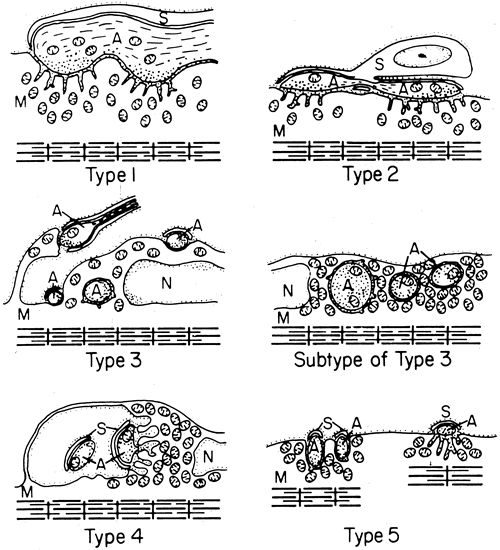
muscles of rhesus monkeys. J Neurocytol 7:693, 1978.

276. Richmond FJR, Johnston WSW, Baker RS et al.: Palisade endings in human extraocular muscles. Invest Ophthalmol Vis Sci 25:471, 1984.

277. Sodi A, Corsi M, Faussone Pellegrini MS, Salvi G: Fine structure of the receptors at the myotendinour junction of human extraocular muscles. Histol Histopath 3:103, 1988.

278. Alvarado-Mallart RM, Pinĉon-Raymond M: The palisade endings of the cat extraocular muscles, a light and electron microscopic study. Tissue Cell 11:567, 1979.

279. Golgi C: Sui nervi dei tendini, e di un nuovo organo nervaso terminale muscula tendinea. Mem Accad Sci Torin 32:359, 1880.

280. Marchi V: Ueber die Terminolorgan der Nerven (Golgi's Nervenkorperchen) in den Schnen der Augenmuskeln. Albrecht von Graefes Arch Klin Exp Ophthalmol 28:203, 1882.

281. Ciaccio GV: Sur les plaques nerveuses finales dans des vertèbres. Arch Ital Biol 14:31, 1891.

282. Ruskel GL: The incidence and variety of Golgi tendon organs in extraocular muscles of the rhesus monkey. J Neurocytol 8:639, 1979.

283. Huber C, DeWitt LMA: A contribution on the nerve terminations in neurotendinous end organs. J Comp Neurol 10:159, 1900.

284. Merrillees NCR: Some observations on the fine structure of a Golgi tendon organ of a rat. In Barker D (ed): Symposium on Muscle Receptors, pp 199–205. Hong Kong, Hong Kong University Press, 1962.

285. Cooper S, Eccles JC: The isometric responses of mammalian muscles. J Physiol (Lond) 69:377, 1930.

286. Duke-Elder WS, Duke-Elder PM: The contraction of the extrinsic muscles of the eye by choline and nicotine. Proc R Soc [B] 107:332, 1930.

287. Hofmann H, Lembeck F: The response of neuromuscular blocking agents on extraocular muscle and intraocular pressure. Proc R Soc Med 62:1217, 1969.

288. Eakins KE, Katz R: The pharmacology of extraocular muscle. In Bach-y-Rita P, Collins CC (eds): The Control of Eye Movements, pp 237–258. New York, Academic Press, 1971.

289. Hill AV: Series elastic component of muscle. Proc R Soc Lond 137:273, 1950.

290. Sakmann B, Neher E (eds): Single-Channel Recording. New York, Plenum Press, 1983.

291. Neher E, Sakmann B: Single-channel currents recorded from membrane of denervated frog muscle fibers. Nature 260:799, 1976.

292. Liebovitch LS, Tóth TI: Using fractals to understand the opening and closing of ion channels. Ann Biomed Eng 18:177, 1990.

293. Liebovitch LS: Analysis of fractal ion channel gating kinetics: Kinetic rates, energy levels, and activation energies. Math Biosci 93:97, 1989.

294. Nachsen DA, Blaustein MP: Some properties of potassium-stimulated calcium influx in presynaptic nerve end ings. J Gen Physiol 76:709, 1980

295. Rahamimoff R, DeRiemer SA, Ginsburg S, Kaiserman I, Sakmann B, Shapira R, Stadler H, Yakir N: Ionic channels in synaptic vesicles: Are they involved in transmitter release?Q J Exp Physiol 74:1019, 1989

296. Scott AB: Botulinum toxin injection into extraocular muscles as an alternative to strabismus surgery. Ophthalmology 87:1044, 1980

297. Eggers HM: Oculinum for strabismus. In Spaeth G, Katz L (eds): Current Therapy in Ophthalmic Surgery, pp 288–291. Philadelphia, BC Decker, 1989

298. Burgen ASV, Dickens F, Zalman LJ: Action of botulinum toxin. J Physiol (Lend) 109:10, 1949.

299. Black JD, Dolly JO: Interaction of I25I-labelled botulinum neurotoxins with nerve terminals: II. Autoradiographic evidence for its uptake into motor nerves and acceptor-mediated endocytosis. J Cell Biol 103:535, 1983.

300. Donovan JJ, Middlebrook JL: Ion-conducting channels produced by botulinum toxin in planar lipid membranes. Biochem 25:2872, 1986.

301. Koniarek J, Welter P, Liebovitch LS, Eggers HM: Botulinum toxin induced ion channels in extraocular muscle fiber membranes. Invest Ophthalmol Suppl 33:1099, 1992.

302. Thesleff S, Molgó J: A new type of transmitter release at the neuromuscular junction. Neuroscience 9:1, 1983.

303. Kim YI, Lφmo T, Lupa MT, Thesleff S: Miniature end-plate potentials in rat skeletal muscle poisoned with botulinum toxin. J Physiol (Lond) 356:587, 1984.

304. Katz B: The Release of Neural Transmitter Substances. Liverpool, Liverpool University Press, 1969.

305. Fertuck HC, Salpeter MM: Localization of acetylcholine receptor by 125I labelled α-bungarotoxin binding at mouse motor end plates. Proc Natl Acad Sci USA 71:1376, 1974.

306. Takeuchi A, Takeuchi N: On the permeability of end-plate membrane during the action of transmitter. J Physiol (Lond) 154:52, 1960.

307. Burton F, Dörstelmann U, Hutter OF: Single-channel activity in sarcolemmal vesicles from human and other mammalian muscles. Muscle Nerve 11:1029, 1988.

308. Koniarek J, Welter P, Liebovitch LS et al: Ion channels in extraocular muscle fibers and cultured myoblasts. Invest Ophthalmol Suppl 32:1242, 1991.

309. Jacoby J: Single channel currents recorded from singly-and multiply-innervated fibers of extraocular muscle. Invest Ophthalmol Suppl 32:1242, 1991.

310. Kuffler SW, Vaughan Williams EM: Properties of the “slow” skeletal muscle fibres of the frog. J Physiol (Lond) 121:318, 1953.

311. Lannergren J: The structure and function of twitch and slow fibers in amphibian skeletal muscle. In Lennerstrand G, Bach-y-Rita P (eds): Basic Mechanisms of Ocular Motility and Their Clinical Implications, pp 63–84. Oxford, Pergamon Press, 1975.

312. Pilar G: Further study of the electrical and mechanical responses of slow fibers in cat extraocular muscles. J Gen Physiol 50:2289, 1967.

313. Stefani E, Steinbach AB: Resting potential and electrical properties of frog slow muscle fibres: Effect of different external solutions. J Physiol (Lond) 203:383, 1969.

314. Kuffler SW, Vaughan Williams EM: Small-nerve junction potentials: The distribution of small motor nerves to frog skeletal muscle, and the membrane characteristics of the fibers they innervate. J Physiol (Lond) 121:289, 1953.

315. Kuffler SW, Gerard RW: The small-nerve motor system to skeletal muscle. J Neurophysiol 10:383, 1974.

316. Ozawa T, Cheng-Minoda K, Davidowitz J, Breinin GM: Correlation of potential and fiber type in extraocular muscle. Doc Ophthalmol 26:192, 1969.

317. Ridgeway EB, Gordon AM: Muscle activation: Effects of small length changes on calcium release in single fibers. Science 189:881, 1975.

318. Hellam DC, Podolsky RJ: Force measurements in skinned fibres. J Physiol (Lond) 200:807, 1969.

319. Chiarandini DJ: Activation of two types of fibers in rat extraocular muscles. J Physiol (Lond) 259:199, 1976.

320. Huxley HE: The structural basis of muscle contraction. Proc R Soc Lond 178:131, 1971.

321. Huxley AF: Muscular contraction. J Physiol (Lond) 243:1, 1974.

322. Huxley AF, Simmons RM: Mechanical transients and the origin of muscular force. Cold Spring Harbor Symp Quant Biol 37:669, 1973

323. Barany M: ATPase activity of myosin correlated with speed of muscle shortening. J Gen Physiol 50(Suppl):197, 1967

324. Gordon AM, Huxley AF, Julian FJ: The variation in isometric tension with sarcomere length in vertebrate 
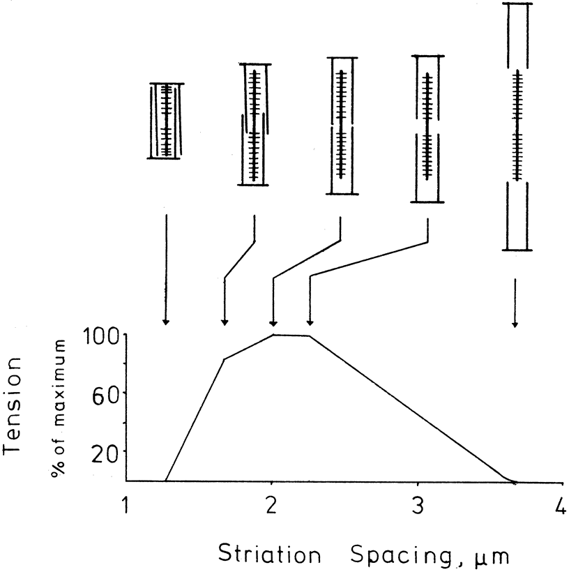
muscle fibers. J Physiol (Lond) 184:170, 1966

325. Close RJ, Luff AR: Dynamic properties of the inferior rectus muscle of the rat. J Physiol (Lond) 236:259, 1973

326. Browne JS: The contractile properties of slow muscle fibers in sheep extraocular muscle. J Physiol (Lond) 254:535, 1976.

327. Goldberg SJ, Lennerstrand G, Hall CD: Motor unit responses in the lateral rectus muscle of the cat: Intracellular current injection of abducens nucleus neurons. Acta Physiol Scand 96:58, 1976.

328. Nelson JS, Goldberg SJ, McClung JR: Motoneuron electrophysiological and muscle contractile properties of superior oblique motor units in the cat. J Neurophysiol 55:715, 1986.

329. Lennerstrand G: Motor units in eye muscles. In Lennerstrand G, Bach-y-Rita P (eds): Basic Mechanisms of Ocular Motility and Their Clinical Implications, pp 119–143. Oxford, Pergamon Press, 1975.

330. Fuchs AF, Luschei ES: Development of isometric tension in simian extraocular muscle. J Physiol (Lond) 219:155, 1971.

331. Barmack NH, Bell CC, Rence BG: Tension and rate of tension development during isometric responses of ex traocular muscle. J Neurophysiol 34:1072, 1971

332. Close RI, Luff AR: Dynamic properties of inferior rectus muscle of the rat. J Physiol (Lond) 236:259, 1974.

333. Close RI: Dynamic properties of fast and slow skeletal muscles of the rat after nerve cross-union. J Physiol (Lond) 204:331, 1969.

334. Robinson DA, O'Meara DM, Scott AB, Collins CC: Mechanical components of human eye movements. J Appl Physiol 26:548, 1969.

335. Miller JM, Robins D: Extraocular muscle forces in alert monkey. Vision Res 32:1099, 1992

336. Buchthal F, Kaiser E, Knappeis GG: Elasticity, viscosity and plasticity in cross striated muscle fiber. Acta Physiol Scand 8:16, 1944.

337. Buchthal F, Rosenfalck P: Elastic properties of striated muscle. In Remington JW (ed): Tissue Elasticity, pp 73–93. Washington, DC, Am Physiol Soc, 1957.

338. Collins CC, Scott AB, O'Meara DM: Elements of the peripheral oculomotor apparatus. Am J Optom 46:510, 1969.

339. Collins CC: Orbital mechanics. In Bach-y-Rita P, Collins CC, Hyde JE (eds): The Control of Eye Movements, pp 283–325. New York, Academic Press, 1971.

340. Hill AV: Energy liberation and “viscosity” in muscle. J Physiol (Lond) 93:4P, 1938.

341. Katz B: The relation between force and speed in muscular contraction. J Physiol (Lond) 96:45, 1939.

342. Robinson DA: The mechanics of human saccadic eye movement. J Physiol (Lond) 174:245, 1964.

343. Robinson DA: The mechanics of human smooth pursuit eye movement. J Physiol (Lond) 180:569, 1965.

344. Robinson DA: A quantitative analysis of extraocular muscle cooperation and squint. Invest Ophthalmol 14:801, 1975.

345. Eckmiller R: Hysteresis in static characteristics of eye position coded neurons in the alert monkey. Pflugers Arch 250:249, 1974.

346. Hilding AC: Normal vitreous, its attachments and dynamics during ocular movement. Arch Ophthalmol 52:497, 1954.

347. Berlin E: Beitrag zur Mechanik der Augenbewegungen. Albrecht von Graefes Arch Klin Exp Ophthalmol 17(2):154, 1871.

348. Fry GA, Hill WW: The mechanics of elevating the eye. Am J Optom 40:707, 1963.

349. Park R, Park G: The center of ocular rotation in the horizontal plane. Am J Physiol 104:545, 1933.

350. Fry GA, Hill WW: The center of rotation of the eye. Am J Optom 39:581, 1962.

351. Enright JT: Saccadic anomalies: Vergence induces large departures from ball-and-socket behavior. Vision Res 24:301, 1984.

352. Miller JM: Functional anatomy of normal human rectus muscles. Vision Res 29:223, 1989.

353. Simonsz HJ, Harting F, de Waal BJ et al: Sideways displacement and curved path of recti eye muscles. Arch Ophthalmol 103:124, 1985.

354. Enright JT: The aftermath of horizontal saccades: Saccadic retraction and cyclotorsion. Vision Res 26:1807, 1986.

355. Donders FC: Beitrag zur lehre von den Bewegungen des menschlichen Auges. Holländische Beitrage zu den ana-tomischen und physiologischen Wissenschaften1:105, 379, 1846.

356. Tweed D, Vilis T: Implications of rotational kinematics for the oculomotor system in three dimensions. J. Neurophysiol 58:832, 1987

357. van den Berg AV: Kinematics of eye movement control. Proc R Soc Lond B Biol Sci 260:190, 1995.

358. Listing JB, cited in Reute CGT: Lehrbuch der Ophthalmologie fur Aerzte und Studirende, pp 37ff. Vol 1, 2nd ed. Braunschweig, Friedreich Vieweg und Sohn, 1855 (Listing did not publish his law).

359. von Helmholtz H: Handbuch der Physiologischen Optik. 3rd ed. Hamburg, Voss, 1910. (English translation by JPC Southall for the Optical Society of America, 1925.)

360. Howard IP, Templeton WB: Human Spatial Orientation, p 46. New York, John Wiley & Sons, 1966.

361. Landolt E: Table of torsion measurements with convergence in the horizontal plane and in planes above and below. Cited in Aubert H: Physiologische optik. In Graefe A, Saemisch T (eds): Handbuch der gesammten Augenheilkunde, p 662. Vol 2. Leipzig, Engelmann, 1876.

362. Westheimer G, McKee SP: Failure of Donder's law during smooth pursuit eye movements. Vision Res 13:2145, 1973.

363. Ferman L, Collewijn H, Van der Berg: A direct test of Listing's law:II. Human ocular torsion measured under dynamic conditions. Vision res 27:939, 1987.

364. Haslwanter T, Straumann D, Hepp K et al: Smooth pursuit eye movements obey Listing's law in the monkey. Exp Brain Res 87:470, 1991.

365. Straumann D, Zee D, Solomon D, Kramer PD: Validity of Listing's law during fixations, saccades, smooth pursuit eye movements and blinks. Exp brain Res 112:135, 1996.

366. Tweed D, Vilis T: Rotation axes of saccades. Ann NY Acad Sci 545:128, 1988.

367. Tscherning M: La loi de Listing. These de Paris, 1887.

368. Quereau JVD: Some aspects of torsion. Arch Ophthalmol 31:783, 1954.

369. Quereau JVD: Rolling of the eye around its visual axis during normal movements. Arch Ophthalmol 53:807, 1955.

370. Hering E: Die Lehre vom binocularen Sehen. Leipzig, Engelmann, 1868. English translation: BridgemanB, StarkL: The Theory of Binocular Vision. New York, Plenum Press, 1977.

371. Tweed D, Cadera W, Vilis T: Computing three-dimensional eye position quaternions and eye velocity from search coil sugnals. Vision Research 30:97, 1990.

372. Tweed D, Vilis T: Geometric relations of eye position and velocity vectors during saccades. Vision Res. 30:111, 1990.

373. Raphan T: Modeling control of eye orientation in three dimensions. I. Role of muscle pulleys in determining saccadic trajectory. J Neurophysiol 79:2653, 1998.

374. Quaia C, Optican L: Commutative saccadic generator is sufficient to control a 3-D ocular plant with pulleys. J Neurophysiol 79:3197, 1998.

375. Robinson DA: Oculomotor unit behavior in the monkey. J Neurophysiol 33:393, 1970.

376. Schiller PH: The discharge characteristics of single units in the oculomotor and abducens nuclei of the unanesthetized monkey. Exp Brain Res 10:347, 1970.

377. Keller EL, Robinson DA: Abducens unit behavior in the monkey during vergence movements. Vision Res 12:369, 1972.

378. Skavenski AA, Robinson DA: Role of abducens neurons in the vestibuloocular reflex. J Neurophysiol 36:724, 1973.

379. Scott AB, Collins CC: Division of labor in the human extraocular muscle. Arch Ophthalmol 90:319, 1973.

380. Henneman E, Somjen G, Carpenter DO: Functional significance of cell size in spinal motoneurons. J Neuro physiol 28:560, 1965.

381. Henneman E, Somjen G, Carpenter DO: Excitability and inhibitability of motoneurons of different sizes. J Neurophysiol 28:599, 1965.

382. Ramon y, Cajal S: Histologie du Systeme de 1'Homme et des Vertebres. Paris, A Maloime, 1909.

383. Sherrington CS: Experimental note on two movements of the eye. J Physiol 17:27, 1894.

384. Breinin G: Electromyography: A tool in ocular and neurologic diagnosis: XI. Muscle palsies. Arch Ophthalmol 57:165, 1957.

385. Strachan IM, Brown BH: Electromyography of extraocular muscles in Duane's syndrome. Br J Ophthalmol 56:594, 1972.

386. Bahill AT, Cuiffreda KJ, Kenyon R, Stark L: Dynamic and static violations of Hering's law of equal innervation. Am J Optom Physiol Opt 53:786, 1976.

387. Bahill AT, Clark MR, Stark L: Glissades-eye movements generated by mismatched componenets of the saccadic motoneuronal control signal. Math Biosci 26:303, 1975.

387a. Optican LM, Robinson DA: Cerebellar-dependent adaptive control of the primate saccadic system. J Neurophysiol 44:1058, 1980.

388. Westheimer G, McKee SP: Visual acuity in the presence of retinal image motion. J Opt Soc Am 65:847, 1975.

389. Hepp K, Henn V, Villis T, Cohen B: Brainstem regions related to saccade generation. In Wurtz RH, Gold ME (eds): The Neurobiology of Saccadic Eye Movements. Reviews of Oculomotor Research III, p 105. Amsterdam, Elsevier, 1998.

390. Nakayama K: Coordination of extraocular muscles. In Lennerstrand G, Bach-y-Rita P (eds): Basic Mechanisms of Ocular Motility and Their Clinical Implications, pp 193–207. Oxford, Pergamon Press, 1975.

391. Goodwin AW, Fender DH: Recognition of component differences in two-dimensional oculomotor tracking tasks. Vision Res 13:1905, 1973.

392. Krewson WE: The action of the extraocular muscles. Trans Am Ophthalmol Soc 48:443, 1950

393. Boeder P: The co-operation of extraocular muscles. Am J Ophthalmol 51:469, 1961.

394. Miller JM, Robinson DA: A model of the mechanics of binocular alignment. Comput Biomed Res 17:436, 1984.

395. Miller JM, Pavlovski , Shaemeva, I: Orbit 1.8 Gaze Mechanics Simulation. Eidactics, SanFrancisco, CA, 1999.

396. Miller JM, Demer JL: Biomechanical Modeling in Strabismus Surgery. In Rosenbaum AL, Santiago P (eds): Clinical Strabismus Management: Principles and Techniques, pp 99–113. St. Louis, Mosby, 1999.

397. Robinson DA: The functional behavior of the peripheral ocular motor apparatus: A review. In Kommerell G (ed): Disorders of Ocular Motility: Neurophysiological and Clinical Aspects, pp 43–61. Presented before the Symposium of Deutschen Ophthalmologischen Gesellschaft, Freiburg, April 1977. Munich, Bergman, 1978.

398. Bahill AT, Adler D, Stark L: Most naturally occurring human saccades have magnitudes of 15 degrees or less. Invest Ophthalmol 14:468, 1975.

399. Matthews BHC: The responses of a muscle spindle during active contraction of a muscle. J Physiol (Lond) 72:153, 1931.

400. Harvey RJ, Matthews PB: The response of de-efferented muscle spindle endings in the cat's soleus to slow extension of the muscle. J Physiol (Lond) 157:370, 1961.

401. Houk J, Henneman E: Responses of Golgi tendon organs to active contractions of the soleus muscle of the cat. J Neurophysiol 30:466, 1967.

402. Cooper S, Daniel P, Whitteridge D: Afferent discharges from extraocular muscles. J Physiol (Lond) 113:463, 1951.

403. Cooper S, Daniel P, Whitteridge D: Nerve impulses in the brain stem of the goat: Responses with long latencies obtained by stretching the extrinsic eye muscles. J Physiol (Lond) 120:491, 1953.

404. Whitteridge D: The effect of stimulation of intrafusal muscle fibers on sensitivity to stretch of extraocular muscle spindles. Q J Exp Physiol 44:385, 1959.

405. Hunt CC, Kuffler SW: Further study of efferent small-nerve fibers to mammalian muscle spindles. Muscle spindle innervation and activity during contraction. J Physiol (Lond) 113:283, 1951.

406. Bach-y-Rita P, Ito F: Properties of stretch receptors in cat extraocular muscles. J Physiol (Lond) 186:663, 1966.

407. Lennerstrand G: Position and velocity sensitivity of muscle spindles in the cat: I. Primary and secondary endings deprived of fusimotor activation. Acta Physiol Scand 73:281, 1968.

408. Lennerstrand G: Position and velocity sensitivity of muscle spindles in the cat: IV. Interaction between two fusimotor fibers converging on the same spindle ending. Acta Physiol Scand 74:257, 1968.

409. Baichenko PI, Matyushkin DP, Suvorov UV: Participation of fast and tonic oculomotor systems in stretch re flexes and labyrinthine reflexes of extraocular muscles. Fiziol Zh SSSR 53:82, 1967.

410. Bach-y-Rita P: Extraocular muscle inhibitory stretch reflex during active contraction. Arch Ital Biol 110:1, 1972.

411. Baker R, Precht W: Electrophysiological properties of trochlear motor neurons as revealed by IV nerve stimulation. Exp Brain Res 14:124, 1972.

412. Keller EL, Robinson DA: Absence of a stretch reflex in extraocular muscle of the monkey. J Neurophysiol 34:908, 1971.

413. Eldred E, Granit R, Merton PA: Supraspinal control of the muscle spindles and its significance. J Physiol (Lond) 122:498, 1953.

414. Marsden CD, Merton PA, Morton HB: Servo action in human voluntary movement. Nature 238:140, 1972.

415. Whitteridge D: The motor nerve supply to extraocular muscle spindles. Electroencephalogr Clin Neurophysiol 10:353, 1958.

416. Steinbach MJ, Kirschner EL, Arstikaitis MJ: Recession versus marginal myotomy surgery for strabismus: Effects on spatial localization. Invest Ophthalmol Vis Sci 28:1870, 1987.

417. Irvine SR, Ludvigh EJ: Is ocular proprioceptive sense concerned in vision?Arch Ophthalmol 15:1037, 1936.

418. Brindley GS, Merton PA: The absence of position sense in the human eye. J Physiol (Lond) 153:127, 1960.

419. Skavenski AA: Inflow as a source of extraretinal eye position information. Vision Res 12:221, 1972.

420. Merton PA: The accuracy of directing the eyes and the hand in the dark. J Physiol (Lond) 156:555, 1961.

421. Hansen RM: Spatial localization during pursuit eye movements. Vision Res 19:1213, 1979.

422. Hansen RM, Skavenski AA: Accuracy of eye position information for motor control. Vision Res 17:919, 1977.

423. Carpenter RHS: Movements of the Eyes. 2nd ed. London, Pion, 1988.

424. von Holst E, Mittelstaedt H: Das Reafferenz-pricip. Naturwissenschaften 37:464, 1950.

425. Ludvigh E: Control of ocular movements and visual interpretation of environment. Arch Ophthalmol 48:442, 1952.

426. Fuchs AF, Kornhuber HH: Extraocular muscle afferents to the cerebellum of the cat. J Physiol (Lond) 200:713, 1969.

427. Baker R, Precht W, Llinas R: Mossy and climbing fiber projections of extraocular afferents to the cerebellum. Brain Res 38:440, 1972.

428. Batini C, Buisseret P, Kado RT: Extraocular proprioceptive and trigeminal projections to the Purkinje cells of the cerebellar cortex. Arch Ital Biol 112:1, 1974.

429. Robinson DA: Adaptive gain control of vestibuloocular reflex by the cerebellum. J Neurophysiol 39:954, 1976.

430. Steinbach MJ: Proprioceptive knowledge of eye position. Vision Res 27:1737, 1987.

431. Crewther DP, Crewther SG, Pettigrew JD: A Role for extraocular afferents in post-critical period reversal of monocular deprivation. J Physiol 282:181, 1978.

432. Graves AL, Trotter Y, Frégnac Y: Role of extraocular muscle proprioception in the development of depth perception in cats. J Neurophysiol 58:816, 1987.

433. Trotter Y, Frégnac Y, Buisseret P: The period of susceptibility of visual cortical binocularity to unilateral proprioceptive deafferentation of extraocular muscles. J Neurophysiol 58:795, 1987.

Back to Top