|
|
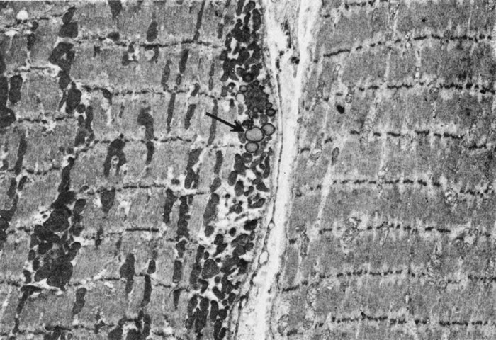
| Fig. 16. Low-power electron micrograph showing two types of Fibrillenstruktur (twitch) fibers. Both fibers show discrete and well-aligned myofibrils. The cell to the left contains many mitochondria and some lipid droplets (arrow) (“red” fiber). The cell to the right has sparse mitochondria (“white” fiber). (Courtesy of Dr. T. Iwamoto.) |