Chapter 72
Pediatric Vitreoretinal Surgery
MICHAEL T. TRESE and PHILIP J. FERRONE
Main Menu   Table Of Contents

Search

EXAMINATION AND PATIENT PRESENTATION
PEDIATRIC RETINAL DETACHMENT
RHEGMATOGENOUS RETINAL DETACHMENT
RETINOPATHY OF PREMATURITY
FAMILIAL EXUDATIVE VITREORETINOPATHY
RETINAL DYSPLASIA
CONGENITAL RETINOSCHISIS
OTHER INDICATIONS FOR VITREOUS SURGERY
REFERENCES

The pediatric patient presents many challenges to the vitreoretinal surgeon that require special consideration relative to surgical circumstances.
Back to Top
EXAMINATION AND PATIENT PRESENTATION
Pediatric vitreoretinal surgical patients are often unable to verbalize their visual complaint. Therefore the examiner must often rely on physical examination techniques. The patient is often referred for one of the following diagnoses: no red reflex, suspected retinal detachment, unexplained strabismus, leukocoria, history of trauma, a change in visual function, or unexplained irritability.

Like adult patients, pediatric patients require a good clinical examination. Unlike adult patients, however, pediatric patients are often uncooperative during the examination. In pediatric patients (especially those aged 1 to 4 years, who are often unable to understand the need for examination but are strong enough to effectively resist examination), the examination must be very directed and complete, yet brief enough so as not to lose the cooperation of the child, family, or other caretaker. Because children often have a very short attention span, it is necessary for you to begin gathering information as soon as you enter the examining room: your initial observations of the child interacting with his or her environment may be the most important piece of information you get from the child.

The physical part of the examination consists of visual acuity assessment. Assessment of vision in preverbal infants is difficult and subjective. Even the Teller and Archer-Droste acuity tests have a subjective component, since the observer must decide whether the infant sees the target.1,2 Given the subjective nature of the testing, it is still necessary to try to judge the presence or absence of light perception. This seems to be most easily assessed by either attraction or aversion to light. As the child gets older, assessment of visual acuity as measured by fixing and following behaviors, followed later by recognition behaviors, becomes possible.

After visual acuity assessment, a history of the child and family are necessary. The purpose of the history is to determine whether an examination under anesthesia, other tests (e.g., electrophysiology, blood workup for genetic markers), or surgical intervention is required. Certainly, one of the greatest risks a systemically compromised child can face is anesthesia; therefore, the surgeon must appreciate the child's systemic condition as well as the level of risk associated with general anesthesia. Often the vitreoretinal surgeon is not one who makes that decision. We prefer to leave that to the neonatologist/pediatrician and anesthesiologist.

The birth history should include the birth weight, conceptual age (i.e., weeks from conception to the present), gestational age (i.e., weeks from conception to birth), the mother's due date, pregnancy data (e.g., illness, trauma, drug use, HIV status), and familial ocular and systemic diseases. The mother's due date is a useful landmark because it gives a reference point as to retinal vascular maturity and foveal formation. Retinal vascular maturity occurs at around the due date to several weeks (rarely even up to 8 weeks) after the due date. Foveal maturation occurs approximately 3 months after the due date. In addition, if retinal detachment occurs as a result of acute retinopathy of prematurity (ROP), it usually occurs 1 or 2 weeks on either side of the due date (see ahead to the Retinopathy of Prematurity section).

EXAMINATION TECHNIQUE

The examination technique varies depending on the child's age. A premature newborn may be quite tolerant of being wrapped and undergoing scleral depression. Caution must be used when performing scleral depression in an infant with extraretinal vascularization, since it is possible to cause retinal hemorrhage during the examination.3 For examination, we use an indirect ophthalmoscope and a 20-diopter lens to assess the anterior segment and a 28- or 40-diopter lens to examine the retina. We prefer to use a small-pupil or spectacle-mounted indirect ophthalmoscope because it gives us greater freedom to move around the child during the examination. We generally have the child lie on his back on an adult's lap with the child's head at the adult's knees. This allows the adult “holder” to control the child's arms and feet while the examiner controls the top of the head and eyelids. We prefer not to use a lid speculum, but some children are unable to be examined without one. In the circumstance that a speculum is needed, a topical anesthetic is often used, and one must be aware of the potential of a corneal abrasion.

Once the examiner is aware of the information that needs to be obtained from the examination and the child is appropriately positioned, the examination can begin. The examiner must realize that the examination that the child will allow will consist of a randomly organized look at the back of the eye. It is the examiner's job to piece together the sum of these views into a useful montage. As a general rule, the more struggling that is necessary for the examination, the more frustrated the child, examiner, and family will become, and the less information will be derived from the examination. It may be necessary to examine such a child under anesthesia or to have the child return another day so that a more complete examination can be performed.

Examination Under Anesthesia

Examination under anesthesia should be reserved for the child whose office examination did not answer the clinical questions. This hopefully is a small number of children, but postponement of diagnosis of treatable retinal pathology in children not only makes repair of the retina more difficult, but also may lead to amblyopia, the need for retinal reattachment, and a poor visual result. Therefore, the decision to perform examination under anesthesia should be made as one would with any other clinical test associated with some risk. Examination under anesthesia should be performed if information can be gained that can help with the child's management.

Back to Top
PEDIATRIC RETINAL DETACHMENT

GENERAL CONSIDERATIONS

All aspects of management of pediatric retinal detachment must be viewed with the realization that all the issues associated with adult retinal detachment are present in the child, but that these circumstances are occurring in an eye that is part of a visual system in the process of learning how to see.

Because of the difficulty of examining the signs as well as recognizing the symptoms of retinal detachment in children, the timing of retinal detachment repair is often unavoidably delayed despite appropriate referral. After retinal detachment or detachment repair, the vigor with which reparative tissue grows may lead to severe stages of proliferative vitreoretinopathy. Our clinical impression, which is in agreement with that of other experienced vitreoretinal surgeons, is that pediatric retinal detachments have a worse prognosis for reattachment and therefore vision, perhaps partly due to severe reproliferation rates. In some series, pediatric retinal detachment is often found in association with some congenital retinal changes that may have an associated retinal or, perhaps even more importantly, vitreous abnormality that predisposes the patient to the retinal detachment.4 In a 1972 study of pediatric aphakic patients in whom a small-wound closed-eye technique was used, 1.5% were found to have retinal detachment at a mean follow-up of 5.5 years.5 In two thirds of these cases of retinal detachments, there was a coexistent anomaly. Other series on retinal detachment, especially those involving children or trauma, also have reported continuing cicatrization and late retinal detachment.6,7

The 1.5% incidence of retinal detachment reported in the 1972 study5 is much reduced from earlier series, which have shown a 3% to 10% incidence of retinal detachments.8,9 The mechanical difference of using a vitreous cutting system to remove a child's soft lens versus the older aspiration technique seem to have led to a more controlled cutting of lens and vitreous. This technique is less likely to transmit the cutting and sucking forces from the lens material to the vitreous base, thus avoiding the creation of retinal tears.

ANATOMIC CONSIDERATIONS

The most obvious anatomic consideration in pediatric retinal surgery is the relatively smaller size of the child's globe and orbit compared with the adult's. There are, however, several other important anatomic considerations in pediatric cataract extraction:

  1. The attachment between the vitreous cortex and retina is very firm.
  2. The soft lens and anterior vitreous are attached.
  3. The secondary vitreous in the term infant without ocular disease consists of a very dense collagenous gel. Some authors believe that in term infants, there is no hyaluronic acid present until 4 years of age.10,11 There are, however, many diseases that are associated with a lack of densely formed vitreous (e.g., myopia, retinitis pigmentosa, ROP, familial exudative vitreoretinopathy, Goldmann-Favre disease, congenital retinoschisis).
  4. A term infant does not have a well-developed pars plana, limiting where the surgeon can safely enter his or her eye for vitreous surgery. As a rule of thumb, an infant 8 months post term has a pars plana approximately 2 mm wide.12 Thus, for vitrectomy, it is necessary to enter much less anterior than the usual adult entry site. Because of the relatively larger size of the lens in relation to the anterior segment in children compared with adults, special care is required if the eye is to be entered without removing the lens.

Back to Top
RHEGMATOGENOUS RETINAL DETACHMENT
Although rhegmatogenous idiopathic retinal detachment is less common in the pediatric population, it is important to consider in the primary repair of pediatric retinal detachments. Rhegmatogenous retinal detachments in children are often found accompanying other vitreoretinal pathology or trauma and has been reported in cases of Marfan's syndrome, Stickler's syndrome, cataract extraction, and trauma.13–15

SCLERAL BUCKLING

A child's eye is prone to more vigorous reproliferation than an adult's. This can be attributed to (1) a biochemistry that supports cell growth more actively or (2) a tendency toward a longer delay between the time of detachment and the determination of a diagnosis and therapy. This has led us to suggest the use of a higher scleral buckle in children than in adults. For primary rhegmatogenous retinal detachments, we use a sculpted 5 × 7 mm sponge with an encircling 2.5-mm solid silicone band. It is often assumed that the child's orbit cannot accommodate a large sponge, but we have found that this type of exoplant is well tolerated. Drainage of subretinal fluid is always controversial. We tend to drain subretinal fluid if the retina is highly detached and minimal retinal traction is present. If fluid is quickly drained while extensive traction remains, other retinal tears can occur. In primary rhegmatogenous detachments, however, drainage is often helpful.

Complications

Postoperative complications of scleral buckling in children range from limitation of eye growth to development of an amblyopic loss of vision due to cycloplegic use of eyedrops. To avoid these pitfalls as much as possible, approximately 3 months after the scleral buckling operation we divide the encircling band in all children who are less than 2 years of age or whose eye growth is retarded. We choose to divide rather than to remove the element because we believe that continued support is given to the retina by the encapsulated exoplant. To reduce the chance of amblyopia development in the postoperative period, we tend to prescribe 1% atropine drops for 5 days; if both eyes have good visual potential, we often prescribe the drops for use in both eyes. In addition to amblyopia therapy, refractive error, which can usually be pursued leisurely in adults, needs to be aggressively treated.

Giant Retinal Tears

Giant retinal tears in adults have been discussed widely in the literature.16 Giant tears in children, however, have received less attention. The problems in treating pediatric giant tears often revolve around positioning of the patient for weeks after surgery as well as complicated surgical techniques. Delay in diagnosis and the fact that pediatric giant retinal tears are often concurrent with other ocular problems or trauma make these retinal tears even more challenging in children than they are in adults. Thus children often have lower anatomic and visual success rates.4 Children's level of physical activity and inability to position postoperatively make the consideration of a long-acting tamponade (e.g., silicone oil) very appealing. This may free the child and caretaker from a rigorous positioning regimen. All of the complications of silicone oil must also be considered, such as cataract, glaucoma, corneal decompensation, and reoperation for removal.4,17 We believe that the advantages of silicone oil far outweigh the disadvantages in this population and will usually use it as our first-line tamponade in giant retinal tears in children.

Back to Top
RETINOPATHY OF PREMATURITY
ROP accounts for a large number of retinal detachments in the pediatric population. The International Classification of Retinopathy of Prematurity helped greatly to improve communication about, and consequently treatment of, ROP. It describes affected eyes in terms of stage, zone, and absence or presence of plus disease18 (Figs. 1, 2, and 3). Screening of premature infants is an important feature in the care of ROP, which is perhaps the most significant and enduring finding of the Cryotherapy for Retinopathy of Prematurity Study (Cryo-ROP Study).19

Fig. 1. Stages of retinopathy of prematurity. A. Stage 1, showing a small white line visible between the avascular and vascularized retina. B. Stage 2, with the junction showing a wider white line between avascular and vascularized retina. C. Stage 3, with frank neovascularization extending into the vitreous cavity from the area posterior to the retinal ridge. D. Stage 4A, showing peripheral retinal detachment with the macula attached. E. Stage 4B, with a partial retinal detachment with the macula detached. F. Stage 5, showing total retinal detachment.

Fig. 2. Standard zoning patterns of retinopathy of prematurity. Zone 1 is defined as a circle with a radius of two times the distance between the center of the optic nerve and the foveola. Zone 2 is a circle whose radius is from the center of the optic nerve to the nasal horizontal ora serrata. All the remaining area of retina is zone 3.

Fig. 3. Dilated and tortuous vessels in the posterior pole, which by definition is plus disease.

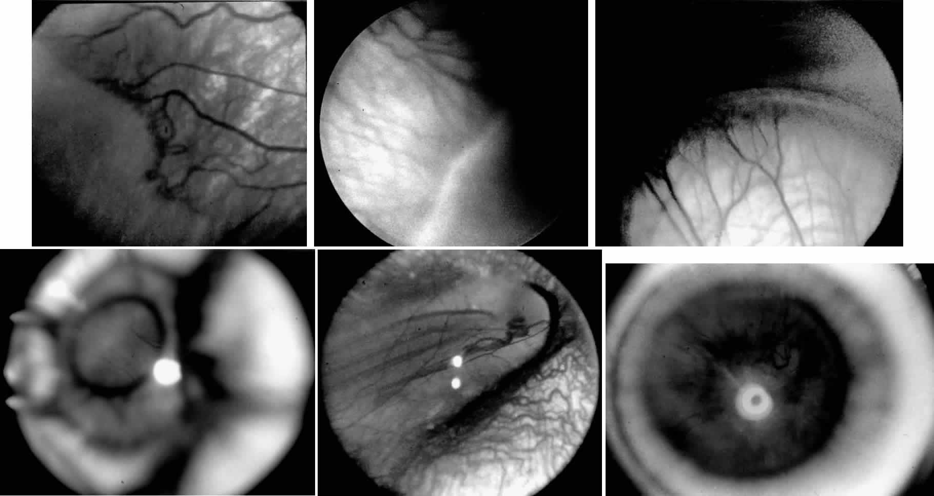
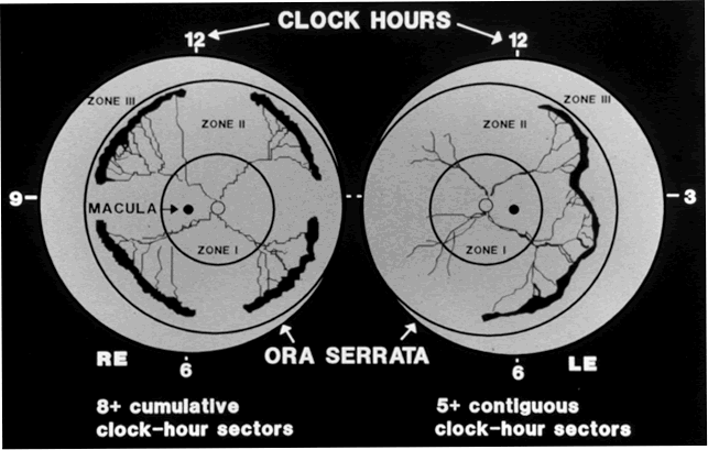
Although several screening protocols have been suggested, we have found that children whose birth weight was 1500 g or less should be screened initially at 4 to 6 weeks after birth, and then every 2 weeks until they reach retinal vascular maturity, which is when nasal vessels in the horizontal meridian have grown to within 1 disc diameter of the ora serrata. During that time, if a child shows threshold disease as defined by the Cryo-ROP Study, namely five clock-hours of contiguous or eight clock-hours of discontiguous neovascularization (stage 3 ROP with plus disease), it is recommended that the child have peripheral ablation with either cryotherapy or laser treatment.19 Newer studies have suggested that zone 1 children may require earlier peripheral ablation.20 The child who fails to respond to peripheral ablation may require further surgical intervention.

Two large series reported that scleral buckling for stages 4A and 4B retinal detachments resulted in a retinal reattachment rate of approximately 70%.21,22 These studies were both retrospective; however, they did show a strong trend toward a higher reattachment rate with scleral buckling than the natural history of these detachments, which have a 55% chance of progression of retinal detachment from stage 4 to stage 5 ROP.23

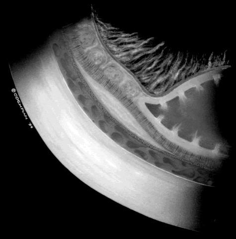
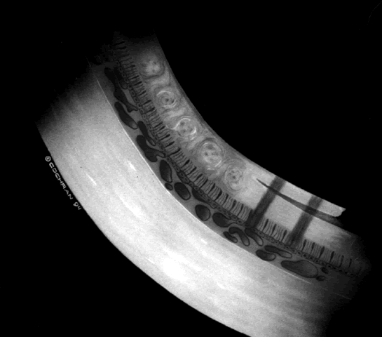
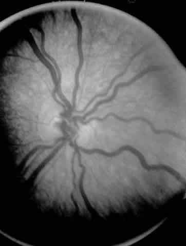
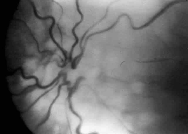
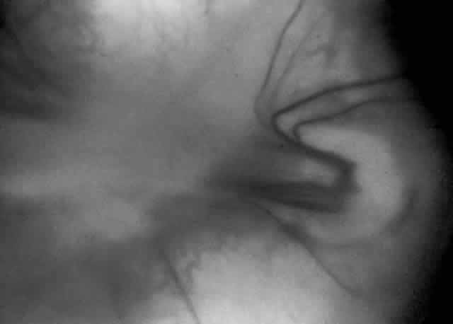
When first assessing a child's retinal detachment, one must judge the amount of effusive versus tractional detachment (Figs. 4 and 5). Scleral buckling would be considered for the child who shows a predominately effusive stage 4B detachment as opposed to a stage 4 predominately tractional detachment, for which lens-sparing vitrectomy may be recommended.24 For a child who has a great deal of retrolenticular touch, lensectomy/ vitrectomy and membrane peeling would be recommended. It appears that the “window” for lens-sparing vitrectomy may be rather brief. In one series, the postconceptual age of the lens-sparing vitrectomized eyes was 42.6 weeks, as opposed to 46.9 weeks for eyes that needed lensectomy/vitrectomy and membrane peeling. This small time difference in the postconceptual age shows the often rapid evolution of this detachment from one in which the lens is salvageable to one in which the lens is unsalvageable. This highlights the need for timely screening of eyes, identification of eyes with progressive disease, and rapid intervention.25

Fig. 4. A predominantly effusive stage 4B retinal detachment.

Fig. 5. A predominantly tractional stage 4B retinal detachment.

The techniques of vitreous surgery in children require an understanding of the anatomy of the pars plicata. A premature child does not have a well-defined pars plana, and therefore pars plicata entry is the only entry possible to avoid the lens upon entering the vitreous cavity. A term infant who is 8 months post term has a 2 mm pars plana.12 Infants who are premature at the time of vitreous surgery require entry immediately posterior to the iris root to avoid damage to the neurosensory retina and crystalline lens.

After vitreous surgery, particularly one that is lens-sparing, the final visual result depends on the child's central nervous system, refractive status, and competition with the fellow eye. With lens-sparing vitrectomy techniques, however, visual acuities can be made as good as 20/60, even in ROP cases.26,27 The child who has lensectomy/vitrectomy as well as membrane peeling with appropriate refractive correction can have a visual acuity as good as 20/200 to 20/400.28 The child's initial aphakic status can greatly affect his or her final visual outcome. This is why we believe strongly that prompt screening in order to time surgical intervention appropriately as well as reacting quickly to the child's surgical need are important means of optimizing final visual outcome. A child's refractive status is always difficult to deal with, especially if the child is aphakic or the red reflex is compromised. In children, we should not forget the need to assess near vision and the use of low vision aids.

A child's retinal detachments progress at variable rates; thus the rate of detachment must be assessed on an individual basis. In children with RUSH disease, the eye has a very immature retina with much of the vascularized/avascular retinal juncture in zone 1. These eyes tend to progress to retinal detachment very quickly, often within 1 to 2 weeks. We have described another uncommon entity called very posterior zone 1 retinopathy of prematurity.29 In this disease, the macula is disorganized and not clearly visible. The posterior pole presents a syncytium of vessels all in zone 1. All of these eyes that we followed have gone on to have tractional retinal detachment. If there is to be any hope of vision in these patients, management requires a very rapid and broad peripheral ablative treatment followed by early vitreous surgery intervention.

The care of ROP patients requires a careful and rapidly performed screening examination, rapid intervention with peripheral ablation, scleral buckling, lens-sparing vitrectomy, or lensectomy/ vitrectomy and membrane peeling. Given a prompt intervention, we have come to believe that ROP can be managed with results comparable to those for patients with diabetes or proliferative vitreoretinopathy. To date, Droste and Trese30 are the only investigators to have reported on a consecutive series managed in that fashion, and this series showed improved visual results. Historically, visual results in ROP have been poor if retinal detachment intervention was delayed. With the advent of appropriate screening and rapid surgical intervention, however, improved visual results are possible.

Back to Top
FAMILIAL EXUDATIVE VITREORETINOPATHY
Familial exudative vitreoretinopathy (FEVR) is a forme fruste of ROP, occurring in a larger and often term infant. It has a variety of genetic inheritance patterns: autosomal-dominant FEVR was the first to be described; more recently X-linked FEVR has been described.31–33 Shastry and Trese34 recently described a point mutation that seems to be associated with X-linked FEVR. Like retinitis pigmentosa, this may represent only one of many mutations that can cause this phenotype. The management of FEVR is similar to that for ROP: in the early stages, peripheral ablation is used to reduce subretinal and intraretinal exudation.35 However, FEVR has a more chronic progression than ROP, showing continued subretinal exudation throughout several decades of life.
Back to Top
RETINAL DYSPLASIA
A child with retinal dysplasia accompanied by tractional or hemorrhagic retinal detachment, as is seen in Norrie's disease, can maintain light perception or minimally better vision if retinal reattachment is accomplished. A child with significant retinal dysplasia may not merit surgical intervention; however, maintenance of eye size should be a consideration, and visual function of even the smallest amount may be helpful in some children.
Back to Top
CONGENITAL RETINOSCHISIS
Congenital retinoschisis is an inherited retinal disease characterized by abnormal Müller's cell pillars that allow schisis cavities to form. These schisis cavities are often described as being present 100% of the time in the foveal area and 50% of the time in the far periphery. They often result in decreased visual acuity by the third decade of life. They certainly can result in reduced visual acuity much sooner than that and may be present either at birth or during the first decade of life.

We believe that congenital retinoschisis is aggravated by vitreoretinal traction and have suggested that surgical therapy may be indicated for congenital retinoschisis under certain specific clinical settings:

  1. A large intraschisis hemorrhage with concurrent extension of vitreous hemorrhage or schisis cavity overhanging the macula
  2. A schisis combined with tractional retinal detachment
  3. A schisis combined with rhegmatogenous retinal detachment

The suggested treatment is scleral buckling or vitreous surgery, including removal of the inner wall of the schisis cavity with extensive panretinal photocoagulation to protect against rhegmatogenous retinal detachment.36–38 The hallmark of this tractional detachment is visual field changes that are larger than expected relative to the schisis cavity.36 Previously, demarcation lines resulting from full-thickness retinal detachment have been reported posterior to the schisis cavity.39 Others have considered this finding to represent an undetected rhegmatogenous retinal detachment, but based on observations during surgery, we believe that this detachment can be reversible and is tractional in origin (Fig. 6).36

Fig. 6. Artist conception of a full-thickness traditional retinal detachment posterior to the schisis cavity before surgical intervention in congenital retinoschisis.

COATS' DISEASE

Coats' disease is an example of a vitreoretinal congenital vascular disease in which vitreous traction often plays a role leading to both preretinal and subretinal hemorrhage with resultant retinal detachment. Children with Coats' disease have been managed with vitreous surgery, scleral buckling, trans-scleral diathermy, cryotherapy, laser therapy alone, and laser therapy accompanied by injectable dyes (Fig. 7).40–42 Many different techniques have been employed because frequently neither the vessel abnormalities nor the vitreous traction can be resolved completely. With the possible advent of enzymatic surgery, such as use of plasmin or chondroitinase, it is hoped that a cleaner and more complete reduction of vitreoretinal traction will help advance the involution of these abnormal vessels without concurrent vitreous or subretinal hemorrhage.43

Fig. 7. Artist conception of resolution of a full-thickness retinal detachment after relief of vitreous traction by vitrectomy and inner-wall retinectomy in congenital retinoschisis.

Back to Top
OTHER INDICATIONS FOR VITREOUS SURGERY
Children who have particularly unilateral premacular hemorrhage persisting for several months and who are less than 1 year old may be susceptible to dense amblyopia. These eyes can be managed with lens-sparing vitreous surgery. This type of eye is often prone to membrane formation beneath the blood. If premacular blood is removed, care should be taken at this time to remove any epiretinal membrane beneath the blood.44

Unfortunately, pediatric retinal surgeons must also deal with the issue of shaken baby syndrome. This syndrome is characterized by blood in the posterior pole and macular area. This blood can be preretinal, intraretinal, or subretinal45; a significantly large component of intraretinal blood may be in the macular area. The differential diagnosis of vitreous hemorrhage in children is quite lengthy, ranging from trauma, to vascular malformations, to congenital retinoschisis, to other forms of retinal tear and detachment. Care should be taken to try to distinguish among these diagnoses in order to identify a potential victim of trauma versus a patient with inherited retinal disease.

Pediatric vitreoretinal surgery strongly requires a team approach among the pediatrician, neonatologist, anesthesiologist, and vitreoretinal surgeon. All aspects of the child's care must be considered before even the most minor surgical examination or intervention can be undertaken. As our knowledge of vitreoretinal surgery increases, the possibility of using a combination of mechanical and enzymatic surgery (e.g., plasmin and chondroitinase) may become particularly helpful in children in whom a vitreoretinal adhesion is exceptionally strong. It is hoped that enzymatic surgery will allow increased safety for the relief of vitreoretinal traction, which may be very helpful in the management of entities such as ROP, FEVR, Coats' disease, and congenital retinoschisis.

Back to Top
REFERENCES

1. McDonald MA, Dobson V, Sebris SL et al: The acuity card procedure: a rapid test of infant activity. Invest Ophthalmol Vis Sci 26:1158, 1985

2. Droste PJ, Archer SM, Helveston EM: Measurement of low vision in children and infants. Ophthalmology 98:1513, 1991

3. Baker J, Trese M, Palmer E: Vitreous hemorrhage during scleral depression. Personal observation, 1996

4. Hassan TS, Trese MT, Blumenkranz MS et al: The surgical management of pediatric giant retinal tears. Unpublished data, 1995

5. Chen YS, Dumas J: The etiology of juvenile retinal detachment. Can J Ophthalmol 7:136, 1972

6. Lemrini F, Dafrallah L, Kabbaj A: Retinal detachment in children. J Fr Ophtalmol 16(3):159, 1993

7. Francois J: Late results of congenital cataract surgery. Ophthalmology 86:1586, 1979

8. Ryan SJ, Blanton FM, von Noorden GK: Surgery of congenital cataract. Am J Ophthalmol 60:583, 1965

9. Shapland CD: Detachment of the retina in the aphakic eye. An Inst Barraquer 3:420, 1962

10. Balazs EA: Fine structure of developing vitreous. In Ocular Fine Structure for the Clinician. Int Ophthalmol Clin 15: 53, 1975

11. Sebag J: Embryology of the vitreous. In The Vitreous: Structure, Function, and Pathobiology, pp 7–16. New York, Springer-Verlag, 1989

12. Hairston RJ, Maguire AM, Vitale S, Green WR: Morphometric analysis of pars plana development in humans. Invest Ophthalmol Vis Sci 33:1197, 1992

13. Greco GM, Ambrosina L: Treatment of retinal detachment in Marfan's syndrome. Ann Ophthalmol 25(2):72, 1993

14. Stickler GB, Belau PG, Farrell FJ et al: Hereditary progressive arthro-ophthalmopathy. Mayo Clin Proc 40:433, 1965

15. Cox MS: Retinal breaks caused by blunt nonperforating trauma at the point of impact. Trans Am Ophthalmol Soc 78:414, 1980

16. Freeman HM: Giant retinal tears: I. Management of giant retinal tear with an inverted retinal flap. In Tasman W, Jaeger E (eds): Duane's Clinical Ophthalmology, Vol 6, Chap 69. Philadelphia, Lippincott-Raven, 1996

17. Ferrone PJ, McCuen WB, deJuan E, Machemer R: The efficacy of silicone oil for complicated retinal detachments in the pediatric population. Arch Ophthalmol 112:773, 1994

18. The Committee for the Classification of Retinopathy of Prematurity: An international classification of retinopathy of prematurity. Arch Ophthalmol 102:1130, 1989

19. Cryotherapy for Retinopathy of Prematurity Cooperative Group: The natural ocular outcome of premature birth and retinopathy: Status at 1 year. Arch Ophthalmol 112:903, 1994

20. The Laser ROP Study Group: Laser therapy for retinopathy of prematurity. Arch Ophthalmol 112:154, 1994

21. Greven C, Tasman W: Scleral buckling in stage 4B and 5 retinopathy of prematurity. Ophthalmology 97:817, 1990

22. Trese MT: Scleral buckling for retinopathy of prematurity. Ophthalmology 101:23, 1994

23. Flynn JT, Bancalari E, Bachynski BN et al: Retinopathy of prematurity: diagnosis, severity, and natural history. Ophthalmology 94:620, 1987

24. Maguire AM, Trese MT: Lens-sparing vitreoretinal surgery in infants. Arch Ophthalmol 110:284, 1992

25. Goyal AK, Trese MT: Surgical management of primary vitreous in tractional stage 4B/5 retinopathy of prematurity. Transactions of International ROP Meeting, Chicago, 1996

26. Maguire AM, Trese MT: Visual results of lens-sparing vitreoretinal surgery in infants. J Pediatr Ophthalmol Strabismus 30:28, 1993

27. Trese MT, El-Rayes EN, Dass AB: Volcano-shaped retinal detachment with attached macula in cicatricial retinopathy of prematurity (ROP) simulating 4B ROP detachment. Retina (in press), 1996

28. Trese MT, Hartzer M, Lin LR et al: Vitreous and retinopathy of prematurity: Vitreous surgery and visual results. In Retinopathy of Prematurity, pp 83–93. New York, Springer-Verlag, 1992

29. Goyal AK, Trese MT: Definition and surgical management of very posterior zone 1 retinopathy of prematurity. Retina (in press), 1996

30. Droste P, Trese M: Management of low vision in advanced cicatricial ROP: An update on strabismus and pediatric ophthalmology. In Lemerstrand G (ed): Proceedings of Joint Congress, June 19–23, 1994. Boca Raton, FL, CRC press

31. Shastry BS, Hartzer MK, Trese MT: Letter to the editor: Familial exudative vitreoretinopathy: Multiple modes of inheritance. Clin Genet 44:275, 1993

32. Lucke KH, Foerster MH, Laqua H: Long-term results of vitrectomy and silicone oil in 500 cases of complicated retinal detachments. Am J Ophthalmol 104:624, 1987

33. Plager DA, Orgel IK, Ellis FD et al: X-linked familial exudative vitreoretinopathy. Am J Ophthalmol 114:145, 1992

34. Shastry BS, Trese MT: X-linked familial exudative vitreoretinopathy (FEVR): results of DNA analysis with candidate genes. Am J Med Genet 45:111, 1993

35. Pendergast SD, Trese MT: Surgical management of familial exudative vitreoretinopathy. Unpublished data, 1996

36. Trese MT, Ferrone PJ: The role of inner wall retinectomy in the management of juvenile retinoschisis. Graefe's Arch Clin Exp Ophthalmol 233:706, 1995

37. Tasman W, Greve CM, Moreno RJ: 1 Nasal retinal dragging in X-linked retinoschisis. Graefe's Arch Clin Exp Ophthalmol 229:319, 1991

38. Schulman J, Peyman GA, Jednock N, Larson B: Indications for vitrectomy in congenital retinoschisis. Br J Ophthalmol 69:482, 1985

39. Ferrone PJ, Trese MT, Lewis H: Vitreous surgery techniques in congenital retinoschisis. Am J Ophthalmol (in press), 1996

40. Machemer R, Williams JM Sr: Pathogenesis and therapy of traction detachments in various retinal vascular diseases. Am J Ophthalmol 105:173, 1988

41. Riffenburgh RS, Sathyavagiswaran L: Ocular findings at autopsy of child abuse victims. Ophthalmology 98:1519, 1991

42. Hassan TS, Trese MT: ICG dye-enhancing diode laser photocoagulation in Coats' disease. Unpublished data, 1996

43. Verstraeten TC, Chapman C, Hartzer M et al: Pharmacologic induction of posterior vitreous detachment in the rabbit. Arch Ophthalmol 111:849, 1993

44. Ferrone P, deJuan E: Vitreous hemorrhage in infants. Arch Ophthalmol 112:1185, 1994

45. Ober RR: Hemorrhagic retinopathy in infancy: a clinico-pathological report. J Pediatr Ophthalmol Strabismus 17:17, 1980

Back to Top