|
|
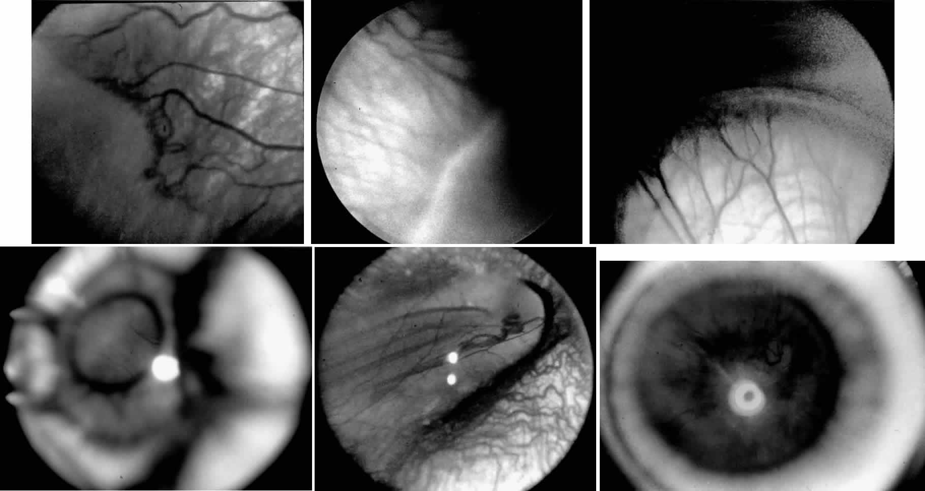
| Fig. 1. Stages of retinopathy of prematurity. A. Stage 1, showing a small white line visible between the avascular and vascularized retina. B. Stage 2, with the junction showing a wider white line between avascular and vascularized retina. C. Stage 3, with frank neovascularization extending into the vitreous cavity from the area posterior to the retinal ridge. D. Stage 4A, showing peripheral retinal detachment with the macula attached. E. Stage 4B, with a partial retinal detachment with the macula detached. F. Stage 5, showing total retinal detachment. |