Chapter 24
Complications of Glaucoma Surgery
AUGUSTO AZUARA-BLANCO and L. JAY KATZ
Main Menu   Table Of Contents

Search

GENERAL COMPLICATIONS OF GLAUCOMA SURGERY
SPECIFIC COMPLICATIONS RELATED TO THE TYPE OF PROCEDURE
REFERENCES

GENERAL COMPLICATIONS OF GLAUCOMA SURGERY

INTRAOPERATIVE AND POSTOPERATIVE SUPRACHOROIDAL HEMORRHAGE

Suprachoroidal hemorrhage is a serious complication that can be seen during or after any intraocular surgery. If it occurs intraoperatively and cannot be controlled (i.e., expulsive hemorrhage), it can lead to loss of vision. The incidence of this complication in the general population after cataract extraction is approximately 0.2%.1–3 The incidence of suprachoroidal hemorrhage in glaucoma patients undergoing various types of intraocular surgery has been reported to be 0.73%.4–6 Ocular risk factors for suprachoroidal hemorrhage include glaucoma, aphakia, pseudophakia, previous vitrectomy, vitrectomy at the time of glaucoma surgery, myopia, and postoperative hypotony. Systemic risk factors are arteriosclerosis, high blood pressure, tachycardia, and bleeding disorders. The source of the hemorrhage usually is one of the posterior ciliary arteries, particularly the point of entrance of the short posterior ciliary vessels into the suprachoroidal space. There seems to be a vascular necrosis and subsequent rupture of the vascular wall.7

Intraoperative suprachoroidal hemorrhage can be associated with sudden collapse of the anterior chamber. The patient may complain of sudden pain breaking through the local anesthesia. If the process is gradual, a dark mass can be observed through the pupil to evolve slowly, but if the process is abrupt the hemorrhage is more expulsive.

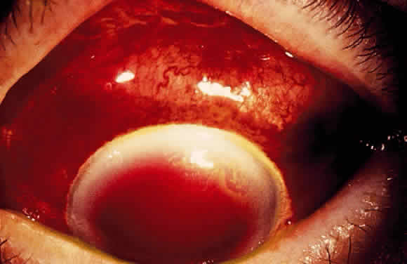
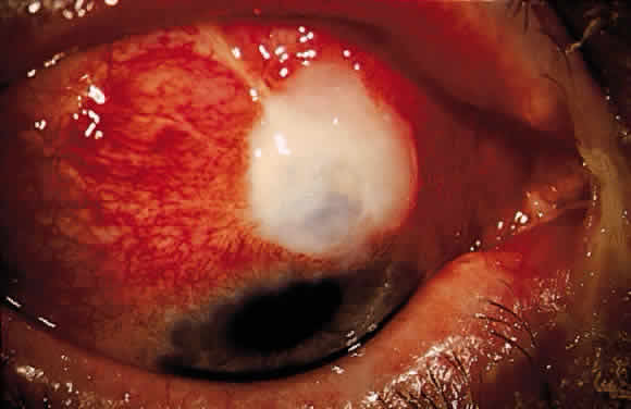
Postoperative suprachoroidal hemorrhage usually occurs within the first week after glaucoma surgery and usually is associated with postoperative hypotony4–6 (Fig. 1). The development of a suprachoroidal hemorrhage typically is acute and associated with the sudden onset of severe pain. Examination of the anterior segment frequently reveals a shallow anterior chamber and a normal or high intraocular pressure (IOP). On fundus examination, a detached and dark choroid is noted. The choroidal elevations have a dark, reddish brown color. Some patients present with bleeding into the vitreous cavity and, uncommonly, retinal detachment. Ultrasonography can be used to diagnose suprachoroidal hemorrhage when fundus examination is not possible.

Fig. 1. Slit-lamp photograph. A. Massive suprachoroidal hemorrhage after Molteno tube shunt implantation. The tube with an intraluminal suture in place can be seen. B. Slit-beam illumination reveals a flat anterior chamber.

Intraoperatively, once a suprachoroidal hemorrhage has been identified, prompt and secure closure of the incision is the first goal of the treatment, with gentle repositioning of prolapsed uvea. The surgeon's finger can be used to tamponade the incision site temporarily while sutures are placed. Meanwhile, intravenous acetazolamide (500 mg) and mannitol 20% (1 to 1.5 g/kg) are administered. Once the eye has been closed, the anterior chamber can be reformed through the incision or a paracentesis tract. After this point, a conservative approach probably is appropriate. Some authors advise immediate drainage of the hemorrhage through posterior sclerostomies (usually not possible because it rapidly clots), sometimes combined with a vitrectomy in aphakic patients, especially if the hemorrhage is large. Prognosis for recovery of vision is good as long as the eye can be closed without loss of uvea.

Treatment of postoperative suprachoroidal hemorrhage is directed toward control of the IOP and relief of pain. Most of these eyes do well with this conservative management, and surgical drainage usually is not necessary.8 The indications for drainage include intolerable pain, a persistent flat anterior chamber, and massive “kissing” choroidal detachments (see later). A waiting period of about 7 days after a suprachoroidal hemorrhage is advised for the fibrinolytic response to liquefy the clot and allow for more effective evacuation of the suprachoroidal space. Drainage through a sclerotomy into the suprachoroidal space reveals liquefied blood, which usually is red or black. Occasionally, the fluid drained is a mixture of clear, straw-colored fluid and reddish to black liquefied blood. Bleeding into the vitreous cavity at the time of the hemorrhage and retinal detachment worsen the visual prognosis.

Prevention

Several steps can be taken in high-risk eyes. Before surgery, correction of bleeding problems and discontinuation of inhibitors of platelet aggregation (i.e., acetylsalicylic acid) is recommended. Preoperative intravenous mannitol at the time of surgery should be used. Prophylactic sclerostomies can be helpful. Use of viscoelastic and tight suturing of the scleral flap to prevent hypotony are recommended. The patient is urged to restrict activities (bending, weight lifting) and to avoid Valsalvapositive conditions (constipation, vigorous coughing, sneezing, or nose-blowing) during the early postoperative period.

HYPHEMA

Hyphema is a common postoperative occurrence in glaucomatous eyes after filtration surgery, surgical peripheral iridectomy, and trabeculotomy. Bleeding commonly arises from the ciliary body or cut ends of the Schlemm's canal, although it also might arise from the corneoscleral incision or iris.

In general, hyphema presents at surgery or within the first 2 or 3 days after surgery. Intraoperatively, if a bleeding spot does not stop spontaneously, it must be identified and coagulated. During filtration surgery, bleeding is decreased by performing the internal sclerostomy as far anteriorly as possible.

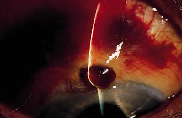
In most cases, no treatment is necessary and the blood is absorbed within a brief period of time. Cycloplegics, corticosteroids, restriction of activity, and elevation of head of the bed 30° to 45° (to prevent blood from obstructing a superior sclerostomy) are recommended. Increased IOP can occur, particularly if the filtering site is obstructed by a blood clot, and it should be treated if necessary with aqueous suppressants. Injection of tissue-plasminogen activator may be considered (see later). Surgical evacuation is considered depending on the level of IOP, size of hyphema, severity of optic nerve damage, likelihood of corneal blood staining, and presence of sickle trait or sickle cell anemia (infarction of the optic nerve can occur at relatively low IOP, and carbonic anhydrase inhibitors are contraindicated). Liquid blood can be easily removed with irrigation. If a clot has formed, it can be removed by expression with viscoelastic or with a vitrectomy instrument set at low vacuum.

HYPOTONY

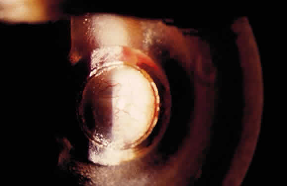
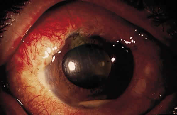
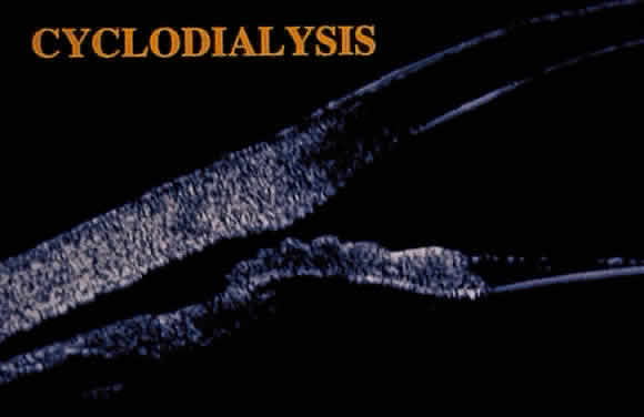
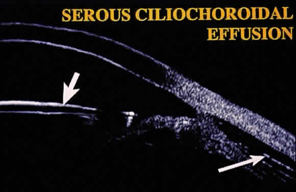
Hypotony (i.e., IOP less than 6 mmHg) after glaucoma surgery can result from excessive aqueous outflow (related to excessive filtration [see later], wound leak, or cyclodialysis cleft) or to reduced aqueous production (related to ciliochoroidal detachment, inflammation, inadvertent use of aqueous suppressants, or extensive cyclodestruction).9 These conditions can coexist. For example, low IOP from overfiltration can induce ciliochoroidal detachment and secondary decreased aqueous production. Possible complications include flat anterior chamber, gradual failure of the bleb, visual loss, cataract, corneal edema, Descemet's membrane folds, choroidal hemorrhage, macular and optic disc edema, and chorioretinal folds (predominantly in young myopic patients). According to Spaeth (Table 1),10 the severity of flat anterior chamber can be classified as grade I when there is peripheral-iris apposition, grade II with pupillary border-corneal apposition, or grade III with lens-corneal touch (see Chapter 15). The central anterior chamber depth also can be described relative to the corneal thickness. Choroidal effusion occurs when fluid collects in the suprachoroidal space (Fig. 2), resulting in forward movement of the lens iris diaphragm with anterior chamber shallowing. On fundus examination, moundlike elevations of the choroid, more commonly in the periphery, are visible.


TABLE 1. Classification of Flat Anterior Chamber

  Grade I, peripheral iris-corneal apposition
  Grade II, pupillary border-corneal apposition
  Grade III, lens-corneal apposition

(Spaeth GL: Glaucoma surgery. In Tasman W, Jaeger EA [eds]: Duane's Clinical Ophthalmology, vol 5, pp 1–53. Philadelphia, JB Lippincott, 1992)


Fig. 2. Composite of ultrasound biomicroscopic examination. A peripheral choroidal effusion (large arrow) is seen as an acoustically hollow area in the suprachoroidal space. The anterior chamber is moderately shallow (small arrow, intraocular lens).

Hypotony Maculopathy

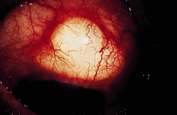
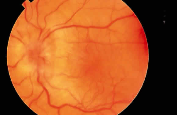
Some patients with intraocular hypotony develop loss of central vision secondary to marked irregular folding of the choroid and retina. Initially, these folds are broad and not sharply delineated. They tend to radiate outward in a branching fashion temporally from the optic disc and concentrically or irregularly nasally to the disc. There may be swelling of the peripapillary choroid simulating papilledema (Fig. 3). The retina often shows a series of stellate folds around the center of the fovea. The retinal vessels are tortuous and sometimes engorged. The primary cause of visual loss is the marked folding of the central retina. Early detection of this condition is important because correction of the cause usually results in visual improvement. In cases of prolonged hypotony, permanent pigmented lines, caused by changes in the retinal pigment epithelium, occur in the macular area and nasally. A postoperative bleb leak and a cyclodialysis cleft were formerly the most common causes of hypotony maculopathy. The incidence of hypotony maculopathy after glaucoma surgery has increased with the use of antifibrotic agents, specifically mitomycin C. A direct toxic of mitomycin cannot be ruled out. The maculopathy is most likely to occur in young myopic patients, who may have a sclera more susceptible to swelling and contraction.11–14

Fig. 3. Fundus photograph. Hypotony maculopathy with choroidal folds, retinal striae, and marked swelling of the peripapillary choroid simulating papilledema.

Phthisis can occur in some complicated cases with severe chronic hypotony. The sclera shrinks and thickens, and the pull of the extraocular muscles deforms and squares the eyeball. There may be retinal gliosis and formation of a cyclitic membrane in the end stage. Ultimately, impairment of intraocular fluid dynamics results in corneal edema, cataract, and calcification of the corneal epithelium, pigment epithelium, and inner choroid.

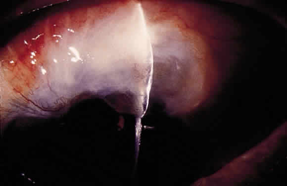
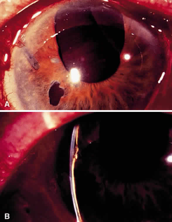
The initial management of early postoperative hypotony with a formed anterior chamber is conservative. Topical steroids and cycloplegics are used. Restrictions in activity (bending, weight lifting) and avoidance of Valsalva-positive conditions are recommended, especially in patients at risk for suprachoroidal hemorrhage (see earlier). If there is hyposecretion related to intraocular inflammation, ciliochoroidal detachment, or both, the initial treatment consists of intense corticosteroid therapy and long-acting cycloplegics, which stabilize the blood-aqueous barrier. Intervention is indicated in patients with hypotony associated with other complications and in persistent, low IOP with loss of visual acuity and hypotony maculopathy. After filtration surgery, prompt management is indicated, also when there is loss of bleb height. Treatment should be aimed at correcting the specific cause of hypotony. When there is lens-corneal touch (flat anterior chamber, grade III) immediate surgical intervention is necessary to prevent endothelial damage and cataract formation (see Chapter 15). Reformation of the anterior chamber with air, balanced salt solution, or, preferably, a viscoelastic can be done at the slit lamp or under the operating microscope through the paracentesis made intraoperatively. Viscoelastic material is best for maintaining, at least temporarily, the anterior chamber depth. When there are large and appositional choroidal effusions, drainage of the fluid also is necessary (Fig. 4). The technique is described in detail in Chapter 15.

Fig. 4. B-scan ultrasound showing hyperreflective dome-shaped echoes corresponding to the detached choroid-retina, with acoustically hollow content corresponding to the fluid accumulated in the suprachoroidal space.

When hypotony results from overfiltration of a filtering bleb, several options are available. A large bandage contact lens,15 symblepharon ring,16 and Simmons' shell17 can be helpful. Bandage contact lenses should have a diameter of at least 16 to 17 mm to cover the filtering bleb. They are not efficient in severe and chronic cases. The Simmons' shell is a 22-mm, dome-shaped rigid shell of transparent polymethylmethacrylate. A raised platform on the concave inner surface of the shell is positioned over the sclerostomy site. The curvature is designed to selectively indent the perilimbal area when pressure dressing is applied. The Simmons' shell usually is effective; however, it may be uncomfortable. Tonometry is not possible to monitor the IOP. Decentration of the shell is frequent unless sutured to the conjunctiva. It requires close (daily) monitoring, and corneal complications (epithelial defects and abrasions) are common. It is particularly difficult for monocular patients. Several chemical and thermal treatments have been used to induce an inflammatory reaction in the filtering bleb, which modify the morphologic features of the filtering blebs and increase the IOP. These procedures include topical application of 0.25% to 1% silver nitrate or 50% trichloracetic acid to the bleb surface,18 cryotherapy,19 diathermy and cauterization,20 argon laser,21 and, recently, the neodymium:yttriumaluminum-garnet (Nd:YAG) thermal laser.22 More than one treatment session might be needed to achieve the desired goal. Possible complications include postoperative discomfort, bleb leak, transient increase in IOP, and corneal edema. Cryotherapy is best done under retrobulbar or peribulbar anesthesia. The probe is applied initially to the lateral borders of the bleb. Before starting the freeze, firm pressure is applied with the cryoprobe to bring the bleb surface tissues into apposition with the underlying sclera. Several applications (two to five) using a temperature of -50° to -80°C and a duration of application of 10 to 30 seconds are used. Nd:YAG thermal laser treatment of overfiltering and leaking blebs has been described more recently. It is best done under regional anesthesia. For this procedure, the continuous-wave mode is required. Energy levels range between 3.0 and 4.0 J, with the laser offset between 0.9 and 1.2 mm and the aiming beam focused in the conjunctival epithelium. The goal is to induce whitening and wrinkling of the conjunctival epithelium. A grid pattern of 30 to 40 spots of laser is placed over the entire bleb. Postoperatively, oral aqueous suppressants and a compressive or “torpedo” (i.e., cotton plug placed directly over the bleb surface) patch are used during the first 48 hours. Injection of autologous blood into the bleb can reduce overfiltration and resolve bleb leaks.23–26 Inflammatory cells and serum proteins from the injected blood may accelerate the inflammatory and healing process, which decreases filtration (Fig. 5). Possible complications include hyphema, endophthalmitis, increase in IOP requiring surgical intervention, bleb failure, corneal blood staining, and corneal graft rejection.27–30 Finally, surgical revision may be needed.31–35 Resuturing the scleral flap and scleral patch grafting (when resuturing is not possible) have been successfully used in patients with hypotony maculopathy associated with overfiltering the filtering bleb. Alternatively, two sets of stitches in the scleral flap, with one set tied tightly, can raise the IOP, stretch the sclera, and flatten chorioretinal folds. Sliding conjunctival flaps or free conjunctival grafts also can be helpful (see later).

Fig. 5. Slit-lamp photograph. Appearance of a filtration bleb after autologous blood injection, which was used to reverse chronic hypotony.

FLAT ANTERIOR CHAMBER AND ELEVATED OR NORMAL INTRAOCULAR PRESSURE

Three conditions should be considered in patients with postoperative flat anterior chamber and elevated or normal IOP: suprachoroidal hemorrhage (see earlier), aqueous misdirection, and pupillary block.

Aqueous Misdirection

Aqueous misdirection, or malignant glaucoma or ciliary block glaucoma, is characterized by a shallowing or flattening of the anterior chamber without pupillary block (i.e., in presence of a patent iridectomy) or choroidal disease (such as suprachoroidal hemorrhage), commonly with an accompanying rise in IOP.36–38 It occurs in 2% to 4% of patients operated on for angle-closure glaucoma but can occur after any type of incisional surgery. The chance of developing malignant glaucoma is greatest in phakic hyperopic (small) eyes with angle-closure glaucoma.

In this condition, aqueous is diverted posteriorly toward the vitreous cavity, increasing the vitreous volume and shallowing the anterior chamber.36 Decompression and shallowing of the anterior chamber appears to be a predisposing factor by inducing forward movement of the peripheral anterior hyaloid. Small choroidal effusions and shallow anterior chamber sometimes occur before the episode of aqueous misdirection. The anterior hyaloid could be placed into direct apposition with portions of the secreting ciliary processes. Thus, aqueous humor might move directly into the vitreous cavity. In hyperopic eyes (with a crowded middle segment), the peripheral anterior hyaloid in its normal position probably is close to the posterior ciliary body. In such eyes, cataract and filtration surgery should be considered as high risk for aqueous misdirection. In aqueous misdirection, a relative resistance to the anterior movement of aqueous humor in the anterior vitreous face or the anterior hyaloid membrane probably occurs. The increased resistance can be related either to abnormal permeability or to available hyaloid surface area for fluid transfer. In normal circumstances, the anterior hyaloid and vitreous offer insignificant resistance to forward fluid flow. In some cases, pupillary block occurs first and is followed by aqueous misdirection. Perhaps a sudden onset of pupillary block forces aqueous humor into the vitreous and expands the vitreous volume, displacing forward the peripheral hyaloid into direct apposition with the ciliary body.39

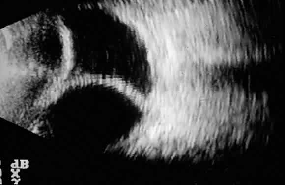
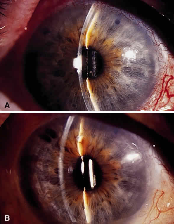
Aqueous misdirection usually occurs in the early postoperative period after filtration or cataract surgery. The anterior chamber is shallow, and the IOP is high (Fig. 6). However, with a functioning filtration bleb, the IOP may not be high. The peripheral iridectomy is patent, and a dilated examination and B-scan ultrasound confirm the absence of choroidal effusion of hemorrhage. If the adequacy of the surgical iridectomy is in doubt and pupillary block is possible, a laser iridotomy should be performed.

Fig. 6. Slit-lamp photographs. A. Aqueous misdirection. The anterior chamber is shallow. Two patent peripheral iridotomies are barely seen at the 10- and 1-o'clock positions. Intraocular pressure was 42 mmHg. B. Same eye 4 weeks after pars plana tube insertion of aqueous shunt with vitrectomy. The anterior chamber is deep. The temporal iridectomy is enlarged. Intraocular pressure was 12 mmHg. (Azuara-Blanco A, Katz LJ, Gandham SB, Spaeth GL: Pars plana tube insertion of aqueous shunt with vitrectomy in malignant glaucoma. Arch Ophthalmol 116:808, 1998)

Medical treatment and laser and vitreous surgery all have been useful options to treat aqueous misdirection. This condition is initially managed with mydriatic-cycloplegic drops,40 aqueous suppressants, and hyperosmotics. Topical 1% atropine or 1% cyclopentolate four times daily and 2.5% phenylephrine four times daily are used. These agents hopefully result in a posterior movement of the lens-iris diaphragm. In cases of aphakic aqueous misdirection, mydriatic-cycloplegic drops are of little benefit. However, it is reasonable to use them for their effect on relaxation of the ciliary body muscle. Systemic carbonic anhydrase inhibitors and topical beta-adrenergic blocking agents in full doses are important. Osmotics (isosorbide, glycerin, or intravenous mannitol) also can be helpful to decrease the fluid content of the vitreous cavity and can be given every 12 hours. If it is well tolerated and there are no contraindications, the medical treatment is tried for 2 to 4 days. If the condition is relieved (i.e., the anterior chamber has deepened), the hyperosmostic agents are discontinued first, and the aqueous suppressants are reduced or stopped over several days. Phenylephrine drops can be stopped, but the cycloplegic drops should be continued for months to years or, in some cases, indefinitely to prevent the recurrence of this condition. Medical treatment relieves about 50% of cases of aqueous misdirection. If medical therapy is unsuccessful and the ocular media are clear, a Nd:YAG laser capsulotomy and hyaloidotomy is used to disrupt the anterior vitreous face, especially in pseudophakic and aphakic patients.41 The usual beginning laser energy is between 2 and 4 mJ. The focus is placed posterior to the anterior hyaloid. After a successful Nd:YAG hyaloidotomy, a slight deepening usually is seen, which increases over the next hours. In pseudophakic eyes, a peripheral hyaloidotomy is more efficient than a central hyaloidotomy because the lens capsule and intraocular lens can prevent communication between the vitreous cavity and the anterior chamber. In phakic eyes, Nd:YAG hyaloidotomy can be tried through the peripheral iridectomy, focusing behind the zonules but in front of the ciliary body. However, a clear view and sharp focusing may not be possible, and there is a risk of lens or zonular injury. Pars plana vitrectomy should be considered when other therapies fail.42–44 A standard three-port pars plana vitrectomy, removing the anterior vitreous and part of the anterior hyaloid, is done. In phakic patients, the lens sometimes can be spared, but the probability of recurrence is higher. Pars plana tube shunt insertion with vitrectomy has been recommended to treat patients with aqueous misdirection, especially in patients with angle-closure glaucoma. The implantation of the tube shunt through pars plana can help to prevent recurrence of this condition and can help in long-term control of IOP.44

PREVENTION. In high-risk eyes undergoing cataract or filtration surgery, the decompression and shallowing of the anterior chamber should be minimized. In filtration procedures, the use of viscoelastic and a large peripheral iridectomy can be helpful. Postoperative overfiltration should be avoided with a thick scleral flap sutured tighter and with more sutures than usual. Postoperatively, judicious suture lysis or cutting/pulling releasable sutures and slow tapering of cycloplegics are recommended. A postoperative shallow anterior chamber caused by overfiltration should be vigorously treated.

Pupillary Block

Pupillary block can be caused by adhesions between the iris and lens, pseudophakos, or vitreous. The inability of aqueous humor to pass from the posterior to the anterior chamber results in the forward movement of the peripheral iris and closure of the drainage angle. Pupillary block typically occurs as a flat or shallow anterior chamber with normal or elevated pressure. It may be difficult to distinguish from malignant glaucoma.

Although a peripheral iridectomy is intended at the time of filtration surgery, in a few patients only the stroma of the iris is removed and the posterior pigment epithelium is left intact. In these patients, blockage may develop. In other patients, the iris may become incarcerated in the wound or the iridectomy may be obstructed by intraocular tissue such as Descemet's membrane, anterior hyaloid surface, vitreous (in aphakic eyes), or ciliary processes.

Therapy with cycloplegic-mydriatics may resolve pupillary block, but a Nd:YAG peripheral iridotomy should be done. The anterior chamber readily deepens after iridotomy is performed, although in the presence of localized compartments of blockage, multiple iridotomies are necessary. This deepening usually is associated with the sudden escape of aqueous humor through the iridectomy and confirms the diagnosis of pupillary block. If laser iridotomy cannot be completed, a surgical iridectomy should be done.

VISUAL LOSS

Unexplained loss of central visual field (i.e., “wipeout”) after glaucoma surgery is rare. Older patients with advanced visual field defects affecting the central field with split fixation are at increased risk. Early, undiagnosed postoperative IOP spikes and severe postoperative hypotony have been suspected as causes for wipeout.45,46

Back to Top
SPECIFIC COMPLICATIONS RELATED TO THE TYPE OF PROCEDURE

COMPLICATIONS OF GUARDED FILTRATION PROCEDURES

Intraoperative Complications of Filtration Procedures

Conjunctival buttonholes and tears can lead to failure of bleb formation and flat anterior chamber. The usual cause for conjunctival buttonholes is penetration of the tissue by the tip of a sharp instrument (needle, scissors, blade) or the teeth of a forceps. Buttonholes and tears are more likely to occur in patients with extensive conjunctival scarring. To diagnose a buttonhole intraoperatively, the conjunctiva should be carefully examined at the end of the procedure by filling the anterior chamber and raising the filtering bleb. If recognized, the buttonhole should be closed at the time of surgery. If it is located in the center of the conjunctival flap, a “purse string” closure is attempted, either internally on the undersurface of the conjunctiva or externally if the flap already has been reapproximated. A 10-0 or 11-0 nylon on a tapered (“vascular”) needle should be used. When the conjunctival buttonhole or tear occurs at the limbus, it can be sutured directly to the cornea, which should be de-epithelialized. A mattress suture or, if large, a running suture with 10-0 nylon can be used. When the buttonhole or tear occurs near the incised edge of a limbal-based conjunctival flap, the sutures used to close the conjunctival incision can be placed anterior to the tear to close it.

SCLERAL FLAP DISINSERTION. A thin scleral flap can be torn or amputated from its base during the surgical procedure. If a sclerostomy has not been performed, a new scleral flap should be dissected in a different area. If a sclerostomy has been done already, reapproximation of the scleral flap can be attempted with 10-0 or 11-0 nylon sutures. If unsuccessful, additional tissue usually is necessary to cover the sclerostomy. This can be obtained by transferring a piece of Tenon's capsule or a flap of partial-thickness sclera from the area adjacent to the defect. Alternatively, donor sclera, fascia lata, or pericardium can be used.

VITREOUS LOSS. Vitreous loss during glaucoma surgery is an uncommon complication, especially in phakic eyes. Conditions that may predispose to vitreous loss include high myopia, previous intraocular surgery, trauma, aphakia, and a subluxated lens. Loss of vitreous can be associated with several complications such as corneal edema, epithelial downgrowth, uveitis, retinal detachment, cystoid macular edema, and endophthalmitis. The vitreous can mechanically plug the sclerostomy and lead to filtration failure. Vitreous should be removed from the surgical site and anterior chamber with a vitrectomy instrument, avoiding damage to the lens in phakic eyes. In the aphakic eye with vitreous filling the anterior chamber, an anterior vitrectomy can be planned as part of the primary procedure. In phakic or pseudophakic eyes with vitreous in the anterior chamber, pars plana vitrectomy may be considered to adequately remove the vitreous from the posterior segment and to avoid lens or intraocular lens subluxation and lens injury.

Postoperative Complications of Filtration Procedures

BLEB LEAK. Bleb leaks can occur early in the postoperative period or months to years after filtration surgery. An inadvertent buttonhole in the conjunctiva during a filtering procedure or a wound leak through the conjunctival incision can be responsible for an early leaking bleb. Spontaneous late bleb leaks are more frequent in avascular, thin blebs, which occur more frequently when antimetabolites are used in the filtering procedure and after full-thickness procedures. The incidence of early and late bleb leaks probably is higher in trabeculectomies supplemented with antimetabolites than nonsupplemented surgeries.47,48 Leakage of the filtering bleb can be associated with hypotony, shallow or flat anterior chamber, and choroidal detachment and may increase the chances for bleb infection and subsequent endophthalmitis. Early leaking can flatten the bleb and lead to subconjunctival-episcleral fibrosis, which would jeopardize a satisfactory long-term filtration.

Bleb leaks are detected with the Seidel test (Table 2). The tear film is stained with fluorescein. For this purpose, a fluorescein strip is applied to the inferior tarsal conjunctiva or, gently, directly to the bleb. Without applying pressure, the eye is examined under cobalt blue illumination. If there is a leak, unstained aqueous humor is seen flowing into the tear film (Fig. 7). If there is no spontaneous leakage, pressure may be gently applied to the globe or to the bleb while the suspicious area is examined.


TABLE 2. Seidel's Test

  1. Cobalt blue illumination
  2. Fluorescein stain; fluorescein strips to gently “paint” the bleb
  3. Aqueous humor flowing into the tear film is seen
  4. In absence of spontaneous leakage, pressure may be carefully applied to the globe or to the bleb


Fig. 7. Slit-lamp photograph of a positive Seidel's test result.

The need and urgency of the management of bleb leaks depend on several factors. Some patients, particularly monocular individuals with leaking blebs that have had previous episodes of bleb-related infections, ocular hypotony, shallow or flat anterior chamber, loss of bleb elevation, or reduced vision always should be treated. However, if there are no complications, such as in patients with late leaks with formed blebs, normal IOP, good central vision, and without previous episodes of bleb-related infection, the leak may not require therapy. Observation sometimes is possible to allow spontaneous closure of the leak. Pharmacologic medical treatment with agents that decrease aqueous secretion (topical beta blockers, carbonic anhydrase inhibitors, or both) and discontinuation of topical steroids, with or without patching, may help the spontaneous closure of these defects by reducing flow of aqueous through the fistula. Prophylactic broad-spectrum antibiotic coverage, alternating different antibiotics, is recommended by some. Patient education regarding symptoms of bleb-related ocular infection is crucial for prompt diagnosis and management.

Therapeutic modalities to treat leaking blebs include pressure patching, bandage contact lens,15 Simmons' shell,17 injection of autologous blood,23–30 cryopexy,19 thermal Nd:YAG laser (see earlier),22 cyanoacrylate glue,49–51 fibrin tissue glue,52–53 and surgical revision.54 A large-diameter (17 to 20 mm) soft therapeutic contact lens can be helpful. The contact lens should be kept in place, if possible, for 1 week to allow reepithelization. Broad-spectrum topical antibiotics should be administered to protect against infection, and close observation is mandatory. Fibrin tissue glue is a mixture of fibrinogen and thrombin that induces the formation of a clot, which can seal bleb leaks.52–57 It is a nonirritating procedure that requires no patching. Tisseel (Immuno AG Industries, Vienna, Austria) is a commercialized fibrin glue—not approved by the Food and Drug Administration—that has the disadvantage of being prepared from pooled plasma and thus may have the potential risk of transmitting blood-borne pathogens.57 Autologous fibrin tissue glue is prepared from the patient's blood, therefore eliminating the risk for disease transmission.53 Cyanoacrylate glue (Histo-acryl, B. Brown Melsungen, Germany) adheres to tissues and can effectively close an early bleb leak seen shortly after surgery.49–51 However, the use of cyanoacrylate glue is not recommended in thin, avascular blebs with leaks developed months to years after surgery. The glue must be applied to a dry conjunctival surface, and only a small amount of glue should be used. The use of a bandage contact lens can prevent the adhesive from being dislodged. When other simpler methods have failed and the bleb leak is persistent, surgical intervention is necessary.58–63 It is important to attempt to save the established initial filtration site. Because of the friable nature of the conjunctiva in long-established filtering blebs, it often is impossible to close the defect directly with sutures and, therefore, healthy conjunctival tissue is needed. First, the ischemic and thin-walled bleb tissue is denuded of conjunctival epithelium by blade debridement or cautery to allow long-term adherence of the grafted conjunctiva. Fresh conjunctiva adjacent to the bleb then is mobilized to cover the previous filtration site by rotational, sliding, or free conjunctival grafts. Separate dissection of conjunctiva and Tenon's capsule sometimes aids in closure because although conjunctiva often can be stretched further than Tenon's capsule, the closure of the latter provides more support. The conjunctiva is sutured over the previously abraded peripheral cornea, providing a watertight seal. Amniotic membrane can be used as an alternative substrate (unpublished data, 1998, Budenz et al). With these methods, bleb function usually can be preserved. The use of a donor scleral flap is likely to compromise the function of the filtering bleb. The latter option is most efficient to treat cases of inadvertent bleb after cataract surgery.

EARLY AND LATE FAILURE OF FILTERING BLEB. Failed blebs are those associated with inadequate IOP control and impending or established obstruction of aqueous outflow. The cause of failure of filtering operations can be divided into intraocular, scleral, and extraocular factors. The extraocular changes account for most failures of external filtering operations.64

Early failure of filtering blebs is characterized by a high IOP, deep anterior chamber, and low and hyperemic bleb.54 Failing blebs should be recognized promptly because if obstruction is not relieved, permanent adhesions between conjunctiva and episclera can lead to closure of the fistula. A tight scleral flap and episcleral fibrosis are the most common causes of early bleb failure. Internal obstruction of the fistula by blood clot, vitreous, iris, or incompletely excised Descemet's membrane also is possible.

To reduce postoperative subconjunctival fibrosis and preserve bleb function, postoperative topical steroids are routinely used65 (see Chapter 15). The use of antifibrotic agents in filtering procedures is associated with a higher success rate but also with a higher complication rate (wound leak and hypotony from overfiltration, hypotony maculopathy, and ocular infection). Therefore, an individualized consideration of the risk-benefit ratio is recommended. 5-Fluorouracil (5-FU) usually is administered in 5-mg aliquots during the first 2 weeks after surgery. The dose is adjusted according to the tolerance of the corneal epithelium of each eye. Intraoperative application of 5-FU also has been described (see Chapter 15). Complications associated with the use of postoperative 5-FU include corneal and conjunctival epithelial toxicity, corneal ulcers, conjunctival wound leaks, subconjunctival hemorrhage, or inadvertent intraocular spread of 5-FU. The frequency of complications is reduced with lower titrated dosages of 15 to 50 mg administered in 3 to 10 injections according to individual response.47,66–68 Mitomycin C is approximately 100 times more potent than 5-FU.69 Postoperative complications associated with overfiltration, hypotony maculopathy, bleb leak, and bleb-related ocular infections are more likely to occur when mitomycin C is used.11,13,14,70–73

Digital ocular compression and focal compression can be used to temporarily improve the function of a nonfunctioning filtering bleb. Digital ocular compression can be applied to the inferior sclera or cornea through the inferior eyelid or to the sclera posterior to the scleral flap through the superior eyelid. Focal compression (Carlo Traverso maneuver) is applied with a moistened cotton tip at the edge of the scleral flap (see Chapter 15 for a detailed description).74,75 Because of potential complications, digital ocular compression is suitable for patients who are physically capable of performing it and had a beneficial response to the initial massage by the physician. In the early postoperative period, laser suture lysis can enhance the filtration.76–81 Gonioscopy performed before the laser can confirm an open sclerostomy with no tissue or clot occluding its entrance. Specially designed lenses such as Hoskins, Ritch, or Mandelkorn lens; the central button edge of the Zeis and Sussman lenses; the Goldmann lens; glass rods; or glass pipettes can be used (Fig. 8). The laser parameters recommended are summarized in Table 3. After the suture is cut, if the bleb and IOP are unchanged, ocular massage or focal pressure can be applied. Usually, only one suture is cut at a time to avoid the possible complications of overfiltration. A hole in the conjunctiva may occur because of trauma from the contact lens or the thermal burn of the laser. If there is subconjunctival hemorrhage, suture lysis can be difficult. In these cases, krypton red or a diode laser should be used because their wavelengths are least absorbed by blood. Some sutures have more influence in restricting the aqueous runoff than others. These “key” sutures should be identified during surgery, and greater caution should be advised when cutting them. The timing of suture release is critical. Suture lysis is effective within the first 2 weeks after surgery without antimetabolites; later, fibrosis of the scleral flap may negate any beneficial effect of this procedure. If antimetabolites have been used at the time of surgery, suture lysis can be effective months after surgery.82,83 Releasable sutures are as effective as laser suture lysis. The use of releasable sutures allows the surgeon to tightly close the scleral flap, knowing that the flow can be increased postoperatively. The externalized sutures are easily removed and are effective in cases of hemorrhagic conjunctiva or thickened Tenon's tissue (which would make suture lysis difficult). The disadvantages of releasable sutures include the need for additional intraoperative manipulation and postoperative discomfort from the externalized suture, corneal epithelial defects, and, possibly, increased risk of ocular infection. Several techniques have been described by Wilson84 (mattress-type suture with an externalized knot on the cornea), Shin85 (removable knot passed through the conjunctival bleb), Cohen and Osher86 (loop-knot suture externalized through the cornea), Hsu and Yarng87 (an externalized hemibow tie in the center of the filtering bleb), Maberley and associates88 (a two-arm “U” suture that leaves no exposed suture end until one arm of the suture is removed), and Johnstone and coworkers89 (releasable “tamponade suture”). In patients with incarceration of iris or vitreous occluding the sclerostomy, Nd:YAG Laser internal revision can be tried.90,91 When the cause of filtration failure is a blood clot or fibrinous clot occluding the sclerostomy, tissue plasminogen activator can be helpful.92–95 Recombinant tissue plasminogen activator is a serine protease with clot-specific fibrinolytic activity. It can be injected into the anterior chamber or subconjunctivally, and the dosage is 7 to 10 μg in 0.1 ml. It works rapidly, so that within 3 hours the effect usually is apparent. Hyphema is the most frequent complication28,92 (Fig. 9).

Fig. 8. Slit-lamp photograph. A Hoskins lens is used to improve the view of the trabeculectomy flap sutures.


TABLE 3. Argon Suture Lysis

  1. Pretreatment with topical phenylephrine 2.5% (optional)
  2. Argon laser parameters:

      Spot size: 50 μm
      Exposure time: 0.05–0.1 second
      Power: 500–700 mW (from 200 to 1000 mW)


  3. Topical anesthesia (proparacaine 0.5%)
  4. Contact lens (Hoskins, Ritch, or other)
  5. Focus the beam posterior to the conjunctiva
  6. If intraocular pressure still elevated and bleb unchanged: gentle ocular massage or focal pressure


Fig. 9. Slit-lamp photograph. Severe intraocular bleeding and subconjunctival hemorrhage after intracameral injection of tissue plasminogen activator.

A late failure of filtering bleb can occur after months to years of adequate function, with subconjunctival-episcleral fibrosis the most common cause.64 Several factors accelerate fibrosis, such as a black race, childhood, postoperative subconjunctival hemorrhage, the presence of reactive sutures, and inflammation.54 Less commonly, internal closure of the sclerostomy can occur. A classic example is late closure of the internal sclerostomy by a membrane in patients with iridocorneal endothelial syndrome. “Warning signs” of failing filtration are increased bleb vascularization, bleb inflammation, and bleb thickening. The IOP typically is high.

In patients with subconjunctival-episcleral fibrosis, an external revision can be tried. A 27- or 30-gauge needle is used to cut the edge of the scleral cap and restore aqueous outflow. Entry into the anterior chamber beneath the flap may be important but should be undertaken with extreme caution in phakic eyes. The technique can be repeated as needed.58,96–98 The outcome may be more favorable if there was a previously well-established filtration bleb before the fistula became occluded. Subconjunctival injections of 5-FU for 2 weeks after revision increase the probability of success.99,100 Mitomycin C also has been used in conjunction with needling of blebs.101 Ab externo holmium:YAG laser sclerostomy can be used to restore the bleb function.102–104 Its advantages compared with filtering procedures include a small conjunctival incision, access to areas difficult to operate, technical simplicity, and shorter operative time. The conjunctiva must be mobile in the proposed sclerostomy site. The probe should be directed to the area where the previous sclerostomy and iridectomy were performed. Subconjunctival 5-FU injections are recommended after laser sclerostomy revision.54 Early postoperative hypotony is the rule if the sclerostomy is open, similarly to other full-thickness procedures. Nd:YAG laser external revision also has been described to externally revise failed filtering blebs.105,106 Finally, a repeat glaucoma surgery with another filtration site can be used.

When there is internal obstruction of the sclerostomy, a Nd:YAG internal revision is best. Transcorneal surgical revision with a curved needle or a goniotomy blade rarely is needed.54

ENCAPSULATED BLEBS. Encapsulated blebs are localized, elevated, and tense filtering blebs with vascular engorgement of the overlying conjunctiva and a thick connective tissue54 (Fig. 10). This type of bleb commonly appears within 2 to 4 weeks after surgery.107–113 Encapsulation of the filtering bleb is associated with a rise of IOP after an initial period of pressure control after glaucoma surgery. They can interfere with upper lid movement and tear film distribution, leading to corneal complications such us dellen and astigmatism. Often, it is seen through the eyelid simulating a lid mass. The frequency of bleb encapsulation after trabeculectomies without antimetabolites ranges from 8.3% to 28%.107–111 In trabeculectomies with postoperative 5-FU, the incidence frequently has been reported to be higher.112 The frequency of encapsulated blebs after guarded filtering procedures with mitomycin C is lower.113 Predisposing factors may include male gender and the use of gloves with powder, as well as prior treatment with sympathomimetics, argon laser trabeculoplasty, and surgery involving the conjunctiva.107 The causes of encapsulation are not clearly identified, but inflammatory mediators probably are involved in their development. The long-term prognosis for IOP control in eyes that develop encapsulated bleb is relatively good.

Fig. 10. Slit-lamp photograph. Encapsulated filtering bleb.

Initial management of encapsulated blebs include antiglaucoma medications in cases of elevated IOP, topical steroids, and digital massage.98,110,111 Deciding between conservative management (medical) or a surgical revision usually is dependent on the severity of glaucomatous damage, the level of IOP, and the response to medical management. When surgical revision is needed, the simplest technique is to cut the fibrotic wall with a 27-gauge needle or a Ziegler knife. This procedure can be done at the slit lamp and, if effective, restores aqueous outflow to a larger subconjunctival area. Alternatively, the fibrous tissue can be excised after reopening the conjunctival incision from the previous filtering operation is reopened.114,115 Subconjunctival injections of 5-FU for 2 weeks after bleb revision increases the chances of success.54,99

SYMPTOMATIC BLEBS. Filtering blebs usually are asymptomatic. Some patients have discomfort, which is most common with nasal,116 large blebs extending onto the cornea54 (Fig. 11). Tear film abnormalities with dellen formation and superficial punctate keratopathy may occur.117 Corneal astigmatism, visual field defect, and monocular diplopia have been described in cases in which large filtering blebs had migrated onto the cornea.118

Fig. 11. Slit-lamp photograph. Large avascular thin-walled filtering bleb with corneal extension.

Artificial tears and ocular lubricants can be helpful, especially in patients with abnormal tear film. Several chemical and thermal methods have been used to shrink blebs (see earlier). A temporary medial tarsorrhaphy can alleviate symptoms of a nasal bleb and shift it superiorly. Large blebs that extend onto the cornea can be freed by blunt dissection. The corneal extension can be excised with a cut parallel to the limbus, usually with excellent results. Partial surgical excision and conjunctival flap reinforcement usually are helpful, although bleb failure is a possibility.54,58,119

CATARACT FORMATION AFTER FILTRATION SURGERY. Cataract formation and progression of preexisting cataract can occur after filtration procedures. The reported incidence varies from 2% to 53%.120–123 Lens opacification is the main cause of early visual loss after filtration surgery. Intraoperative lenticular trauma is possible and can be recognized shortly after surgery. Postoperative flat anterior chamber with lens-corneal touch rapidly precipitates cataract formation. Other probable risk factors include age, presence of exfoliation, use of air to reform the anterior chamber, profound hypotony, use of miotics and topical steroids, and inflammation.120–124

Cataract extraction can be associated with an impairment of the function of the filtering bleb. Phacoemulsification of the lens with a corneal incision induces less conjunctival inflammation than large scleral incisions and may be theoretically the best method to preserve the function of the bleb. Postoperative subconjunctival injections of 5-FU can be considered. If the control of IOP is borderline, a combined cataract extraction and filtration procedure may be the best choice.

BLEB-RELATED OCULAR INFECTION. Ocular infections related to filtration procedures can occur months to years after the initial surgery.54,125–137 The incidence of bleb-related ocular infections after filtration procedures not supplemented with antifibrotic agents ranges from 0.2% to 1.5% after midterm and long-term follow-up.126,127 Inferior filtering blebs128–130 and the use of antifibrotic agents during filtration surgery increase the probability of bleb-related ocular infection.47,73,129–131 Thin bleb wall, frequently seen after full-thickness procedures and trabeculectomies with antimetabolites, and bleb leaks probably are associated with a higher risk of infection. Bleb-related ocular infections can affect three compartments: the subconjunctival space, the anterior segment, and the vitreous cavity. Usually, the spread of infection proceeds in that order. Because the fluid within the bleb is continuous with the anterior chamber, the bleb may be considered an exteriorized portion of the anterior chamber. Therefore, an infection of the bleb affecting the subconjunctival space (“blebitis”) has a real potential to rapidly spread posteriorly. The bacteria that cause bleb-related endophthalmitis certainly arise from the ocular flora. The most commonly involved organisms include Streptococcus species, Hemophilus influenzae, and Staphylococcus species.132–135

Patients with bleb-related ocular infection usually present with ocular pain, blurred vision, tearing, redness, and discharge. Examination often reveals conjunctival and ciliary injection (most intense around the bleb edge), purulent discharge, and variable intensity of periorbital chemosis, corneal edema, and anterior chamber reaction, including keratic precipitates and, in some cases, hypopyon. The bleb typically has a milky white appearance with loss of clarity; a pseudohypopyon within the bleb can be observed. A positive Seidel's test result is common, and some patients may have a substantial leak, hypotony, and even flat anterior chamber. Alternatively, an increased IOP is possible because of internal closure of sclerostomy site with purulence and debris. Vitreous reaction is not evident in early cases of blebitis, but untreated, the infection spreads to the posterior segment.54

Bleb-related ocular infections have been classified into three different stages.54 In grade I, there is only bleb involvement (Fig. 12). Erythema around the bleb and milky white appearance of the bleb with loss of clarity is observed. In grade II, the infection has extended into the anterior chamber, and cells and flare are noted. Hypopyon may be seen (Fig. 13). In grade III, the vitreous is involved. If the media is not clear (i.e., dense cataract), B-mode ultrasonography can be helpful to detect involvement of the retrolental area.

Fig. 12. Slit-lamp photograph. Bleb-related ocular infection, grade I (without involvement of the anterior chamber). There is an intense erythema around the filtering bleb.

Fig. 13. Slit-lamp photograph. Bleb-related ocular infection, grade II (with anterior chamber involvement). A small hypopyon is seen.

The general principles that guide the management of ocular infections apply to this condition.54,136 Identifying It the organism responsible is important. A conjunctival sample is routinely collected, stained, and cultured. However, the value of conjunctival culture in the etiologic diagnosis of bleb-related endophthalmitis has little value. A vitreous sample should be obtained in grade III. Antibiotic sensitivities should be determined on positive cultures. In grade I (blebitis without anterior chamber reaction), frequent topical application of a commercially available broad-spectrum antibiotic can be used with close supervision. Steroids can be considered to reduce the intense inflammation and preserve bleb integrity when the infection appears to be controlled. In grade II (the anterior segment but not the posterior segment is involved), treatment with fortified topical antibiotics around the clock is advisable. Topical fortified cefazolin or vancomycin (25 mg/ml) associated with fortified tobramycin (14 mg/ml) or amikacin (50 mg/ml) are likely to be effective against most gram-positive and gram-negative microorganisms. Additional systemic antibiotics such as ofloxacin (400 mg orally twice daily) can be used. In grade III (bleb-related endophthalmitis), intravitreal antibiotics are required, administered either through a pars plana injection at the time of sampling or associated with a vitrectomy. We are currently using 1 mg of vancomycin (10 mg/ml) and 400 μg of amikacin (5 mg/ml). Oral ofloxacin (400 mg orally twice daily) and fortified topical eye drops of the same antibiotics that were injected into the vitreous cavity should be used.54

After resolution of the infection, the function of the filtration bleb may be impaired, especially in eyes that have not received antifibrosis agents.136 Other possible complications include corneal edema, cataract, vitreoretinal traction, and retinal toxicity from the bacteria's toxins or the antibiotics. The visual outcome usually is good in cases with anterior segment involvement and poor when the vitreous is involved, especially with virulent bacteria such as streptococci, coagulase-positive staphylococci, and gram-negative organisms.

Prevention of bleb-related ocular infection is important.54 Some ophthalmologists use long-term topical antibiotics after filtration procedures,137 although the efficacy of this regimen has been questioned. It seems reasonable to use long-term antibiotics in some cases of leaking blebs, inferior blebs, or recurrent bleb-related infections. Conjunctivitis and blepharitis should be treated promptly, and soft contact lens wear should be avoided. Patient education about early symptoms of infection currently is the most important approach to minimize the chances of severe visual loss.

COMPLICATIONS OF SURGICAL PERIPHERAL IRIDECTOMY

Surgical peripheral iridectomy is necessary in uncooperative patients and those with poor visualization (i.e., angle-closure glaucoma with a hazy cornea), which impedes a laser iridotomy (see Chapter 15). After this procedure, bleeding usually is minimal. Flat or shallow anterior chamber without elevation of IOP usually results from wound leak. It should be reformed promptly because of the risk of aqueous misdirection, which is most likely to occur in patients with angle-closure glaucoma (see earlier).

COMPLICATIONS OF AQUEOUS SHUNT IMPLANTATION

Implantation of aqueous tube shunts is associated with a significant risk of postoperative complications.137–146 Early postoperative complications include hypotony, flat anterior chamber, choroidal effusions, suprachoroidal hemorrhage (see Fig. 1), aqueous misdirection, hyphema, and increased IOP. The use of intraoperative mitomycin C does not influence the success and complication rate of tube shunt implantation.144–146 Hypotony, the most common complication, usually results from excessive outflow of aqueous humor and may result in flat anterior chamber and choroidal detachment. In cases of flat anterior chamber and choroidal effusions, surgical intervention may be needed (see earlier). Recurrent flat anterior chamber requires temporary tube ligation. Restrictive or valved implants may be less commonly complicated with hypotony than unregulated devices.

High IOP can be related to occlusion of the tube by a fibrin or blood clot, iris, or vitreous. The fibrin or blood may resolve spontaneously; however, an intracameral injection of tissue plasminogen activator helps to dissolve the clot within few hours (see earlier). When the iris tissue occludes the lumen of the tube, a Nd:YAG laser iridotomy or an argon laser iridoplasty may reestablish the patency of the tube. Vitreous incarceration can be treated successfully with Nd:YAG laser, but an anterior vitrectomy may be necessary. Recurrence and closure of the tube is common after successful treatment with the Nd:YAG laser.147 The tube can retract from the anterior chamber. If the tube is too short (i.e., not seen in the anterior chamber), the plate should be moved anteriorly. In nonrestrictive devices, high IOP can be related to a tight ligature, internal occlusion, or both. In these cases, aqueous suppressants are used to control the IOP within an acceptable range for the first 3 to 4 weeks. At this time, profound hypotony is least likely to occur, and the device used to increase the outflow resistance can be safely removed.

Late postoperative complications of aqueous shunting procedures include increased IOP, hypotony, implant migration, conjunctival erosion, corneal edema or decompensation, cataract, diplopia, and endophthalmitis. Late failure with increased IOP can be caused by blockage of the anterior chamber tube opening (see earlier) and excessive fibrosis around the plate. Surgical bleb revision (excision or needling of the fibrous wall) with antifibrosis agents can be helpful. A failed procedure also can be managed by implantation of additional episcleral devices. Corneal decompensation may result from direct contact between the tube and the cornea. When there is tube-cornea touch, repositioning of the tube should be considered, especially in cases where there is risk of endothelial failure (i.e., patients with focal corneal edema, or after penetrating keratoplasty). Diplopia results from mechanical restriction of the extraocular muscles.141 If diplopia is persistent, the shunt may need to be removed or relocated.

COMPLICATIONS OF CYCLODESTRUCTIVE PROCEDURES

The use of cyclodestructive procedures in its various forms usually has been restricted to eyes with recalcitrant and end-stage glaucoma because of their limited predictability. Some eyes require multiple treatments to achieve pressure lowering, whereas others become hypotonous or phthisical after a single session. The cyclodestructive procedures that are currently used are cyclocryotherapy,148,149 noncontact Nd:YAG laser cyclophotocoagulation (CPC),150,151 contact Nd:YAG laser CPC,152,153 contact diode CPC,154,155 and endophotocoagulation.156,157 The latter techniques have the potential for more controlled destruction of the ciliary body processes and a lower incidence of complications compared with cyclocryoablation. For example, the 810-nm semiconductor diode laser has the theoretical advantages of good penetration and selective absorption by the pigmented tissues of the ciliary body. Endophotocoagulation offers the possibility of selectively treating the ciliary body epithelium with relative sparing of surrounding tissues.

Possible complications of cyclocryotherapy include severe pain, elevation of IOP, hyphema (common in eyes with neovascular glaucoma), visual loss (wipeout fixation in patients with advanced optic nerve damage), choroidal detachment, retinal detachment, chronic hypotony, cystoid macular edema, anterior segment necrosis, vitreous hemorrhage, aqueous misdirection, cataract, lens subluxation, and phthisis.148–158 Pain often occurs during the first 2 days after cyclocryotherapy, and strong analgesics (narcotics) should be used routinely. Laser cyclodestructive procedures usually do not require strong analgesia. A major concern is the possibility of phthisis bulbi (0% to 7%) after cyclodestructive procedures. It is more common in neovascular glaucoma and in patients who underwent cyclocryotherapy in four quadrants. It is the least common after diode laser CPC. The possibility of sympathetic ophthalmia after cyclodestructive procedures also is of great concern. It has been reported after noncontact and contact Nd:YAG laser CPC.159–162

COMPLICATIONS OF CYCLODIALYSIS

Cyclodialysis is not a popular procedure because it is unpredictable. However, some clinicians still use it, especially in aphakic and pseudophakic glaucomas. After the procedure, miotics are used to maintain an open cleft (Fig. 14). Cycloplegics should be avoided. Cyclodialysis lowers the IOP by increasing uveoscleral outflow and, secondarily, by decreasing aqueous humor formation.

Fig. 14. Composite of ultrasound biomicroscopic examination. A cyclodialysis cleft is seen.

Common complications of cyclodialysis are intraoperative bleeding and hyphema, which may decrease the chances of long-term success. Postoperative hypotony is associated with accumulation of fluid between the ciliary body and sclera. If the accumulation of fluid extends posteriorly, it may reach the macula and impair visual acuity. The degree of hypotony is not related to the length of the cleft in the angle that is observed gonioscopically. If visual function is compromised, cryotherapy can be used to partially close the cleft. Cryotherapy may be ineffective or it may induce complete closure of the cleft and elevation of IOP. Surgical closure of the cleft may be necessary. Spontaneous closure of the cleft may occur months after a successful surgery, producing an acute rise of IOP and pain resembling an attack of acute angle-closure glaucoma. In some cases, intensive miotic treatment associated with phenylephrine can reopen the cleft.

Other possible complications include corneal opacity, injury to Descemet's membrane, iridocyclitis, corectopia, lens subluxation, cataract, loss of vitreous, vitreous hemorrhage, retinal detachment and myopic refractive shift.

COMPLICATIONS OF TRABECULOTOMY AND GONIOTOMY

Trabeculotomy and goniotomy are the first surgical options to treat infants with glaucoma.163–166 Trabeculotomy is preferred when the cornea is so clouded than the angle cannot be properly visualized. Ultrasound biomicroscopy can be used to evaluate the anterior chamber angle before and after surgery in infants with glaucoma and corneal opacity.167 Trabeculotomy also can be a useful option to treat some adult glaucomatous patients.168–169

Severe complications after trabeculotomy and goniotomy are rare. Moderate bleeding into the anterior chamber is common. Blood clots usually resorb within a few days. Intraoperative complications during trabeculotomy can be related to difficulty identifying Schlemm's canal, which is more difficult to locate in infants. The initial goal is to open the outer wall of Schlemm's canal without perforating the inner wall into the anterior chamber. If penetration into the anterior chamber occurs, the iris may prolapse. In this case, an iridectomy may be necessary. If the subciliary space is wrongly probed, forward rotation into the anterior chamber is not possible unless considerable force is exerted, causing cyclodialysis and iridodialysis. If the tip of the trabeculotomy probe is held toward the cornea during rotation, a Descemet's membrane tear can occur, but they usually are small and do not cause corneal edema.

Back to Top
REFERENCES

1. Brubaker RF: Intraocular surgery and choroidal hemorrhage. Arch Ophthalmol 102:1753, 1984

2. Bukelman A, Hoffman P, Oliver M: Limited choroidal hemorrhage associated with extracapsular cataract extraction. Arch Ophthalmol 105:338, 1987

3. Payne JW, Kameen AJ, Jensen AD et al: Expulsive hemorrhage: Its incidence in cataract surgery and a report of four bilateral cases. Trans Am Ophthalmol Soc 83:181, 1985

4. Cantor LB, Katz LJ, Spaeth GL: Complications of surgery in glaucoma: Suprachoroidal expulsive hemorrhage in glaucoma patients undergoing intraocular surgery. Ophthalmology 92:1266, 1985

5. Ruderman JM, Harbin TS, Campbell DG: Postoperative suprachoroidal hemorrhage following filtering procedures. Arch Ophthalmol 104:201, 1986

6. Givens K, Shields MB: Suprachoroidal hemorrhage after glaucoma filtering surgery. Am J Ophthalmol 103:689, 1987

7. Manshot WA: The pathology of expulsive hemorrhage. Am J Ophthalmol 40:15, 1955

8. Frenkel REP, Shin DH: Prevention and management of delayed suprachoroidal hemorrhage after filtration surgery. Arch Ophthalmol 104:1459, 1986

9. Schubert HD: Postsurgical hypotony: Relationship to fistulization, inflammation, chorioretinal lesions, and the vitreous. Surv Ophthalmol 41:97, 1996

10. Spaeth GL: Glaucoma surgery. In Tasman W, Jaeger EA (eds): Duane's Clinical Ophthalmology, vol 5, pp 1–53. Philadelphia, JB Lippincott, 1992

11. Costa VP, Wilson RP, Moster MR et al: Hypotony maculopathy following the use of topical mitomycin C in glaucoma filtration surgery. Ophthalmic Surg 24:389, 1993

12. Shields MB, Scroggs MW, Sloop CM, Simmons RB: Clinical and histopathologic observations concerning hypotony after trabeculectomy with adjunctive mitomycin C. Am J Ophthalmol 116:673, 1993

13. Stamper RL, McMenemy MG, Lieberman MF: Hypotonous maculopathy after trabeculectomy with subconjunctival 5-fluorouracil. Am J Ophthalmol 114:544, 1992

14. Zacharia PT, Deppermann SR, Schuman JS: Ocular hypotony after trabeculectomy with mitomycin C. Am J Ophthalmol 116:314, 1993

15. Blok MDW, Kok JHC, van Mil C et al: Use of the megasoft bandage lens for treatment of complications after trabeculectomy. Am J Ophthalmol 110:264, 1990

16. Hill RA, Aminlari A, Sassani JW, Michakski M: Use of a symblepharon ring for treatment of over-filtration and leaking blebs after glaucoma filtration surgery. Ophthalmic Surg 21:707, 1990

17. Simmons RJ, Kimbrough RL: Shell tamponade in filtering surgery for glaucoma. Ophthalmic Surg 10:17, 1984

18. Gehring JR, Ciccarelli EC: Trichloroacetic acid treatment of filtering blebs following cataract extraction. Am J Ophthalmol 74:622, 1972

19. Douvas NG: Cystoid bleb cryotherapy. Am J Ophthalmol 74:69, 1972

20. Kirk HQ: Cauterization of filtering blebs following cataract extraction. Trans Am Acad Ophthalmol Otolaryngol 77: (Op)573, 1973

21. Fink AJ, Boys-Smith JW, Brear R: Management of large filtering blebs with the argon laser. Am J Ophthalmol 101: 695, 1986

22. Lynch MG, Roesch M, Brown RH: Remodeling filtering blebs with the Neodynium:YAG laser. Ophthalmology 103: 1700, 1986

23. Leen MM, Moster MR, Katz LJ et al: Management of overfiltering and leaking blebs with autologous blood injection. Arch Ophthalmol 113:1050, 1995

24. Smith MF, Magauran RG III, Doyle JW: Treatment of postfiltration bleb leak by bleb injection of autologous blood. Ophthalmic Surg 25:636, 1994

25. Wise JB: Treatment of chronic postfiltration hypotony by intrableb injection of autologous blood. Arch Ophthalmol 111:827, 1993

26. Chen PP, Palmberg PF, Culbertson WW et al: Management of overfiltering and leaking blebs with autologous blood injection. Arch Ophthalmol 114:633, 1996

27. Alward WLM: Marked intraocular pressure rise following blood injection into a filtering bleb. Arch Ophthalmol 113: 1232, 1995

28. Lu DW, Azuara-Blanco A, Katz LJ: Severe visual loss after autologous blood injection for mitomycin-associated hypotony maculopathy. Ophthalmic Surg Lasers 28:244, 1997

29. Flynn WJ, Rosen WJ, Campbell DG: Delayed hyphema and intravitreal blood following intrableb autologous blood injection after trabeculectomy. Am J Ophthalmol 124: 115, 1997

30. Zaltas MM, Schuman JS: A serious complication of intrableb injection of autologous blood for the treatment of postfiltration hypotony. Am J Ophthalmol 118:251, 1994

31. Cohen JS, Shaffer RN, Hetherington J Jr, Hoskins HD Jr, Revision of filtration surgery. Arch Ophthalmol 95:1612, 1977

32. Nuyts RM, Greve EL, Geijssen HC, Langerhorst CT: Treatment of hypotonous maculopathy after trabeculectomy with mitomycin C. Am J Ophthalmol 118:322, 1994

33. Schwartz GF, Robin AL, Wilson RP et al: Resuturing the scleral flap leads to resolution of hypotony maculopathy. J Glaucoma 5:246, 1996

34. Haynes WL, Alward WLM: Rapid visual recovery and long-term intraocular pressure control after donor scleralpatch grafting for trabeculectomy-induced hypotony maculopathy. J Glaucoma 4:200, 1994

35. Suner IJ, Greenfield DS, Miller MP et al: Hypotony maculopathy after filtering surgery with mitomycin C: Incidence and treatment. Ophthalmology 104:207, 1997

36. Shaffer R: Role of vitreous detachment in aphakic and malignant glaucoma. Trans Am Acad Ophthalmol Otolaryngol 58:217, 1954

37. von Graefe A: Beitrage zur pathologie und therapie des glaucoms. Arch Ophthalmol 15:108, 1869

38. Weiss DI, Shaffer RN: Ciliary block (malignant) glaucoma. Trans Am Acad Ophthalmol Otolaryngol 76:450, 1972

39. Epstein DL: The malignant glaucoma syndromes. In Epstein DL (ed): Chandler and Grant's Glaucoma, 4th ed, pp 285–303. Baltimore, Williams & Wilkins, 1997

40. Chandler PA, Simmons RJ, Grant EM: Malignant glaucoma: Medical and surgical treatment. Am J Ophthalmol 66:495, 1968

41. Epstein DL, Steinert RF, Puliafito CA: Neodynium:YAG laser therapy to the anterior hyaloid in aphakic malignant (ciliary block) glaucoma. Ophthalmology 87:1155, 1980

42. Lynch MG, Brown RH, Michels RG et al: Surgical vitrectomy for pseudophakic malignant glaucoma. Am J Ophthalmol 102:149, 1986

43. Byrnes GA, Leen MM, Wong TP, Benson WE: Vitrectomy for ciliary block (malignant) glaucoma. Ophthalmology 102:1308, 1995

44. Azuara-Blanco A, Katz LJ, Gandham S, Spaeth GL: Pars plana tube insertion of aqueous shunt with vitrectomy in malignant glaucoma. Arch Ophthalmol 116:808, 1988

45. Lichter PR, Ravin JG: Risk of sudden visual loss after glaucoma surgery. Am J Ophthalmol 78:1009, 1974

46. Costa VP, Smith M, Spaeth GL et al: Loss of visual acuity after trabeculectomy. Ophthalmology 100:599, 1993

47. Belyea DA, Dan JA, Stamper FL et al: Late onset of sequential multifocal bleb leaks after glaucoma filtering surgery with 5-fluorouracil and mitomycin C. Am J Ophthalmol 124:40, 1997

48. The Fluorouracil Filtering Surgery Study Group: Three-year follow-up of the Fluorouracil Filtering Surgery Study. Am J Ophthalmol 115:82, 1993

49. Awan KJ, Spaeth PG: Use of isobutyl-2-cyanoacrylate tissue adhesive in the repair of conjunctival fistula in filtering procedures for glaucoma. Ann Ophthalmol 6:851, 1974

50. Weber PA, Baker ND: The use of cyanoacrylate adhesive with a collagen shield in leaking filtering blebs. Ophthalmic Surg 20:284, 1989

51. Zalta AH, Wieder RH: Closure of leaking filtering blebs with cyanoacrylate tissue adhesive. Br J Ophthalmol 75: 170, 1991

52. Aminlari A, Sassani JW: Tissue adhesive for closure of wound leak in filtering operations. Glaucoma 11:86, 1989

53. Asrani SG, Wilenski JT: Management of bleb leaks after glaucoma fitering surgery: Use of autologous fibrin tissue glue as an alternative. Ophthalmology 103:294, 1996

54. Azuara-Blanco A, Katz LJ: Dysfunctional filtering blebs. Surv Ophthalmol 43:93, 1998

55. Kajiwara K: Repair of a leaking bleb with fibrin glue. Am J Ophthalmol 109:599, 1990

56. Wilensky JT: Management of late bleb leaks following glaucoma filtering surgery. Trans Am Ophthalmol Soc 93:161, 1992

57. Eder G, Numann M, Cerwenka R et al: Preliminary results of a randomised controlled study on the risk of hepatitis transmission of a two component fibrin sealant. In Schlag G, Redl H (eds): Fibrin Sealant in Operative Medicine, vol 2, pp 51–59. New York, Springer-Verlag, 1989

58. Fitzgerald JF, McCarthy JL: Surgery of the filtering bleb. Arch Ophthalmol 68:453, 1962

59. Galin MA, Hung PT: Surgical repair of leaking blebs. Am J Ophthalmol 83:328, 1977

60. Hyams S: Repair of a leaking filtering bleb after trabeculectomy. Glaucoma 10:148, 1988

61. O'Connor DJ, Tressler CS, Caprioli J: A surgical method to repair leaking filtering blebs. Ophthalmic Surg 23:336, 1992

62. Petursson GJ, Fraunfelder FT: Repair of an inadvertent buttonhole of leaking filtering bleb. Arch Ophthalmol 97:926, 1979

63. Sugar HS: Complications, repair and reoperation of antiglaucoma filtering blebs. Am J Ophthalmol 63:825, 1967

64. Maumenee AE: External filtering operations for glaucoma: The mechanism of function and failure. Trans Am Acad Ophthalmol Soc 58:319, 1960

65. Starita RJ, Fellman RL, Spaeth GL et al: Short- and long-term effects of postoperative corticosteroids on trabeculectomy. Ophthalmology 92:938, 1985

66. Dietze PJ, Feldman RM, Gross RL: Intraoperative application of 5-fluorouracil during trabeculectomy. Ophthalmic Surg 23:662, 1992

67. Goldenfeld M, Krupin T, Ruderman JM et al: 5-Fluorouracil in initial trabeculectomy: A prospective, randomized, multicenter study. Ophthalmology 101:1024, 1994; Ophthalmic Surg 15:731, 1984

68. Mora JS, Nguyen N, Iwach AG et al: Trabeculectomy with intraoperative sponge 5-fluorouracil. Ophthalmology 105: 963, 1996

69. Jampel HD: Effect of brief exposure to mitomycin C on viability and proliferation of cultured human Tenon's capsule. Ophthalmology 99:1471, 1992

70. Kitazawa Y, Kawase K, Matsushita H, Minobe M: Trabeculectomy with mitomycin: A comparative study with fluorouracil. Arch Ophthalmol 109:1693, 1991

71. Kitazawa Y, Suemori-Matsushita H, Yamamoto T, Kawase K: Low-dose and high-dose mitomycin trabeculectomy as an initial surgery in primary open-angle glaucoma. Ophthalmology 100:1624, 1993

72. Skuta GL, Beeson CC, Higginbotham EJ et al: Intraoperative mitomycin versus postoperative 5-fluorouracil in high risk glaucoma filtering surgery. Ophthalmology 99:438, 1992

73. Greenfield DS, Suner I, Miller MP et al: Endophthalmitis after filtering surgery with mitomycin. Arch Ophthalmol 114:943, 1996

74. Weiland M, Spaeth GL: Use of digital compression following glaucoma surgery. Ophthalmic Surg 19:350, 1988

75. Traverso CE, Greenidge KC, Spaeth GL, Wilson RP: Focal pressure: A new method to encourage filtration after trabeculectomy. Ophthalmic Surg 15:62, 1984

76. Chopra H, Goldenfeld M, Krupin T, Rosenberg LF: Early postoperative titration of bleb function: Argon laser suture lysis and removable sutures in trabeculectomy. J Glaucoma 1:54, 1992

77. Hoskins HD, Migliazzo C: Management of failing filtering blebs with the argon laser: Management of failing filtering blebs with the argon laser. Ophthalmic Surg 15:731, 1984

78. Melamed S, Ashkenazi I, Glovinski J, Blumenthal M: Tight scleral flap trabeculectomy with postoperative laser suture lysis. Am J Ophthalmol 109:303, 1990

79. Savage JA, Condon GP, Lytle RA, Simmons RJ: Laser suture lysis after trabeculectomy. Ophthalmology 95: 1631, 1988

80. Bluestein EC, Stewart WC: Tight versus loose scleral flap closure in trabeculectomy surgery. Doc Ophthalmol 84: 378, 1993

81. Ritch R, Potash SD, Liebmann JM: A new lens for argon laser suture lysis. Ophthalmic Surg 25:126, 1994

82. Morinelli EN, Sidoti PA, Heuer DK et al: Laser suture lysis after mitomycin C trabeculectomy. Ophthalmology 103:306, 1996

83. Pappa KS, Derick RJ, Weber PA et al: Late argon laser suture lysis after mitomycin C trabeculectomy. Ophthalmology 100:1268, 1993

84. Wilson RP: Technical advances in filtration surgery. In McAllister JA, Wilson RP (eds): Glaucoma, pp 243–350. Boston, Butterworths, 1986

85. Shin DH: Removable-suture closure of the lamellar scleral flap in trabeculectomy. Ann Ophthalmol 19:51, 1987

86. Cohen JS, Osher RH: Releasable scleral flap suture. Ophthalmol Clin North Am 1:187, 1988

87. Hsu CT, Yarng SS: A modified removable suture in trabeculectomy. Ophthalmic Surg 24:579, 1993

88. Maberley D, Apel A, Rootman DS: Releasable “U” suture for trabeculectomy surgery. Ophthalmic Surg 25:251, 1994

89. Johnstone MA, Wellington DP, Ziel CJ: A releasable scleral-flap tamponade suture for guarded filtration surgery. Arch Ophthalmol 111:398, 1993

90. Cohn HC, Aaron-Rosa D: Reopening blocked trabeculectomy sites with the YAG laser. Am J Ophthalmol 95: 293, 1983

91. Praeger DL: The reopening of closed filtering blebs using the neodynium:YAG laser. Ophthalmology 91:373, 1984

92. Lundy DC, Sidoti P, Winarko T et al: Intracameral tissue plasminogen activator after glaucoma surgery: Indications, effectiveness, and complications. Ophthalmology 103:269, 1996

93. Ortiz JR, Walker SD, McManus PE et al: Filtering bleb thrombolysis with tissue plasminogen activator [letter]. Am J Ophthalmol 106:624, 1988

94. Piltz JR, Starita RJ: The use of subconjunctivally administered tissue plasminogen activator after trabeculectomy. Ophthalmic Surg 25:51, 1994

95. Tripathi RC, Tripathi BJ, Park JK et al: Intracameral tissue plasminogen activator for resolution of fibring clots after glaucoma filtering procedures [letter]. Am J Ophthalmol 111:247, 1991

96. Ferrer H: Conjunctival dialysis in the treatment of glaucoma recurrent after sclerectomy. Am J Ophthalmol 24:788, 1941

97. Greenfield DS, Miller MP, Suner IJ, Palmberg PF: Needle elevation of the scleral flap for failing filtration blebs after trabeculectomy with mitomycin C. Am J Ophthalmol 122: 195, 1996

98. Costa VP, Correa MM, Kara-Jose N: Needling versus medical treatment in encapsulated blebs. Ophthalmology 104: 1215, 1997

99. Ewing RH, Stamper RL: Needle revision with and without 5-fluorouracil for the treatment of failed filtering blebs. Am J Ophthalol 110:254, 1990

100. Shin DH, Juzych MS, Khatana AK et al: Needling revision of failed filtering blebs with adjunctive 5-fluorouracil. Ophthalmic Surg 24:242, 1993

101. Mardelli PG, Lederer CM, Murrary PL et al: Slit-lamp needle revision of filtering blebs using mitomycin C. Ophthalmology 103:1946, 1996

102. Schuman JS, Stinson WG, Hutchinson T et al: Holmium laser sclerectomy: Success and complications. Ophthalmology 100:1060, 1993

103. McAllister JA, Watts PO: Holmium laser sclerostomy: A clinical study. Eye 7:656, 1993

104. Iwach AG, Hoskins HD, Drake MV, Dickens CJ: Subconjunctival THC:YAG (“holmium”) laser thermal sclerostomy ab externo: A one-year report. Ophthalmology 100:356, 1993

105. Latina MA, Rankin GA: Internal and transconjunctival neodynium:YAG laser revision of late failing filters. Ophthalmology 98:215, 1991

106. Van Rens GHMB: Transconjunctival reopening of an occluded fistula with the Q-switched Nd:YAG laser. Doc Ophthalmol 70:205, 1988

107. Feldman RM, Gross RL, Spaeth GL et al: Risk factors for the development of Tenon's capsule cysts after trabeculectomy. Ophthalmology 96:336, 1989

108. Ophir A: Encapsulated filtering bleb: A selective review. New Deductions. Eye 6:348, 1992

109. Scott DR, Quigley HA: Medical management of a high bleb phase after trabaculectomies. Ophthalmology 95:1169, 1988

110. Sherwood MB: Tenon's cysts (encapsulated filtering blebs). In Sherwood MB, Spaeth GL (eds): Complications of Glaucoma Therapy, pp 293–300. Thorofare, NJ, SLACK Inc, 1990

111. Sherwood MB, Spaeth GL, Simmons ST et al: Cysts of Tenon's capsule following filtration surgery: Medical management. Arch Ophthalmol 105:1517, 1987

112. Ophir A, Ticho U: Encapsulated filtering bleb and subconjuntival 5-fluorouracil. Ophthalmic Surg 23:339, 1992

113. Azuara-Blanco A, Bond BJ, Wilson RP et al: Encapsulated filtering blebs after trabeculectomy with mitomycin C. Ophthalmic Surg Lasers 28:805, 1997

114. Pederson JE, Smith SG: Surgical management of encapsulated filtering blebs. Ophthalmology 92:955, 1985

115. Van Buskirk EM: Cysts of Tenon's capsule following filtration surgery. Am J Ophthalmol 94:522, 1982

116. Sanders R, MacEwen C, Haining WM: Trabeculectomy: Effect of varying surgical site. Eye 7:440, 1993

117. Soong HK, Quigley HA: Dellen associated with filtering blebs. Arch Ophthalmol 101:385, 1983

118. Greenfield DS, Budenz DL, Curtin VT: Late visual loss secondary to filtering bleb exuberance. Arch Ophthalmol 14:772, 1996

119. Scheie HG, Guehl JJ: Surgical management of overhanging blebs after filtering procedures. Arch Ophthalmol 97:325, 1979

120. Keroub C, Hyams SW, Rath E: Study of cataract formation following trabeculectomy. Glaucoma 6:117, 1984

121. Sugar HS: Postoperative cataract in successfully filtering glaucomatous eyes. Am J Ophthalmol 69:747, 1970

122. Spaeth GL: A prospective, controlled study to compare the Scheie procedure with Watson's trabeculectomy. Ophthalmic Surg 11:688, 1980

123. Watson PG, Jakeman C, Ozturk M et al: The complications of trabeculectomy: A 20-year follow-up. Eye 4:425, 1990

124. Spaeth GL, von Sallmann L: Corticosteroids and cataracts. Int Ophthalmol Clin 6:915, 1966

125. Azuara-Blanco A, Katz LJ: Infectious keratitis in a paracentesis tract. Ophthalmic Surgery Lasers 28:332, 1997

126. Katz LJ, Cantor LB, Spaeth GL: Complications of surgery in glaucoma: Early and late bacterial endophthalmitis following glaucoma filtering surgery. Ophthalmology 92:959, 1985

127. Freedman J, Gupta M, Bunke A: Endophthalmitis after trabeculectomy. Arch Ophthalmol 96:1017, 1978

128. Caronia RM, Liebmann JM, Friedman R et al: Trabeculectomy at the inferior limbus. Arch Ophthalmol 114:387, 1996

129. Higginbotham EJ, Stevens RK, Musch DC et al: Bleb-related endophthalmitis after trabeculectomy with mitomycin C. Ophthalmology 103:650, 1996

130. Wolner B, Liebmann JM, Sassani JW et al: Late bleb-related endophthalmitis after trabeculectomy with adjunctive 5-fluorouracil. Ophthalmology 98:1053, 1991

131. Rockwood EJ, Parrish PR II, Heuer DK et al: Glaucoma filtering surgery with 5-fluorouracil. Ophthalmology 94: 1071, 1987

132. Mandelbaum S, Forster RK: Endophthalmitis associated with filtering blebs. Int Ophthalmol Clin 27:107, 1987

133. Mandelbaum S, Forster RK, Gelender H, Culbertson W: Late onset endophthalmitis associated with filtering blebs. Ophthalmology 92:964, 1985

134. Philips WB, Wong TP, Bergren RL et al: Late onset endophthalmitis associated with filtering blebs. Ophthalmic Surg 25:88, 1994

135. Kangas TA, Greenfield DS, Flynn HW Jr et al: Delayed-onset endophthalmitis associated with conjunctival filtering blebs. Ophthalmology 104:746, 1997

136. Brown RH, Yang LH, Walker SD et al: Treatment of bleb infection after glaucoma surgery. Arch Ophthalmol 112: 57, 1994

137. Wand M, Quintiliani R, Robinson A: Antibiotic prophylaxis in eyes with filtration blebs: Survey of glaucoma specialists, microbiological study, and recommendations. J Glaucoma 4:103, 1995

138. Molteno ACB, Van Roogen MMB, Bartholomew RS: Implants for draining neovascular glaucoma. Br J Ophthalmol 61:120, 1977

139. Lloyd MAE, Baerveldt G, Fellenbaym PS et al: Intermediate-term results of a randomized clinical trial of the 350- versus the 500-mm2 Baerveldt implant. Ophthalmology 101:1456, 1994

140. Smith MF, Doyle JW, Sherwood MB: Comparison of the Baerveldt glaucoma implant with the double-plate Molteno drainage implant. Arch Ophthalmol 113:444, 1995

141. Nguyen QH, Budenz DL, Parrish RK: Complications of Baerveldt glaucoma drainage implants. Arch Ophthalmol 116:571, 1998

142. Krupin Eye Valve Filtering Study Group: Kruin eye valve with disk for filtration surgery. Ophthalmology 101:651, 1994

143. Molteno ACB, Straughan JL, Ancker E: Control of bleb fibrosis after glaucoma surgery by anti-inflammatory agents. S Afr Med J 50:881, 1976

144. Sussana R Jr, Nicolela MT, Takahashi WY: Mitomycin C as adjunctive therapy with glaucoma implant surgery. Ophthalmic Surg 25:458, 1994

145. Perkins W, ardakli UF, Eisele JR et al: Adjunctive mitomycin C in Molteno implant surgery. Ophthalmology 102:91, 1995

146. Azuara-Blanco A, Moster MR, Wilson RP et al: Simultaneous use of mitomycin C with Baerveldt implantation. Ophthalmic Surg Lasers 28:992, 1997

147. Singh K, Eid TE, Katz LJ et al: Evaluation of Nd:YAG laser membranectomy in blocked tubes after glaucoma tube-shunt surgery. Am J Ophthalmol 124:781, 1997

148. de Roetth A: Cryosurgery for the treatment of advanced simple glaucoma. Am J Ophthalmol 66:1034, 1968

149. Bellows AR, Grant WM: Cyclocryotherapy in advanced inadequately controled glaucoma. Am J Ophthalmol 75: 679, 1973

150. Dickens CJ, Nguyen N, Mora JS: Long-term results of non-contact transscleral neodymium:YAG cyclophotocoagulation. Ophthalmology 102:1771, 1995

151. Shields MB, Shields SE: Non-contact transscleral Nd:YAG cyclophotocoagulation: A long-term follow-up of 500 patients. Trans Am Ophthalmol Soc 92:27, 1994

152. Brancato R, Leoni G, Trabbuchi G et al: Contact trans-scleral cyclophotocoagulation with Nd:YAG laser in uncontrolled glaucoma. Ophthalmic Surg 20:547, 1989

153. Schuman JS, Bellows AR, Shingleton BJ et al: Contact transscleral Nd:YAG laser cyclophotocoagulation. Ophthalmology 99:1089, 1992

154. Bloom PA, Tsai JC, Sharma K et al: “Cyclodiode”: Trans-scleral diode laser cyclophotocoagulation in the treatment of advanced refractory glaucoma. Ophthalmology 104: 1508, 1997

155. Kosoko O, Gaasterland DE, Pollack IP, Enger CL: Long-term outcome of initial ciliary ablation with contact diode laser transscleral cyclophotocoagulation for severe glaucoma. Ophthalmology 103:1294, 1996

156. Uram M: Combined phacoemulsification, endoscopic ciliary process photocoagulation, and intraocular lens implantation in glaucoma management. Ophthalmic Surg 26: 346, 1995

157. Zarbin MA, Michels RG, de Bustros S et al: Endolaser treatment of the ciliary body for severe glaucoma. Ophthalmology 95:1639, 1988

158. Azuara-Blanco A, Dua HS: Malignant glaucoma after diode cyclophotocoagulation. Am J Ophthalmol 117:397, 1999

159. Bechrakis NE, Muller-Stolzenburg NW, Helbig H et al: Sympathetic ophthalmia following laser cyclocoagulation. Arch Ophthalmol 112:80, 1994

160. Singh G, Lam S, Lam BL et al: Sympathetic ophthalmia after Nd:YAG cyclotherapy. Ophthalmology 100:798, 1993

161. Lam S, Tessler H, Lam BL et al: High incidence of sympathetic ophthalmia after contact and noncontact neodymium:YAG cyclotherapy. Ophthalmology 99:1818, 1992

162. Edward DP, Brown SV, Higginbotham E et al: Sympathetic ophthalmia following nedymium:YAG cyclotherapy. Ophthalmic Surg 20:544, 1989

163. Anderson DA: Trabeculotomy compared to goniotomy for glaucoma in children. Ophthalmology 90:805, 1983

164. Shaffer RN, Hoskins HD: Goniotomy in the treatment of isolated trabeculodysgenesis (primary congenital [infantile] developmental glaucoma). Trans Ophthalmol Soc UK 103: 581, 1983

165. McPherson SD, Berry DP: Goniotomy vs. external trabeculotomy for developmental glaucoma. Am J Ophthalmol 95: 427, 1983

166. Debnath SC, Teichman KD, Salamah K: Trabeculectomy versus trabeculotomy in congenital glaucoma. Br J Ophthalmol 73:608, 1989

167. Azuara-Blanco A, Spaeth GL, Araujo SV et al: Ultrasound biomicroscopy in infantile glaucoma. Ophthalmology 104: 1116, 1997

168. Tanihara H, Negi A, Akimoto M et al: Surgical effects of trabeculotomy ab externo on adult eyes with primary open angle glaucoma and pseudoexfoliation syndrome. Arch Ophthalmol 111:1653, 1993

169. Chihara E, Nishida A, Kodo M et al: Trabeculotomy ab externo: An alternative treatment in adult patients with primary open angle glaucoma. Ophthalmic Surg 24:735, 1993

Back to Top