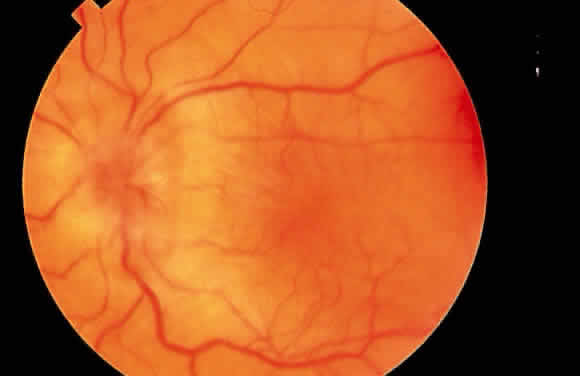
Fig. 3.
Fundus photograph. Hypotony maculopathy with choroidal folds, retinal striae, and marked swelling of the peripapillary choroid simulating papilledema.