Chapter 11
Intraocular Lenses
JAMES A. DAVISON, GUY KLEINMANN and DAVID J. APPLE
Main Menu   Table Of Contents

Search

HISTORICAL OVERVIEW
INTRAOCULAR LENS MATERIAL CHEMISTRY
INTRAOCULAR LENS TYPES, CLINICAL CHARACTERISTICS, AND USE
PSEUDOPHAKIC INTRAOCULAR LENS WITH SPECIAL FEATURES
PHAKIC INTRAOCULAR LENSES
COMPLICATIONS
CONCLUSION
ACKNOWLEDGMENTS
REFERENCES

This chapter describes the evolution, chemistry, and clinical characteristics of pseudophakic and phakic intraocular lenses (IOLs). Surgical strategies and complications are also discussed, as well as future trends. Cataract surgery, secondary lens implantation, IOL power calculation, and pediatric lens implantation are discussed in other chapters.
Back to Top
HISTORICAL OVERVIEW
The history of the IOL is interesting and colorful. It is a classic example of the improvement of medicine with the active cooperation of science and industry. It involves a reciprocating but overlapping evolutionary relationship of cataract removal technology with IOL design. Cataract surgery evolved through extracapsular cataract extraction (ECCE), intracapsular extraction (ICCE), machine-assisted ECCE, phacoemulsification by external nuclear attack, and phacoemulsification-assisted internal nuclear disassembly. For IOL fixation, the evolution has been posterior chamber, anterior chamber (AC), pupil and iris, iridocapsular, ciliary sulcus, asymmetric placement, and capsular bag. As with any evolutionary process, this has been and still is a leapfrogging phenomenon, so that at any one point in time several cataract surgery strategies and IOL implantation techniques can be considered good science and good medicine. The process continues as microincision phacoemulsification procedures gain sophistication in search of an IOL to be inserted through a sub–2.0-mm incision.

SIR HAROLD RIDLEY

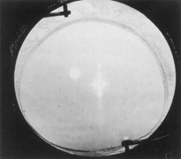
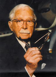
Credit for the invention and first implantation of the IOL is given to Sir Harold Ridley of London (Fig. 1)1–8 Details regarding Ridley and his invention are provided in a 1996 biography (Apple DJ, Sims JD: Harold Ridley and the Invention of the Intraocular Lens). Ridley's first IOL surgery was accomplished as a two-step procedure. The ECCE was performed on November 29, 1949. He waited for the eye to become quiet and stable and implanted the IOL secondarily a few months later on February 8, 1950. During the next 12 years, approximately 1000 Ridley IOLs were implanted. These operations were described as successes in 70%, failures caused by dislocation in 20%, and secondary glaucoma in 10%, which sometimes required explantation.9

Fig. 1. Sir Harold Ridley, father of the intraocular lens (IOL). (Courtesy of Rayner Surgical Inc., Los Angeles, CA.)

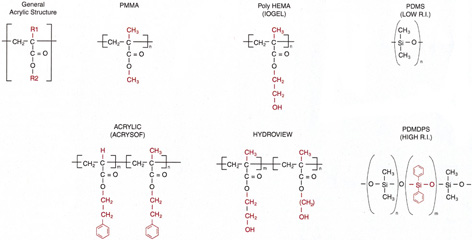
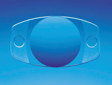
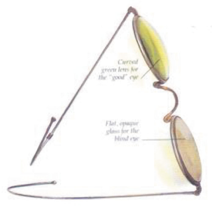
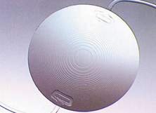
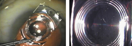
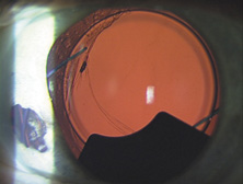
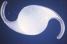
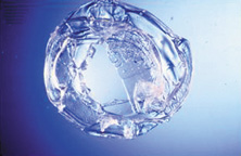
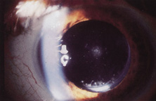
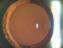
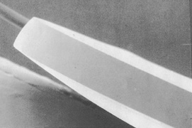
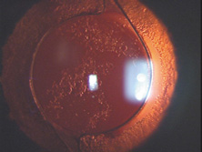
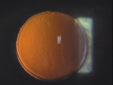
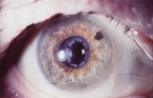
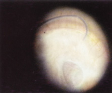
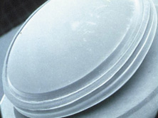
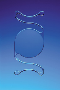
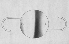
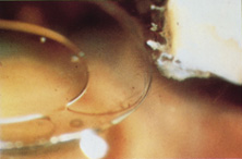
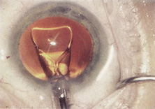
Ridley had been inspired by the tolerance of British fighter pilots' eyes to plastic fragments, which had lodged in them after their canopies, made of polymethylmethacrylate (PMMA; Perspex), had shattered. He worked with the Rayner Company and Imperial Chemical Industries, both in Great Britain, to develop Perspex CQ, a more purified “clinical-quality” PMMA. He used the human lens as his model and selected similar radii of curvature to create a biconvex disc while using approximately half the thickness and weight (∼5 mm thick and 230 mg for the human lens). One of his original lenses made by Rayner, a 23.00 diopter (D), was measured at 8.5 mm in diameter and 2.4 mm thick, with a weight of 108 g. Modern analysis has demonstrated that this IOL passes modern optical bench examinations10 (Fig. 2).

Fig. 2. This scanning electron micrograph (SEM) of an original Ridley IOL shows compound, relatively square edges. (Courtesy of David J. Apple, MD, Charleston, SC.)

Ridley said that the cataract operation “without a replacement lens was an incomplete, only half finished operation”11 and that he would like to be remembered as the man who cured or at least initiated the cure for aphakia. He saw aphakic vision as a highly significant but unnecessary disability. Now only a memory, at least in the modern world, aphakic vision contained many disturbing visual side effects. The roving scotoma, Jack-in-the-box phenomenon, 30% magnification, distortion, loss of side vision, and extreme spectacle dependence were disabling for many patients. Because of this, even as late as the mid-1970s, surgery was typically performed only when the patient's vision decreased to 20/70 or worse in the better eye, with that eye undergoing operation 3 days after the first eye.

In recognition of his pioneering efforts in IOL development and implantation, Mr. Ridley was knighted on February 19, 2000, by Queen Elizabeth II, 50 years after his first implantation. Warren Reese was the first American surgeon to perform the first IOL surgery in the United States at the Wills Eye Hospital, Philadelphia, in 1952. Mr. Ridley himself performed the first operation in America and gave the lenses to him after delivering a lecture at the Chicago Ophthalmological Society. Despite that early beginning, it would take more than 25 years for IOL implantation to become the dominant and standard accepted method of curing aphakia in the United States.

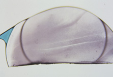
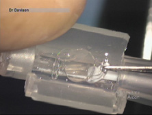
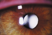
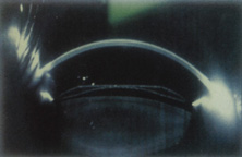
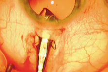
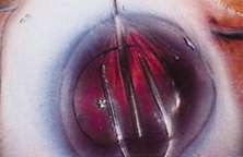
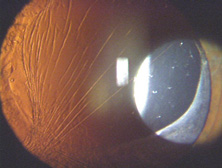
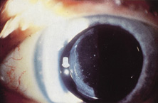
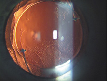
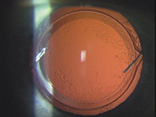
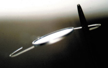
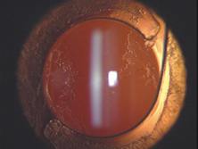
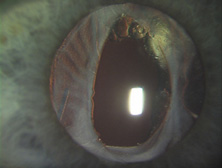
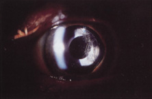
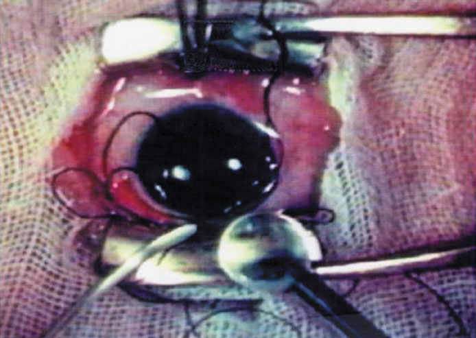
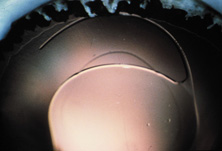
The Ridley lens was placed in the posterior chamber after ECCE (Fig. 3). The anterior capsulectomy of the day was very large, and thus zonular support was poor. Some Ridley lenses dislocated into the vitreous because of poor zonular support, and partially because of their weight, which was approximately eight times that of current IOLs.

Fig. 3. Photograph of Sir Harold Ridley's IOL implant operation taken from his original movie taken during surgery. (Courtesy of David J. Apple, MD, Charleston, SC.)

Ridley's achievements were finally and belatedly celebrated in numerous tributes. His first and perhaps most-prized honor was his election to the Royal Society in 1986. Dr. Apple presented him his first University Doctorate at the Medical University of South Carolina in 1988. He received another honor at the Science Museum in London on November 29, 1999, the 50th anniversary of the first part of the first IOL implantation. In the Flight Room, with airplanes suspended overhead, Ridley was honored by fellow pioneers and colleagues from around the world, as well as by the Rayner Corporation and government representatives from the United Kingdom and United States. The same year, the American Society of Cataract and Refractive Surgery (ASCRS) honored him as one of the 10 most influential ophthalmologists of the 20th century. In 1990, he was guest of honor at the Annual Meeting of the American Academy of Ophthalmology. In recognition of his unique efforts in IOL development and implantation, Ridley was knighted on February 9, 2000, by Queen Elizabeth II. On May 25, 2001, at the age of 94 years, Sir Harold died in Salisbury, England, after a cerebral hemorrhage.

Because of the difficulty with posterior chamber IOL placement, pioneering surgeons would spend the next two decades trying to find a better place to fixate the IOL. The AC lens, pupil-fixated IOL, iris-fixated IOL, and iridocapsular IOL would be placed in large numbers, only to return to the posterior chamber in the 1970s. This made for very interesting surgical residencies during these times. We were always learning something completely new. For example, during J.A.D.'s 3-year residency starting in 1977 at the Mayo Clinic, we evolved through cryoextraction ICCE using no IOL, cryoextraction ICCE with Medallion iris-sutured IOLs, machine-assisted ECCE with iridocapsular fixation using metal and then plastic clips through the iridectomy, and machine-assisted ECCE with posterior chamber IOL implantation.

ANTERIOR CHAMBER AND PUPIL-SUPPORTED LENSES

Apple and associates have described in detail the evolution of the IOL, describing six generations. AC IOLs were generation two.12

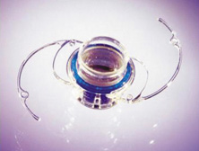
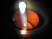
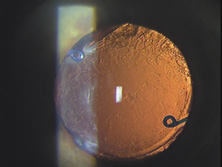
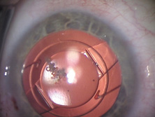
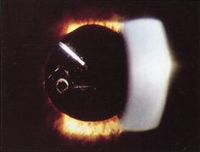
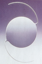
The first AC IOL was implanted by Baron of France in 1952. This lens failed primarily because of excessive anterior vaulting, which caused contact with the corneal endothelium. Mr. Ridley's good friend and long-time defender, Peter Choyce, also of the United Kingdom, was one of the developers and certainly the greatest champion of the AC lens (Fig. 4). These lenses could be placed after ECCE or ICCE procedures. His models were very successful with his last models, the Mark VIII and Mark IX, used until the mid-1970s in the United States.

Fig. 4. Choyce anterior chamber (AC) lens. (Courtesy of Department of Ophthalmology, Mayo Clinic, Rochester, MN.)

AC lenses had problems as well. Ellingson's uveitis, glaucoma, hyphema syndrome13 was associated with their use. Chronic irritation of the delicate structures of the angle caused this problem, and even without it, pain could sometimes be elicited by simply touching the eye. Later, similar rigid AC IOLs would have similar problems. Some had to do with sizing difficulties; others were problems of poor finish and the misapplication of polypropylene haptics in the AC, where they underwent ultraviolet (UV) degradation.

Ultimately the flexible AC IOL was developed, most commonly the Kelman Multiflex (Fig. 5). This style of IOL features rather broad area smooth footplates, which can be placed in the angle without causing the chafe and erosion that small-sized loop-shaped haptic IOLs did. The Multiflex-type lens has provided excellent performance with similar and sometimes lower long-term corneal endothelial cell loss in secondary implantation than has the sutured ciliary sulcus posterior chamber IOL.14

Fig. 5. Kelman multiflex. (Courtesy of Alcon Laboratories, Fort Worth, TX.)

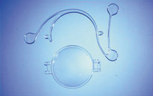
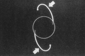
Taking another path, Cornelius Binkhorst of Holland developed a lens that involved pupil fixation with pairs of horseshoe-shaped haptics in front of and behind the iris (Fig. 6). This lens was associated with total dislocation into the AC or vitreous after pupil dilation, so miotics were many times given on a prophylactic basis indefinitely. Also, after ICCE, there was so much iridodonesis, especially with the initially used metal haptics, that the anterior aspects of the anterior haptics could touch the endothelial surface of the cornea, leading to localized corneal decompensation. Also, the weight of metal haptics could erode the iris. It was this style of lens that was implanted by early American implant surgeons Jaffe, Hirschman, Byron, and Kwitko in 1967.15

Fig. 6. Binkhorst lens with polymethylmethacrylate (PMMA) haptics in front of and behind the iris. Prolene suture has been placed through the superior anterior haptic to prevent dislocation. (Courtesy of Department of Ophthalmology, Mayo Clinic, Rochester, MN.)

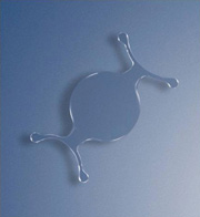
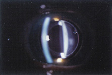
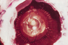
Introduced in 1967, Stanislav Fyodorov's “sputnik” IOL design achieved stabilization without larger anterior haptics (Fig. 7). In the mid-1970s, the Worst medallion IOL also featured an anterior optic with two horizontally oriented horseshoe-shaped looped posterior haptics similar to the Binkhorst structure. A Prolene suture was passed horizontally through the superior iris and then threaded between through two small holes in the superior optic. Because iris suture had to be preplaced, it always seemed to become entangled with the haptics during insertion making it difficult to place smoothly under air after ICCE, which featured a 180-degree corneal incision and an unprotected anterior hyaloid membrane. The suture was tied loosely so that the optic was secured to the superior iris. This would still allow the posterior haptics to dislocate anteriorly (embarrassingly, many times after having sex) or create a partial papillary capture, but at least it prevented total dislocation. Because surgical manipulation was expensive and carried the risk of infection, we would spend hours positioning patient's heads and bodies after pharmacologic weak dilation so that gravity would reposition the IOL. Then, when the IOL fell into position, we would reposition the patient and administer a topical miotic to capture the appropriate part of the IOL with the pupil. Aside from partial dislocation, this type of lens performed very well, but eye movement after ICCE still generated substantial irido-pseudophakodonesis.

Fig. 7. The pupil-supported Sputnik (named for its resemblance to the first Russian satellite launched in space) featured relatively long PMMA spokes extended radially from behind optic to rest anterior to the iris with the loop haptics posterior. Courtesy of American Society of Cataract and Refractive Surgery, with permission.

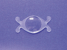
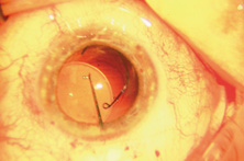
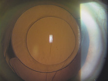
ICCE was difficult and time-consuming and carried a high risk. Even when done well, the procedure left an eye not able to support an IOL in stable fashion. The structural diaphragm of the posterior capsule after ECCE was rediscovered and appreciated because it contained the vitreous and created compartmentalization and stabilization of the AC, iris, and posterior chamber. Iridocapsular fixation was the next step. The lenses designed for this technique had a small platinum or plastic rod attached to the superior optic that could be clipped to the superior posterior haptic through a superior peripheral iridectomy (Fig. 8). The inferior haptic would be inserted, and eventually fibrose, between a leaflet of remaining anterior capsule and the posterior capsule. When properly secured, the pupils of these eyes could be dilated without fear of superior or inferior IOL dislocation. Because of capsule fibrosis and stabilization, there was also a substantial reduction in pseudophakodonesis. Surgeons who had taken up phacoemulsification, which had been introduced in 196716 and adopted by a fair number by 1972, could enjoy the small-incision control aspect of cataract removal but still had to enlarge the incision for IOL insertion. Things were improving.

Fig. 8. Worst iridocapsular lens with PMMA clasp clipped through the peripheral iridectomy to the superior posterior haptic. (Courtesy of Department of Ophthalmology, Mayo Clinic, Rochester, MN.)

RETURN TO POSTERIOR CHAMBER LENSES

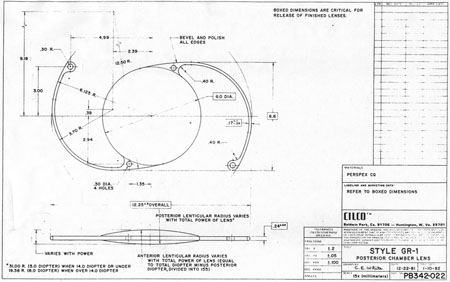
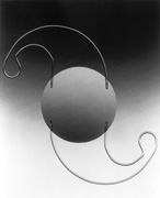
John Pearce of the United Kingdom returned implantation to the posterior chamber by developing a rigid tripod-shaped PMMA IOL designed for implantation there,17 but it was Steve Shearing who would have the greatest influence on the future development posterior chamber IOLs. In March 1977, he introduced his IOL, which featured flexible posterior haptics (Fig. 9).18 His original intention was to have the haptics placed within the capsular bag remnant. The lens had a 5-mm optic (because that was the size of the Binkhorst optic, which was already in production at the time) and an overall length of 12 mm and was flat.

Fig. 9. Shearing posterior IOL with two positioning holes. (Courtesy of David J. Apple, MD, Charleston, SC.)

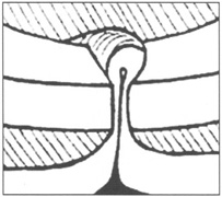
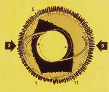
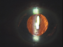
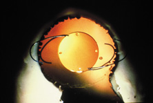
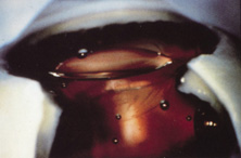
This lens design revolutionized the concept of IOL placement, but the need for a supportive capsular bag architecture was not fully appreciated, nor had the techniques to preserve it been developed. Most dislocations were incomplete (i.e., nonintravitreal). For example, the sunset syndrome (Fig. 10) resulted from occult anterior radial capsular tears (ARTs), which extended through the equator or inferior zonular disruptions and allowed the inferior haptic to sink through the defect. However, in most inferior dislocations, the IOL optic was still contained within the ciliary sulcus. The IOL's overall length was too short for symmetric sulcus fixation, so the whole IOL structure gravitated inferiorly with sulcus placement. The “windshield wiper syndrome” resulted from sulcus contact inferiorly but none superiorly. Surgeons tried to retain at least a leaflet of anterior capsule inferiorly in which to place the inferior haptic. When that was accomplished, the IOL stayed fairly well centered. However, if asymmetric placement occurred with the inferior haptic contained within the capsular bag remnant and the superior haptic loose in the ciliary sulcus, there would be opportunity for another type of decentration, the sunrise syndrome.

Fig. 10. Sunset syndrome with posterior chamber IOL sinking through an inferior zonular defect. (Courtesy of Department of Ophthalmology, Mayo Clinic, Rochester, MN.)

Because the capsular bag was such a hard place to find, the IOL was soon modified to include a 6-mm optic and 13-mm overall length. Even then, fixation was not perfect, and pupillary capture, just as Dr. Shearing had experienced in his fourth case, was not rare (personal communication, Steve Shearing, MD, 1998).

However, an important step had been taken, and during the next two decades, the Shearing posterior chamber IOL would evolve into three basic styles. The first was designed primarily for ciliary sulcus fixation but could also be used for capsular bag fixation. It was the result of the tremendous influence of two popular, committed, prestigious California phacoemulsification surgeons who had trained together at Duke University, Dick Kratz and Bob Sinskey. The second was the short haptic diameter modified C-loop IOL designed for capsular bag placement. The third featured larger haptic and optic diameters and was designed for surgeons still practicing planned ECCE who might place the IOL within the sulcus or asymmetrically with one haptic in the sulcus and one in the capsular bag.

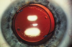
The Kratz IOL was compatible with Kratz's phacoemulsification technique, which involved a prolapse of the superior nuclear pole into the iris plane for external attack phacoemulsification. In this technique, remaining anterior capsule was a problem because it could produce asymmetric placement and decentration. Therefore, anterior capsulotomies were large so that the IOL could be placed in the ciliary sulcus. Kratz developed a “tap test” that involved tapping the eye over the ciliary sulcus with a Weck sponge to see whether the IOL would move, which would indicate that the haptic was in contact with the structures of the ciliary sulcus and not hung up in the capsular bag remnant. Through the Precision Cosmet Company (Minneapolis, Minnesota), Kratz introduced a 10-degree optic posterior angulation as an effective way to prevent pupillary capture. The resistance to compression of the haptics was softened significantly by a side optic mounting modification with the overall length of 13.5 mm. Actually, before that, more than a few surgeons had been bending the Shearing haptic at the optic haptic junction to try to reduce compression resistance. Sinskey's lens was very similar and was introduced at about the same time, but by a different company, IOLAB (San German, Puerto Rico). Because of the nearly simultaneous introduction and similarity, the lens ultimately has been called the Kratz-Sinskey IOL (Fig. 11).

Fig. 11. The Kratz-Sinskey IOL involved haptic anchoring to the optic sides and a more gentle modified J-loop haptic. (Courtesy of Robert M. Sinskey, MD, Sinskey Eye Institute, Santa Monica, CA.)

Sinskey's phacoemulsification technique was basically one-handed and allowed a more generous anterior capsular remnant to exist after nucleus removal. Symmetric capsular bag fixation was possible a fair amount of the time. Optic posterior angulation was incorporated in his lens to prevent pupillary capture, which was still possible.

The concept of intracapsular phacoemulsification and the desire to fixate the IOL within the capsular bag ultimately led to the second type of posterior chamber IOL, the minimally compression-resistant, short haptic configuration created for capsular bag fixation that exists today.19

The third style was a transitional lens that was oversized and was to be used with planned ECCE. It eventually measured 14 mm in haptic diameter and 7 mm in optic diameter. It was large and stiff in its eventual one-piece design. The problem was that after ECCE, the capsular bag did not exist in a structural sense. With broad contact, at least one haptic would be captured by some partially unrolled capsular flap remnant, usually the inferior. If it were asymmetrically placed, which was the expectation, the lens was so long that capsular fibrosis would not decenter the very large optic too much because of the contact created in the superior ciliary sulcus by the high resistance to compression. Because of their large size, these lenses were very difficult to place symmetrically within the capsular bag but are still in use today as ciliary sulcus placed secondary IOLs in patients with very large eyes.

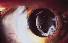
As an aside, glass IOLs were marketed for a short period. Even in aqueous, they were heavier than plastic. Glass also broke if hit by the neodymium:yttrium-aluminum-garnet (Nd:YAG) laser (Fig. 12).20 Plastic IOLs inadvertently hit by the YAG laser during capsulotomy developed small cracks or pits that did not affect vision and did not completely fracture. Glass was eventually abandoned because of its weight and YAG intolerability. The right to use polyamide framing and haptic material was purchased from Lynell Optics by STAAR Surgical (Monrovia, CA) and is in use today by that company as a haptic material combined with silicone optics.

Fig. 12. The glass IOL by Lynell Optics worked well but could break with yttrium-aluminum-garnet (YAG) laser treatment. The polyamide haptics are used in the haptic structure of some modern silicone lenses. (Courtesy of Charles D. Fritch, Superior Eyecare Centers, Bakersfield, CA.)

ENDOTHELIAL CELL DAMAGE, VISCOELASTICS, ANTERIOR CAPSULAR TEARS, AND SUTURELESS CLOSURE

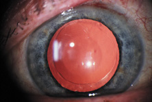
With the introduction of clinical corneal endothelial photography 1976, Bourne and Kaufman heightened our awareness of quantitative and qualitative endothelial damage associated with IOLs.21 In 1980, Miller and Stegmann, working with the Pharmacia Company of Uppsala, Sweden, introduced the first viscoelastic, 1% sodium hyaluronate (Healon).22,23 This not only protected the corneal endothelium during IOL implantation but also made anterior capsulotomy much easier to perform. The control of the anterior capsular surface with viscoelastic, pushing it back and making it flat, was an important aid in the prevention of unwanted ARTs during the can-opener capsulotomy process.

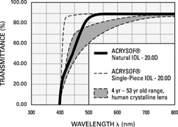
The number of central corneal endothelial cells after IOL implantation decrease at rates greater than those of healthy unoperated corneas24–29 (normal loss ranged from 0.3% to 1.0% per year).

The only randomized trial of lens implantation, the Oxford Cataract Treatment and Evaluation Team found a higher rate cell loss in eyes with implants than those without in the first 3 postoperative years.30 Unfortunately the type of surgical technique and IOLs (ICCE and Binkhorst 4-loop lens) are no longer being used.

A 5-year prospective study from the Mayo Clinic31 reported 23% to 28% of endothelial cell loss after cataract surgery over the 5-year postoperative period. The rate of cell loss was not found to be influenced by the surgical technique (ICCE vs. ECCE), implantation of an IOL, and type of IOL implanted (medallion iris suture IOL, transiridectomy clip implant, or posterior chamber IOL). Correlation was found between the endothelial trauma judged at surgery and the long-term endothelial cell loss. Extension of the follow-up time to 10 years32 demonstrated that the eyes continued to lose endothelial cells from the central cornea at an average rate of 2.5% per year (2.5–8.0 times the rate in healthy unoperated eyes); also in this study the type of IOL implanted did not influence the rate of cell loss. These studies had two major caveats: (a) A significant percentage of patients were lost to follow-up. (b) The study represents the early cases in the implant experience of the surgeons (1976–1982) and do not represent the currently surgical technique and IOL technology.

In the early days of the phacoemulsification the corneal cell loss rate was high because of the long phaco time and high energy that were commonly used. In recent years, damage to corneal endothelial cells during cataract extraction has been minimized as a result of better instrumentation,33 newer viscoelastic materials,34,35 and improved surgical technique such as phaco chop,36 which aims to reduce machine measured phaco time.

Several preoperative and intraoperative parameters can influence the risk for endothelial cell loss after phacoemulsification. A high nucleus grade,37 old age, long phaco time,38 and short axial length39 are associated with an increased risk for endothelial cell damage. Ravalico and coauthors40 reported that AC IOLs implantation did not appear to alter corneal endothelial function over 5.2 years of postoperative follow-up period. As a summary, it seems that the endothelial cell loss after cataract surgery is related to the surgical trauma and maybe to the absence of the crystalline lens but not to the IOL implantation.

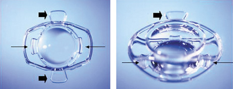
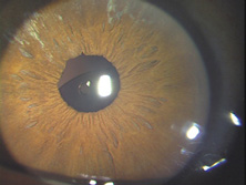
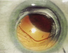
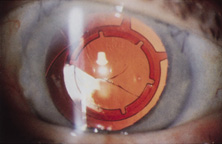
Surgeons who performed one-handed phacoemulsification were preserving the capsular bag better than two-handers. In 1981, John Graether,41 a left-handed one-hander, designed the first one-piece all-PMMA IOL that was 12.25 mm in overall length designed specifically for implantation in the capsular bag (Fig. 13). He had invented a “collar button” retractor and described a way to use it by pushing on the superior optic-haptic junction to compress the inferior haptic and dial the IOL into the capsular bag, the method still used today.41 Later, a reduction in the amount of nuclear prolapse into the iris plane made it possible for surgeons who used the two-handed procedure to retain more anterior capsule by creating only one ART most of the time.42 Even if two superior tears were noted, posterior chamber IOLs could be placed in what was left of the capsular bag with minimal or no decentration.43 Sometimes lenses with broader haptics, as designed by Bill Simcoe, were used to bridge the gap created by ARTs in the capsular bag equator. Even though surgery and implantation had improved, significant IOL optic decentration, with its attendant optical and physical complications, was not uncommon (Fig. 14). Given the anatomy of the eye with possible multiple ARTs after iris-plane phacoemulsification or ECCE, two schools of thought developed. The first embraced larger optic and haptic diameters, which provided greater stability and increased resistance to compression and were designed in an attempt to center IOL optics in capsular bag, ciliary sulcus fixation, or asymmetric fixation and prevent pupillary capture (Fig. 15). The other school fashioned 6.0-mm optics and reduced 12.0-mm haptic diameters with a very low resistance to compression to try to keep them within the capsular bag or capsular bag remnant (Figs. 16 and 17).19 In an attempt to custom tailor IOLs to patient anatomy, J.A.D. created the “graduated length method” through the D.G.R. Company of Clearwater, Florida. This IOL line featured shorter 12.0-, 13.0-, and 14.0-mm haptic structures for small, medium, and large eyes.

Fig. 13. One-piece all PMMA IOL designed specifically for implantation within the capsular bag that was 12.25 mm in overall length and had an equally proportioned biconvex optic. Dr. Graether is left-handed and drew this IOL in what would look like an upside-down view. He actually implanted it with a technique that would look like an upside-down implantation. (Courtesy of John M. Graether, MD, Wolfe Eye Clinic, Marshalltown, IA.)

Fig. 14. Asymmetric bag–sulcus posterior chamber lens fixation with capsular remnant and anterior optic precipitates. (Courtesy of James A. Davison, MD, Wolfe Eye Clinic, Marshalltown, IA.)

Fig. 15. The Graether ScrollFlex IOL from 1984 designed for sulcus or capsular bag placement was 14.0 mm in length and had a relatively stiff resistance to compression. Four-point optic fixation made it resistant to tilt or optic capture. Notice the interesting similarity of the compound curves in the C-flex IOL from Rayner (Fig. 35). (Courtesy of John M. Graether, MD, Wolfe Eye Clinic, Marshalltown, IA.)

Fig. 16. Early modified J-loop IOLs grew to 14 mm in length so they could be placed in the ciliary sulcus or tolerate asymmetric bag–sulcus placement. In the early 1980s, reduced diameter IOLs with softer flex characteristics were developed specifically for placement within the capsule bag. Both lengths are seen overlapping in this open-sky keyhole demonstration. (Courtesy of James A. Davison, MD, Wolfe Eye Clinic, Marshalltown, IA.)

Fig. 17. State of the art postoperative IOL appearance in the early 1980s. Note the can-opener anterior capsulotomy with an anterior radial tear superior temporal in this right eye. Note the IOL is well centered within the pupil and the mostly overlapping anterior capsular remnant. The haptic orientation is 90 degrees from the ART, and a slight stress line can be seen in the posterior capsule. Two positioning holes are present, and the dioptric power (17.5) is stamped on the peripheral optic of this J-looped IOLAB IOL. (Courtesy of James A. Davison, MD, Wolfe Eye Clinic, Marshalltown, IA.)

Because of these compounding developments, David Apple began his important work with Randy Olson and in 1983 founded the Center for Intraocular Lens Research at the University of Utah. They were a uniquely qualified combination. Dr. Apple was board certified as both a pathologist and ophthalmologist; Dr. Olson was an academic ophthalmic surgeon. Much like Harold Ridley and Peter Choyce, they began their dedicated quest to improve the condition of pseudophakia. They solicited autopsy specimens and wrote innumerable articles about IOL design, centration, and complex IOL–ocular interactions. One of their most important articles, published in 1985, was a position article citing the advantages of capsular bag placement and recommending it over ciliary sulcus placement, still a controversial issue at the time.44 The eyes submitted to them that year demonstrated capsular bag fixation in 31%, ciliary sulcus fixation in 11%, and asymmetric bag–sulcus fixation in 58%.45 At that time, 85% of surgeons still preferred planned ECCE as their surgical technique.46

While in Utah, Apple and Olson saw the problems created by ARTs solved with the invention of capsulorrhexis, almost simultaneously reported in 1986 by four surgeons from around the world. Two presented articles at the Welch Cataract Congress in Houston that year, and two reported their techniques independently. The discovering surgeons in alphabetical order are Drs. Calvin Fercho (Welsh Cataract Congress, Houston, 1986), Howard Gimbel (video presentation at the annual meeting of the ASCRS in Boston, 1985),47 John Graether (Welch Cataract Congress, Houston, 1986),48 and Thomas Neuhann (video presentation at the meeting of the German Ophthalmological Society in Heidelberg, 1985).47 With this technique, and the creation of an approximate diameter of 5 mm, symmetric placement of any IOL could be guaranteed. It still took several years for the majority of ophthalmologists to incorporate continuous curvilinear capsulorrhexis (CCC) into their surgical routines.

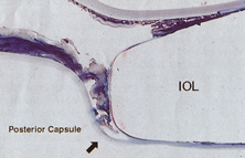
In 1988 Dr. Apple relocated the laboratory to the Storm Eye Institute at the Medical University of South Carolina in Charleston. Reflecting new challenges, his laboratory name would be changed to the Center for Research on Ocular Therapeutics and Biodevices. From there, he and his staff, residents, and research fellows continued to receive autopsy specimens from around the world and eventually demonstrated, in the largest autopsy specimen study to date, a decline in asymmetric placement to only 10% of eyes with foldable lenses submitted in 1998.45 Early in his work there, Dr. Apple recruited Dr. Kensaku Miyake's retrociliary photographic analysis method to locate and analyze IOL placement within the eye (Fig. 18). With increasing video sophistication and use of the process by Dr. Apple and his colleagues, Dr. Miyake himself generously expanded the name of the procedure to be called the Miyake-Apple technique.

Fig. 18. First picture from Dr. Miyake of a Shearing lens in the ciliary sulcus. Note the profound capsular bag shrinkage and abundant Soemmering's ring. (Courtesy of Kensaku Miyake, MD, Miyake Eye Hospital, Japan.)

After cancer was diagnosed in Dr. Apple and he was successfully treated, the center was relocated to Salt Lake City in 2002, where it has been permanently designated as the David J. Apple, MD, Laboratories for Ophthalmic Devices Research.

In the days when the laboratory was in Charleston, Dr. Apple worked with industry representatives and surgeons to refine IOL design to ensure that capsular bag residence would be as consistent as possible, thereby reducing lens contact with other eye structures in both routine and complicated situations (Figs. 19 and 20). J.A.D. had the great pleasure of working with him in his laboratory to help improve an already sophisticated haptic configuration in a one-piece all-PMMA IOL. At that time, we thought that the entire haptic should be C-shaped so that even its distal end could be recruited for capsular equatorial support (Fig. 21). We studied resistance to haptic compression, attempting to make it softer and more uniform through diameter reductions from 13.0 to 11.5 mm 2.5 mm (Fig. 22). These efforts contributed to the development of the Pharmacia model 811, which, along with others of its day, may have represented the height of single-piece PMMA IOL development (Fig. 23).49

Fig. 19. Sulcus placement required long haptic structure and posterior angulation to avoid pupil capture. (Courtesy of James A. Davison, MD, Wolfe Eye Clinic, Marshalltown, IA.)

Fig. 20. The shallow disc of a posterior chamber IOL fills only the anterior portion of the capsular bag. (Courtesy of James A. Davison, MD, Wolfe Eye Clinic, Marshalltown, IA.)

Fig. 21. Haptic support “steering wheel” type of PMMA IOL was tried to eliminate the noncontribution of the far distal haptic to centration. The result was that there was no distal haptic. Perfect equatorial contact was possible without zonular stress or ciliary body contact. (With permission from Tetz M, O'Morche D, Gwin T, et al: Posterior capsular opacification and intraocular lens decentration. Part II: Experimental findings on a prototype circular intraocular lens design. J Cataract Refract Surg 14:614, 1988.)

Fig. 22. Early single-piece PMMA IOL modified C-loop haptics exhibited too much resistance to compression proximal, which resulted in contact only of the midhaptic (similar to the J-loop they replaced) and noncontact with the peripheral haptic. If the capsular bag was small and the IOL relatively large, this could result in an ovaling of the capsulorrhexis. Modifications of the haptic configuration resulted in more even distribution of contact of the mid- and peripheral haptic. (Courtesy of James A. Davison, MD, Wolfe Eye Clinic, Marshalltown, IA.)

Fig. 23. PMMA Pharmacia model 811 IOL designed for capsular bag placement. (Courtesy of Pharmacia and Upjohn, Uppsala, Sweden.)

Inevitably, with the new technique of capsulorrhexis, the improved haptic architecture was actually not as critical as it had been earlier, when ARTs were universal. It was still helpful in difficult situations in which capsular support was anomalous (e.g., pseudoexfoliation and trauma). Coatings of heparin and Teflon were developed to increase the biocompatibility of PMMA IOLs.50 However, the benefits of the sophisticated one-piece PMMA capsular bag design would soon be replaced by the even greater benefits of foldable IOLs, the use of which would overtake PMMA in 1998.46

Sutured superior corneoscleral incisions with round PMMA IOLs were standard throughout the 1980s, until the self-sealing incision was described by Mike McFarland in 1991.51 In an attempt to integrate the smaller phacoemulsification incision and PMMA technology, ovoid posterior chamber IOLs had been produced since 1980 and reached their peak in popularity (35%) in 1991 (Fig. 24).46 But, because of their truncated edge, ovoid IOLs introduced a higher incidence of glare, streaks, and halos52 (pseudophakic dysphotopsia); for this reason, they never reached great popularity. Attempting to reduce edge glare and still maintain optic truncation, Charles Kelman created the PhacoFlex IOL. Two side portions of the optic had opaque silicone wings, which would fold on top of the PMMA central optic during insertion through a 3.5-mm incision, and then unfold in the eye. Many years later, Howard Fine would recommend using such a textured finish in plate haptic silicone IOL haptics, not for optical reasons, but to enhance capsular fixation to prevent IOL movement within the capsular bag.

Fig. 24. An ovoid IOL is seen within the capsular bag. A diamond-shaped neodymium (Nd):YAG laser posterior capsulotomy has been created. The truncated optic of these lenses was more prone to create dysphotopsia. (Courtesy of James A. Davison, MD, Wolfe Eye Clinic, Marshalltown, IA.)

FOLDABLE LENSES, CLEAR CORNEAL INCISIONS, AND TOPICAL-INTRACAMERAL ANESTHESIA

In 1984, Mazzocco et al introduced the first foldable plate haptic silicone lens produced by STAAR Surgical.53 It became known as the “Mazzocco Taco” because of the way it appeared when folded. It was delivered through an injector, whose cartridge went through a 3-mm incision allowing the IOL to unfold in the posterior chamber (Fig. 25). This changed cataract surgery forever because it made possible the full appreciation of phacoemulsification. Cataract surgery had become the first microscope-accomplished, machine-assisted “small-incision surgery” in medicine. All of the benefits of improved surgery safety, improved postoperative state quality, and shorter patient recovery were realized.

Fig. 25. Plate haptic silicone lens unfolds into the capsular bag after it leaves the injector cartridge. (Courtesy of James A. Davison, MD, Wolfe Eye Clinic, Marshalltown, IA.)

This IOL did not fixate very well in the sulcus, and asymmetric bag–sulcus fixation was the standard of the day. An intact or almost intact (one or two opposing ARTs) capsular bag was necessary for acceptable centration performance of this IOL. Concerned about this, the designers made the earliest models too long, so that when they were well fixated within the capsular bag they could actually wrinkle centrally because of capsule contraction, causing the “Z phenomenon.” The silicone material was hydrophobic and did not bond to the capsule during fixation, so that if asymmetric placement occurred, it could be squeezed in one direction out of the bag. Significant decentration was likely when only anterior capsular leaflets remained for fixation. Intravitreal dislocation was also possible after YAG laser capsulotomy.

Nevertheless, these IOLs became very popular, especially in the United States, because they were relatively easy to insert with an injector, could be implanted through a small incision, and were relatively inexpensive.

Next, three-piece foldable silicone optics with polypropylene haptics emerged. These IOLs centered better and could be used in cases with ARTs. Popular “small-incision” cataract surgery had finally arrived. Surgeons no longer had to enlarge the incision much after cataract removal to accommodate the IOL, so one of the main advantages of phacoemulsification could be realized. However, increased AC reaction, capsular fibrosis, and optic decentration compared with single-piece PMMA IOLs kept some surgeons from using early three-piece silicone IOLs.54

In 1992, Kimya Shimizu reintroduced a very clear corneal incision only 3 mm wide to accommodate phacoemulsification and foldable IOL insertion.55 One suture was placed. That same year, Howard Fine showed that the temporal clear corneal incision could be left unsutured and consistently perform well, with an extremely low incidence of incision complication and very little astigmatic consequence.56 Although this incision was not as strong as one created with an additional scleral shelf,57 it was strong enough for normal clinical application. Foldable silicone lenses could be placed without using scissors, cautery, or sutures.

Cataract surgery using only topical anesthesia was introduced by Richard Fichman in 1992. This was substantially improved with the addition of intracameral anesthesia by Jim Gills in 1995.58 As of 2003, the topical anesthesia technique was adopted by 61% of American surgeons (38% of surgeons performing one to five operations per month and 76% of surgeons performing more than 75 surgeries per month); 73% of surgeons used intracameral lidocaine with the technique.59

Also, in 1995, the acrylic IOL (AcrySof®, Alcon Surgical, Fort Worth, TX) was approved for use in the United States. Since its introduction, the acrylic lens has had substantial popularity. With self-sealing temporal clear corneal incisions and topical-intracameral anesthesia available, surgeons who had objected to foldable lenses made of silicone found their last obstacle to small-incision surgery removed. However, the acrylic lens had to be withdrawn temporarily because of glistenings within the optic.60 These turned out to be water vacuoles, which were associated with packaging materials. When the lens was repackaged, the glistenings were reduced.61 Through manufacturing improvement, the glistenings continue to be reduced to lower levels, but they still may be present in many patients to a trace to −1 degree, but without visual consequences.

An important part of the evolution of the IOL has been the propagation and dissemination of information as well as the continuous development of educational processes, ensuring that the latest techniques and materials were available to surgeons. The first International Intraocular Implant Club meeting was held on July 14, 1966, with Mr. Ridley presiding. The American Intraocular Implant Society was founded in 1974 by its first president, Kenneth Hoffer, MD, of Santa Monica, California. In 1985, the name was changed to the American Society of Cataract and Refractive Surgery, and in 1988 its annual meeting outgrew its traditional location, the Century Plaza Hotel in Century City, California. This organization has grown to 5000 U.S. and 2000 international members, and in 1996, it combined its journal publication with that of the European Society of Cataract and Refractive Surgery. This open climate of information sharing ultimately has benefited patients greatly.

CATARACT SURGERY 2004

The changes in cataract removal technique and IOL implantation have been gradual, with considerable overlap of individual preferences. As can be seen in any evolutionary activity, there is never just one right answer. That is, at any one point in time, there exist multiple materials and surgical techniques that have their individual and combined inherent advantages and disadvantages. In fact, as of 2004, there were 1548 IOLs available from 33 different manufacturers.62 In 1998 many surgeons preferred PMMA (33%) and superior corneoscleral incisions, but momentum was shifting away from those methods toward the use of silicone (22%) and acrylic (42%)19 and clear corneal incisions. By 2003, PMMA had decreased to 6%, silicone had stabilized to 21%, and foldable acrylics had grown to 69%. Acrylic IOLs have evolved and are now made by manufacturers other than Alcon. The Alcon acrylics continue to evolve as well (Fig. 26).

Fig. 26. Alcon AcrySof® three-piece, single-piece, and single-piece light-normalizing designs. (Courtesy of Alcon Laboratories, Fort Worth, TX.)

Substantial industry consolidation has occurred so that, in the United States at least, there are four major manufacturers with commercially available IOLs in 2004: Alcon Surgical of Fort Worth, Texas; Advanced Medical Optics (AMO) of Irvine, California; Bausch and Lomb of Clearwater, Florida; and STARR Surgical of Monrovia, California. Although many company executives and factory representatives from earlier companies have been integrated into the contemporary organizations, gone are the days of the prominent IOL lines of the 1980s and 1990s: CooperVision, Ciba Vision, O.R.C., Cilco, IOLAB, Precision Cosmet, Lynell Optics, and Pharmacia.

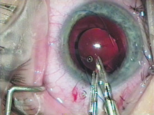
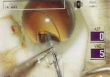
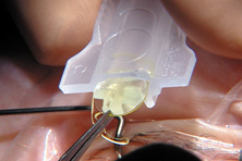
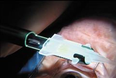
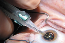
Modern lensectomy lens replacement surgery consists of removal of the nucleus, cortex, and as much remaining lens epithelium on the posterior capsule as possible while avoiding other ocular structures. It should leave a central circular anterior capsular opening so that the anterior capsule remnant can overlap the peripheral IOL optic by approximately 0.25 to 0.5 mm for a complete 360 degrees. Surgery is usually accomplished under topical-intracameral anesthesia through a temporal clear corneal incision of less than 3.0 mm in length, permitting a foldable IOL to be placed using an adjunctive viscoelastic device. If well executed with either forceps (Fig. 27) or more commonly with an injector using a disposable cartridge (Figs. 28, 29, 30), this strategy provides good IOL centration and minimal unwanted side effects regardless of the foldable IOL material or design used (Figs. 31 and 32).

Fig. 27. With a Buratto direct action forceps, the acrylic lens is placed through a self-sealing temporal clear corneal incision. (Courtesy of James A. Davison, MD, Wolfe Eye Clinic, Marshalltown, IA.)

Fig. 28. An AcrySof® SN60WF is loaded into the cartridge by grasping the peripheral optic with forceps. The trailing haptic will be tucked over the top of the left side of the optic so that the plunger can engage the rim of the edge of the optic without becoming entangled with the trailing haptic. (Courtesy of James A. Davison, MD, Wolfe Eye Clinic, Marshalltown, IA.)

Fig. 29. The SN60AT can be seen resting flat within the central cartridge cavity. The cartridge has been placed in the Monarch injector, and the plunger is about ready to engage the peripheral optic. (Courtesy of James A. Davison, MD, Wolfe Eye Clinic, Marshalltown, IA.)

Fig. 30. The curled SN60WF opens slowly as it is injected through the end of the cartridge to the capsular bag. A small air bubble is visible just to the left of the IOL. (Courtesy of James A. Davison, MD, Wolfe Eye Clinic, Marshalltown, IA.)

Fig. 31. Alcon SN 60 WF in good position with respect to the dilated pupil and the capsule opening. The overlap is approximately 0.5 mm. Greater overlap tends to create a more prominent anterior capsular opacification and a tendency toward capsular contraction. A smaller amount of intended overlap can result in incomplete overlap so that a portion of the IOL is not covered by the anterior capsule remnant. Note the slight irregularity in the lower right quadrant. This is the initiation and finish quadrant where such irregularities are more common. (Courtesy of James A. Davison, MD, Wolfe Eye Clinic, Marshalltown, IA.)

Fig. 32. An SI40NB is in perfect position with approximately 0.4-mm capsular overlap 360. The overlap is at the junction of the functional optic and peripheral carrier except for superior-temporal where the capsulorrhexis opening is a little smaller. The slight irregularity in portion has occurred in the initiation-completion quadrant. (Courtesy of James A. Davison, MD, Wolfe Eye Clinic, Marshalltown, IA.

Back to Top
INTRAOCULAR LENS MATERIAL CHEMISTRY
A variety of materials have been used for IOLs, including glass, but most have been made from plastics, also known as polymers. The name polymer is derived from Greek poly (many) and mer (unit). The polymer is a long chain structure, composed of many units. The properties of polymers are derived both from the units in the structure and the relation of the chains to each other.

Polymerization is the process by which the repeating units (the monomers) forming a polymer are linked by covalent bonds, which renders a stable bond between molecules. The basic methyl methacrylate structure, for example, is the monomer used for the manufacture of PMMA (Fig. 33). PMMA is a rigid, linear acrylic polymer. Three-dimensional, flexible acrylic polymers can be created by a process known as cross-linking. When different monomers are polymerized together, the process is called copolymerization. Each currently available foldable acrylic lens design is manufactured from a different copolymer acrylic, with different refractive index, glass transition temperature (above which the polymer exhibits flexible properties and below which it remains rigid), water content, mechanical properties, and so forth.

Fig. 33. Basic formula of the acrylic family. (Courtesy of Alcon Laboratories, Fort Worth, TX.)

Biomaterials currently used for the manufacture of IOL optics can be divided into two major groups, acrylics and silicone, or according to the rigidity of the materials.

The rigid material for IOL optic usage is PMMA. PMMA is a “glassy” material at room and body temperature, that is, it is both rigid and brittle. It exhibits these properties because of its structure. Because the individual chains are inflexible and the chains are tightly packed together, the glassy properties are manifest.

A variety of different flexible polymers have been used for IOLs. They fall into three categories: silicones, hydrophobic acrylics, and hydrophilic acrylics (aka hydrogels), and the flexibility of each of these materials is based on three factors in the polymer's structure: flexibility of the molecular chain, interchain flexibility, and flexibility as the result of the presence of other materials.

The silicone group and the two acrylic groups comprise the basis of foldable IOLs implanted worldwide today.

SILICONES

Silicones are known chemically as polysiloxanes. They possess a silicone-oxygen molecular backbone, which confers mechanical flexibility to the material. The first silicone material used in the manufacture of IOLs was polydimethylsiloxane, which has a refractive index of 1.41. This was termed “first-generation silicone.” Poly(dimethyl diphenyl siloxane) is a later second-generation silicone IOL material in use today. It has a higher refractive index of 1.46, so IOL optics can be thinner.

Silicones derive their flexibility from both their chain structure, which links silicon and oxygen in a very flexible bond, and an intermolecular structure, which is highly cross-linked. Substitutional modifications along the chain can be made to modify material properties, most notably flexibility and refractive index. These are the only nonhydrocarbon polymers in general use and were developed primarily because of their ease of fabrication and thermal stability. They incite little inflammatory reaction and are used in scleral buckling implant materials, heart valves, shunts, and other surgical devices.

Major manufacturers of silicone materials include AMO, Santa Ana, California (SI40® series and Clariflex®); STARR Surgical (AA4203); and Bausch and Lomb (SoFlex™ series).

ACRYLICS

”Acrylic” is actually defined as any compound derived from acrylic acid. In general terms, it is used to apply to any type of plastic, for example, acrylic resin used by artists. The PMMA of Ridley's first implant was designated as an “acrylic” material.

With the introduction of the AcrySof® IOL, the term “acrylic” was expanded to define a new type of foldable IOL optic material, which had come into existence, as opposed to the other foldable material of the time, “silicone.” The two adjectives hydrophobic and hydrophilic, which modify the term acrylic as it pertains to IOL chemistry, are based on the wet ability or more accurately the contact angle measurement of the material.

Flexible Hydrophobic Acrylic Polymers

Flexible hydrophobic acrylic polymers are nearly identical to the glassy PMMA used for IOLs for many years. However, substitutional changes to the units used to fabricate the chains, the use of two different units in a controlled composition, and control of the intrachain structure provide the desired flexibility of these acrylic materials. Hydrophobic acrylic lenses have a very low water content, usually less than 2%.

Major manufacturers of hydrophobic acrylic materials include Alcon Laboratories (AcrySof®), AMO (Sensar™), and Hoya (AF Series; Hoya, Japan).

Flexible Hydrophilic Acrylic Polymers

Hydrophilic acrylic polymers consist of hydrophilic crosslinked polymers and water. They are insoluble in water but have the ability to swell like a sponge in water and retain a significant amount of water in their structure while not dissolving. Their equilibrium water content depends on their composition and dictates their bulk and surface properties.

The currently available hydrophilic acrylic lenses are manufactured from copolymers of acrylic with water contents ranging from 18% to 38%. One exception is represented by a lens manufactured in Brazil (Acqua™, Mediphacos, Belo Horizonte, MG, Brazil), which has a water content of 73.5%. This expandable lens, based on the concept of the full-sized lens (Assia et al,63 and Siepser and Wieland64), is inserted in the dry state and attains its final dimension of the original crystalline lens within the capsular bag after hydration and expansion.

The hydrophilic lenses got off to a slow start in the United States because some of the early designs brought to the international market were poorly designed and fabricated, and inadequately tested. Some of these designs had unanticipated surface and interior calcification. This has largely been eradicated by careful contemporary manufacture performed by established companies. Some lenses never had the problem of primary calcification of the material. For example, the biomaterial used to fabricate the Rayner Centerflex IOL (Rayner, London, England) series has not produced any cases with this complication in almost 1,000,000 implantations over 5 years.

Three hydrophilic lenses are presently available in the United States. The Bausch and Lomb Hydroview™ IOL (18% water), the IOLtech MemoryLens™ (La Rochelle, France) (20% water) hydrophilic acrylic designs, and the Collamer™ material (34% water) (STAAR Surgical), used for manufacture of phakic PC-IOLs or the phakic intraocular contact lens (ICL). The Collamer™ material is composed of a proprietary copolymer of hydrophilic acrylic material and porcine collagen, with a water content of 34%.

The Rayner C-flex IOL (Rayner) (26% water) design is presently under Food and Drug Administration (FDA) investigation in the United States. Clinical and laboratory studies as well as preliminary results of the FDA study have shown excellent results with low rates of posterior capsule opacification (PCO).

Major manufacturers of hydrophilic acrylics include Rayner Intraocular Lenses Ltd., Brighton Hove, East Sussex, England (C-Flex IOL, formerly Centerflex™); Bausch and Lomb, Rochester, New York (Hydroview™); STAAR Surgical (Collamer™ IOL); IOLtech (MemoryLens™); and a wide variety of European lenses that are not available in the United States.

LIGHT FILTERS

Two classes of UV-absorbing chromophores are used in general for the manufacture of pseudophakic IOLs, namely, benzotriazole and benzophenone.

More recently, IOLs that filter both UV and short wavelength visible violet and blue light have been introduced. A yellow chromophore is incorporated into the IOL optic, which represents an attempt to more accurately mimic the light transmission characteristics of the normal crystalline lens.

HAPTIC MATERIALS

Four materials are used at present for the manufacture of the haptic component (loops) of three-piece lenses: PMMA, polypropylene (Prolene), polyamide (Elastamide), and polyvinylidene fluoride (PVDF).

MATERIAL SUMMARY

The following is a summary of some of these materials:

  • Polymer: a compound of high molecular weight derived from the addition of many small molecules forming chains.
  • Elastomer: a material that returns to its original shape after folding.
  • Acrylic resin: any of a group of thermoplastic resins formed by polymerizing the esters of acrylic or methacrylic acid.
  • Acrylics: polymers incorporating methacrylic or acrylic esters, including PMMA, hydrogels, and foldable acrylic (Fig. 21).
  • PMMA: polymethylmethacrylate, a hydrophobic acrylic, with R1 = CH3 and R2 = CH3 (Fig. 21).
  • Acrylic PMMA IOLs are acrylic in nature even though the term “acrylic IOL” has come to define a foldable IOL with an acrylic structure, such as the Alcon AcrySof® series. Technically acrylics are composed of long-chain methacrylate and acrylate co-monomers (2-phenylethyl acrylate and 2-phenylethyl methacrylate) (Fig. 33). The addition of the phenyl ring is key to the increased refractive index of 1.55. Acrylic IOLs have a glass transition temperature of +18°C, which accounts for their slow folding at room temperature and faster unfolding at body temperature. The unfolding rate is considerably slower than that of silicone IOLs. It is hydrophobic, with a water content of less than 1%.
  • Hydrophilic acrylic, aka hydrogels: a family of polymers that swell in water and retain a significant amount of water in their hydrated structure without dissolving. These copolymers of methacrylate esters mostly consist of a hydrophilic co-monomer with a hydroxyl functional group such as HEMA of 6-hydroxyethyl methacrylate (HOHEXMA). The presence of this hydroxyl group is responsible for absorbing and retaining water. They may also include a second hydrophobic co-monomer such as methylmethacrylate. The ratio of hydrophilic to hydrophobic co-monomers determines the water content of the hydrogel, and it may vary from 18% to 38%. Its formula is based on the acrylic polymer formula (Fig. 33), where R1 is CH3 and R2 is CH3, Ch2Ch2OH, or (Ch2)6OH. There are many other variations of hydrogel IOLs used outside the United States.
  • HEMA: 2-hydroxyethylmethacrylate (Fig. 33). The original IOGEL lens was composed of HEMA, called poly-HEMA, and is a hydrogel of high water content (38%). This hydrophilic material damages corneal endothelial cells to a lesser degree than PMMA on contact66 and incites less giant cellular reaction on their surfaces.67
  • HOHEXMA: 6-hydroxyhexylmethacrylate combines with HEMA in the formation of the Hydroview IOL.
  • Hydroview: a hydrogel-type IOL made by Bausch and Lomb Surgical. It has a low-water-content hydrophilic optic of 18% and PMMA haptics. The optic contains HEMA, HOHEXMA, and 1, 6-hexanedioldimethacrylate (Fig. 21).
  • Elastamide: a polyamide haptic structure used in three-piece silicone optic IOLs manufactured by STAAR Surgical; originally used by Lynell Optics to frame and provide a haptic for its glass implants.
  • Silicones: silicone–oxygen chains containing organic groups such as methyl or phenyl form an elastomer (Fig. 33). Polysiloxane with two methyl groups forms polydimethylsiloxane, a first-generation silicone material. If the organic groups are two phenyl groups alternating with polydimethylsiloxane, then a second-generation silicone is created, polydimethyldiphenylsiloxane. The refractive index is 1.41 for the first generation (STAAR AA4203VF), which accounted for the relatively thick optic. Because of the addition of the phenyl groups, the refractive index of the thinner second-generation IOLs is 1.47 (AMO SI40 and SA40). Silicone lenses have a glass transition temperature of approximately −100°C, making them very flexible at room temperature. They are hydrophobic in nature.
  • UV blocker: benzophenone or benzotriazole groups.
  • Collamer™: Trademarked by STAAR Surgical, this hydrophilic hydrogel comprises p-HEMA containing 34% water and 0.3% porcine collagen with a benzophenone UV blocker. The porcine tissue component was chosen to enhance biocompatibility. The collagen is not subject to biodegradation because of the high degree of material cross-linkage.

Back to Top
INTRAOCULAR LENS TYPES, CLINICAL CHARACTERISTICS, AND USE

POLYMETHYLMETHACRYLATE

Until 1997, PMMA was the preferred optic material for IOL manufacture. In 1998 acrylic became the preferred optic material and the PMMA IOL was the second most popular choice (33%) for use in primary implantation.44 As of 2003, PMMA was preferred by only 6% of the surgeons.59 It is available in an almost infinite variety of haptic configurations. It is available as a one-or three-piece IOL with PMMA or polypropylene haptics usually available in a modified-C configuration. It is available in positive and negative optic powers and because of its hardness can be piggybacked without optical degradation, which may be encountered in foldable IOLs.68

Optic sizes range from 5 to 7 mm. The 5-mm optic has been used by some surgeons in carefully crafted clear corneal incisions. A 5.5- to 6-mm optic is standard for those who want to place the IOL through a traditional superior corneoscleral incision. The usual overall haptic length of the IOL for intracapsular fixation is 12 to 13 mm. PMMA optics may be used for primary implantation in some very high myopes and are even available in negative power.69 These are available in larger 13.5- to 14-mm diameters and are good choices for ciliary sulcus-fixated secondary IOL implantation in very large eyes in which a sufficient posterior capsular structure still exists; when it does not, these larger lenses can be secured within the sulcus and fixated with sutures anchored internally to the iris or externally to the sclera.

SILICONE

Effective since its introduction in 1984, silicone, the original foldable IOL material, is still very popular, preferred by 22% of American surgeons in 1998 for use in small-incision cataract surgery46 and by 21% of American surgeons in 2003.59 It is found in two designs, the plate haptic and the modified C-loop haptic. The early silicone plate designs appeared to be difficult to manufacture, and problems with molding edge finishing, optic opalescence, and surface irregularities were not uncommon. By the late 1980s, the design and manufacture had markedly improved and modern foldable plate IOLs, first characterized by the presence of small positioning holes on either side of the optic, emerged. With an overall length of 10.8 mm (11.2 mm measured corner to diagonal corner), the plate haptic lens produced by STAAR Surgical is designed specifically for use within an intact capsular bag. It originally had small 0.3-mm diameter round fenestrations in each fixation plate. Apart from the main body of the lens itself, the plate design had no specific haptic elements designed for fixation. They were therefore not only prone to decentration within the capsular bag but also occasionally underwent significant dislocations when the surrounding capsular bag was disturbed, in particular after Nd:YAG laser posterior capsulotomy. The openings have been enlarged to 1.15 mm in diameter in these plate haptic silicone IOLs to promote a more significant fibrosis within them and produce greater long-term IOL stability within the capsular bag.70,71 As of 2004, the haptic surfaces have also received a textured finish to promote capsular fixation and rotational stabilization, which is important for the toric model (AA-4203TL).

This IOL is commonly delivered through an injector device using viscoelastic and a disposable plastic cartridge. An incision size of 2.8 to 3 mm is necessary.

The plate haptic IOL is not recommended for placement in eyes that have anterior radial capsular defects because of its strong tendency for clinically significant decentration and dislocation in such situations.

Although ACO is a common occurrence with a potential complication of phimosis and decentration, the rate of central PCO as measured by the Nd:YAG laser posterior capsulotomy rate is relatively low (15.2%).72

An important modification of this design is the toric IOL. For this design to be effective, the lens should not rotate within the eye after implantation.

For the modified C-loop silicone IOL, available haptic materials include polypropylene, polyamide, PMMA, or PVDF in overall haptic diameters of 12.5 to 14.0 mm overall lengths. These 5.5- to 6.3-mm optic diameter lenses can also be delivered with an injector through a 3-mm incision or folded and placed with instruments through a 3.2-mm incision. Care must be taken with the haptics so they are not overly deformed or broken.

The posterior surface of silicone optics immediately fogs when in contact with intravitreal gas. Silicone optics create an interface with silicone oil, which makes fundus observation impossible during pars plana vitrectomy.73 Condensation compromises postoperative checks as well. Because of this difficulty, silicone lenses are not generally recommended for patients in whom vitrectomy may be more likely.

Earlier generations of three-piece silicone lenses seemed to produce greater and longer-lasting inflammatory changes. Increased cells and flare and chronic long-term uveitis seemed more common. PCO and capsule contraction appeared to be more common as well. All of these problems have been more prevalent in patients with blood–aqueous barrier defects. Second-generation, silicone preparations (RMX 3 for STAAR and SLM2 for AMO) seem to produce these problems less often, and the Allergan SI40 IOL has been reported to produce similar PCO rates as Alcon Surgical's acrylic IOL, the AcrySof®.74 A study of Nd:YAG laser posterior capsulotomy rates of human eyes obtained postmortem by Dr. Apple and his group showed that the PCO rate of the earlier SI-30 design was 23.3% in total, whereas the PCO rate noted with the AMO SI40 design was 14.5% in total.72

ACRYLIC

1998 was the first year that the acrylic IOL became the first choice (42%) among American surgeons,46 and it was increased to 69% by 2003.59 The acrylic IOL provides all the advantages of a foldable optic with none of the problems associated with silicone. Optically, it has all the attributes of PMMA. It can be placed with folder instruments or through an injector. Because of the material and squared optic edge, capsular opacification and capsule contraction are generally reduced, compared with PMMA and silicone optics. Variable amounts of glistenings (water vacuoles) become apparent within the optics, but they usually appear to cause no loss of vision. The substantial advantage of this IOL is the reduction in the epithelial inflammatory consequences of capsular opacification and contraction.

Hydrophobic Acrylics

As of 2004, the acrylic IOLs available to U.S. surgeons is the AcrySof™ (Alcon Surgical) and the Sensar® AR40 and AR40E (AMO Surgical).

The AcrySof® IOL is manufactured from hydrophobic acrylic materials with crosslinked polymers or copolymers of acrylic esters and is less than 1% water. It is available in 5.5-, 6-, and 6.5-mm optics with bonded PMMA modified-C haptics in overall lengths of 12.5 and 13.0 mm (the MA series: 30, 60, and 50), and as a one-piece lens (the SA series). All IOLs feature a nonreflective surface of the optic edge. This treatment is applied to the haptics in the single-piece design as well. There is a 5.50 D base curve on the posterior surface, with the remaining refractive power applied to the anterior surface.

Alcon has incorporated two additional features as combinable options into its acrylic IOLs: a light-normalizing blue blocker (SN) and the posterior aspheric surface (WF). As of 2004, the toric IOL (SA60TT) and pseudo-accommodative IOL (ReSTOR™) are awaiting final FDA approval and are not available in the United States.

In the initial models, the high-index plastic and squared edge combined to cause an increased incidence of unwanted photic phenomenon, such as glare, rays, and temporal dark shadows (dysphotopsia) in a small number of patients. The glare and rays have almost been eliminated after Alcon engineers placed the power curve on the anterior optic surface and made the optic edge thinner and nonreflective. The incidence of temporal shadows or temporal obscurations of vision do not seem to have been influenced as much by those design modifications.

In 2000, the FDA approved the second foldable acrylic IOL, the Sensar AR40 (AMO Surgical) three-piece foldable IOL. This IOL featured a round edge optic. The edge was modified incorporating a PCO inhibiting posterior square edge and a compound rounded middle and anterior edge design to reduce the incidence of light reflections.

Hydrophilic Acrylics (Hydrogels)

For the last three decades IOLs fabricated from hydrophilic materials have occupied a back seat to silicone and hydrophobic acrylic because of technical complexities, varying degrees of bioincompatability, and several cases in which the whitish discoloration on the optic surface or within the optical component occurred and required explanation. It is now known that these problems occurred only in poorly fabricated designs. Well-manufactured IOLs, for example, the series of Rayner IOLs, properly manufactured with their Rayacryl material, have not had that problem. More than one million of these lenses have been implanted over the past 6 years without a single report of calcification that had given this category of lenses a poor reputation.

The high-water-content acrylic hydrogels are the descendants of the 38% high-water-content IOGEL composed of HEMA, which was not approved by the U.S. FDA (circa 1992) because of its tendency for intravitreal dislocation after YAG laser capsulotomy. It should be emphasized that the demise of this IOL and some others in this era was not because of lack of biocompatibility, but rather design problems that precluded good fixation of the IOL.

Hydrophilic acrylic IOLs are packed in fluid (wet packed). They are packed in a vial containing distilled water or balanced salt solutions; thus they are already in the hydrated state in their final dimensions within the container. These lenses are flexible, enabling the surgeon to fold and insert/inject the lens through small incisions.

The Bausch and Lomb Hydroview™ entered the international market in 1995. It was the first hydrophilic acrylic IOL to be approved by the U.S. FDA. However, in May 1999, the manufacturer first received reports about “clouding” of a small number of Hydroview™ IOLs. Analysis performed at Dr. Apple's laboratory revealed that the optical surfaces of the IOLS were covered almost completely by a layer of granular deposits on both anterior and posterior surfaces. The granules noticed on the optical surfaces stained positive with several calcium stain (1% Alizarin red, von-Kossa) (Fig. 34). Surface chemistry studies performed by Bausch and Lomb identified the lens deposits as a layered mixture of calcium phosphate, fatty acids, salts, and small amounts of silicone. This model, according to the manufacturer, revealed a migration of silicone from the gasket in the lens packaging onto the surface of the IOL. The models also indicated a possibility that in addition to silicone, fatty acids needed to be present to attract calcium ions to the lens surface. A compromised blood-retinal barrier also seemed to be associated with the appearance of calcified deposits. Therefore, fatty acids and silicone, perhaps in association with a metabolic disease in the affected patient, could result in the calcification.

Fig. 34. Hydroview IOL. (A) Extensive surface calcification. (B) Calcification shown by positive staining with Alizarin red. (Courtesy of David J. Apple, MD, Charleston, SC.)

On the basis of this evidence, the manufacturer has changed the packaging of the Hydroview™. The new packaging retained the ease-of-use of the previous SureFold™ components, but it was sealed with a gasket made from a perfluoroelastomer. The company states that calcification has not been reported in any of these IOLs. However, a new “epidemic” of more than 100 cases has now been seen in England, and further studies will be necessary to evaluate the pathogenesis of these cases.

The MemoryLens™ by IOLtech (formerly by Ciba Vision, Duluth, GA) was introduced in 1989. The thermoplastic properties of the IOL are unique. The polymer used for the manufacture of the optic of this lens contains 59% HEMA, 16% methyl methacrylate, 4% 4-methacryloxy 2-hydroxy benzophenone UV absorber, and 1% ethylene glycol dimethacrylate. The haptics are made of polypropylene (Prolene). The lens is prefolded and remains glassy and stiff at room temperature. The prefolded MemoryLens™ can be implanted directly from the container without any requirement of folding instruments. The container with the lens is kept at a temperature of 8°C. After intraocular insertion, and under the influence of body temperature, the lens unfolds slowly (∼15 minutes) providing an atraumatic and controlled implantation.

Reports on granular deposits on the optical surface component of the MemoryLens™ led Ciba Vision to voluntarily withdraw the lens from the market in April 2000.75 Analyses performed in Dr. Apple's laboratory on some of those lenses demonstrated that the deposits were in part composed of calcium/phosphate. Analyses performed by the manufacturer revealed that a “biofilm,” composed of different proteins, in addition to calcium/phosphate, was covering the optic surfaces of the affected lenses. The company modified their tumbling process used in lens polishing, thus changing the surface characteristics of the lens in the hope of avoiding the unwanted “biofilm.” No new cases of this problem have been reported with this new lens design. After identifying and correcting the problem, the manufacturer received approval from both the U.S. FDA and the European regulatory authorities to return the lens to the market. Ciba Vision re-released the MemoryLens™ as model CV232. This maintains the same basic characteristics of the previous models, but in addition has an incorporated square posterior optic edge for PCO prevention. The new CV 232 model allows surgeons to place this IOL through a 3.2-mm incision.

The STAAR Surgical Collamer™ IOL (CC4204BF) is a plate haptic, single-piece foldable lens manufactured from a “collamer material” (Fig. 35). The overall length of the IOL is 10.8 mm (11.2 mm corner to diagonal corner) with an optic diameter of 6.0 mm. The haptic design has two 0.9-mm fenestrations to facilitate capsular fixation. These fenestrations are smaller than those of 1.15 mm incorporated into STARR's silicone plate haptic IOL.

Fig. 35. The remodeled STARR AA4203 plate haptic Collamer™ IOL features enlarged positioning holes (0.9 mm diameter) to provide a larger area for fibrosis and capsular fixation. It shows the textured surface of the haptics and the 1.15-mm diameter fenestrations. (Courtesy of STAAR Surgical, Monrovia, CA.)

This acrylic IOL is composed of a hydrophilic collagen polymer (copolymer of 63% hydroxyl-ethyl-methyl-acrylate, 0.3% porcine collagen, and 3.4% of a benzophenone for UV absorption), with a water content of 34%, a light transmission of 99%, and a refractive index of 1.45 at 35°C.

A newly designed hydrophilic acrylic IOL has escaped the stigma associated with some of the pervious IOLs. The Rayner C-flex™, formerly Centerflex™ (Rayner Intraocular Lenses Ltd.), is a newly developed one-piece, hydrophilic acrylic IOL, which began FDA trials in the United States in 2004, with excellent results to date. Studies in Dr. Apple's laboratory have confirmed that this IOL and its basic polymer material have not been associated with the opacification/calcification and decentration problems seen with the previously mentioned IOLs, as well as the others that unfortunately have, until now, given this category of material a bad name. The C-flex lens has extended closed loops or haptics that render a general configuration of standard one-piece modified C-loop IOL designs. This type of platform helps provide stability of the lens optic in all three axes, an advantage that should be useful as new refractive or other specialized elements are added to this basic lens. The optic size of this lens is 5.75 mm with an overall diameter of 12.00 mm. The lens is made of a copolymer of hydrophilic and hydrophobic methacrylates with a water content of 26%, namely, HEMA and methyl methacrylate. Its material incorporates a benzophenone UV-absorbing agent, and it is inserted into the eye by means of a disposable cartridge-injector system (Fig. 36).

Fig. 36. The Rayner C-flex 570 lens is placed in a disposable cartridge, which has been coated with Viscoat®. The cartridge door is being held open with J.A.D.'s finger and will be closed once the IOL is seated within. The cartridge is part of a completely disposable single-use injector system. (Courtesy of James A. Davison, MD, Wolfe Eye Clinic, Marshalltown, IA.)

The C-flex original Rayner Centerflex™ IOL had a correctly manufactured square, truncated edge around most of the optic edge. The optic edge remained continuous with the haptics, and therefore, it was rounded and continuous where the haptics attached to the optic (Fig. 37). Dr. Michael Ammon (Vienna, Austria), in observations of clinical cases, and Dr. David J. Apple, in observations of pathologic specimens, postulated that these foci might represent a functional vulnerability to epithelial cell invasion, or as termed by Dr. Apple, an “Achilles heel,” in which the lack of the 360-degree square edge may inhibit the IOLs ability to block ingrowth of lens epithelial cells (LECs) over the visual.76 Both of these authors have furthermore postulated that the continuation of the square edge in the region of the optic-haptic junction would provide a complete 360-degree physical barricade against invading epithelial cells that cause PCO.

Fig. 37. Scanning EM of Rayner Centerflex™ IOL shows a smooth physical transition from haptic to optic. (Courtesy of David J. Apple, MD, Charleston, SC.)

In rabbit studies completed at Dr. Apple's laboratory, it was noted that the 570C model with the discontinuous “enhanced edge” provided the best barrier effect against cell migration/proliferation and PCO formation.65 Analyses of the scanning electron photomicrographs demonstrate this model's complete ridge or “enhanced edge” extending for 360 degrees around the lens optic (Fig. 38). It is this enhanced-edge model 570C that is undergoing FDA trial with the hope that epithelial cell growth has in fact been seen to be stopped at the edge at the optic-haptic junction.

Fig. 38. SEM of a Rayner C-flex model 570 IOL demonstrates the raised square edge of the optic at the optic haptic junction. The optic is equi-convex with raised edges on both the anterior and posterior surfaces. There is a slight concavity central to the edge, but the posterior capsule applies itself to the concavity in continuous fashion without leaving a space. (Courtesy of Rayner Surgical Inc., Los Angeles, CA.)

In summary, over the past few years the faulty hydrophilic designs have been largely weeded from the market, and more recent experiences such as noted with the Rayner design have altered our negative thinking about this category.

As time has passed, several advantages of these materials have been established that support their continued usage. Blanket condemnation of these materials is unwarranted, and excellent lens design fabricated from well-formulated hydrophilic materials are now available. To repeat and emphasize once more: Mistakes or missteps by individual manufacturers should not have resulted in a condemnation of this category as a whole, which has unfortunately happened. The tide has now turned, and vastly improved hydrophilic IOLs have now emerged that warrant serious consideration by surgeons and manufacturers. We believe that hydrophilic lenses have a useful place in the armamentarium of surgeons for the future for several reasons, including both medical reasons and financial/logistical reasons.

Back to Top
PSEUDOPHAKIC INTRAOCULAR LENS WITH SPECIAL FEATURES

LOW/HIGH DIOPTER INTRAOCULAR LENSES

STARR Surgical created low dioptric power IOLs with an overall optic diameter of 6.3 mm and overall length of 14.0 mm of 10-degree angled polyamide modified C-loop haptics (model AQ501OV). Powers available are +1.0 to +4 D in a convex/plano optic, 0 D in a plano/plano optic, and −1 to −4 D in a plano/concave optic. These can be used primarily in the capsular bag or in the ciliary sulcus as a piggyback IOL in situations in which IOL power calculation results have resulted in residual postoperative refractive error. They also have been used to physically push back the Crystalens™ optic if the optic or haptic became involved in asymmetric anterior capsular fibrosis and the fibrosis had to be physically disrupted as a secondary procedure.

Similar products are available from other manufacturers, for example, the AMO:DL60L with a positive dioptric power range from 1 to 5 D and the DL60N with a minus dioptric power range from −10 to −1 D. Both of these are 13-mm overall length one-piece PMMAs. Alcon: The AcrySof® Expand® Series MA60MA is available with a diopter range from +5.0 to −5.0 in whole diopter steps and 13.0-mm overall length. Bausch and Lomb: The P574UV and BVR165XL are available in −18 D to +3 D in whole diopter steps. Models P359UV and P389UV are available in plano to +45 D in half diopter steps. These are all single-piece PMMA lenses of 13.0 mm overall length. The three-piece silicone LI61AO is available in positive dioptric powers: 0 to 4.0 D in whole diopters and 5.0 to 30.0 D in 0.5 diopters.

LIGHT-NORMALIZING INTRAOCULAR LENSES

Background

Concerns about human eye retinal damage by UV radiation emerged because of the results of animal light toxicity studies in the late 1970s.77–79 This awareness stimulated the IOL industry to develop ultraviolet radiation (UVR)-filtering (300–400 nm) IOLs, especially because there should be no downside, that is, UVR did not contribute to vision. But, when these IOLs were introduced in the early 1980s, questions were raised regarding toxicity of the UVR-filtering chromophore80 and its potential for long-term leaching and possible release during Nd:YAG laser posterior capsulotomy. Unproven stability, safety, and lack of clinically demonstrated benefit of safety for these IOLs added to initial skepticism. The U.S. FDA testing for of the new UVR-filtering material addressed these concerns, as did demonstration of protection by such filtering in animal experiments.81 Human studies82,83demonstrated a need for protection from UVR. As a result, by the mid-1980s, virtually all manufacturers decided to offer only IOLs with an incorporated UVR-filtering chromophore.

In 1978 Mainster78 discussed retinal damage from intense near-UVR light sources after providing measurements of a non–UVR-filtering PMMA IOL and comparing it with that of human crystalline lens data from the literature.84 He recognized the need for tinting IOL material. But because of the excellent visual results of non-blue light-filtering IOLs at that time, and to avoid new materials, he suggested continued formulation of permissible exposure levels of UV radiation and continued counseling pseudophakic patients about possible occupational or environmental hazards to excessive visible light. Zigman79 supported yellow tinting of IOLs, citing his own earlier experiments in animals, which showed retinal damage even with non-intense near-UVR sources. He cited references85,86 on the yellow color of the crystalline lens of several species including human. He gave other circumstances such as light sensitivity, exposure to light sensitizing drugs, and excessive glare, for which he suggested tinting of IOLs. He even created light-normalizing PMMA by a degradation process with UV radiation on an existing IOL material and IOLs made from it.87 In 1983, Sliney88 reported that removing wavelength of light less than 500 nm would filter out only 10% of useful visible light but would remove 97% of harmful UV and blue radiation. In 1985, Marshall reviewed the issues of radiation and the aging eye89 and suggested tinted IOLs so that they would transmit light in similar fashion as the natural human lens. Filtering of UVR and blue light for pseudophakic eyes was also suggested in 1986.90,91 Similarly in 1987 Mainster92 reviewed the possible link between light and macular degeneration and recommended UVR and deep blue protective sunglasses for aphakes and pseudophakes without any blue light-filtering IOL as precaution until the relationship between photic retinopathy and age-related macular degeneration (AMD) might be better understood. In addition to these studies, animal electroretinography (ERG) research,93,94 laboratory lipofuscin fluorophore A2E assessments,95 and human vitreous autofluorescence and fluorophotometry96 all suggest a potential benefit by reducing the amount of blue light overexposure experienced in UV-filtering IOLs.

In 1987, Young97 wrote a review on pathophysiology of AMD and followed up next year with a review98 on the hypothesis that solar radiation plays a key role in AMD and suggested both antioxidants and protective radiation filters be intrinsic components of a program of preventive medicine. As of 1988, it was recognized that although the need for sunglasses in very bright sunlight could not be met by an IOL, at least filtering as much as possible damaging UV and blue light for all light conditions in pseudophakic eyes might be reasonable.

Anil Patel, PhD, was the chief scientist at CooperVision in the early 1980s and contributed significantly to the development of UVR blocking and blue-light attenuating IOLs. At CooperVision he also worked on phototoxicity safety issues for its Argon laser delivery system for retinal photocoagulation. He had become part of the CooperVision organization through its acquisition in 1979 of the dental company Cavitron (New York). Before the acquisition, he made presentations to the FDA about phototoxicity considerations of a new UVR light source (Quicklite), which was used in the curing of dental material.99 Of course, CooperVision acquired Cavitron because it was Cavitron that manufactured the ultrasonic teeth-cleaning machine on which Dr. Charles Kelman based his first phacoemulsifier.100

Before the introduction of the foldable Acrysof® Natural IOL, Hoya Corporation of Japan had created a similar yellow IOL in PMMA with chemically unbound yellow chromophores, in the early 1990s with a Japanese patent application date of early 1988.101 It had yellow dye but was not foldable; thus it was not significantly used inside or outside of Japan. It is interesting to note that Hoya created it primarily to reduce the frequency of clinical cyanopsia noted in pseudophakic patients. There exist several publications102–105 reporting that patients with pseudophakic vision with clear IOLs have color distortion because of excessive blue light, a characteristic called cyanopsia or dyschromatopsia. It is interesting to mention that the French artist Claude Monet chose yellow cataract spectacles (in one side) as he continued painting in old age after his cataract removal circa 1920 (Fig. 39). There are three publications on clinical studies of the Hoya PMMA IOL, which showed safety and normal color vision,106 improved contrast sensitivity,107 and long-term efficacy in decreasing blood-retinal barrier disruption.96

Fig. 39. It is interesting that the French artist Claude Monet chose yellow cataract spectacles (in one side) as he continued painting in old age after his cataract removal circa 1920. (Courtesy of David J. Apple, MD, Charleston, SC.)

Menicon Corporation introduced a yellow-tinted IOL in Japan in 1994. It too was nonfoldable and thus not used in any significant numbers inside or outside Japan. Recently Hoya Healthcare Corporation introduced a foldable Hoya Acryfold-1 (UY) IOL, which is a UVR- and blue light-filtering IOL composed of Hoya's hydrophobic acrylic material. At the 2004 ASCRS meeting in Ichikawa,108 a color vision expert from Japan presented a new foldable light-normalizing silicone IOL from Canon.

Light-normalizing IOLs were also used in Sweden, and although ultimately no product came from Pharmacia, it patented109 a yellow PMMA material. ERG studies in rabbits93,94 in Sweden in the late 1980s showed that yellow filters protected the rabbit retina from light damage. Light-normalizing IOLs have been used in Russia as well. Since the mid-1980s, approximately 500,000 yellow-tinted PMMA IOLs have been implanted in patients at the S.N. Fyodorov Eye Microsurgery State Institute in Moscow, Russia (Fig. 40) (personal communication, Boris E. Malyugin, MD, PhD, September 2004).

Fig. 40. Early light-normalizing IOL invented by Professor Fyodorov and used at his institute in the mid-1980s. (Courtesy of Boris Malyugin, MD, S. Fyodorov Eye Microsurgery Complex State Institution, Moscow, Russia.)

In the fall of 2003, Alcon Surgical introduced the AcrySof® Natural IOL, the first foldable light-normalizing IOL. The AcrySof® Natural IOL with Alcon's proprietary blue light-filtering chromophore filters light in a manner that approximates the human crystalline lens in the 400 to 475 nm blue light wavelength. In addition to standard UV-light filtering, the AcrySof® Natural IOL reduces transmission of blue light wavelengths by 71% at 400 nm and by 22% at 475 nm. The details of the creation of the bondable nonleaching, nontoxic, inert biocompatible patented dye for the foldable light-normalizing AcrySof® Natural IOL material are proprietary to Alcon with several patents for the dye and material. The transmission characteristic for the light-normalizing single-piece AcrySof® Natural IOL is on average similar to that of a 25-year-old natural crystalline lens. Figure 2 provides comparative transmission spectra for the current AcrySof® Natural IOL, the UVR-filtering single-piece AcrySof® IOL, and the range of a 4- to 53-year-old human crystalline lens (Fig. 41).

Fig. 41. Transmission curve of the AcrySof® Natural is, on average, similar to that of a 25-year-old patient. With current formulation, which is the same for all diopter (D) powers (and thus optic thicknesses), the range is approximately age 4 to 53 years depending on IOL power 10 to 30 D. (Courtesy of James A. Davison, MD, Wolfe Eye Clinic, Marshalltown, IA, and Anil S. Patel, PhD, Seattle, WA.)

This lens is manufactured by using the platform of the single-piece AcrySof® and has been shown to be safe and effective in terms of biocompatibility, but the protective effect of this lens is still controversial and needs to be further investigated.

Questions about the accuracy of color vision perception and the quality of vision in photopic, mesopic, or scotopic circumstances seem to have been raised for three reasons.

First, the proprietary nature of the chromophore bonding chemistry gave a marketing exclusivity to the Alcon Company, which is the only manufacturer able to provide a foldable light-normalizing IOL.

Second, although there are some studies that suggest that there may be an increase in the progression of macular degeneration after cataract surgery,110–113there are no studies demonstrating a reduction in the incidence of new macular degeneration or its progression in patients receiving the Acrysof® Natural IOL versus those receiving conventional UVR-filtering clear IOLs. Existing population studies are difficult to interpret because of vastly different study methods of phakic eyes; a lack of uniformity in what defines macular degeneration; inherent differences in genetics, diet, and general health standards; and environmental experiences of the subjects. Because of these problems many of the current large epidemiologic studies are in fact contradictory. Epidemiologic studies are difficult to interpret because some studies have shown a correlation between macular degeneration and light exposure, whereas others have not.114–121 Studies of the retina and macular degeneration after cataract surgeries have also produced conflicting results.110–112,122–127 A number of reports,111–113,128,129 a postmortem study,110 and data from the Beaver Dam Eye Study130 have raised concerns that cataract surgery could increase the development of late age-related maculopathy (ARM) (neovascular ARM or central geographic atrophy) or progression to late ARM in eyes with early late-stage ARM lesions (soft drusen and retinal pigmentary abnormalities). Other studies have demonstrated a non-relationship between cataract surgery and AMD.126,131,132

Third, there has been a value theorized for the relatively excessive short-wavelength blue light transmitted by non–light-normalizing IOLs for actually improving vision in scotopic conditions.133 There is a concern that patients might not function as well in scotopic conditions if this short-wavelength blue light is removed.78,133,134 Older adults have difficulty seeing in dim environments,135–138 but the practical utility of scotopic vision is interesting to consider. It is colorless, pure rod night vision ranging in luminance from 0.000,001 cd/m2 to 0.001 cd/m2 (a moonless overcast night). Scotopic sensitivity is available when light energy is insufficient to stimulate cone photoreceptors. At these levels of illumination, which would be exemplified by a moonless overcast night, only rod receptors are active.

However, mesopic vision is much more valuable and practical for humans as it is experienced within intermediate luminance levels.139 It starts at as a moonlit night extending to the levels of light experienced at early twilight or dusk. It is a common misconception that cones function only in the day and rods only at night. Both rods and cones function simultaneously within mesopic limits and color is just beginning to become visible. From a practical standpoint, the levels of illumination encountered during night driving represent mesopic conditions.140

As to vision in mesopic and photopic ranges of luminance, a well-controlled comparative study with control of pupillary aperture has been reported.107 This study demonstrated improvement in contrast sensitivity for the midspatial frequencies of 6°C and 12°C and a reduced effect of central glare on the contrast sensitivity for light-normalizing IOLs.

Considering non–light-normalizing IOLs, there may be a tradeoff for any relative potential increase in scotopic sensitivity. Photochemical damage potential increases as wavelength shortens from green to higher energy blue light. The potential of a few more photons to pass through a UVR-filtering IOL for scotopic night vision as a benefit in the aging pseudophakic eye needs to be balanced with the consideration of allowing the potential of exposure to 1,000,000,000 times more blue-light photons during bright daylight vision.

There have been measurements made in several categories of scientific study that support the use of spectrum-normalizing IOLs. In addition to studies already mentioned, animal ERG research,93,94 laboratory lipofuscin fluorophore A2E assessments,95 and human vitreous autofluorescence and fluorophotometry96 all suggest a potential benefit by reducing the amount of blue-light overexposure experienced in UV-filtering IOLs.

Currently, scientific literature does not directly or indirectly prove the effectiveness of light-normalizing IOLs in preventing AMD or maintaining functional scotopic vision. A provisional argument has been made to acknowledge the general principle that the body's repair processes become increasingly less efficient as exposure to environmental stimuli accumulates and that protection against those stimuli may be beneficial. But in the eye, it is difficult to discriminate between problems produced by a lifetime of light stimulation and those that may be seen as a result of a normal reduction in the effectiveness of repair mechanisms over time. They may be actually inseparable viewpoints of the senescent effects of the passage of time moderated by variations in the predispositions of genetics, nutrition, and lifetime radiation exposure.

Even internationally known experts do not agree as to the value of light-normalizing IOLs. Mainster and Sparrow133 reached different but somewhat similar conclusions about light protection in the bright outdoor sunshine. Mainster preferred a UV-filtering IOL plus sunglasses for use outdoors, whereas Sparrow preferred a UV and blue light-filtering IOL and sunglasses.

At this point it is unknown whether blue light-normalizing IOLs will have any effect on the incidence or progression of AMD. In trying to consider this question, scientists and surgeons have begun to consider not only the concept of cumulative exposures but also the cumulative risks and benefits. The benefits of light-normalizing IOLs might be (a) elimination of cyanopsia, (b) improvement of contrast sensitivity and reduced glare effect in mesopic and photopic conditions, and (c) a potential protective effect of foveal function. The risk might be the loss of photons, which may have the potential for creating improved scotopic vision.

The validity of the potential long-term benefit of light-normalizing IOLs as a preventive measure for the preservation of macular vision needs long-term comparative clinical trials in pseudophakic eyes to be carried out similar to the AREDS study141 for establishing the role of UVR- and blue light-filtering IOLs as a potential preventive measure against known loss of vision with further aging for all eyes and AMD for susceptible eyes. It may be discovered that the current blue light-normalizing IOLs may not have a preventive effect on the occurrence or progression of AMD. If no preventive effect is found, it is possible that an increased concentration of dye could be used to possibly render an effect. If an effect is found, it might be possible that a lesser concentration of dye might still be salutary. Long-term trials should yield answers as to the potential benefits of light-normalizing IOLs.

AcrySof® Natural

This study of the AcrySof® Natural SN30AL was carried out, presented, and accepted by the U.S. FDA as part of the process to gain pre-market approval. Study results showed no significant differences in performance between SN30AL and SA30AL models with regard to visual acuity, contrast sensitivity, or color perception as measured by the Farnsworth-Munsell D-15 test. The material has passed all requisite biologic testing for degradation, leaching, mutagenesis, and carcinogenesis. This IOL was approved for commercial use in the United States by the FDA in 2003. Since then, more than 500,000 cases have been performed with no report of IOL exchange because of color vision misperception (personal communication, Alcon Surgical, August 2004).

As an aside, in J.A.D.'s clinical experience, color misperception after cataract surgery has been rarely observed in people in whom we thought might have the potential for ultrafine color perception (i.e., artists and engineers). Sometimes after surgery with conventional UVR-filtering IOLs, a rare number of those patients had difficulty with color-matching components of their wardrobes or work materials. They would report that things had a greater blue tint than it had before surgery. Likewise, in 200 cases of implanting a conventional IOL in one eye and an AcrySof® Natural in the other, J.A.D. has seen only two patients who noticed that colors seemed “whitish blue” with the conventional IOL versus “skin toned” with the AcrySof® Natural IOL. Neither perception was favored, and the combination was not bothersome to that patient.

TORIC INTRAOCULAR LENSES: 2004

Toric IOLs are a valuable method for treating medium degrees of astigmatism. They usually contain the cylindrical correction on the posterior optic surface and the spherical correction on the anterior optic surface. Several models are currently available in Europe and the United States. Cataract surgery with a toric IOL implantation demands careful evaluation of corneal parameters and accurate toric IOL calculation. Before surgery, astigmatism should be designated by marking axis of alignment on the cornea or conjunctiva with the patient in a seated position.

For the manufacture of lenses to reduce preexisting astigmatism in patients with cataracts, it is very important to use a design that provides appropriate centration, fixation, and stability, without rotational movements. Rotational movement of 10 degrees can reduce astigmatic correction by 25%, and, in a worst-case scenario, a 90-degree rotation increases astigmatism.

STAAR Toric™ Lenses

The classic STAAR Surgical silicone posterior chamber plate-style IOL has been modified to create with toric optic capability, for example, models AA-4203TF and AA-4203TL approved by the FDA in 1998. These are designed to reduce preexisting astigmatism in patients with cataracts. This is STARR's single-piece plate design: a silicone lens, with biconvex optic, 1.15-mm fixation fenestrations, and textured haptics, designed to be implanted within the capsular bag. The cylindrical powers of the IOLs are of 2 and 3.5 D in the long axis of the lens. The cylindrical power of the toric IOL at the corneal plane for a 2 D lens is approximately 1.54 D, and approximately 2.3 D for the 3.5 D IOL. Rotation of these IOLs within the capsular bag after implantation has been a problem for IOLs of lower power, that is, less than 17 D, and rotation of the IOL from the intended alignment will reduce the effectiveness of the toric correction.

A toric design is not yet available in its Collamer™ design hydrophilic IOL.

AcrySof® Toric Intraocular Lens

With approval pending by the FDA, the toric model SA60TT (Alcon Laboratories) lens is manufactured by using the platform of the single-piece AcrySof® IOL (Alcon Laboratories). This design, now under investigation, has been chosen because of its reported excellent stability within the capsular bag, without significant problems with postoperative rotation. The SA60TT is available in three models for the ongoing study: 1.50 D (SA60T3), 2.25 D (SA60T4), or 3 D (SA60T5) power at the IOL plane. These IOLs feature three alignment marks on each side of the lens to assist with axis orientation. Implantation is also performed by injection with the Monarch™ II system.

Interim FDA data have been presented demonstrating that mean rotation at 3 months was 4 degrees, 83% of optics rotated less than 5 degrees, and none rotated more than 15 degrees. (See complication section, “Rotation of Toric Plate Haptic Silicone Intraocular Lens.”)

C-Flex Toric Intraocular Lens

The Rayner toric IOL model 571T (Rayner Intraocular Lenses Ltd.), using the C-Flex lens as a platform, is well suited for this purpose because it is manufactured with a specific haptic design that should allow the IOL to stay centered and avoid the effects of capsular contraction or optic rotation. It should be resistant to rotation because the space between the “closed-loop” type haptics functions in a fashion similar to the large holes or “foramen” seen with modern plate IOLs, in which fibrous tissue can grow. The toric model 571T has demonstrated good performance in European clinical trials.

MicroSil® Lens

The MicroSil® toric IOL (Dr. Schmidt, formerly of HumanOptics, Erlangen, Germany) is a three-piece silicone-optic, PMMA-haptic lens. It has truncated edges, a spherical front surface, and a toric back surface. The overall diameter of the lens is 11.6 mm, with an optical diameter of 6 mm. The haptics have a special “Z”-shaped design, which is purported to influence the rotational stability of the lens within the capsular bag and to balance any mechanical forces during postoperative capsular bag shrinkage.

Artisan Toric Intraocular Lens

The Artisan phakic toric IOL is now designated as the Verisyse™ lens (AMO, Santa Ana, CA). This is a modification of the early Iris claw lens of Worst-Fechne-Sirghx now manufactured by AMO (formerly Ophthtec Corporation). It is useful for astigmatism correction in myopic and hyperopic eyes. It is undergoing clinical trials and is not available in the United States as of the end of 2004

ASPHERIC INTRAOCULAR LENSES

Tecnis® Z 9000

The Z-Sharp™ Optic Technology is a recent technology invented by Sverker Norrby, PhD, of AMO (formerly of Pfizer Corp. and Pharmacia Corp.). It is designated as the Tecnis® Z 9000 IOL. It has been created using the engineering chemistry and square-edge design featured in the CeeOn™ Edge IOL, silicone model 911 platform (Fig. 42).

Fig. 42. This Tecnis™ IOL has been in place for 2 years. Note the complete absence of posterior capsular opacification even though there is abundant lens epithelial cell (LEC) proliferation under the anterior capsule and peripheral to the optic. Overlap is perfect at 0.4 mm. (Courtesy of James A. Davison, MD, Wolfe Eye Clinic, Marshalltown, IA.)

The principle of this technology is based on the fact that spherical aberrations of the human eye vary with age. The cornea has a positive spherical aberration, which means that light rays passing through the central cornea are focused on the retina and those passing through the peripheral cornea are bent more and actually focused in front of the retina. The positive spherical aberration of the cornea remains throughout life; however, in young individuals, the crystalline lens compensates for this corneal feature because, although it exhibits many aberrations, the dominant form of the lens is a negative spherical aberration. One of the changes the crystalline lens undergoes with age is a shift toward positive spherical aberration. The negative spherical aberration of the young lens gradually approaches zero at approximately 40 years of age and then continues to become increasingly positive as aging continues. This adds to the positive spherical aberration of the cornea, which can result in glare and reduced contrast sensitivity.

Most currently available IOLs are spherical lenses, which impart positive spherical aberration with other aberrations being close to zero. Because of that, the eyes' positive spherical aberration will be increased in patients receiving a normal IOL. Application of the “Z-Sharp™ Optic Technology” modifies the anterior optic surface to produce a negative spherical aberration, which helps to compensate for the positive aberration of the cornea. The Tecnis® lens has an aspheric anterior surface, more specifically a modified prolate profile. This means that the lens has less refractive power at the periphery, in contrast with spherical lenses, which have more refractive power at the periphery. Therefore, more light rays converge at the same point on the retina, which leads to higher contrast sensitivity.

The optic component of the CeeOn™ Edge is manufactured from a third-generation silicone material, poly(dimethyl diphenyl siloxane) developed by Pharmacia and Pfizer and now, after corporate consolidation, manufactured by AMO with a high refractive index (1.46). The optic rim has square truncated edges. The haptics are manufactured from PVDF. The haptic design is the cap configuration with a 90-degree exit and an angulation of 6 degrees. The lens has an incorporated UV absorber, benzotriazole.

A new haptic material, PVDF was found to present good rigidity and retentive memory after compression compared the shape recovery of silicone lenses having different haptic materials, that is, PMMA, polypropylene, polyamide, and PVDF after compression.142 Silicone-PMMA, silicone-polyamide, and silicone-PVDF lenses presented similar loop memories, which were significantly better than with silicone-polypropylene lenses (P < .05). The haptic “cap C design” is stated to maintain the shape of the capsular bag offering more clock hours of contact between the haptics and the capsule. These characteristics help in the prevention of lens decentration and tilt in cases of capsular bag contraction. This is very important because, qualitatively, any aberration correction is sensitive to decentration and tilt. Patients will benefit from the advanced technology of Tecnis® only if lens decentration is less than 0.4 mm and lens tilt is less than 7 degrees.

Initial clinical results with these lenses have shown improvement in contrast sensitivity under low luminance and high spatial frequencies when compared with fellow eyes implanted with conventional IOLs. In a randomized study including 40 patients, Neuhann and Mester et al and Neuhann et al found that spherical aberration was markedly reduced in eyes implanted with the Tecnis® Z 9000 IOL.75,143 They also found that low contrast visual acuity and mesopic contrast sensitivity were significantly better in eyes implanted with this lens. Packer and associates144 compared the Tecnis® lens with the AcrySof® acrylic IOL and demonstrated that the modified prolate Tecnis® optic surface has the potential to improve contrast sensitivity under both mesopic and photopic conditions. Mester et al143 evaluated 45 patients with bilateral cataract implanted with the Tecnis® lens in one eye and a three-piece silicone lens (SI40, AMO, Santa Ana, CA) in the other. Their clinical results confirmed the theoretic preclinical calculations that the spherical aberration of the eye after cataract surgery can be reduced by modifying the anterior surface of the IOL.

Superior contrast sensitivity results of the Tecnis® in one eye versus an AcrySof® IOL in the contralateral eye has been confirmed by J.A.D. in an unpublished study of 10 patients. Although all patients had better test results, none could discern a subjective difference in vision one eye to the other. This improved performance was observed in clinical trials as reduced postoperative spherical aberration and increased night driving performance in driving simulator testing. Because of this, current labeling can state the lens provides reduced spherical aberration and has improved patients' night driving simulator performance.

AcrySof® Higher Order Aberration WF Series

In the fall of 2004, Alcon Surgical introduced a posterior aspheric surface on its single-piece AcrySof, the higher order aberration model designated with the postscript “WF” (e.g., SN60WF). This IOL should provide similar correction of positive spherical aberration and provide superior modulation transfer function compared with normal IOLs.

Sofflex Advanced Optics

Bausch and Lomb will add this new silicone IOL (model LI61AO) (AO stands for Advanced Optics) to their extensive line of round-edge (model LI61U) and square-edge (model LI61SE) silicone optic three-piece lenses. The SofPort AO IOL model LI61AO is FDA approved. It is a square-edge 6.0-mm silicone optic and 13-mm PMMA haptic structure, which has prolate posterior and anterior surfaces, and no inherent spherical aberration. It neither adds to nor subtracts from the natural spherical aberration of the cornea. Because the IOL is aberration neutral, it should perform well and be independent of the patient's individual corneal aberration. Its performance should be unaffected by decentration, tilt, or pupil size. The IOL can be implanted with Bausch and Lomb's Mport SI injector.

PSEUDOPHAKIC MULTIFOCAL INTRAOCULAR LENSES: 2004

The definition of the term “pseudoaccommodation” preserves the term “accommodation” as that which is defined by accommodation by the natural crystalline lens. Although pseudoaccommodation has been recognized as a feature produced by normal IOLs, true and consistent multifocality of the IOL as a prosthetic device can be currently achieved by one of two methods. First, the optical engineering of the IOL may be such as to incorporate a multiple zone refractive optic or feature a combination of refractive and diffractive effects. Second, the optic, or components of a dual optic, must actually move with respect to the cornea and retina to render an accommodative-like effect. The physical and performances of IOLs in this second category have helped define one of the key factors of pseudoaccommodation, namely, the forward movement of the IOL-capsule-anterior vitreous diaphragm during ciliary body contraction. This and the results of scleral band implants have sparked a discussion about the theories of normal accommodation.

Optical Multifocal Intraocular Lenses

Early attempts at manufacture of a multifocal IOL were made by 3-M Corporation in the 1980s. They made a design based on diffractive lens technology. IOLAB Corp, also in the early 1980s, fabricated a bifocal disc IOL based in part on a design of Dr. John Pearce. The bifocal addition was placed centrally. In 1989, Pharmacia introduced a multizonal IOL as well (Fig. 43).

Fig. 43. An early multifocal lens from Pharmacia 1989. The large central zone was dedicated for near vision, whereas the peripheral zone was dedicated for distance. (Courtesy of James A. Davison, MD, Wolfe Eye Clinic, Marshalltown, IA.)

Because of decreased contrast sensitivity, glare, and other visual aberrations, multifocal technology was withdrawn from the market until 1997 when Allergan Corporation's Array™ lens was approved by the FDA. This was the first multifocal design to be placed on a foldable IOL platform. Multifocal IOLs (distinctive from accommodative IOLs) are based on one or both of two mechanisms, refraction and diffraction.

ARRAY™ MULTIZONAL REFRACTIVE INTRAOCULAR LENS.

FDA approved in 1997, the Array™ lens by AMO is a three-piece multifocal multizonal IOL manufactured from a second-generation silicone material (Fig. 44). The optic material has a refractive index of 1.46. It has angulated “C” haptics made of extruded PMMA. The optical design of this lens is a zonal-progressive type with five concentric zones. It is a distance-dominant, zonal-progressive optic. The center of the lens is primarily for distance, with the other zones having distance and near vision in different proportions: 50% of the available light is devoted to distance vision, 13% to intermediate vision, and 37% to near vision. The addition for near vision is +3.5 D.

Fig. 44. The AMO Array™ features PMMA haptics and a 6.0-mm silicone optic diameter. It is a multizonal, refractive, multifocal IOL. There are six zones altogether with dedicated far and near points of focus. The dedicated diameters in millimeters are from central: 2.1 distance, 2.1 to 3.3 near, 3.4 to 3.9 distance, 3.9 to 4.6 near, 4.6 to 4.7 transitional, and from 4.7 to the edge is distance focused. (Courtesy of David J. Apple, MD, Charleston, SC.)

There have been concerns related to a higher incidence of (a) decreased contrast sensitivity, (b) glare, and (c) suboptimal near vision performance with the Array™ IOL. Especially in consideration of the accommodative pupil constriction reflex, patients with smaller pupils might not benefit from the peripheral portions of the IOL devoted to near vision. However, Javitt and Steinert145 compared bilateral implantation of the Array™ lens versus a monofocal lens with respect to visual function, patient satisfaction, and quality of life. They found that those patients who had bilateral implantation of the Array™ obtained better uncorrected and distance-corrected near visual acuities and reported better overall vision, less limitation in visual function, and less spectacle dependency than patients with bilateral monofocal lenses. In a study by Schmitz and colleagues146 reporting on contrast sensitivity and glare disability by halogen light after monofocal and multifocal lens implantation, reduced contrast sensitivity was found in the multifocal group only at the lowest spatial frequency without halogen glare. The monofocal and multifocal groups of patients studied by Schmitz et al had no statistically significant differences in contrast sensitivity with moderate and strong glare.

The Array™ IOL reached a maximum popularity in 2000, with use in 35% to 50% of some ophthalmologists' practice, but had substantially decreased in use by 2004.

REZOOM™ MULTIZONAL REFRACTIVE INTRAOCULAR LENS.

ReZoom™ is planned to be presented by the AMO in 2005. It is similar to the Array™ in its multizonal design but will be created using a foldable hydrophobic acrylic optic featuring the optiedge and PMMA haptics.

RESTOR™ REFRACTIVE-DIFFRACTIVE INTRAOCULAR LENS.

Overview and Food and Drug Administration Study Results.

In 1980, 3M introduced a diffractive multifocal IOL. It was eventually withdrawn because of poor performance and side effects, but Alcon purchased the patent rights and in 2002 began testing on its diffractive IOL, the ReSTOR™. The ReSTOR™ lens (Alcon Laboratories, Fort Worth, TX) is a refractive-diffractive multifocal IOL manufactured by initially using the platform of their three-piece AcrySof™ lens and later their one-piece lens. The lens is designed so that the diffractive grating is present only in the central 3.6 mm of the optic. The largest diffractive step is at the lens center and sends the greatest portion of energy to the near focus. As the steps move away from the center, they gradually decrease in size, blending into the periphery, thereby sending a decreasing proportion of energy to the near focus and a greater proportion to the distance focus. From the 3.6-mm diameter to the periphery, the optic is dedicated to all distance focus. As a result of this design, there is equal potential for good vision for either distance or near focus in small pupil situations, such as distance vision in daylight or during near tasks such as reading (Fig. 45). However, in large pupil situations, such as night driving, the ReSTOR™ lens becomes a distant-dominant lens, providing good distance vision while minimizing unwanted visual phenomena because of the decreased energy contributing to the near focus point (Fig. 46).

Fig. 45. This is a well-centered ReSTOR™ IOL seen through an approximate 1.5-mm pupil. With this small pupil size, three discontinuities are visible and distance and near vision are contributed to an equal fashion. (Courtesy of James A. Davison, MD, Wolfe Eye Clinic, Marshalltown, IA.)

Fig. 46. A view of a ReSTOR™ IOL through an approximately 4.0-mm pupil reveals all 12 discontinuities and some of the peripheral optic where no discontinuities are present. (Courtesy of James A. Davison, MD, Wolfe Eye Clinic, Marshalltown, IA.)

The Alcon ReSTOR™ IOL pseudo-accommodative IOL was brought to testing first in Europe and later in the United States. The results from both trials were compiled into one document and submitted to the U.S. FDA in 2004, which included 127 European and 633 U.S. cases including 194 distance-corrected monofocal controls. The IOL is pending approval at the time of this writing.

The IOL uses a combined refractive-diffractive design to focus distant and near points of focus onto the retina. Details of the design and explanation of its method of action follow.

ReSTOR™ Food and Drug Administration Testing.

Testing accomplished and data assembled for U.S. FDA pre-market approval presentation included corrected and uncorrected distance vision, near acuities, combined functional acuities, contrast sensitivities, and patient satisfaction surveys.

Respective percentages of patients achieving level of testing performance of ReSTOR™ versus monofocal control were the following: similar for uncorrected distance vision, 20/20 = 63.7% versus 70.7%, 20/40 = 99.3% versus 97.5%, but different for uncorrected near vision, 20/20 = 40% versus 3.2%, 20/25 = 73.7% versus 14%. A measure of what we might call good “functional vision” can be obtained by tabulating respective percentages of patients who achieved both distance vision of 20/25 and near vision of J2: uncorrected = 84.3% versus 22.7%, distance corrected = 92.4% versus 15.9%, and best corrected for distance and near = 95.4% versus 94.7%.

Distance contrast sensitivity was tested without correction in monocular and binocular fashion under photopic and mesopic conditions with and without glare using the CSV 1000 contrast sensitivity test. For the purposes of the FDA study, a significant difference between contrast sensitivity levels was defined to be greater than 0.3 cpd at any frequency. Contrast sensitivity testing under all conditions revealed no significant difference in performance between the ReSTOR™ group and the control IOL group.

Eighty percent of patients implanted with the ReSTOR™ IOL never wore glasses after surgery versus 8% of control patients.

Night vision problems, glare, and halos were graded as none to severe on a 0 to 7 scale by patients in the study. For purposes of presentation, these have been grouped into three groups: none to mild, moderate, and severe. A similar number of respective patients in the ReSTOR™ IOL versus monofocal group had problems with night vision (90, 8, and 2 vs. 95, 3, and 2, respectively), but there was a significantly greater frequency of problems with glare (84, 13, and 3 vs. 94, 5, and 1, respectively) and halos (85, 12, and 3 vs. 97, 2, and 1, respectively). Despite this, only one patient underwent IOL exchange because of glare or halos during the study. Three of J.A.D.'s 31 study patients had a moderate level of rings perceived around lights after surgery. After 1-year follow-up, one patient had the same level of side effect but believed that it was a good tradeoff for the reward of near and far uncorrected vision. Two patients had their rings totally resolve after 1 year.

How the ReSTOR™ Refractive-Diffractive Optic Works.

Light travels in a straight line, but when it encounters the edge of an obstruction it slows and spreads out slightly. This effect is called diffraction. Diffraction cannot be adequately explained with a ray-tracing model because ray tracing should only be applied to smooth and continuous optical surfaces. Rays are normally perpendicular to a wavefront at every point, but if the wavefront encounters an edge, simple ray tracing gives misleading results because diffractive effects become dominant.

The ReSTOR™ refractive-diffractive IOL uses a set of circular zones to focus light at two foci. The far focus is at the foveola, which is the high-resolution central region of the fovea, and the near focus is approximately 1 mm in front of the foveola. There are a number of discontinuities, or steps, that define zone boundaries. There are zones between the discontinuities, but these do not act like the zones of a refractive multifocal IOL, that is, these zones do not alternate far-near-far-near. Instead, the diffractive effects created by the geometry of the steps combine with the refractive lens properties to create a unique design.

This is inherently difficult to represent in a simple fashion for two reasons. First, there are distractions of too much detail, like the thickness of the optic, the range of wavelengths for visible light, the refractive indices of the media, and the anatomy of the fovea. Second, there are a very large number of light waves involved. Without these distractions, Figure 47 schematically represents the distance from the lens to the foveola (19 mm) and the distance from the lens to the point of near focus (18 mm) in an emmetropic model eye.

Fig. 47. Light travels slower on the plastic side of the step near the lens compared with the speed at which it moves through aqueous. This results in a difference in the total number of wavelengths from those points to the fovea and back. It is this phase delay that creates two points of focus, one for distance and one for near. (Courtesy of Mike Simpson, Alcon Laboratories, Fort Worth, TX, and James A. Davison, MD, Wolfe Eye Clinic, Marshalltown, IA.)

The distances can be measured in wavelengths of light as well. By using green light as an example, there are approximately 46,000 wavelengths of 550 nm light in vitreous from the lens to the foveola, the far image, and approximately 44,000 wavelengths from the lens to the add power focus, the near image, which is approximately 1 mm in front of the foveola. Starting at the first diffractive step of the ReSTOR™ lens, and moving radially outward until the difference of the distances between the two foci changes by just one wavelength, that position on the IOL optic defines the location of the second zone boundary. The other 11 zone boundaries are placed at additional locations of one wavelength change. As it turns out, to keep the same two points of focus (far and near) as zones are added, zone boundaries become progressively closer together peripherally. If the add power was lower, the second focus would move closer to the foveola (e.g., it would be at 18.5 mm for a 2 D add), and the family of zones would move outward on the lens to maintain the one wavelength requirement.

The magnitude of these numbers seems incredible, but changes in optical path length of one wavelength relating points that are thousands of wavelengths away specify the widths of the optical zones that lie within the zone boundaries. The engineering with regard to these numbers is equally fascinating. That is, a cast molding process had to be created that can reliably produce plastic lenses out of foldable AcrySof® material that have step heights of approximately 1.3 μm gradually decreasing in stepwise fashion to 0.2 μm peripherally. Recall that 1.3 μm equal 1300 nm. This is little more than two wavelengths of green light in air, or approximately three wavelengths in aqueous; 0.2 μm is 200 nm or 1/2 a wavelength of blue light in air.

Diffractive Steps.

The diffractive steps can also be called zone boundaries or discontinuities. The steps introduce phase delays for light at the zone boundaries. The height of the step is a measure of the phase delay, although the entire zone surface changes if the step height is changed. This is because, although the optical zones themselves have generally spherical surfaces, they each are composed of individually different curvatures that are also different from the underlying optical base curve of the lens. The characteristics of a diffractive lens can be seen in the ReSTOR™ lens surface shown in Figure 48. The shape of the surface profile of each zone determines the predominant direction for light passing through the zone while tiny steps at the zone boundaries adjust the phase of the light. The combination of the zone boundary placement, zone surface profile, and phase delay at the steps creates the overall optical properties.

Fig. 48. SEM of the ReSTOR™ shows the apodized diffractive anterior surface. The steps become continuously less tall peripherally with the first discontinuity at 1.3 μm, whereas the last is only 0.4 μm. (Courtesy of Alcon Laboratories, Fort Worth, TX.)

The step height can be used as a simple descriptor of overall optical properties. If there were no steps at the zone boundaries, for example, all the light would go to the lens base power because it would be a monofocal lens. If the step heights all increased the optical path by one wavelength, the lens would be a monofocal lens with all the light going to the add power, in which case the curvatures of the individual zones would all be identical, and similar to those of a refractive lens.

Something that is less obvious is that if the step heights all increase the optical path by 1/2 wavelength, then approximately 41% of the light goes to each of the two primary lens powers. This is theoretically and practically the best division of light that can be achieved by diffraction alone for two lens powers, and it results from the complex interaction between the zone boundary locations and the zonal structure. The step height essentially determines how much light goes to each image, and this provides control over energy balance. The additional light energy goes into other lens powers of –4 D, +8 D, −8 D, and +12 D, which is related to other image distances geometrically having integral multiples of optical path distance (three wavelengths, five wavelengths, and so forth). The images are not perceived because they are extremely defocused and their energy is very low.

Apodization.

The diffractive structure of the ReSTOR™ lens is apodized to control light energy. This is illustrated in Figure 49, in which the exaggerated surface is compared with the approximate energy balance between the two images. Previous diffractive lenses have used the same step height for all the zone boundaries, which also makes the zone curvatures all similar to each other. For the ReSTOR™ lens, the phase delay at the central steps is approximately 1/2 wavelength with small pupils, which divides the light energy fairly equally between the base power and add power. As pupils become larger, additional zones are used, with the step heights becoming progressively smaller and the zones less steeply curved. The phase delay at the steps is reduced because the step heights get shorter, which results in less light going to the add power, and correspondingly more light being used for far vision. The step heights actually straddle the base curve, so that the zone boundary location calculation is not affected by step-height changes.

Fig. 49. The apodization of the ReSTOR™ optic features taller steps centrally with progressively shorter steps peripherally. This results in a contribution of virtually equal portions of light for distance and near with small pupil diameters and increases the contribution for distance vision as the pupil gets larger and more of the peripheral optic is used. (Courtesy of Mike Simpson, Alcon Laboratories, Fort Worth, TX, and James A. Davison, MD, Wolfe Eye Clinic, Marshalltown, IA.)

Two factors were found to be complementary when considering the design: the situations in which near vision are used and the visibility of photic phenomena. In general, near vision is important primarily when the pupil is smaller because of normal illumination levels for near work and the accommodative reflex, whereas photic phenomena caused by the second image are more likely at night when the pupil is larger. The apodization profile for the ReSTOR™ lens provides an equal distribution of energy between the two primary images for smaller pupils, but as the pupil becomes larger, more of the light goes to the far lens power.

The zone boundary locations determine the add power of 4 D in the optic plane, which is 3.20 D in the spectacle plane. The zone surfaces and the step heights at the zone boundary locations determine how much light goes to each primary image from different radial locations on the lens.

Pseudoaccommodative Performance of the ReSTOR™ Intraocular Lens.

Another definition of pseudoaccommodation has been used by Alcon Surgical in referring to the ReSTOR™ IOL. In this case, pseudoaccommodation has been defined as the observation of accommodative-like visual performance. The ReSTOR™ lens has two fixed primary powers in which retinal focus is sharpest and best acuity can be achieved. It also provides “good” visual acuity for two ranges of object distances. The midpoint of each range of “good” vision is each of the primary best focusing distances. Figure 50 shows defocus curves measured in the phoropter that compare the ReSTOR™ lens with a monofocal control. Far vision ranges are comparable for both lenses, but the ReSTOR™ lens provides an additional range of vision at approximately the best near focus of 32 cm. Both primary lens powers of the ReSTOR™ lens contribute to intermediate distance vision.

Fig. 50. The monofocal IOL has its peak performance with 0 D in the phoropter. The ReSTOR™ IOL has two performance peaks, one with 0 D and the other with −3 D dialed into the phoropter because the ReSTOR™ lens possesses a plano correction and a +3.00 correction. A range of pseudoaccommodation can be defined as the range of defocus in diopters through which a person can see 20/40 or better. For the ReSTOR™ lens this pseudoaccommodation range is 6 D, whereas for the monofocal IOL it is 3.5 D. (Courtesy of Alcon Laboratories, Fort Worth, TX.)

The intensity of the second image is such that is not seen. That is, the change in intensity of defocused light varies with the type of object and other parameters. For a point source the light from the second power has less than 1% of the intensity of the focused spot. For a line or other object, it may be 2% to 3%. The human visual system is designed to pick out structure in a scene and to ignore other optical phenomena, which arise normally in the phakic eye because of corneal tear film effects, corneal dryness, corneal swelling, floaters, and other phenomena. Faint secondary defocused light is rarely noticed, although it is more likely to be visible at night when there are bright point sources against a dark background.

Patients do not report a loss of light even though the focus of the energy is split between the two images. Human vision is very tolerant of changes in light energy, which continuously occur when the pupil diameter changes and as a person moves about under different illumination conditions. The eye looks for detail of interest, and modest intensity changes are ignored. Objects are visible for luminance changes of more than a factor of 1 million from a lighted office to outdoor sunshine. An intensity change of a factor of 2 is not normally noticed.

Potential Problems.

Rings Around Point Sources of Light.

Faint rings are also sometimes visible around point sources of light, and these are caused by the steps that separate the diffractive zones. This perception is different from glare or halos, which can otherwise be commonly described by pseudophakic patients. In J.A.D.'s experience with 31 patients in the FDA study, there were no reports of ghost images or halo images in daylight or at night, but two patients reported moderate rings around point sources of light that were bothersome. Neither brimonidine nor refinement of refraction with overcorrection helped. Interestingly, the rings became fainter and less noticeable with time. In both cases, the perceptions of rings around lights were gone after 1 year. There must be some habituation in the visual system that makes the patient unaware of them after time, but as part of the patient selection and informed consent process, candidates need to be able to tolerate an approximate 25% chance of seeing some rings around lights.

Poor performance because of imperfect Intraocular Lens power selection.

It is important to accomplish perfect biometry and calculations to achieve the performance this IOL can deliver. Surgeons should round down to the next lower power IOL if calculations show emmetropia could be obtained with a power between the half diopter available powers. That is, patients should be left slightly hyperopic rather than myopic. The near focus point will be too close if patients are left myopic.

Poor performance because of Intraocular Lens decentration.

The diffractive rings can be perceived as an optic-centering measurement device. Biologic variations demonstrate that the capsule-IOL optic complex may not be perfectly centered, but that performance will be good up to decentration of 1.0 mm (Fig. 51). A single ART should most likely not interfere with visual performance.

Fig. 51. This ReSTOR™ IOL is decentered superior and nasal within the pupil of this left eye by approximately 0.8 mm. The other eye of the same patient has a similar optic-pupil decentration. Both eyes demonstrate excellent ReSTOR™ performance. Because of the target-like nature of the ReSTOR™ lens, the asymmetry of patients' pupil and lens anatomy with respect to each other is apparent. In laboratory bench testing, the ReSTOR™ lens continues to function well and maintain its modulation transfer function with up to 1 mm decentration (personal communication, Alcon Surgical, 2003). (Courtesy of James A. Davison, MD, Wolfe Eye Clinic, Marshalltown, IA.)

TECNIS™ REFRACTIVE-DIFFRACTIVE INTRAOCULAR LENS.

AMO is conducting studies on a refractive-diffractive IOL based on its Tecnis™ platform (Fig. 52).

Fig. 52. The Tecnis™ diffractive-refractive multifocal IOL maintains two separate points of focus by reducing the distance between discontinuities peripherally. (Courtesy of Burkhard Dick, MD, Johannes Gutenberg University, Mains, Germany.)

PSEUDOACCOMMODATIVE INTRAOCULAR LENSES

This category comprises IOLs, which move with respect to the cornea and retina in some fashion with accommodative effort created by ciliary body contraction. There are two basic types of pseudoaccommodative IOL: the single optic type and the dual optic type. The former type has been designed to attempt to restore some accommodative capacity by enabling a forward movement of the optic during accommodative effort. In the latter type, a component or components of the dual optic move with respect to each other and with respect to the cornea and retina.

The Helmoltz theory of accommodation dictates a change of lens shape and power as a result of varying zonular tension regulated by varying states of ciliary body contraction. Another mechanism was described by Mueller147 with his observation that the most external layers of the ciliary muscle contraction could act as a means of creating pressure of the vitreous humor pushing the lens forward. In 1986, Coleman published his article, which demonstrated a simultaneous increase in vitreous pressure, and decrease in AC pressure with contraction of the ciliary muscle in 10 primates.148 The theory has been expanded and detailed.147 In the same year, Spencer Thornton described pseudoaccommodation accomplished by posterior placement of a 7-degree posterior angle-thin PMMA IOL that moved forward by increased vitreous pressure during ciliary body contraction.149 Factors that can improve pseudoaccommodative ability are posterior placement of the IOL,150 creating residual myopic astigmatism151 and small pupil size reduction during accommodation.152 Also, higher-powered IOLs will have a greater effect and lower-powered IOLs a lesser effect.

Some of these additional pseudoaccommodation factors must be contributing because numerous studies have shown that the forward movement of the capsular bag after lens implantation is only approximately 0.5 mm during accommodative effort,153–155 which is not enough to restore full accommodation in most scenarios.

Crystalens™

Approved by the FDA on November, 14, 2003, the Eyeonics, Inc. (Aliso Viejo, CA) Crystalens™ (model AT-45) is a modified plate haptic lens designed for placement in the capsular bag manufactured from a high-refractive index (1.430), third-generation silicone material (Biosil™) that contains a UV filter (Fig. 53).

Fig. 53. Crystalens™ IOL in good position. Note the hinges at the optic haptic junction of the relatively small 4.5-mm diameter optic. The anterior capsule barely overlaps the superior optic and is just peripheral to the inferior optic border. (Courtesy of Mark Packer, MD, Casey Eye Institute at Oregon Health and Science University, Portland, OR.)

The Crystalens™ was developed by Stuart Cumming after his observations of greater pseudoaccommodation in eyes implanted with plate haptic silicone IOLs compared with three-piece silicone IOLs in 1989. He noted a substantially more posterior location with the plate lenses and some forward movement with accommodation.156 He conducted ultrasonic AC–vitreous chamber length study confirming the optic movement during accommodation.157 It is hinged adjacent to the optic and has small looped polyamide haptics, which are intended to fixate firmly in the capsular bag. The grooves across the plates adjacent to the optic make the junction of the optic with the plate haptic the most flexible part of the optic-haptic design (Fig. 54). The overall length of the lens is 10.5 mm (11.5 mm measured diagonally). The optic is biconvex and 4.5 mm in diameter; the recommended A-constant is 119.24. The theoretic mechanism of efficacy of this lens is based on the concept that with accommodative effort, redistribution of the ciliary body mass will result in increased vitreous pressure that will move the optic forward anteriorly within the visual axis, creating a more plus-powered lens (Fig. 55). Anterior capsulorrhexis is recommended to be larger than the 4.5-mm optic to prevent midperipheral capsular attachment and asymmetric fibrosis, which could result in optic tilt and poor accommodative performance. One drop of atropine administered at the time of the surgery and one drop on the first day after surgery allow the lens to remain in the maximal posterior position within the capsular bag and not move forward during the period of fibrosis around the lens haptics. This resulted in a greater potential for forward movement of the lens on ciliary body constriction. The hinge is the key feature used to facilitate the forward movement of the optic by minimizing the resistance to the possible pressure exerted on the lens by the forward movement of the vitreous body by contraction of the ciliary muscle.

Fig. 54. The thin hinges and optic posterior configuration are visible in this SEM of the Crystalens™. The optic and hinged wings are single-piece silicone, and the peripheral t-like haptics are polyimide. (Courtesy of Burkhard Dick, MD, Johannes Gutenberg University, Mains, Germany.)

Fig. 55. In this Scheimpflug photograph, the Crystalens™ is seen in its posterior position (distance focus) relative to the anterior capsule remnant and iris. With ciliary body contraction the vitreous pushes on the anterior vitreous-zonule-capsule complex causing it to move forward, resulting in higher effective plus power and improved near vision. (Courtesy of Steven J. Dell, MD, Texan Eye Care, Austin, TX.)

Results submitted to the FDA on 369 eyes of 242 subjects implanted with this lens reported encouraging results regarding the accommodative effect achieved by the patients at 1-year follow-up. More than 90% of subjects achieved near visual acuity through the distance correction of J3 or better when measured monocularly. This increased to more than 98% when measured binocularly; 50% were able to achieve 20/30 uncorrected vision distance, intermediate and near. Night-time glare/flare was graded as moderate by 13.8% and severe by 5.4%. Halos (rings around lights) were reported as moderate by 12.3% and severe by 6.2%. Night vision (difficulty driving at night) was reported as moderate by 11.6% and severe by 3.3%; 25.8% did not wear spectacles at all after surgery, and an additional 47.7% reported that they wore them infrequently.

A study reported by Steven J. Dell, MD, at the American Academy of Ophthalmology meeting in 2004, was performed in 10 eyes at least 3 years after Crystalens™ implantation. All patients had “excellent distance vision,” with a mean uncorrected distance acuity slightly better than 20/20. Their uncorrected near visual acuity was also good, with a mean of ?2.1.

Immersion ultrasound was performed in each eye under the influence of cyclopentolate and pilocarpine to determine the maximum range of movement of the IOL. He found that the lenses moved anteriorly a mean of 0.84 mm under cyclopentolate compared with their position under pilocarpine; 1 mm is the maximum movement one might expect.158

Taking into account the IOL powers implanted, this movement would correspond to a mean accommodative change of 1.79 D. Also, at the 2004 American Academy of Ophthalmology meeting, George I. Papastergiou, MD, reported a study with a 36-month follow-up of 84 pseudophakic eyes implanted with the Crystalens.™ All eyes achieved uncorrected distance visual acuity of 20/40 or better, and 93% had uncorrected near acuity of J3 or better. The postoperative accommodative range was from 0.75 to 2.00 D (Siganos DS, Papastergiou GI, Bessis N: Clinical evaluation of the Crystalens™ accommodating IOL: Three-year follow-up. Presented at the American Academy joint meeting, October 23–26, 2004, New Orleans, LA).

Dr. Papastergiou cautioned that the lens should not be implanted in eyes with mesopic pupils larger than 6 mm and that the long-term effects of capsular fibrosis still must be studied.

OTHER PSEUDOACCOMMODATIVE INTRAOCULAR LENSES IN CLINICAL TRIALS OR AVAILABLE IN EUROPE BUT NOT IN THE UNITED STATES

Akkommodative® 1CU

The Akkommodative® 1CU is not available in the United States as of 2004, but it is available in Europe, Russia, India, China, Mexico, Taiwan, Korea, and Singapore.

The Akkommodative® 1CU, manufactured by Dr. Schmidt Corporation, formerly HumanOptics (Erlangen, Germany), is also manufactured from a hydrophilic acrylic material. The optical diameter of this lens is 5.5 mm with an overall diameter of 9.8 mm (Fig. 56). The refractive index of the lens material is 1.46. The special design and mechanical properties of this IOL are such that the manufacturer claims that the lens enables the lens to change power by a forward movement of the optic during the contraction of the ciliary muscle.159 Kuchle et al have implanted this lens design in more than 90 patients since June of 2000.160

Fig. 56. SEM of the Akkommodative® 1CU. (Courtesy of Burkhard Dick, MD, Johannes Gutenberg University, Mains, Germany.)

Their results seem to indicate good and safe implantability, good centration, no IOL-related complications, and good distance visual acuity. In comparison with control groups with standard lenses, patients with the Akkommodative® 1CU enjoyed significantly better distance-corrected near visual acuity, a larger accommodative range, and increased anterior and posterior axial movement of the lens optic after medical stimulation or inhibition of the ciliary muscle. The authors interpret their results as a confirmation of the optic-shift concept of this lens design.

BioComFold™ Intraocular Lens

The BioComFold™ is not available in the United States as of 2004, but it is available in Europe.

The BioComFold™ lens (Fig. 57), manufactured by Morcher GmbH (Stuttgart, Germany), is composed of a hydrophilic copolymer of PMMA and poly(2-hydroxyethyl methacrylate) with a water content of 28% and a refractive index of 1.46. Its overall length is 10.0 mm, and its biconvex optic is 5.8 mm. Because of the special design and the compressible material, it is stated that a pseudoaccommodative effect can be achieved with this lens. The square edge that was applied to the lens components is designed to prevent PCO formation. A peripheral bulging ring is connected to the optic by an intermediate, forward-angled (10 degrees) perforated ring section. With accommodation efforts for near vision, the centripetal force of the elastic hollow ring of the equator narrows the peripheral ring, thereby steepening the intermediate ring section of the lens, which pushes the optical part forward. For distance vision the elastic properties of the bulging ring and the intermediate ring section of the lens return the optic to its primary position. The results of clinical studies are unclear; the forward movement of this lens during maximal pupil constriction was documented by ultrasound. However, the pseudoaccommodative effect, expected with this movement, could not be clearly demonstrated. Other modifications have been made on the BioComFold™ design. The current version, the 43E, has an overall length of 10.20 mm and an optic diameter of 5.8 mm. The diameter of the perforations in the perforated ring section is also larger. Another model (43S) has a refractive zone of +2 D.161

Fig. 57. Gross photograph of the BioComFold™ lens. (Courtesy of David J. Apple, MD, Charleston, SC.)

Synchrony™ Lens

This IOL was implanted in more than 100 human eyes outside of the United States. Clinical trials in the United States will begin during 2005.

The Synchrony™ IOL (Visiogen Inc., Irvine, CA) is a one-piece lens manufactured from silicone (Fig. 58). The lens is a dual-optic design composed of two optical components, a plus-powered lens anterior, and a minus-powered lens posterior. These are connected by a spring-like haptic structure. The posterior aspect of the device is designed with a significantly larger surface area than the anterior, to maintain stability within the capsular bag during the accommodation/un-accommodation process. The anterior optic has two expansions oriented parallel to the haptic component that lift the capsulorrhexis edge up, thereby preventing a complete contact of the anterior capsule with the anterior surface of the lens. The lens is designed to work in concert within the capsular bag, according to the traditional Helmholtz theory of accommodation. In the nonaccommodative state, the tension of the capsular bag and zonule keeps the two optics close to each other. With accommodation, the ciliary muscle contracts, the zonule relaxes, and the capsular bag expands with the springs pushing the two optics away from each other.

Fig. 58. Gross photographs showing the anterior (A) and lateral (B) views of the Synchrony™ lens. The expansions on the posterior surface (small arrows, A) provide lens stability and prevent posterior excursion of the posterior optic. The expansions on the anterior surface (large arrows, B) prevent complete contact of the anterior capsule with the anterior lens surface. (Courtesy of Visiogen Inc., Irvine, CA.)

The first Miyake-Apple analyses demonstrated that this lens could be implanted without distortion/ovalization of the capsulorrhexis opening and the capsular bag. Also, significantly less ACO and PCO were observed in rabbit eyes after implantation of the Synchrony™ lens in comparison with plate haptic silicone lenses.

A total of 100 lenses have been implanted as of the end of 2004 in non-U.S. clinical studies.

Sarfarazi Lens

This IOL is undergoing laboratory study.

Another dual-optic, accommodating IOL system was invented by Dr. Faezeh M. Sarfarazi, and it is now being developed by Bausch and Lomb (Rochester, NY). This is a single-piece molded silicone lens, with two optics connected by three haptic components. Preliminary experimental results with the latest version of this design are not yet available.

This lens, with any dual optic system, has the possibility of an interlenticular opacification (IOL). This is because of an ingrowth of residual/retained equatorial epithelial cells into the space between the lens. The best means to prevent this is to achieve an excellent cortical clean-up.

SmartIOL™

This IOL (Medennium Inc. Irvine, CA) fills the capsular bag and may have the ability to provide accommodation as well (see “Pseudophakic Intraocular Lenses for Very Small Incisions”).

ANIRIDIA INTRAOCULAR LENSES: 2004

Aniridia IOLs are available in Europe, but they are only available in the United States on a compassionate use request to the FDA.

Morcher GmbH also manufactures posterior chamber aniridia IOLs from black PMMA. These are available in various forms, sizes, and dioptric powers, and are best used if they can be secured within the capsular bag or its remnants or when suturing of the lens is required (Fig. 59).

Fig. 59. Morcher GmbH (Stuttgart, Germany) aniridia lenses manufactured from black PMMA for sulcus/scleral fixation. (Courtesy of David J. Apple, MD, Charleston, SC.)

Two basic models are available. For partial iris damage, a ring with one black segment added to it can be implanted to cover the damaged area. For extensive iris damage, a multisegmented ring is available. One should always remember that other noninvasive solutions such as colored contact lenses and tattoos are possible.

Ophtec (Groningen, The Netherlands) manufactures iris reconstruction implants as well.

PSEUDOPHAKIC INTRAOCULAR LENSES FOR VERY SMALL INCISIONS

The advent of microincision surgical techniques rendered cataract removal through clear corneal incisions as small as 1.1 to 1.6 mm possible. The natural consequence of this advance is the development of IOLs that can be inserted through such small incisions.

UltraChoice™ ThinLens

The UltraChoice™ ThinLens is not available in the United States. ThinOptX (Abingdon, VA) received CE Mark approval for the UltraChoice™ monofocal cataract lens on September 9, 2002.

One of the recently developed lenses that can be inserted through a sub–2.0-mm incision (1.45-mm) is the UltraChoice 1.0 Rollable™ ThinLens (ThinOptX) lens (Fig. 60). It is manufactured from a hydrophilic acrylic material with 18% water content. The refractive index of the material is 1.47. The dioptric power of this lens ranges from +15 to +25 D, as of April 2003. The optical thickness is 300 to 450 μm, with a biconvex optical configuration having a meniscus shape. The overall diameter of the lens is 11.2 mm, and the optical diameter is 5.5 mm.

Fig. 60. Gross photographs showing the UltraChoice™ lens and its optic steps. (Courtesy of ThinOptX, Abingdon, VA.)

The ultrathin properties of the lens are attributable to its optical design. The optic features three to five concentric optical zones with steps of 50 μm. Each Fresnel-like ring or segment of the lens has a small change in the radius to correct for spherical aberration. The difference in radius is stated to ensure that each ring of the lens focuses light at nearly the same point as the prime meridian. According to the manufacturer, by making the lens thinner, other aberrations such as coma and the potential for distortion and glare are reduced. The four tips of the haptic component are very thin, measuring only 50 μm. They can roll once in the capsular bag, absorbing capsular contraction forces. The edge of the lens is also 50 μm thick, which is stated to reduce the potential for halos and glare.

In the first clinical studies involving this lens design, many of the patients presented with better than expected near vision. It has been hypothesized that the thin nature of this design provides increased amplitude of pseudoaccommodation, which will be further investigated. One explanation could be that the thin lens is associated with increased depth of field. Another possibility is that the lens would move with the capsular bag during efforts for accommodation, because it is thin and light, and exerts little force against the equator.

At first the UltraChoice™, also called the ThinLens, was folded manually. As of 2004, a roller/injector system with an autoclavable, reusable cartridge made of Teflon is available. Cadaver eye studies conducted in Dr. Apple's laboratory demonstrated excellent centration of the UltraChoice™ ThinLens IOL.

Acri.Smart™

Another recently developed lens for insertion through very small incisions is the Acri.Smart™ lens (Acri.Tec GmbH, Berlin, Germany). This is a one-piece, plate, planar lens with an optical diameter of 5.5 mm and a total length of 11.0 mm (model 48S). The first Acri.Smart™ lens was rolled onto itself to create a prefolded lens that was shorter in diameter. A folded +19 D lens had a width of approximately 1.2 to 1.3 mm. During the minutes after implantation in the capsular bag, the Acri.Smart™ unfolded gradually, and was completely unfolded after 23 to 30 minutes. The more recent models of the Acri.Smart™ lens (model 48S with a 5.5-mm optic and model 46S with a 6.0-mm optic) have been developed for implantation with a specially designed injector through a 1.4- to 1.5-mm incision. These are hydrophilic acrylic (25% water content) lenses with a hydrophobic coating. The overall design is that of plate haptic lenses with square edges, which are loaded into the injector already in a hydrated state; thus the unfolding is faster. Model 36A, with a special aspherical design, has also been developed to compensate for the positive spherical aberration of the cornea, in a mechanism probably similar to that of the Tecnis® lens.

SmartIOL™

Currently in laboratory study, a new concept of small-incision IOLs is being developed at Medennium Inc. (Irvine, CA), called the SmartIOL™.

The SmartIOL™ uses a thermodynamic hydrophobic acrylic material that is packaged as a solid rod approximately 3.0 mm long and 2.0 mm wide. The refractive index of the hydrophobic (0% water) material is 1.47 and the glass transition temperature is 20°C to 30°C. When the lens is implanted through a small incision, body temperature transforms the solid into a soft gel-like material, which has the shape of a full-sized biconvex lens that completely fills the capsule. The entire transformation takes less than 30 seconds and results in a lens approximately 9.5 mm wide and 3.5 mm thick at the center, depending on dioptric power. The lens is highly flexible, more closely resembling a gel, and it recovers its full shape when not compressed. Before it forms into a rod, the precise dioptric power and dimensions that the transformed material will take upon thermal activation in each eye can potentially be imprinted.

In addition to being implanted through a small incision, another potential advantage is to restore accommodation. By combining a full-sized optic with a very flexible material, Medennium scientists hope to be able to mimic the accommodative action of the young, natural lens and achieve a larger potential accommodation than other optical-mechanical designs, according to the classic Helmholtz theory. Also, complete filling of the capsular bag eliminates space for cell growth. The hydrophobic acrylic material of this lens exhibits high tackiness, which might promote its attachment to the capsular bag, further enhancing PCO prevention. A new design of this lens is being developed, which is a three-piece lens with PVDF haptics. This will probably eliminate potential problems related to the different diameters of the capsular bags in different eyes.

ADJUSTABLE-POWER PSEUDOPHAKIC INTRAOCULAR LENSES

Light-Adjustable Lens™

This IOL is under clinical study.

Calhoun Vision (Pasadena, CA) is developing a three-piece silicone IOL with photosensitive silicone subunits that move within the lens on fine-tuning with a low-intensity beam of near-UV light (light-adjustable lens [LAL]) (Fig. 61). The refractive power of the lens can be adjusted noninvasively after implantation to give the patient a definitive refraction. This new technology is being studied to provide a potential way to adjust incorrect lens power noted after IOL implantation, a significant reason for explantation of modern foldable IOLs.

Fig. 61. Proposed mechanism of optic swelling. Selective irradiation of the central zone of the IOL polymerizes the macromer creating a chemical potential between the irradiated and nonirradiated regions. To reestablish equilibrium excess macromer diffuses into the irradiated region causing swelling. Irradiation of the entire IOL “locks” the macromer and shape change. (Courtesy of Calhoun Vision, Pasadena, CA.)

The LAL is a foldable modified C haptic three-piece lens. The optic component is manufactured from a silicone material, poly(dimethyl siloxane), with a refractive index of 1.43. The optic rim of this lens has square truncated edges. The haptics are manufactured from PMMA. The optic lens material has an incorporated UV absorber to protect the retina from radiation in the 300 to 400 nm range.

When the eye is healed 2 to 4 weeks after implantation, the refraction is measured and a low intensity beam of UV light is used to correct any residual error. The application of the appropriate wavelength of light onto the central optical portion of the LAL polymerizes the macromere in the exposed region, thereby producing a difference in the chemical potential between the irradiated and nonirradiated regions. As a consequence of the diffusion process and the material properties of the host silicone matrix, the LAL will swell producing a concomitant decrease in the radius of curvature of the lens. This process may be repeated if further refractive change in the LAL is desired or an irradiation of the entire lens may be applied consuming the remaining, undiffused, unreacted macromer, and photoinitiator. This action has the effect of “locking” in the refractive power of the LAL.

It should be noted that it is possible to also induce a myopic change by irradiating the edges of the LAL to effectively drive macromer and photoinitiator out of the central region of the lens, thereby increasing the radius of curvature of the lens and decreasing its power. Astigmatism can be treated by using a band-shaped pattern of irradiation across the center of the lens, orienting the light beam along the astigmatic axis. After verification of the new refraction, the surgeon “locks in” the power by irradiating the entire lens optic, a procedure that does not affect the final lens power obtained. During the interval between lens implantation and light adjustment, patients need to wear sunglasses with UV absorbers while performing outside activities. This is necessary to avoid unwanted, noncontrolled polymerization of the silicone macromers with unpredictable results regarding change in the IOL power.

IMPLANTABLE MINIATURE TELESCOPE

This IOL is under clinical study.

The Implantable Miniature Telescope (IMT™), manufactured by VisionCare Ophthalmic Technologies Inc. (Saratoga, CA) and invented in 1997 by company founders Dr. Isaac Lipshitz and Yossi Gross, is a unique visual prosthetic device designed specifically to improve vision of patients with late-stage AMD (Fig. 62) and other maculopathies.162 It is a miniature Gallilean telescope that functions in conjunction with the dioptric power of the cornea. The second-generation telescope, currently in U.S. clinical trials, is a wide-angle version of the device that allows for a larger view of the central visual field than previous models. It provides 3.0× magnifications from a distance of 50 cm. The magnified image is projected onto a 20-degree central field of the retina. Its optics consist of an anterior plus lens and a posterior minus lens with a bubble of air between. The optical components of the telescope are made of glass. The difference in the index of refraction of the lenses and the air increases the magnification power of the telescope. The carrying device is composed of a clear carrier and a blue-light restrictor. The carrier has two modified C-loops PMMA that hold the prosthetic device in the capsular bag. Once secured inside the bag, the anterior window of the optic extends through the pupil. It is designed to allow a clearance of approximately 2.0 to 2.5 mm to the corneal endothelium.

Fig. 62. Gross photograph showing the current version of the telescope (lateral view). (Courtesy of VisionCare, Saratoga, CA.)

The IMT™ is implanted in one eye to improve central vision, whereas the other eye remains as is to continue to provide peripheral vision. This is designed to improve activities of daily living, and reading may be accomplished with standard spectacles to bring the enlarged retinal image into focus. A patient with an IMT™ should be able to scan the field of view for reading and distance visual activities through natural eye movements, because the device is placed entirely in the eye.

In Alio et al's study, patients had significantly better visual acuity with the device after surgery. A total of 14 of 36 eyes had adverse effects including three with patient dissatisfaction requiring exchange and implantation of a PC IOL, two with air bubbles in the telescope, one with diplopia, and several with iris or zonule damage.163

Another model of the telescope is being specially modified for patients who are pseudophakic.

Back to Top
PHAKIC INTRAOCULAR LENSES

SIZING AND GENERAL

The advent of LASIK has allowed good surgical correction of refractive error to approximately –12.00 D. There is simply not enough corneal tissue to correct greater degrees of myopia. Patients may or may not be candidates for LASIK with lesser amounts of myopia depending on their corneal thickness as measured by central pachymetry or pupil size. There have been suggestions that phakic IOL performance may exceed that of LASIK for high dioptric corrections. Phakic IOLs have evolved in four basic styles: iris fixation (Verisye™, AMO), posterior chamber (ICL, STAAR Surgical), AC (NuVita, Bausch and Lomb), and Barraquer posterior chamber lens placement. Phakic IOLs can be used in myopia or hyperopia without clear lens extraction, thus preserving accommodation and preventing the complications associated with cataract surgery. Some of the advantages in phakic IOL surgery are the preservation of the normal aspheric cornea, no risk of irregular astigmatism, and the use of familiar, relatively inexpensive surgical techniques. It can also be considered a reversible procedure.

Phakic IOLs are associated with some complications. Cataract formation with progression has been noted with all three types. All have undergone modifications in design and the implantation method to reduce that complication substantially, but it still may occur in a significant number of patients.

Phakic IOL designs are critically dependent on sizing. To date, there is no perfect system to determine the internal diameter of important tissues and landmarks (e.g., the AC angle or the ciliary sulcus). This evaluation is only approximate and depends on the estimation of the white-to-white distance externally, which more often than not may be inaccurate.

Pop and colleagues164 have evaluated 43 eyes of 24 patients measuring white-to-white with surgical calipers and sulcus diameter with composites of ultrasound biomicroscopy photographs (50 MHz). The authors could not find a correlation between the white-to-white and the sulcus-to-sulcus measurements. They concluded that traditional estimation of sulcus size through limbal measurement is inadequate, and that limbus size alone cannot predict sulcus size. In an experimental study by Werner and colleagues,165 a positive correlation was found between the white-to-white measurements and the AC diameter in the 10 eyes studied at the 6 o'clock to 12 o'clock meridian, but not in the 12 eyes studied at the 3 o'clock to 9 o'clock meridian. The latter is the meridian frequently used by surgeons to perform white-to-white measurements and thus be able to choose the overall size of the phakic IOL to be implanted. No correlation was found between the white-to-white measurements and the ciliary sulcus diameter in the two meridians.

Manfred Tetz, MD, of Berlin, Germany, has proposed a two-piece device to obtain measurements of the angle-to-angle distance intraoperatively.

A plastic sizer manufactured by IOLtech with steps of 0.5 mm has been evaluated. This device was designed to be used with the Vivarte™ lens.

Imaging of the anterior segment with high-frequency ultrasound is available in the Artemis™ system, manufactured by Ultralink LLC (St. Petersburg, FL). It was developed by J. Coleman, R. Silverman, and D.Z. Reinstein at the Cornell University (New York, NY) and approved by the FDA in August 2002. The system has a 50-MHz probe as does the ultrasound biomicroscopy, but imaging is enhanced by digital signal processing. It can create accurate angle-to-angle and sulcus-to-sulcus measurements, in microns, by a noncontact immersion technique. As of 2004, it is available on a build-to-order basis.

Another technology in development for the measurement of anterior segment dimensions for phakic IOL sizing is AC optical coherence tomography Carl Zeiss Meditec (Dublin, CA).

IRIS-FIXATED PHAKIC INTRAOCULAR LENSES

Artisan/Verisyse

This lens became available for use in the United States in 2004. The principle of iris fixation by “enclavation” as a method of clipping an IOL to the iris was defined by the predecessor of the Artisan lens designed as the Worst Claw lens in 1978 by Jan Worst of The Netherlands. The lens was used extensively as a secondary IOL by surgeons around the world. It has been used in Europe and elsewhere, but not in the United States since 1984 as a phakic IOL for the treatment of myopia. In 1986, Drs. Worst and Fechner updated the original lens to a biconcave design. In 1991, after a 5-year study, the lens evolved into the ARTISAN phakic IOL with a concave-convex configuration. The current design, the Worst-Fechner phakic iris claw IOL, is currently manufactured and distributed by Ophtec in Europe under the name of Artisan™ (Fig. 63). As of December 2004, it accounted for 59% of phakic IOLs used worldwide.166 Receiving FDA approval in September 2004, it became the first phakic IOL to become available in the United States and is distributed by AMO in the United States under the trade name Verisyse™. It is a one-piece all PMMA lens with two overall optic diameters of 5 and 6 mm with each having the same overall haptic diameter of 8.5 mm. A small knuckle of peripheral iris tissue is gathered into, or enclavated, within in the delicate haptic claw for fixation (Fig. 64).

Fig. 63. Artisan phakic IOL with iris enclavation. (Courtesy of Ophtec USA, Inc., Boca Raton, FL.)

Fig. 64. Enclavation process showing the amount of iris stroma, which is swept into the claw-like haptic end. (Courtesy of Advanced Medical Optics [AMO], Santa Ana, CA.)

The 5.0-mm optic is available from −3.00 to −23.0 D in 0.50 D steps and the 6.0-mm optic available from −3.00 to −15.50 D in 0.50 D steps.

It is tempting to think of this IOL system as not requiring too much thought as to sizing and placement, but there are several exacting design features and demanding implantation techniques that are in fact very important to provide good results.

The IOL is designed to be clipped to the iris and sit perfectly within the AC so that it does not contact the natural lens or the corneal endothelium (Fig. 65). Phacodonesis is thought not to be an issue because the 8.5-mm diameter enclavation points are relatively close to the iris root. Central clearance from the natural lens is achieved by 0.8-mm anterior vaulting. There is no contact with the peripheral lens because the enclavated iris acts as a fixation clip and also insulates the human lens from IOL contact. Peripheral optic clearance from the corneal endothelium varies with IOL power. The higher the myopic power correction the IOL has, the thicker the peripheral optic must be, and this increased thickness projects forward toward the corneal endothelium. It is important to allow 1.5 mm from the peripheral optic to the corneal endothelium. Tables have been prepared with an assumption of an average corneal power of 43.00 D to allow the surgeon to preliminarily estimate this critical clearance for central AC depths from 2.6 to 4.0 mm for all of the IOL powers available for each IOL optic size. A computer program created by Ophtec called VeriCalc® is provided to accurately calculate the exact IOL power and model recommended by inputting axial length, AC depth, and keratometry readings. Because the 6.0-mm optic is wider, it has a greater peripheral thickness and is limited in power availability to make sure adequate distance between the optic and corneal endothelium is maintained.

Fig. 65. Diagram of 0.8-mm clearance of the posterior optic surface from the anterior crystalline lens surface and the critical distance measured between the corneal endothelial surface to the edge of the IOL optic. This distance must not be less than 1.5 mm. Higher minus-powered IOLs have edges that project further forward. (Courtesy of AMO, Santa Ana, CA.)

To reduce the chances for pupillary block, a superior peripheral iridotomy is performed on all eyes before surgery.

Implantation is usually accomplished using peribulbar anesthesia and a miotic to constrict the pupil. Two superiorly oriented side paracentesis incisions are made in exact locations to accommodate the enclavation instrument. A cohesive viscoelastic is introduced because more complete removal can be accomplished at the end of surgery. A 6.0-mm superior corneoscleral incision is made, and the IOL is placed over the pupil. It must be decentered 0.5 mm inferior because the three or four sweeping motions of the enclavation process will gather and accumulate superior iris and cause the IOL to actually move superior. The IOL will have to be decentered 0.5 mm temporal to accommodate for the usual nasal decentration of the pupil with respect to the dome of the cornea to help decrease the chances for nasal corneal endothelial contact. The main incision is closed with sutures, and a bimanual irrigation/aspiration (I/A) technique is used to remove viscoelastic.

Numerous studies have been reported generally demonstrating the effectiveness and safety of the Artisan phakic IOL with qualities of vision in patients sometimes superior to those of patients undergoing the LASIK procedure.167–181

In addition to requiring a substantially sophisticated surgical technique, other studies have catalogued some of the difficulties associated with iris fixated IOLs, including localized iris ischemia.182

Food and Drug Administration Clinical Study

Phase I was initiated in 1997, and Phase III was initiated in 1999. Enrollment Criteria in Phase III, which corrected axial myopia of −5.00 to −20.00 included best corrected vision of at least 20/40, an AC depth of 3.2 mm or greater, preoperative endothelial cell count greater than 2000 c/mm2, pupil under mesopic conditions smaller than the optic size, and refractive cylinder less than 2.50 D (limbal relaxing incisions were not allowed).

Performance

Interim results of 560 subjects (971 implants) showed that the mean refractive error was −12.6 D. At 3 years after surgery 88% of eyes achieved a best corrected visual acuity of 20/40 or better, and 35.6% achieved 20/20 or better. Interestingly, 60% of patients gained lines of best corrected visual acuity, whereas only 6% of patients lost them. Refractions were stable over 3 years. For uncorrected visual acuity, 87% of patients achieved 20/40 or better and 60% achieved 20/25 or better. No significant changes in contrast sensitivity were noted preoperatively to postoperatively.

A high percentage of subjects had no change reported in visual symptoms postoperatively versus preoperatively: glare 73.6%, halos 72.0%, starbursts 78.5%. A smaller percentage of subjects reported a change in visual symptoms: glare 26.4%, halos 28%, starbursts 21.5%. Some had new symptoms occur, and others had symptoms go away. Only halos were net significantly increased. Glare: preoperative no and postoperative yes = 51% versus preoperative yes and postoperative no = 49%. Halos: preoperative no and postoperative yes = 65% versus preoperative yes and postoperative no = 35%. Starbursts: preoperative no and postoperative yes = 55% versus preoperative yes and postoperative no = 45%.

Complications

The available 3-year data from the clinical study indicate a continual steady loss of endothelial cells of 1.8% per year, and this rate has not been established as safe. If endothelial loss continues at that rate, 39% of patients would be expected to lose 50% of their corneal endothelial cells within 25 years of implantation.166 Because of this and considering that the normal ECD loss per year is approximately 1%, a table has been supplied for the surgeon to use to determine minimal endothelial cell density according to a patient's age at surgery. The table is summarized for age range in years and minimum endothelial cell density in cells/mm2: 21 to 25 years = 3550; 26 to 30 years = 3175; 31 to 35 years = 2825; 36 to 40 years = 2500; 41 to 45 years = 2225; and more than 45 years = 2000.166 The FDA suggests long-term monitoring of ECD for all patients.

  • IOL exchange—1.36% of patients required IOL exchange because of inadequate surgical fixation, small optic size, or power calculation error.
  • IOL removal—1.51% of patients required IOL removal because of inflammatory response, patient anxiety, lens optic smaller than pupil, postoperative trauma, or surgical trauma.
  • IOL reattached—0.60% of patients required IOL reattachment because of inadequate surgical fixation or postoperative trauma.
  • Retina repair—0.60% of patients required repair of retinal tear or detachment.
  • Previous reports—In Europe, this fixation principle is available for astigmatic correction with a toric model because the lens may be fixated horizontally, vertically, or obliquely. A foldable model, the Artiflex (Opthec BV, Groningen, The Netherlands), is currently under investigation as well.

ANGLE-FIXATED PHAKIC INTRAOCULAR LENSES

Although Dr. Benedetto Strampelli of Rome was the first to implant a minus power anterior phakic IOL designed for phakic refractive purpose this lens style received its greatest early advocacy from Peter Choyce. His first implantation of a phakic IOL was not only important and unique from the standpoint of a refractive procedure, but also because it was performed in a child's eye, one of the first pediatric implants (1957).

Strampelli, Barraquer, and Choyce made many design modifications and reported their cases in the 1950s and 1960s citing some cases of explantation because of corneal decompensation, chronic iridocyclitis, and hyphema.182A

Eventually, the optimal fixation elements for such lenses featured broad, relatively large plate-like fixation points, similar to those pioneered by Choyce in his initially rigid designs. These lenses tend to be designated as Kelman-Choyce designs regardless of whether they are used for phakic or aphakic implantation. Kelman recommended both 3- and 4-point fixation styles and used the broad footplates in his popular Multiflex™ AC IOL designs.

Complications include corneal endothelial damage, pupil ovalization, iris atrophy, anterior uveitis, and glaucoma.183–185

NuVita

The NuVita is not available in the United States, but it is available in Europe.

The original NuVita (Bausch and Lomb, Claremont, CA) was a flexible PMMA AC IOL that resembled a Kelman Multiflex™ (Fig. 66). An experimental hydrogel NuVita with a 6-mm optic designed to be folded and inserted with an injector through a 3.5-mm clear corneal incision is in clinical trials. To avoid peripheral rubbing of the implant on the iris, the profile of the haptics was modified to include an arcuate shape in each peripheral contact point. The connecting footplate bridges were thus moved further from the angle, which decreases the potential for iris contact. The footplates were redesigned and broadened to improve conformity to the angle geometry and to promote better distribution of compression forces. To reduce the glare/halo effect, the effective optical diameter of the lens was increased from 4 to 4.5 mm, while keeping the diameter of the optic at 5 mm. The optic edge thickness was reduced by 20%. The overall optical profile design was modified from biconcave to meniscus, with the anterior face of the optic rendered flat or slightly convex. According to the manufacturer, a peripheral detail technology was applied to the optic edge to reduce the incidence of refracted and reflected glare to undetectable levels as measured by laboratory tests. This antireflective procedure is purported to significantly reduce nocturnal glare under scotopic pupillary conditions. Despite all of the improvements, this design continued as a long 4-point fixation rectangular design, with the potential dangers of ovalization. Preliminary findings suggest little pupillary ovalization, no endothelial damage, good stability, good night vision, and no reduction in the AC angle. Cumulative cell loss during a 7-year period was 8.37%, with approximately 0.5% cell loss in years 3 to 7.183

Fig. 66. NuVita AC phakic IOL. Note the similarity to the Kelman Multiflex. (Courtesy of Bausch and Lomb Surgical, Claremont CA.)

The Kelman Duet™

The Kelman Duet™ is not available in the United States, but it is available in Europe.

Manufactured by Tekia Inc. (Irvine, CA), this is another phakic angle-fixated AC IOL (Fig. 67) designed for insertion through a small incision. This lens has two components: an independent Kelman tripod PMMA haptic, with an overall diameter of 12.0, 12.5, or 13.0 mm, and a 5.5-mm monofocal silicone optic, with an incorporated UV absorber. Dioptric powers will be available from –8.0 to –20.0 D. The haptic of this lens is implanted first into the AC through a sub–2.0-mm incision. The optic is then inserted using an injector onto the previously implanted haptic and then fixated to it by the means of the optic eyelets and haptic tabs using a Sinskey type hook.

Fig. 67. The principles gained in the earlier studies by Choyce, Kleman, Clemente, Apple and associates, and others on initially nonfoldable PMMA designs have been successfully applied to modern foldable designs that may be inserted through much smaller incisions with regard to the three-point fixation designs that have now been fabricated for small-incision procedures. Kelman Duet™ IOL, with PMMA haptics and a silicone optic (manufactured by Tekia Corp.). (Courtesy of Tekia Inc., Irvine, CA.)

Alcon Anterior Chamber Phakic Intraocular Lens

This anterior chamber phakic IOL (Alcon, Dallas, TX) is an FDA Investigational device currently undergoing clinical evaluation. It is a hydrophobic, UV-absorbing acrylate/methacrylate copolymer and refractive lens similar to an AcrySof® single-piece IOL (Fig. 68). The investigational product specifications vary according to power with two optic sizes of 5.5 mm and 6.0 mm and overall lengths ranging from 12.5 mm to 14.0 mm.

Fig. 68. Investigational Alcon acrylic AC phakic IOL. (Courtesy of Alcon Laboratories, Fort Worth, TX.)

Vision Membrane

The Vision Membrane (Vision Membrane Technologies, Inc., Carlsbad, CA) is a silicone AC phakic IOL that uses a combination refractive–diffractive optic to provide multifocality by creating increased depth of field (Fig. 69). The optic is 600 μm thick for all powers and can be placed through a 2.5-mm incision.

Fig. 69. Investigational Vision Membrane IOL. (Courtesy of Vision Membrane Technologies Inc., Carlsbad, CA.)

The design features an anterior vaulting profile sufficient to obviate the need for a peripheral iridotomy yet still provide clearance from the corneal endothelium. The single-piece IOL features a 6.0-mm diameter optic and a broad haptic design to prevent formation of anterior synechiae. The large-diameter optic is thought to be critical in eliminating halos, which may be noticed with the phakic IOLs of smaller diameter. One size of the device fits nearly all eyes, but two sizes will eventually be available.

Six lenses have been implanted in patients in Mexico with application for European and FDA trials scheduled for late 2005.

POSTERIOR CHAMBER PHAKIC INTRAOCULAR LENSES

In 1986, in an attempt to reduce the incidence of potential problems with phakic AC IOLs, especially such sequelae as corneal endothelial damage and pupil ovalization, Dr. Svyatoslav N. Fyodorov in Moscow, Russia, introduced a phakic posterior chamber IOL made of silicone to be inserted between the iris and the crystalline lens.174 He performed the first implantation of this silicone-design IOL in a phakic eye to correct high-degree myopia in August 1986. The first models were in fact pupil-fixated lenses with the optic projecting slightly through the pupil, sometimes called “mushroom lenses.” Fyodorov reported his work on silicone posterior chamber phakic IOLs in the late 1980s. Cataract has been reported with the use of the Fyodorov-type silicone IOL model 094M-1, with one study reporting a cataract incidence of 82%, almost all of which were anterior subcapsular cataracts.186

This Fyodorov IOL was modified by Chiron-Adatomed (Munich, Germany) under the guidance of Zuev and Fechner. The Chiron-Adatomed IOL was a single-piece, plate, boat-shaped lens with planar haptics, also made of silicone. The possibility of crystalline lens damage (i.e., the formation of cataract) and IOL dislocation probably represent the most controversial issue of phakic posterior chamber IOL implantation.

STAAR Surgical

The STAAR Surgical lens is not available in the United States.

The well-known phakic posterior chamber IOL, termed the “implantable contact lens,” was developed by STAAR Surgical AG (Nidau, Switzerland). It was first implanted in a patient in Italy in 1992. The lens material is a proprietary hydrophilic collagen polymer (copolymer of 63% hydroxy-ethyl-methyl-acrylate, 0.3% porcine collagen, and 3.4% of a benzophenone for UV absorption) know as Collamer™, with a water content of 34%, light transmission of 99%, and refractive index of 1.45 at 35°C. According to the manufacturer, Collamer™ is highly biocompatible and permeable to gas (oxygen) and metabolites. These characteristics associated with the space ideally left between the ICL and the crystalline lens, which is filled by aqueous humor, would allow the crystalline lens to maintain a normal metabolism, avoiding the development of cataracts. This lens has undergone six revisions since then and can be used for hyperopia or myopia. The first version was simply a vaulted plate lens. The more recent models added footplates, larger optical zones, greater vaulting (Figs. 70 and 71), and toric astigmatism correction.

Fig. 70. STARR implantable contact lens. (Courtesy of STAAR Surgical, Monrovia, CA.)

Fig. 71. Scheimpflug photo of ICL vaulting away form anterior lens surface. (Courtesy of Roberto Zaldivar, MD, Instituto Zaldivar, Mendoza, Argentina.)

The most recent lens has had problems, but earlier models had ametropia, pupillary block, decentration, pigment dispersion, increased intraocular pressure, and cataract formation. The ICL is a single-piece plate design. The lens can be folded and inserted through a suture less corneal incision smaller than 3.0 mm. The myopic lens has an optical zone varying between 4.5 and 5.5 mm according to the power required. It is 7.0 mm wide and 11.0 to 13.0 mm long, with differences of 0.5 mm between different lengths. The lens is supplied in powers ranging from –3.00 to –20.50 D. Required are high predictive accuracy of the ciliary sulcus dimension, preoperative peripheral iridotomies, an AC depth working space of 2.8 mm, and a 3-mm clear corneal incision. It has been used successfully to improve vision in children with refractive amblyopia in whom traditional treatment had failed.187 It is currently awaiting FDA approval in the United States.

Medennium Phakic Refractive Lens

The Medennium phakic refractive lens is not available in the United States, but it is available in Europe.

The phakic posterior chamber IOL manufactured by IOLtech (formerly Ciba Vision Corp.) is termed the phakic refractive lens (PRL) (Duluth, GA). It is manufactured from a clear silicone, with a refractive index of 1.46 and an overall length of 10.9 and 11.3 mm.

A major feature of this lens is said to be that the hydrophobic nature of its material associated with aqueous flux dynamics would maintain the lens away from contact with the anterior capsule of the crystalline lens. The IOL would “float” on the natural lens, and “no touch” on this ocular structure would be observed even during accommodation.

Lens opacification has been shown to occur in up to 14.5% of patients with other minor complications less frequent.188,189 Dislocation into the vitreous cavity because of zonule disruption is rare but has been reported in several cases190 (Figs. 72 and 73).

Fig. 72. Posterior chamber phakic IOL (PIOL model 101; PRL, Medennium, CIBA Vision) is sinking inferior through inferior zonular defects. After surgical luxation into the anterior vitreous, the IOL was retrieved through an extension to 3.5 mm of one of the pars plana vitrectomy incisions. (Courtesy of Vincente Martinez-Castillo, MD, Barcelona, Spain.)

Fig. 73. PIOL model 101 (PRL, Medennium, CIBA Vision) is removed from the retinal surface by diamond-dusted forceps through the nasal pars plana sclerotomy. This forceps will hand off the IOL to another forceps through a 3.5-mm extension of the temporal pars plana sclerotomy. (Courtesy of Vincente Martinez-Castillo, MD, Barcelona, Spain.)

BIOPTIC

A serial combined procedure called Bioptic combines the creation of a corneal flap with a microkeratome and phakic IOL implantation.191 Three to five months later, the flap is lifted and a final refinement is added to the overall correction with the excimer laser (Fig. 74). The flap is made before IOL implantation because of the risk of endothelial damage when the AC shallows when the suction ring is placed and the suction is activated. LASIK or IntraLASIK after implantation with the Verisyse IOL should not present such a problem because the AC deepens with application of the suction device.

Fig. 74. The excimer energy has been delivered, and the corneal flap is about to be replaced. Note the peripheral iridotomies from the previous placement of a posterior chamber phakic ICL. (Courtesy of Roberto Zaldivar, MD, Instituto Zaldivar, Mendoza, Argentina.)

Back to Top
COMPLICATIONS
Considering its importance, cost, availability, recovery, and a success rate of 98%, modern cataract surgery has become the best surgical procedure in medicine today. Because of these factors, it the most common surgery performed in the United States today with more than 2 million cataract surgeries accomplished annually. Surgeons and their patients tolerate this low risk of complication because the reward of successful surgery is so great. There is an expectation of even lower risk of complications during clear lensectomy and phakic IOL implantation surgeries because of their inherent promise for substantial uncorrected vision improvement with continued maintenance of best corrected vision. By definition, an IOL is used in virtually 100% of lens replacement surgeries, and the IOL is rarely responsible for complications, but complications can still occur. Some of the more common complications are discussed in the following section.

INTRAOCULAR LENS MODEL SELECTION ERROR

Wrong Monofocal Versus Multifocal, Accommodative, or Pseudoaccommodative Decision

When considering this option, surgeons need to ask their patients whether decreased spectacle dependence would be important to them. If they say yes, then the side effects of multifocal IOLs, such as circles around lights192,193 and/or reduced contrast sensitivity,194,195 should be emphasized as well as their chances for becoming apparent. Many patients get used to these untoward phenomena, and they become only minor and occasional distractions. However, some patients do not adapt and require IOL exchange. For the multifocal pseudophakic visual system to function optimally, multifocal type lenses should be implanted in both eyes. Proper IOL power selection is critical to multifocal type IOL performance. Patient selection and counseling are of key importance as well. Lifestyle quality is generally improved with the tradeoff the potential for reduced contrast sensitivity, glare, halos, and night vision problems, but many patients are willing to consider this possibility.196

Wrong Optic Material

This is a controversial topic, with studies supporting multiple opinions. As mentioned earlier, patients with diabetes, retinitis pigmentosa, pseudoexfoliation, or a history of uveitis; patients undergoing combined procedures; or patients with other blood—aqueous barrier compromise should be better served with acrylic IOLs. These patients may develop more optic glistenings, but acrylics perform better because of their tendency to incite less inflammatory reaction and cystoid macular edema. Patients with primarily nighttime glare symptoms may have less glare and photic phenomenon after surgery if a rounded-edge silicone IOL is used. The risk of PCO, capsular contraction, rebound uveitis, cystoid macular edema, and difficulty during vitrectomy using air or silicone oil must be balanced against the increased risk of dysphotopsia. These selection difficulties can be complicated in situations of primary implantation into the ciliary sulcus or secondary IOL implantation in that location in patients with diabetes or high myopia. The rounded anterior surface and broad PMMA haptic configuration make the AMO Sensar IOL appealing in these situations.

Silicone Water Droplet Condensation

An inherent problem with silicone material is vapor condensation, which appears immediately on the posterior optic surface when air or gas is placed within the vitreous cavity during pars plana vitrectomy.73,197 This condensation makes subsequent visualization of the retina impossible. For this reason, silicone optics are generally not recommended for patients who might be at higher risk for requiring pars plana vitrectomy (e.g., patients with diabetes, previous retinal tear history, or very high myopes).

Silicone Oil Adherence to Intraocular Lenses

The interaction of silicone oil, used in vitreoretinal surgery, with silicone IOLs is a well-documented clinical complication.198,199 Irreversible adherence of silicone oil to the IOL optic may lead to sequelae, including visual disturbances and visual loss for the patient, as well as obstruction of the vitreoretinal surgeon's view into the eye. Apple and associates compared the degree of silicone oil adherence occurring with several IOLs fabricated from various biomaterials. The study demonstrated that the more hydrophobic materials with higher dispersive energy and relatively higher contact angle had more silicone oil adherence. In contrast, hydrophilic biomaterials with relatively low contact angles and low dispersive surface energy demonstrated less silicone oil adherence. Furthermore, silicone oil coverage of PMMA IOLs was found to be significantly decreased once the latter were heparin-surface-modified.200 This phenomenon might be explained by the fact that coating of PMMA IOL with heparin converts its hydrophobic surface into a hydrophilic one. The same phenomenon was observed after covering silicone IOLs with heparin.201–203 Several solvents were investigated in an attempt to remove the silicon oil from the lens.204

Wrong Power Selection

Sources of error include keratometry, A-scan biometry, data entry, data copying, formula selection, opening the box of the wrong IOL power or type, wrong IOL sterile packaging in correct box, wrong IOL in sterile packaging, and use of the wrong record at the time of surgery. Most formulas will give satisfactory results for most eyes, but many surgeons favor the use of the Hoffer Q, Holladay II, and SRK-T on a discretionary basis, depending primarily on axial length variation. Keratometry and A-scan biometry are difficult skills with somewhat subjective end points. Newer technologies, such as laser interferometry with automated keratometry, should allow more accurate and consistent results.

A disciplined routine should still be followed during every operation, including a check of the record by the surgeon at the moment immediately before implantation. This check includes confirmation of the patient's name, the eye to be implanted, and the power and type of IOL.

Calculation of IOL power after keratorefractive procedure (i.e., LASIK, PRK) is even more difficult. These procedures create an alternation of the front curvature of the cornea with unpredictable small changes in the posterior curvature. In these situations the assumptions that are used by standard measuring devices are no longer valid. The problem is that third- and fourth-generation IOL calculation formulas use corneal power values in two different ways in the vergence part of the formula and in estimating the effective lens position. Insertion of the postoperation K readings will lead to a wrong estimation of the position of the IOL. Several solutions have been developed to deal with these difficult situations including the historical method, the contact lens over refraction method, topography methods, splitting the K reading in the formula, and other theoretic calculations.205–214

Mixing Tinted and Nontinted Intraocular Lenses in the Same Patient

Two patients who have been seen by J.A.D. noted a substantial difference in their visual perception, one eye compared with the other. These differences were not problems with color matching. Both patients described their perception from their Alcon Natural IOL eyes as having a “flesh tone” quality. Both patients described their perception from the eyes implanted with AMO SI40s as though everything had a “whitish blue” cast.

Aniridia Intraocular Lens Misapplication

Because the pigmented plate of an aniridia segment has weight, the segment may rotate inferiorly if the ring is placed in the ciliary sulcus (Fig. 75).

Fig. 75. The subincisional iris was traumatized during cataract surgery with glare and polyopia after surgery. The aniridia segment was placed secondarily into the ciliary sulcus with the opaque segment under the defect. After time the seqment rotated inferior. It was surgically repositioned and once again rotated inferior. (Courtesy of James A. Davison, MD, Wolfe Eye Clinic, Marshalltown, IA.)

PROCEDURAL PROBLEMS

Endothelial Touch

The use of viscoelastic agents has made implantation much safer and easier; still, touch of the endothelial surface by the IOL optic, haptics, or implantation instruments can damage these vitally important cells. Evidence of touch may be seen in the early postoperative period as localized Descemet's folds, increased stromal thickness, and microcystic edema. If touch is substantial in persons with already compromised corneas, eventual corneal endothelial decompensation may develop even if viscoelastic or surface modification IOL technology50 was used.

Iris Damage

If too much viscoelastic is placed in the AC or if the incision is not performed properly, the iris may be prolapsed through the incision at the time of introduction of the IOL. This is more likely to be seen in hyperopic patients with already shallow compact anterior segment anatomy. The AC should not be overfilled. It is helpful to inject viscoelastic just over the subincisional iris surface as the final procedure in viscoelastic injection. It is important not to force the IOL through the iris, because traction on it may lead to hemorrhage from iris root stretching or localized iridodialysis.

If Grieshaber iris retractors are used, it is important to release some of the tension on the proximal iris hooks before phacoemulsification. This reduces friction-induced iris trauma during this step and in the subsequent IOL insertion step, where the stretched pupillary sphincter muscle may be caught by the leading haptic or optic.

Haptic Damage

The single-piece AcrySof® square profile haptics have a stronger tensile strength, better memory, and lower resistance to compression than any other material available in 2004.215 Even though they are strong, they can still be torn from their optic during the injection process. PMMA haptics are still an excellent material because of their flexibility and excellent memory. However, if bent enough, PMMA haptics may be fractured anywhere along their length.

Polypropylene haptics are still available in many products. These haptics can be more easily bent and thus not fractured. But because of their relatively poor memory, they may be permanently deformed and can produce optic decentration (Figs. 76 and 77).54

Fig. 76. The relatively poor memory of polypropylene is demonstrated by the permanent distortion of the haptic, which leads to optic decentration. (Courtesy of James A. Davison, MD, Wolfe Eye Clinic, Marshalltown, IA.)

Fig. 77. Substantial distortion of the trailing polypropylene haptic is seen in this implantation of an AMO SI18B IOL. (Courtesy of James A. Davison, MD, Wolfe Eye Clinic, Marshalltown, IA.)

Optic Damage

PMMA optics may be scratched, silicone optics torn, and acrylic optics slightly cracked during implantation. Care in handling the optic is key. Proper loading of injectors is important, but even if well done by experienced doctors, technicians, and nurses, tears in delicate silicone are possible. Acrylic lenses should be folded slowly and carefully at approximately room or body temperature. It is possible to damage the central acrylic optic if folded too sharply for too long.216 Damage can also occur in silicone optics during the unfolding process.217 Of course, all instruments should be cleaned and polished free of debris before optic contact, or debris may be imprinted onto the optic. The Monarch injector by Alcon gently curls the acrylic optic, avoiding the acute folding required by instruments. The chance for optic trauma is higher if the lens is tightly rolled in the distal cartridge for a longer time.61 Optic torque has been found to be less if a dispersive viscoelastic such as Viscoat was used during the injection process.218

In general, if defects are small and peripheral, they may be tolerated: The risk of exchange is greater than the risk of leaving them in place. If the tears are substantial and may affect vision or structural performance, the IOLs need to be exchanged. PMMA lenses can simply be removed as they were implanted. Silicone lenses may be divided with scissors and removed (Fig. 78), whereas acrylic IOLs may be refolded within the eye and then removed from the original incision (Fig. 79).57

Fig. 78. Under a retentive viscoelastic, this Clariflex IOL optic is bisected with scissors and then will be removed. (Courtesy of James A. Davison, MD, Wolfe Eye Clinic, Marshalltown, IA.)

Fig. 79. A cyclodialysis spatula is introduced so that the optic can be refolded over it and then removed through the original incision. (Courtesy of Paul H. Ernest, MD, Eye Care Physicians of Michigan, Jackson, MI.)

INTRAOCULAR LENS LOCATION PROBLEMS

In general, IOL location problems are caused by zonule or capsule defects. These defects may consist of tears created at the time of surgery in the anterior or posterior capsules or they may involve both the anterior and posterior capsules. Zonular fibers may be defective in localized fashion because of preoperative trauma or in a generalized fashion from trauma or more commonly, pseudoexfoliation syndrome. Zonular fibers may also be ruptured in diffuse fashion during surgery to the point that capsule contraction or pseudophakodonesis is encountered after surgery.

Capsular Tears in General

Small insignificant tears in the anterior or posterior capsule may be extended to the point of clinical significance during IOL implantation. Most commonly, a small ART may extend to the inferior (surgeon's view) capsular equator. This is usually created during phacoemulsification. Superior tears (surgeon's view) are more likely to be generated in attempts to aspirate subincisional cortex. If the tears do not extend through the equator, the IOL can be placed in the capsular bag with haptics oriented 180 degrees to the tear. The danger is that the tear can extend through the equator into the posterior capsule. This rarely happens superiorly because of the inferiorly directed implantation force.

If an anterior capsular tear exists inferiorly (surgeon's view), the leading IOL haptic may be inserted into the capsular bag as usual. A Lester hook can be used to dial the IOL optic to the point that the leading haptic is in the bag but 180 degrees away from the tear. At this point, pressure is placed on the optic so that the leading haptic is compressed while simultaneously dialing the trailing haptic through the inferior anterior capsular defect, then dialing it also into the capsular bag.

Single or Multiple Anterior Radial Capsular Tears

Lenses with a modified J-loop or C-loop may be used in eyes with single or multiple ARTs. However, asymmetric bag–sulcus placement may still occur if insufficient capsule (<4 clock hours) is present to contain both haptics (Fig. 80).43 Often, Simcoe-style lenses are not immediately available, and if too many defects are present so that a bag as such no longer exists, a larger ciliary sulcus design IOL should be placed, remembering to subtract 0.50 to 0.75 D from the IOL power to adjust for its forward placement.

Fig. 80. Four clock hours are necessary to embrace a modified J- or C-loop haptic within the capsular bag remnant. (From James A. Davison, MD, Wolfe Eye Clinic, Marshalltown, IA, with permission J Cataract Refract Surg, 1986.)

Single Posterior Capsular or Equatorial Defect with Intact Capsulorrhexis

If the defect is small and noncircular (e.g., the triangular type generated when a tear is made during the capsular vacuum process) or if it is small and round (e.g., from capsular aspiration into the phacoemulsification tip or created during IOL insertion), a capsular bag dimension IOL, PMMA or silicone, can be placed into the capsular bag. The very soft single-piece AcrySof® IOL placed within the capsular bag remnant works well in these situations. Rarely an equatorial defect can be created during insertion of a single-piece AcrySof® IOL. If the defect is small, the haptic can be brought back into the capsular bag and the IOL rotated so that the haptics are located 90 degrees from the defect. If the defect is larger than 1.5 mm and noncircular, the IOL optic can be placed posterior to and subsequently captured by the anterior capsulorrhexis with the haptics embraced within the ciliary sulcus, except for the single-piece AcrySof® IOLs. Because of their thickness, hose haptics should not be placed in the sulcus because they will contact the posterior iris contact and create pigment dispersion. Other capsular bag or ciliary sulcus dimension lenses of acrylic, PMMA, or nonplate silicone can be used. Posterior tear enlargement is a risk if capsular bag placement is attempted in these situations.

Combined Anterior Radial, Equatorial, and Posterior Capsular Defects

These tears frequently are in the same clock location. If the defects occur at the 12 o'clock position (patient's orientation) and the eye is of normal dimension, a larger acrylic optic may be placed within the capsular remnant with the PMMA haptics in the sulcus. If the capsular anatomy is even less, or if the eye is unusually large, a longer haptic dimension acrylic should be placed within the sulcus and secured by one or preferably two McCannel sutures. The sutures are necessary because the IOLs, especially the soft smaller ones, will tend to dial themselves out of an inferior or horizontally oriented posterior defect, producing a sunset syndrome or complete intravitreal dislocation. McCannel sutures can also be used after ciliary sulcus placement if zonular anatomy is so compromised that a capsular tension ring seems ill advised (Fig. 81). The AMO AR40E three-piece IOL is ideal for this situation (as well as for posterior chamber secondary IOL implantation) because of its rounded anterior optic surface as well as its acrylic composition so that it will not interfere with air used in vitrectomy or silicone oil.

Fig. 81. In a case that preceded the availability of endocapsular rings, preoperative trauma created a 5 o'clock hour zonular dialysis in this eye. Despite this, phacoemulsification was accomplished and most of the cortex was removed. An IOL was placed within the ciliary sulcus using a double McCannel suture technique. The inferior suture can be seen at the inferior border of the slit-lamp beam. Because there is no zonular support, the unsupported capsule has contracted and is beginning to encroach on the visual axis. A capsular tension ring placed at the time of surgery would prevent the central encroachment of this contracture. (Courtesy of James A. Davison, MD, Wolfe Eye Clinic, Marshalltown, IA.)

Asymmetric Bag–Sulcus Placement

Before capsulorrhexis was introduced in 1985, asymmetric IOL placement with one haptic in the capsular bag and the other in the ciliary sulcus occurred in 58%55 of cases, making it perhaps the most common complication of IOL surgery. As of 1998, it still occurred in 23% of eyes submitted for study, but in only 10% of eyes with foldable IOLs.55 It is most likely to occur if ARTs exist and go unrecognized. Regardless of IOL type, PCO is more common in eyes with asymmetric bag–sulcus implantation than with symmetric capsular bag fixation.219 Most asymmetric placement is the result of management problems associated with capsular tears. It is possible to miss placing one haptic through an intact capsulorrhexis, particularly if surgery is made difficult by patient movement, positive pressure, or small pupil. Asymmetric placement may induce the uveitis, glaucoma, hyphema syndrome in patients with posterior chamber IOLs.13,220

Sunset Syndrome

Since Ridley's early IOLs, this classic malposition syndrome is produced by loss of inferior capsule and zonular support. It is usually caused by the extension of an inferior ART through the capsular bag equator, and is usually noticed within 2 to 4 weeks of surgery. This defect allows the inferior haptic to fall through the affected area, and because of this, the IOL optic appears to sink like a setting (Fig. 82). The capsular defect may be small enough to stop the optic from falling through it, so it is tempting to leave these as is if the patient can see through the top of the optic. Incredibly, sometimes patients can appear to see well even if a positioning hole is present within the visual axis (Fig. 83). It is usually better to consider performing a double McCannel suture under viscoelastic right at the time of discovery of the complication. If repositioning and iris fixation sutures are not accomplished, some of these sunset lenses will set completely, requiring a pars plana vitrectomy to acquire, position, and secure the IOL. If the lens has set completely with the optic and haptics not visible with the slit lamp, and is fixed in the vitreous base and not moving or causing problems, it can probably safely be observed after placing a secondary AC IOL. Other less common malpositions include the sunrise caused by asymmetric bag–sulcus placement and inferior capsule fibrosis and contraction pushing the IOL optic superior. The lens can also dislocate within the capsular bag mainly in pseudoexfoliation cases or postoperative ocular trauma cases.

Fig. 82. Sunset syndrome with posterior chamber IOL sinking through an inferior zonular defect. (Courtesy of Department of Ophthalmology, Mayo Clinic, Rochester, MN.)

Fig. 83. Figure of positioning hole. The inferior haptic has fallen through an anterior radial capsular tear (ART), which has extended into the equator. The IOL optic is large enough not to have fallen through the defect and has stabilized. The patient reports some modest glare from headlights at night but otherwise sees well with 20/25 vision. (Courtesy of James A. Davison, MD, Wolfe Eye Clinic, Marshalltown, IA.)

Rotation of Toric Plate Haptic Silicone Intraocular Lens

Plate haptic toric IOLs produced by STAAR Surgical are 11.2 mm long for powers of 22.00 D and lower. The remainder are 10.8 mm long. The day after surgery, 90% of plate haptic toric IOLs are in good position relative to the axis marked with the patient upright before surgery. Up to 10% of them have rotated enough so that their power may be affected. Only rotations of 20 degrees or more are repositioned, usually 1 week after surgery to allow some capsular reaction to take place, reducing the risk of rerotation. A 10-degree rotation reduces effective astigmatic correction by 25%, and, in a worst-case scenario, a 90-degree rotation increases astigmatism. To achieve appropriate correction, these IOLs need to have their position adjusted. Rotation may be lessened by the newer 10.8-mm diameter (up from 10.5 mm). They are available in 2.00 D and 3.50 D models, which can correct approximately 1.50 D and 2.50 D of refractive cylinder. Alcon's single-piece toric model has been shown to exhibit very little rotation.221 (Fig. 84).

Fig. 84. This is an image of two photographs that are overlapping. They are photographs of an SA30AL taken the day of surgery and 6 months after surgery. The photographs have two points for orientation, which are constant. One is a small iris defect marked with a circle on the left, and the other is a small central notch in the capsulotomy marked with a square on the right. On the day of surgery the optic haptic junctions were marked with a dot, but 6 months later they were marked with an X demonstrating an approximate 15-degree rotation from its position the day of surgery. (Courtesy of James A. Davison, MD, Wolfe Eye Clinic, Marshalltown, IA.)

Pupillary Capture

After normal intraocular pressure is obtained, most clear corneal incisions will eventually show signs of leakage at surgery, if they are observed long enough. A very rare patient may experience an incision leak in the hours or days after surgery significant enough to shallow the AC. This may be the result of incision imperfection or a blow to the eye. If the pupil is still dilated, it will be possible to capture the optic partially or completely. Pupillary capture implies anterior capsulorrhexis capture. Also, it would be impossible to capture the optic by the pupil unless it had escaped the capsular bag.

Patients with plate haptic lenses, regardless of the status of the incision or AC depth, will have to return to sterile conditions to have their IOLs placed back within the capsular bag.

For patients with C-loop haptics, if the incision is still seen to be leaking with accompanying low intraocular pressure and shallow chamber, a return to sterile conditions will be required so that the incision can be closed and the IOL repositioned.

In patients in whom the incision is no longer leaking, with normal IOP and a normal depth AC, pupillary dilation and then constriction after the IOL has fallen posterior may allow appropriate pharmacologic repositioning. The IOL will still be captured within the capsulorrhexis opening, but that should be of little consequence.

Substantial changes to the iris and capsular bag architecture may occur if pupillary capture goes unnoticed for some time (Fig. 85).

Fig. 85. Pupillary capture was undiagnosed for some time. The IOL was eventually placed behind the iris but not within the capsular bag that had fibrosed closed. The inflammatory reaction also caused substantial pigment from the posterior iris pigment epithelium to become adherent in a large sheet-like posterior synechia. Although the synechia was mechanically broken, much posterior pigment stayed behind. (Courtesy of James A. Davison, MD, Wolfe Eye Clinic, Marshalltown, IA.)

It is important to check the IOP and examine with the indirect ophthalmoscope because a shallow chamber may be produced by choroidal effusion or suprachoroidal hemorrhage immediately after surgery.

Anterior Position of Crystalens™

Atropine is given after surgery to make sure that the optic is flexed posteriorly against the posterior capsule. An anterior position, which would change functional IOL power and pseudoaccommodative performance, could occur with postoperative wound leakage and chamber shallowing. Because of this, Dr. Cumming recommends the use of a scleral tunnel incision with the Crystalens™. He also recommends the SRK/T formula for axial lengths of 22.0 mm or longer and the Holladay II formula for shorter eyes.156

Capsular Bag Distention/Capsular Block Syndrome

Capsular bag distention syndrome may have several different causes. Usually seen several days to weeks after surgery, it is characterized by a distended capsular bag with the IOL optic pushed forward slightly, usually creating a low-grade myopia (Fig. 86). It has been thought that trapped viscoelastic material could trigger this phenomenon. Micropipette studies have demonstrated residual viscoelastic within the capsular bag.222 Protein production by residual epithelial cells has also been mentioned as a possible cause. Distention can be created during the final intraocular pressure adjustment and stromal hydration incision sealing procedure. Finally, a bench test model experiment has demonstrated that fluid from the AC can be pushed back behind the IOL into the intracapsular space by saccadic eye movements.223 Treatment can be accomplished with a single 1.6-mJ shot from the YAG laser, making a small opening in the peripheral anterior capsule that allows egress of the trapped fluid.

Fig. 86. Three slit-lamp beam reflections are seen. Cornea, IOL optic, and posterior capsule (right to left). The photographer has focused on the posterior capsule, which is posterior to the defocused IOL. (Courtesy of James A. Davison, MD, Wolfe Eye Clinic, Marshalltown, IA.)

Pigment Dispersion and Uveitis, Glaucoma, Hyphema Syndrome

This was a more common problem in the days of ciliary sulcus placement and asymmetric placement. It could occur with both posterior and AC IOLs.220,224 It can still be observed in capsular bag placement especially in cases of zonular laxity (e.g., pseudoexfoliation). In these cases, there can be substantial iridopseudophakodonesis, which may lead to transillumination defects in the shape of the haptics (Fig. 87). Deposition of pigment occurs in the angle over the trabecular meshwork and may be associated with increased intraocular pressure. Observation of pressure and optic nerve head anatomy should be conducted regularly because the outflow channels in patients with pseudoexfoliation may have been compromised previously. Because of the iridophakodonesis, intermittent bleeding and episodes of uveitis may also be noted independent of increased pressure, and IOL exchange may be necessary.

Fig. 87. Extensive posterior capsule opacification (PCO) exists in this patient with pseudoexfoliation who received an implant of a single-piece AcrySof® IOL implanted in the capsular bag. The posterior capsule was flaccid because of weak zonular attachment, so no capsular vacuuming had been performed, which permitted early PCO. This contributed to capsule contraction and pseudophakodonesis, which resulted in chafing of the posterior iris pigmented epithelium and consequent iris transillumination. (Courtesy of James A. Davison, MD, Wolfe Eye Clinic, Marshalltown, IA.)

There are several reports of such complication after implantation of both single- and three-piece Acrysof® IOLs into the ciliary sulcus.225,226 The AMO Sensar with a rounded anterior edge should be better for sulcus implantation of acrylic IOLs.

Post-Nd:YAG Dislocation

Nd:YAG laser capsulotomy has been performed in X, cross, triangular, and circular patterns. When using one of the linear patterns, the posterior capsular opening could extend to the capsular bag equator. Because plate haptic silicone IOLs are weakly held in place by capsular fibrosis, some of these were found to dislocate into the vitreous cavity after Nd:YAG laser capsulotomy (Fig. 88).227,228 This was a problem with the early hydrogel IOLs as well. This may be avoided by creating a relatively small, 3.5-mm, round posterior capsulotomy with multiple low-energy applications (1.2–1.6 mJ). A large floating sheet of posterior capsule can be prevented by starting central and spiraling out, similar to making an anterior capsulorrhexis. Epithelial cell proliferation into a “string of pearls” may interfere with vision and require enlargement (Fig. 89).229

Fig. 88. This plate haptic lens is about to fall into the vitreous. The posterior YAG capsulotomy has extended to the equator allowing the IOL to dislocate. (Courtesy of James A. Davison, MD, Wolfe Eye Clinic, Marshalltown, IA.)

Fig. 89. “String of pearls” consisting of epithelial cell Elschnig pearls after small central posterior capsulotomy. (Courtesy of James A. Davison, MD, Wolfe Eye Clinic, Marshalltown, IA.)

“Pits” on the optical component of the IOL are another common complication of Nd:YAG laser capsulotomy. This is usually the result of a close relation between the posterior capsule and the posterior optic surface of the IOL, or problem with focusing of the laser. These “pits” usually have no influence on the patients' visual acuity, and exchange of the damaged IOL is rarely required.

INTRINSIC INTRAOCULAR LENS PROBLEMS

Sterility

We take for granted the sterility of the delivered packaged product. Each outside box and inside sterile package should be free of defect. If the box appears to be damaged or if the internal package is suspect, the IOL should not be used.

Haptic Defects

Haptics may be deformed or their angulation with respect to the optic may be disturbed during the manufacture and packaging process. A careful inspection before implantation usually reveals any significant defect.

Optic Defects

It is highly unusual to find an optic defect before implantation, but a brief inspection is good. No fluid should be placed on the IOL before inspection, because defects will not be seen. It is so unusual to find a defect that inspection by a properly trained technician or nurse loading the IOL injector is acceptable.

Optic Discoloration

Some earlier plate haptic lenses manufactured by both STAAR Surgical and AMO have been noted to become evenly brown with time.230–233 This color change has not been associated with decreased vision. It is less common with the latest formulation of silicone, RMX 3, which STAAR is currently using. Extremely rare cases of minimal browning of the central two-thirds thickness and 4.0 mm diameters of Alcon SA60AT and SN60WF optics has also been observed (Fig. 90) (JAD unpublished observations). The silicone optics manufactured by AMO more commonly appears blue when seen at the slit lamp. These color changes are intrinsic and not progressive, and do not normally interfere with visual performance. The exception to that rule is the extremely rare patient who has one eye implanted with a light-normalizing IOL (Alcon Surgical SN60WF) and the other with an AMO S140. Differences in color perception can become apparent with the perception through the AMO eye appearing “bright and white” and the Alcon eye appearing “flesh toned.”

Fig. 90. Subtle central browning is seen in the central ½ thickness of a 4 mm diameter zone of this Alcon SN60WF acrylic IOL optic. (Courtesy of James A. Davison, MD, Wolfe Eye Clinic, Marshalltown, IA).

Acrylic Glistenings

The appearance of glistenings60 caused the acrylic lens to be temporarily withdrawn from the U.S. market shortly after its introduction in 1995. These translucencies accumulated in the optic over the first few months of implantation but subsequently were found to be nonprogressive (Fig. 91). They were actually found to be water vacuoles drawn in from the aqueous. They were associated with another plastic in the presterilized packing material,234 and they decreased markedly when the packaging was changed. Manufacturing process has improved so that the amount of glistenings has been dramatically reduced to trace amounts but can still be seen in a fair number of patients. They are more prominent in eyes with blood-aqueous barrier abnormalities like diabetes. Interestingly, these are just the eyes that benefit the most from the biocompatibility of the acrylic material. Glistenings can also occur in silicone IOLs, although they are much less obvious.

Fig. 91. Optic glistenings are water vacuoles in the acrylic material. They usually cause no problem with vision. (Courtesy of James A. Davison, MD, Wolfe Eye Clinic, Marshalltown, IA.)

The glistenings may be uniform or appear to be denser in one part of the optic than another. They rarely have been associated with reduced vision and explantation.

”Snowflake” Alteration of Polymethylmethacrylate Intraocular Lens Optic

Beginning with a gradual accumulation of anecdotal reports in the mid-1990s, opacifications of PMMA lenses that had developed in the late postoperative period started to appear.235 All IOLs were three-piece designs with PMMA optics and blue polypropylene or PMMA haptics. The majority were Ioptex and Surgidev models. All were implanted between the early 1980s to the mid-1990s. In some cases the implant duration was 10 years or more. Although the Surgidev IOL cases were not progressive, most of the others, especially the Ioptex models, caused a gradual but progressive decrease in vision as the lesions increased in volume and intensity. Most examiners described the white-brown opacities within the IOL optics as “crystalline deposits.” In addition to visual loss, the reported symptoms included decrease in contrast sensitivity and various visual disturbances and aberrations, including glare. A correlation of the clinical, gross, light, and electron microscopic profiles of all cases showed a distinct pattern and revealed almost identical findings, differing only in the degree of intensity of the snowflake lesions. The recurrent and interconnecting finding in all cases was the presence of the roughly spherical snowflake lesion, which was interpreted as foci of degenerated PMMA biomaterial. It was suggested that manufacturing variations in some lenses fabricated in the 1980s and early 1990s may be responsible. It is possible that the late change in the PMMA material process is facilitated by long-term UV (solar) exposure. Potential causes of a snowflake lesion include (a) insufficient post-annealing of the cured PMMA polymer; (b) excessive thermal energy during the curing process leaving voids in the polymer matrix; (c) nonhomogeneous distribution of the UV chromophore and/or thermal initiator into the polymer chain; and (d) poor filtration of the precured monomeric components (MMA, UV blocker, thermal initiator). Another possible pathogenic factor could be an inadvertent use of excessive initiator substance during the polymerization process that may be disrupted by gradual UV exposure with a release of nitrogen gas (N2) and that may facilitate the formation of the snowflake lesions.236

Optic Calcification

Several hydrophilic acrylic IOLs have presented with opacification on the surface or within the IOL optic (Fig. 34). This opacification can be divided into the following:

  1. Primary calcification that is related or caused by the IOL itself, namely, properties of the polymer or its surface. This may relate to problematic material associated with packaging or problems with the manufacturing process. Such calcification was seen in several designs including the SC60B-OUV® (MDR, Clearwater, Florida), the Aqua-Sense® (Ophthalmic Innovations International, Ontario, Canada) (Fig. 92), the MemoryLens™ (IOLtech), and the Hydroview™ (Bausch and Lomb).

Fig. 92. Gross photograph of Aqua-Sense™ lens analyzed in our laboratory. This explant presented with total opacification of the optic and haptic components. (Courtesy of David J. Apple, MD, Charleston, SC.)

  1. Secondary calcification refers to deposition or crystallization of calcium onto the surface of the IOL because of environmental circumstances within the surrounding ocular anatomy, for example, in cases of disruption of the blood aqueous barrier, after ruptured capsule, or where a preexisting disease exists. It is not dependent on the IOL itself. Examples of secondary calcification/opacification were noticed in several cases of lenses that were implanted in children with persistent hyperplastic primary vitreous (PHPV) (personal communication, D.J. Apple, MD), plate haptic silicon IOLs that were implanted in patients with asteroid hyalosis,237 and a few cases of contamination of three-piece silicon IOL by aerosolized chemicals before implantation.238
  2. False positive form which was seen in cases in which dried viscoelastic materials and dried BSS were confused with calcification, or in some cases in which the nature of the polymer caused a positive staining with 1.0% alizarin red but actual calcification had clearly not occurred.

With most of the lenses, the onset of the opacification occurred from 6 months to years past the operation; the lens opacifications have occurred in only a handful of lenses. This has, unfortunately, at present served to give this material a poor reputation. Virtually all problems seen were based on faulty manufacturing, either at the time of formation of each company's polymer or during fabrication and packaging of the lens. For example, a hydrophilic acrylic material manufactured by Rayner Corporation has been implanted in approximately 1 million cases in 5 years with no report of primary calcification (data in file at Rayner Corporation).

Pseudophakic Dysphotopsia

The potential for flare, streaks, halos, shadows, and other abnormal visual perceptions239 in the pseudophakic patient (termed pseudophakic dysphotopsia (Olson RJ: IOL exchange to correct severe glare in 5.5 mm AcrySof patients. Presented at the Symposium on Cataract, IOL and Refractive Surgery, San Diego, CA, April 1998) have been present in every IOL design since Ridley's first implantation in 1949. The phenomenon is increased in incidence and severity by the use of small-diameter optics or truncated ovoid optics.60,240–245 Pseudophakic dysphotopsia is underdiagnosed because many patients will tolerate mild to moderate symptoms without ever saying much of anything about them. Other patients are more persistent in their willingness to articulate their experiences, which makes it easier to diagnose pseudophakic dysphotopsia. Some patients feel absolutely possessed by their visual distractions and find them virtually unbearable. If asked about them in a specific questionnaire, 20% of pseudophakic patients will report some type of abnormally imaged side effect.240 Although sometimes severe, these symptoms have usually been transient and clinically insignificant. However, these abnormal secondary perceptions attained contemporary interest because of their apparent increased incidence and severity in the very popular AcrySof® IOL.134,221,246–263This continues to be a reason for IOL exchange in a small but significant number of patients with this lens design and in patients implanted with similar designs as well. The dysphotopsia can be divided into “positive,” bright spots and streaks (Fig. 93),240 and “negative,” straight or slightly curved temporal dark shadows that appeared to be dark to the patient (Fig. 94).260

Fig. 93. Patient's drawings of streaks emanating from point light sources. Her right eye was implanted with an Alcon AcrySof® SN60AT. After she experienced streaks around lights, an AMO SI40NB was implanted in her left eye. Unfortunately, the result was positive pseudophakic dysphotopsia of similar amount and character in each eye. (Courtesy of James A. Davison, MD, Wolfe Eye Clinic, Marshalltown, IA.)

Fig. 94. A drawing created by a patient with both positive and negative pseudophakic dysphotopsia. Because they are perceived centrally, the ring and rays around a light source can be drawn centrally in the illustration. But the patient is unable to draw the concave temporal dark shadow because it is so far peripheral that he cannot create a rendition showing it in his visual field on a piece of paper held in front of him because that sheet of paper is actually too central to the perception, which is more located in his far temporal field near his temporal orbit. (Courtesy of James A. Davison, MD, Wolfe Eye Clinic, Marshalltown, IA.)

The combination of high refractive index hydrophobic acrylic material, square edge, and relatively flat anterior surface is especially prone to this complication. The square edge, which is a key feature in reducing central epithelial proliferation and resultant PCO, has been incriminated in a ray tracing analysis study. The internal reflection from the IOL edge diverts light rays to the opposite retinal field, casting a shadow where they otherwise would have struck the retina (Figs. 95 and 96).134 Other ray-tracing studies found that the relatively flat anterior surface of the high index refraction IOL may contribute to dysphotopsia.251,258An external light source at approximately 1 to 25 degrees to the optical axis can result in an internal surface reflection that is round or oval in shape and occurs with pupil sizes as small as 3.2 mm (Fig. 97).

Fig. 95. Selected ray tracing through four edge designs using three groups of rays: (1) rays that miss the lens; (2) rays that are refracted by the anterior surface, reflected internally by the edge, and then refracted by the posterior surface; and (3) rays that are refracted by both surfaces. Selected ray tracings are shown for nonlenticular lenses. Note the dispersion of the internally reflected rays in the design with rounded corners. (From Holladay J, Lang A, Portney V: Analysis of edge glare phenomena in intraocular lens edge designs. J Cataract Refract Surg 25:748, 1999, with permission.)

Fig. 96. Pseudophakic edge glare: A glare source at a given angle to the visual axis at the nodal point of the pseudophakic eye will produce a refracted and a reflected image if rays are able to reflect internally from the edge of the lens. The unwanted reflected glare image will appear as a thin crescent of partial ring on the side of the retina opposite the glare source. (From Intraocular lens surfaces and their relationship to postoperative glare. J Cataract Refract Surg 29:336, 2003, with permission.)

Fig. 97. Anteriorly reflected light from the fundus can be redirected posteriorly by a reflection from the anterior surface of the human lens or IOL to form an internally reflected retinal glare image. A. The unaccommodated human lens, with a steep anterior surface curvature (10 mm), defocuses internal surface reflections to a large area that is of very low intensity and not noticeable. B. An IOL with a flat anterior surface focuses internal surface reflections to a small, concentrated area that is of high intensity and may be noticeable. (From Intraocular lens surfaces and their relationship to postoperative glare. J Cataract Refract Surg 29:336, 2003.)

Alcon engineers responded with three key design modifications that were ultimately introduced in its single-piece models and later in its MA30AT and MA60AT three-piece models to improve optical performance. First, the power curves of the optics were reversed so that the relatively flat 5.5 D, which had defined the anterior surface, was placed on the posterior surface with the relatively more round dioptric power curve on the anterior surface. Second, the edge was modified in a proprietary process so that it was rendered antireflective (Fig. 98). And third, the edge was thinned to 0.22 mm from its immediately preceding 0.3 mm on the MA30AL and MA60AL (it was 0.4 mm thick at its introduction on the MA60AL) (personal communication, Alcon Surgical, November 2002).

Fig. 98. The Alcon one-piece acrylic with 5.5-mm optic is next to an AcrySof® IOL with a 6.0-mm optic. (Courtesy of Alcon Laboratories, Fort Worth, TX.)

The incidence of substantial positive and negative dysphotopsia with the single-piece AcrySof® design is very low and the frequency of IOL exchange required is even lower. In J.A.D.'s series of 7234 consecutive single-piece AcrySof® IOLs from April 2001 to December 2004, the following was recorded prospectively: positive dysphotopsia, 11 cases (0.15205%); negative dysphotopsia, 26 cases (0.35941%); total dysphotopsia, 39 cases (0.53912%). Nine IOL exchanges for dysphotopsia (four positive and five negative) were accomplished (0.12%) cases. All cases were exchanged with AMO SI40s. Of those with positive dysphotopsia, three patients had symptoms improve, and one patient experienced no relief. Of those with negative dysphotopsia, three patients had symptoms totally resolve, and two patients had only improvements. Of the seven patients with bilateral surgery, five had AcrySof® IOLs implanted in their fellow eye with no dysphotopsia symptoms. There were no patients with an IOL exchange who had bilateral symptoms. During that time one Clariflex IOL was exchanged for negative dysphotopsia and one AMO AR40E was exchanged for positive dysphotopsia.

AMO engineers created a round-edge acrylic foldable design, the AR40. This IOL performed well but was eventually found to have a higher incidence of PCO.260 Aware of the attributes and problems associated with the square edge, AMO engineers created a modification so that only the intuitively critical posterior edge would be square but the middle and anterior portions would be rounded (the OptiEdge™) (Fig. 99) to reduce the ability to create reflections. This IOL, the AR40E, has been found to produce negative dysphotopsia (peripheral dark shadows) in approximately 1% of patients.260 Confident in their design, AMO engineers adapted this compound edge to their silicone optic product, producing the Clariflex IOL. This IOL is not free of dysphotopsia; the only Clariflex J.A.D. ever implanted had to be exchanged (Fig. 79) because of the perception of a temporal and inferior dark arc-like shadow. An SI40 resolved the problem.

Fig. 99. SEM photograph of compound edge of AMO AR40E acrylic IOL. The posterior edge is square to inhibit LEC invasion. The remaining edge is designed so that it does not provide an evenly flat reflective surface. (Courtesy of AMO, Santa Ana, CA.

Pharmacia's engineers wanted to recruit the apparent advantage of the square-edge design also, and it was incorporated into several models including the Technis IOL. Because of the lower index of refraction, these optics are thicker and have thicker edges, for example, 0.6 mm in the case of the Technis IOL. Peripheral dark shadows are also visible in approximately the same proportion of patients when using the Technis IOL as well.

Although the problems with dysphotopsia are rare, they are extremely troublesome for some patients. Because of this some surgeons have continued or renewed their interest in round-edged silicone IOLs such as the three-piece design SI40 or the STAAR plate haptic design AA4203. But even round-edge silicone IOLs can produce dysphotic symptoms in predisposed patients.260

Round-edge silicone IOLs, three-piece or plate haptic, are good choices for most patients just as are square-edge acrylics. There is a balance of risks of problems associated with each; increased inflammatory sequelae and PCO with one style versus pseudophakic dysphotopsia with the other. It is preferred to consider implantation of a round-edge IOL in patients under two circumstances. First, some patients reveal a lifetime history of glare (day and/or night) and/or night driving difficulty because of headlight glare. It is likely that they will be more prone to the same glare symptoms after surgery as well as being more prone to pseudophakic dysphotopsia. This would be particularly true if patients have corneal endothelial dystrophy. Second, this style optic may perform better if patients have as their subjective chief symptom blur and glare symptoms out of proportion to their objective cataract appearance, even in the absence of a lifetime glare history. Consideration for the rounded-edge IOL is even greater if moderate corneal guttata are present.

Most patients adapt to the dysphotopsia and find that it resolves for the most part within a few months. This may be from adaptation, fatigue, or capsular fibrosis.257 Sometimes posterior capsular striae are present, but they have not been found to affect vision.264 Rarely, the phenomena are so disturbing that IOL exchange for an IOL made of another material may be required. This usually but not always solves the problem. Although rarely seen with improved design acrylics, if positive pseudophakic dysphotopsia symptoms are substantial, an IOL exchange can be contemplated within 1 or 2 weeks or after surgery because these symptoms will not improve enough to satisfy a patient with time. If negative pseudophakic dysphotopsia symptoms are still bothersome after 2 months, an IOL exchange should be contemplated (Fig. 100).

Fig. 100. AcrySof® SA30AL encased in contracted capsule after explantation as a single unit. (From Izak AM, Werner L, Pandey SK, et al: Single piece hydrophobic acrylic intraocular lens explanted within the capsular bag:case report with clinicopathological correlation.; J Cataract Refract Surg 30:1356,2004

In rare cases, Alphagan P has provided some varying relief in symptoms while waiting for definitive improvement in positive or negative dysphotopsia symptoms.

Nd:YAG laser treatment does not resolve this symptom and makes IOL exchange more difficult if it is found to be necessary. Nd:YAG laser posterior capsulotomy has occasionally brought relief to the patient who sees a line of light that seems to be at right angles to a very prominent fold or folds in the posterior capsule intuitively creating a kind of Maddox rod-type effect (Fig. 101).

Fig. 101. Pseudoexfoliation syndrome has created lax zonular tension so that the posterior capsule tension is flaccid, which makes cortex removal incomplete and capsular vacuuming impossible. As fibrosis progressed, substantial oblique striae developed with their apex at the area of retained cortex. Such striae sometimes can create subjective oblique streaks perceived by the patient to be oriented 90 degrees from the direction of the observed striae. In some of these cases, posterior Nd:YAG capsulotomy might help reduce the streaks, but if not, IOL exchange would be made even more difficult. (Courtesy of James A. Davison, MD, Wolfe Eye Clinic, Marshalltown, IA.

The edge and anterior surface reflection theories are probably both correct; positive dysphotopsia is predominantly caused by anterior surface reflections, and negative dysphotopsia is predominantly caused by edge reflections. Perhaps some reflection from the corneal endothelial surface is involved. Maybe light is just missing the optic and passing between the space between the anterior optic surface and the posterior iris and shining on the retina creating an obscuration or shadow. As of 2004, the exact cause of dysphotopsia is unknown, but the varying circumstances make understanding difficult: Dysphotopsia occurs only in eyes requiring normal to high-power IOLs; positive and/or negative dysphotopsia can be unilateral or bilateral; square edge is more frequent for negative dysphotopsia, but round edge can also cause negative dysphotopsia; positive dysphotopsia more frequently occurs with high index of refraction plastic and flat anterior IOL surface; IOL exchange may not solve problem. Some consideration needs to be given to the importance of the dysphotically predisposed individual and/or his or her individual ocular anatomy. Because of the infrequency of severe symptoms, this suggests that clinically significant dysphotopsia must represent some kind of idiosyncratic reaction in one or both eyes of an anatomically vulnerable individual. It may be generated by a loss of transmitted light and a reflection of the temporal corneal endothelial surface (Fig. 102).

Fig. 102. Intraoperative video capture of the reflection of the corneal endothelium from the anterior acrylic IOL surface. Note residual folding marks and darker shadow of nonreflection from the inferior portion especially near the haptic junction. (Courtesy of James A. Davison, MD, Wolfe Eye Clinic, Marshalltown, IA.)

Studies are ongoing using the Artemis technology to see whether the architecture of certain patients' anterior segments permits light to pass between the posterior iris and the anterior surface of the IOL.

INFLAMMATION-MEDIATED COMPLICATIONS

Methods of Assessment

AC cell and flare, corneal endothelial dysfunction creating Descemet's folds, anterior optic surface precipitates, PCO, capsule contraction, iris synechia to the capsulorrhexis edge, and cystoid macular edema are all phenomena that occur as a direct or indirect result of inflammation-mediated responses. From the measurements of these phenomena, investigators have attributed different levels of biocompatibility to different IOLs. The IOL materials considered are PMMA, hydrophobic acrylic, hydrophilic acrylic, and second-generation silicone. Enhanced biocompatibility has been attributed to some of these lenses because of their shape and chemistry. These features seem to actively inhibit LEC proliferation and metaplasia, which cause central PCO and capsular contraction.112 Because of variations in the study methods and our understanding of the significance of clinical findings, results can appear to be contradictory in terms of not only the data measurement but also the biocompatibility inference.

There is an assumption of a normal postoperative course in usual clinical and pathologic studies. That normal course is initiated by carefully delivered delicate surgery. It is substantially facilitated by the suppression of inflammation after surgery. For cataract surgery by phacoemulsification and acrylic posterior chamber IOL implantation, J.A.D. uses what many surgeons use: prednisolone acetate 1% four times daily for 2 weeks and then twice daily for 2 weeks. (A higher and longer dosage schedule was required for similar suppressive effect with earlier silicone IOLs.) Although some minimal cell and flare occur, this dosage is adequate to suppress abnormal amounts of inflammation in most but not all patients. Eyes that are predisposed to excessive inflammation, either with a tendency toward inflammation like previous iridocyclitis or after experiencing more difficult manipulative surgery, should be treated more aggressively or break-through of increased levels of inflammation may occur. The surgeon will react to this increased inflammation by increasing anti-inflammatory medication, but although cell and flare will resolve with this increased reactive treatment, the sequelae of epithelial metaplasia and consequent accelerated and ultimately excessive fibrosis that occur with inflammation break-through cannot be reversed. Patients sometimes do not adequately self-dose or forget their suppressive anti-inflammatory medication altogether, and some of those patients will experience greater levels of inflammation and fibrotic inflammatory sequelae.

Short-term corneal endothelial cell loss after direct contact with lens materials has been measured, but long-term endothelial cell loss, which should also be a good measurement of long-term inflammatory consequences, has not yet been accomplished with the new IOLs materials and designs. This is because of the relatively short time that these materials have been used and the lack of comparative study controls.

Long-Term Endothelial Cell Loss

Corneal endothelial cell loss after contemporary phacoemulsification surgery using PMMA IOLs averages approximately 6%. In most studies, this determination has been the mean percentage lost at approximately 8 to 12 weeks after surgery. In one of the few studies to follow patients with IOLs over years, it was found that there is a continued 2.5% cell loss per year long term.32 In this study the 10-year analysis was conducted on 67 (26%) of the 253 total eyes in the beginning of the study. The patients underwent ECCE or ICCE with an IOL implantation (medallion iris suture IOL, transiridectomy clip implant, or posterior chamber IOL). This is an increased rate over the approximate 1% ECD loss per year in unoperative eyes.265 This has little practical effect, given that the average patient with cataracts is 72 years old, but it may have significance years from now for our younger patients. It is unknown whether IOL material selection or surface modification has any influence on long-term endothelial cell loss. ECD loss with the Verisyse IOL was measured at 1.6% per year over 3 years in the U.S. FDA study.

Optic Precipitates

Precipitates may form on the anterior IOL surface in patients with Fuchs' heterochromic iridocyclitis. Sometimes they are visually significant and recurrent and can be treated with anteriorly focused YAG laser dusting (Figs. 103 and 104). The impact of the shock wave blows the precipitates off the optic surface without affecting the optic itself. Low-dose topical steroid treatment can help prevent recurrences as well.

Fig. 103. Abundant pigmented anterior optic precipitates in Fuchs' heterochromic iridocyclitis. The posterior capsule is intact, with a small residual plaque. (Courtesy of James A. Davison, MD, Wolfe Eye Clinic, Marshalltown, IA.)

Fig. 104. The anterior surface has been dusted with YAG laser energy. The precipitate material is in the AC and posterior plaque remains. (Courtesy of James A. Davison, MD, Wolfe Eye Clinic, Marshalltown, IA.)

These precipitates can also be seen in patients with blood–aqueous barrier defects such as other types of uveitis, diabetes, and glaucoma, and in patients subjected to more surgical trauma, as in cataract surgery combined with trabeculectomy. They can be treated in similar fashion. The precipitates are the result of a variety of cellular reactions that may include epithelioid histiocytes and inflammatory giant cells.266

In some studies, patients implanted with the AcrySof® acrylic IOL had fewer giant cell deposits than those implanted with first-generation silicone or PMMA lenses.267 However, in a series of patients with combined cataract and trabeculectomy, second-generation silicone has been reported to incite even fewer giant cell deposits than acrylic IOLs, which both outperformed first-generation silicone lenses.268

Some of these differences may be resolved when considering the type of precipitate observed. PMMA and acrylic optics seem to display a zone of epithelial cell growth just peripheral to the capsulorrhexis border. This seems to peak at 2 to 4 weeks and then resolves so that it is not visible 1 year later (Fig. 105).269–271 The close adhesion of the capsule to the optic creates a framework for the epithelial cell growth, but the material itself may prevent its extension and permanence. Some silicone IOLs show fewer anterior optic precipitates, and there is less extension from the anterior capsule in the early stages because of the lack of adhesiveness there. However, later there may be more epithelial cell proliferation creating PCO centrally.

Fig. 105. LECs are proliferating from the capsulorrhexis border onto the surface of a PMMA optic. (Courtesy of James A. Davison, MD, Wolfe Eye Clinic, Marshalltown, IA.)

The hydrophilic acrylic IOLs (Hydroview, Bausch and Lomb) have shown the highest predilection for epithelial cell ingrowth, anterior optic precipitates,67 and formation of anterior surface white membranes in 33%.272

Surface deposits in pediatric patients have been reported to occur less frequently with PMMA IOLs treated with heparin surface modification versus untreated PMMA IOLs.273

Posterior Capsule Opacification

PCO has existed since the beginning of ECCE. Indeed, Harold Ridley documented this complication in his very first cases: “The principal complication has been thickening of the posterior capsule.”274 This complication was particularly common and severe in the early days of PC IOL surgery (late 1970s, early 1980s), when the importance of LEC and cortex removal, as well as other important factors, was much less understood than it is today. Clinical and pathologic observations have shown that this nagging and expensive complication of ECCE PC IOL surgery occurred at an incidence of between 30% and 50% through the 1980s and early 1990s. In fact in the early 1980s, it was common to perform a primary posterior capsulotomy with a needle at the end of the procedure. It has been a major hindrance to implementation of pediatric IOLs and cataract IOL surgery in the underprivileged world to the 25 million people blind from cataract. Cataract is by far the most common cause of visual impairment in the developing world; complications of cataract surgery represent the second most common cause of blindness in these regions. It is important that high rates of PCO do not compromise the results of the increased numbers of ECCE/Phaco surgeries now being conducted in underprivileged regions.275,276 Until the development of the Nd:YAG laser by Aron-Rosa et al in 1980,277 the treatment of PCO was posterior capsulotomy performed with a Ziegler knife under the microscope in sterile conditions. Although commonly performed, Nd:YAG laser secondary posterior capsulotomy is still a procedure associated with some risk: intraocular pressure elevation, cystoid macular edema, IOL injury, retinal detachment, and iridocyclitis. It is also a significant cost burden to the U.S. health care system. By the turn of the 20th century, Nd:YAG laser treatments of approximately 1 million patients with PCO per year have cost the U.S. health care system up to $0.25 billion ($250 million) annually. With the improvement of surgical technique and IOL technology the rates of PCO have decreased significantly, showing a very low Nd:YAG laser posterior capsulotomy incidence of approximately 15.4% with modern foldable lenses, as opposed to 30.9% with rigid lenses.

Principles of prevention of PCO can be formulated on what we now know regarding the pathogenesis and cellular origin of PCO. Two major principles that may be applied to prevention of PCO were defined. These are categorized as follows: (a) Minimize the number of retained/regenerated cortex and LECs (especially equatorial “E” cells) after cortical cleanup. This is the first line of defense against this complication. (b) Create a secondary line of defense against unwanted, proliferative cells that had remained by erecting a barrier to block growth and movement of cells/cortex from the equatorial region (Soemmering's ring) toward the center of the visual axis.278

Seven Factors to Reduce the Incidence of Posterior Capsule Opacification

Beginning in 1982 in Salt Lake City, transferring in 1988 to Charleston, and then continuing in Utah since 2002 at the Moran Eye Center in the David J. Apple Laboratories for Biodevices Research, Dr. Apple and his many associates have conducted continuous laboratory research to eradicate the persistent problem of PCO. Much of this has been summarized in a 1992 review article.279 It was known that PCO was more likely in patients with pseudoexfoliation, glaucoma, uveitis, retinitis pigmentosa, and high myopia.280 The Apple laboratory identified evidence that some architectural and chemical features could help reduce the incidence of PCO by inhibiting the survival and migration of LECs.

Over the years, bumps and circular ridges have been added to the posterior surface of PMMA posterior chamber IOLs in an effort to reduce PCO or at least make Nd:YAG procedures easier. There was conflicting evidence on the possible contribution of heparin surface modification.281,282 For PMMA, nothing seemed outstandingly effective,283–286 and PCO eventually required Nd:YAG laser posterior capsulotomy in an average of 25% of patients.

Dr. Apple's laboratory as well as the rest of the scientific community, and probably even the researchers at Alcon Laboratories, were not prepared for the contribution that the Alcon AcrySof® IOL was going to make to the reduction of, and exploration of, the multifactorial cause of PCO.287 Substantial interest was renewed in contemplating the cause of PCO because it looked like its incidence could be changed.

Soon after the introduction of the AcrySof® IOL in 1995, observations were made about how quiet these eyes were after surgery, and soon after that, reports started being published demonstrating that PCO had been substantially reduced in patients receiving the AcrySof® acrylic IOL.46,288–296 In one landmark photographic study with 2 years of follow-up, PCO rates were reported as 44% in PMMA, 34% in silicone, and 12% in AcrySof® acrylic IOLs (Figs. 106, 107, 108).294 Another photographic study compared PCO rates for PMMA, silicone, and AcrySof® IOLs.288 There was a significant difference at 3 years of follow-up. The AcrySof® acrylic lenses were associated with less opacification (10%) than silicone (IOLAB LI41U) (40%) and PMMA (Alcon MC60BM) (56%).

Fig. 106. Two-year retroillumination photograph of PCO behind PMMA IOL. (From Ursell PG, Spalton DJ, Pande MV, et al: Relationship between intraocular lens biomaterials and posterior capsule opacification. J Cataract Refract Surg 24:352, 1998.)

Fig. 107. Two-year retroillumination photograph of PCO behind a silicone IOL. (From Ursell PG, Spalton DJ, Pande MV, et al: Relationship between intraocular lens biomaterials and posterior capsule opacification. J Cataract Refract Surg 24:352, 1998.)

Fig. 108. Two-year retroillumination photograph of PCO behind an acrylic IOL. (From Ursell PG, Spalton DJ, Pande MV, et al: Relationship between intraocular lens biomaterials and posterior capsule opacification. J Cataract Refract Surg 24:352, 1998.)

The Scheimpflug videophotography system was used in another study to measure the density of the posterior capsular reflection of eyes implanted with acrylic (Alcon MA60BM), silicone (Allergan SI-30NB), and PMMA (Alcon MZ60BD) IOLs.74 The reflectivity of the PMMA and silicone eyes was much higher than that observed in the eyes implanted with the AcrySof® IOLs. Some eyes in that study with 2 years of follow-up had Nd:YAG laser capsulotomy: 30% in the PMMA group, 6% in the silicone group, and 3% in the acrylic group.

By 1998, many investigators had concluded that the variations in published PCO observations and Nd:YAG rates must be the result of multiple factors, some IOL factors, and some surgeon-accomplished factors.297

In 2001, Dr. Apple and associates enumerated six clinically relevant factors (three surgical and three IOL) as particularly important in relation to preventing or at least delaying this complication.72 A fourth surgeon/patient-associated factor of adequate postoperative suppressive anti-inflammatory treatment can be added (discussed previously) to create a total of seven factors.

  1. Hydrodissection-enhanced cortical cleanup
  2. In-the-bag (capsular) fixation
  3. Capsulorrhexis edge on IOL surface
  4. IOL biocompatibility
  5. Maximal IOL optic-posterior capsule contact—”shrink-wrap”
  6. Barrier effect of the IOL optic
  7. Suppression of postoperative inflammation

FACTOR 1. HYDRODISSECTION-ENHANCED CORTICAL CLEANUP.

The surgeon must provide careful meticulous atraumatic surgery to provide a successful operation and to help reduce the chances for developing PCO.219,298One of the steps in successful extracapsular cataract surgery is hydrodissection. Hydrodissection has been used for many years by many surgeons, including pioneers such as Cornelius Binkhorst, Henry Claymon, Aziz Anis, and many others. The first formal literature documentation and the naming of this procedure was published by Dr. Kenneth Faust of Leesburg, Florida in 1984.299 Successful hydrodissection is difficult with this capsulotomy technique because the jagged edges of the capsular edge formed after the can-opener cut would often tear radially because of the pressure of the fluid entering the eye during injection. However, the principles of direct subcapsular injection as illustrated by Faust, in which he showed the movement of fluid along the posterior subcapsular region, define the principle of today's hydrodissection.

Dr. I. Howard Fine of Eugene, Oregon, popularized the term “cortical cleaving hydrodissection.” Many surgeons appreciate hydrodissection's physical effect of freeing the lens nucleus to facilitate nucleus rotation. Although this is important to gain access to the nucleus and prevent capsule and zonule damage, the longer-term advantage of hydrodissection is a more efficient removal of cortex and the LECs, which in turn is essential to reducing PCO.72,219,300–303 Areas of incomplete cortex removal, usually subincisional, eventually allow LECs to proliferate under the edge of the optic surface (Fig. 109). Capsular vacuuming makes the posterior capsular surface optically more clear early and late because it removes residual cortex and bladder cells.

Fig. 109. Some posterior capsular opacification has occurred under this Alcon MA60 IOL. The capsular opacification is most substantial where cortex remains inferior. The whitish cortex can be seen peripheral to the optic and is a good example of incomplete cortical cleanup causing posterior capsular opacification. The point of invasion of LEC superior is in the area of the optic haptic junction in this situation where there is no anterior capsule overlap 360 degrees. (Courtesy of James A. Davison, MD, Wolfe Eye Clinic, Marshalltown, IA.)

As a surgical tip, stubborn subincisional cortex may be removed more easily and safely after IOL implantation. The IOL can be “walked' into position by alternating compression of each haptic by pushing on the optic edge with a Lester hook, then slightly rotating the noncompressed haptic around the capsular equator (Fig. 110). In this way, the haptics of the IOL, three-piece PMMA, or single-piece acrylic, can be used as a tool to loosen cortical attachments. This should be done under viscoelastic reinjection with single-piece AcrySof® IOLs because the haptics have enough friction under balanced salt solution insufflations that they may grab the capsule and tear it. The I/A tip (or a cannula can be used if material is still stuck deep in the equatorial capsule) then can safely aspirate the remaining cortex because the optic protects the posterior capsule from aspiration.

Fig. 110. To dial in the trailing haptic, compression of the inferior haptic is more than usually imagined. (Courtesy of James A. Davison, MD, Wolfe Eye Clinic, Marshalltown, IA.)

FACTOR 2. IN-THE-BAG (CAPSULAR) FIXATION.

In-the-bag capsular IOL fixation is the hallmark of modern extracapsular cataract surgery with posterior chamber IOL implantation. An early publication by Apple and associates, at a time when many surgeons preferred ciliary sulcus (uveal) fixation, demonstrated the many advantages of in-the-bag fixation.44 The most obvious advantage of in-the-bag fixation is the accomplishment of good optic centration.45 However, an equally important advantage is often overlooked. In-the-bag placement is extremely important, indeed mandatory, in reducing the incidence of PCO.72,219,300,304

FACTOR 3. CAPSULORRHEXIS EDGE ON INTRAOCULAR LENS SURFACE.

One of the most important factors is ensuring that a rim of anterior capsule completely overlaps the entire peripheral portion of the optic. After 1 year, the average area of posterior opacification was 33% for overlapping 4.5 to 5.0 mm rhexes and 66% for larger 6 to 7 mm non-overlapping rhexes.305 A CCC with a diameter slightly smaller than that of the IOL optic, so anterior capsule completely overlaps the entire peripheral portion of the optic, is efficacious in decreasing the rate of PCO.72,300 In fact, in one study, the overlap status was of greater importance in preventing PCO than was the square-edge design of the IOL optic. But this study did not include the single-piece 6.0-mm acrylic IOL from Alcon.306

Any clock hours circumference of incomplete overlap may become areas for LEC invasion (Figs. 111 and 112). The first 90 degrees of the capsulorrhexis is the quarter of the circumference where most surgeons have the most difficulty achieving perfect dimensions. Once underway, the capsulorrhexis can be completed with an appropriate diameter.

Fig. 111. Incomplete capsular overlap in this left eye has allowed epithelial cell proliferation underneath the optic surface. Cells can be seen invading the midperipheral posterior capsule. No invasion is seen in the areas of overlap. The area of incomplete overlap is the transition zone of the first 90 degrees of capsulorrhexis creation and completion. (Courtesy of James A. Davison, MD, Wolfe Eye Clinic, Marshalltown, IA.)

Fig. 112. Early epithelial cell invasion can be seen in the left eye of this patient in an area where the anterior capsule just touches the peripheral optic and does not overlap it. Every surgeon has his or her own pattern of capsulorrhexis creation, but all patterns involve a 90-degree transition zone of initiation and completion. For J.A.D., who makes a counterclockwise tear, the transition is inferior-temporal for left eyes and superior-temporal for right eyes. (Courtesy of James A. Davison, MD, Wolfe Eye Clinic, Marshalltown, IA.)

There is probably a “just right” size of anterior capsule overlap: 0.25 to 0.5 mm. That is, simply making the capsulotomy small is probably not the best answer either. Smaller diameter CCCs seem to be subject to greater anterior capsule fibrosis and contraction of the anterior capsular opening.

FACTOR 4. BIOCOMPATIBILITY.

The importance of the IOL biocompatibility was described as early as the mid-1980s by Apple and associates.307,308 The general definition of biocompatibility is the “Capability of a prosthesis implanted in the body to exist in harmony with tissue without causing deleterious changes” (International Dictionary of Medicine and Biology, 1986). Some of the surface properties of IOL materials that are related to its ability to interact with the capsular bag, and consequently influence its reactive behavior, include contact angle (a measure of hydrophobicity/hydrophilicity), protein binding, adhesiveness, and the ability to facilitate and support LEC growth. Dick et al measured contact angles of various IOLs and found values ranging from 56.5 for heparin-coated IOLs to 119.0 for silicone lenses.309 PMMA lenses had a contact angle of approximately 75. The hydrogel acrylics had the lowest contact angle of 60 to 69. The contact angles were 73 and 82 for the AcrySof® and the fluorinated acrylics (AMO AR40), respectively.

The biocompatibility of IOLs can be divided into the following: uveal biocompatibility, level of foreign-body reaction to implant, capsular biocompatibility, and level of anterior and posterior LEC outgrowth.310

Uveal biocompatibility is evaluated according to the cellular reaction on the surface of the IOL (mainly foreign body reaction), inflammatory reaction in the AC, and synechia formation. Capsular biocompatibility is evaluated according to the anterior and posterior capsule fibrosis/opacification. The hydrophobic acrylic material seems to have the best capsular biocompatibility, but has slightly reduced uveal biocompatibility (at least by this limited definition and probably without clinical significance) in comparison with the hydrophilic acrylic and the new generation of silicone materials.311–315

Silicone IOLs seem to incite a more vigorous capsular reaction with Nd:YAG rates of 24% to 40%.61,316 Some studies suggest that modern second-generation silicon may be associated with a PCO rate similar to acrylic.74 It seems as though the pattern of growth and vigor of growth is different for rounded-edge silicone versus square-edge acrylic (Figs. 113, 114, 115). Silicone material has a tendency to create a more pronounced ACO reaction, but the biocompatibility of the new silicone generation has been reported to be similar to that of the acrylic material.311,312,317 LEC regression of growth on the posterior capsule has been observed in AcrySof® implanted eyes.318 This phenomenon had not been observed with PMMA, silicone, or hydrophilic acrylic IOLs.

Fig. 113. SI40MB in good position after 1 year. Note that the anterior capsular overlap is a perfect 0.4 mm, 360 degrees. There is early peripheral posterior capsular opacification 360 degrees, but because of the greater contact central, this epithelial proliferation has not yet affected the central visual axis. (Courtesy of James A. Davison, MD, Wolfe Eye Clinic, Marshalltown, IA.)

Fig. 114. Substantial capsular opacity has occurred in this patient with an SI 18 NB in place. Note that the capsular opacity is more dense peripherally and less dense centrally where contact is greater but still dense enough to require an Nd:YAG laser capsulotomy. (Courtesy of James A. Davison, MD, Wolfe Eye Clinic, Marshalltown, IA.)

Fig. 115. Track-like proliferation of epithelial cells underneath the AcrySof® single-piece IOL is different than the diffuse granule-like proliferation normally seen with silicone lenses. (Courtesy of James A. Davison, MD, Wolfe Eye Clinic, Marshalltown, IA.)

Regeneratory opacification subsequent to Nd:YAG posterior capsulotomy has been compared with respect to IOL material: hydrogel, silicone, PMMA, and AcrySof® hydrophobic. The posterior capsules of eyes containing hydrogel lenses had the highest rate of regeneratory opacification, whereas silicone and PMMA IOLs provided a high degree of epithelial cell pearl formation at the capsulotomy margin. The posterior capsules containing AcrySof® lenses showed no regeneratory opacification or pearl formation.319

Hydrophilic acrylic materials have been reported to have excellent biocompatibility, perhaps so good that it may permit the growth of LECs over its surface and allow a higher rate of PCO and ACO311,313,315,317,320

FACTOR 5. MAXIMAL INTRAOCULAR LENS OPTIC-POSTERIOR CAPSULE CONTACT—“SHRINK-WRAP.”

The reaction of the capsule to the IOL optic after surgery is significantly associated with the biocompatibility factors previously listed. Other than the origin of their descriptions, the two factors, biocompatibility and capsular shrink-wrap, at least with respect to PCO and character of capsule fibrosis, are inherently inseparable.

The best means of attaining a tight contact between the IOL optic and posterior capsule, a state required to inhibit ingrowth of cells across the visual axis (the “no space, no cells” concept), is to achieve in-the-bag fixation with 360-degree anterior capsule overlap.

The inherent “stickiness” of the hydrophobic material creates an adhesion of the capsule and IOL optic. There is evidence that at least some hydrophobic acrylic IOL materials may provide enhanced adhesion through a bioadhesive factor, helping bind the capsule to the IOL optic. The “sandwich theory” has been postulated as an explanation for the appearance of AcrySof® IOL within the capsular bag because its anterior and posterior capsular surfaces appear to come together like shrink-wrap.295 Cellular adhesion has been shown to occur with acrylic and not with silicone, PMMA, or hydrogel in an in vitro rabbit model.290 Adhesion of soluble fibronectin, laminin, and collagen is better to acrylate and PMMA than silicone or hydrogel.289 Bioadhesion of the capsular surfaces to the presenting optic surfaces provides a more intense active proximity so that the active inhibition of cellular proliferation can be produced and appreciated. A “sandwich theory” has been postulated as an explanation by Linnola.295 The acrylic material may produce enhanced adhesion, a bioadhesion that allows remaining LECs to bond to both the optic and the posterior capsule surfaces. This produces a sealed sandwich unit composed of the IOL, a monocell layer of inhibited cells, and the posterior capsule.321,322

Incomplete inhibition of cellular proliferation is seen if the optic is not shrink-wrapped completely. Where anterior capsular optic overlap is present, virtually no cells are able to grow on the posterior capsular surface, but where overlap is not present, some cell growth occurs.

Silicone IOL optic materials do not possess the same bioadhesive characteristic, but it appears that this may be compensated for by the contribution of retained anterior LECs (“A” cell), which may provide some adhesion of the capsule onto the anterior IOL optic. Excessive anterior subcapsular proliferation and fibrosis of the “A” cells facilitated by the silicone material can lead to shrinkage of the anterior capsulectomy opening (Fig. 116).323

Fig. 116. Anterior capsule reaction typical of silicone optics includes reduction in continuous curvilinear capsulorrhexis (CCC) diameter and thickened anterior capsular edge. (Courtesy of Hideyuki Hayashi, MD, Fukuoka University School of Medicine, Department of Ophthalmology, Fukuoka, Japan.

In regard to the hydrophilic acrylic lenses, the modern Rayner design exemplified a means of creating the required tight fit by a totally different mechanism. The design used relatively low refractive index material with a substantially thicker optic. The presence of this thick optic causes the tight fit by means of its larger vallum within the capsule bag.

FACTOR 6. SQUARE EDGE.

One of the defining differences in the AcrySof® IOL versus other IOLs at the time was its square optic edge (Fig. 117). By the late 1980s and early 1990s, thanks to the pioneering work by Hoffer (Figs. 118 and 119)324 and Hara et al325,326 in Japan, the evolution of a modem concept of a square edge began. Studies in rabbits by Nishi and associates327,328 in Japan, and similar rabbit studies and studies of human eyes obtained postmortem by Peng et al,278 have shown that IOL optics with rounded edges, when placed in the capsular bag, may allow some ingrowth of cells onto the posterior capsule as they migrate under the tapered, peripheral-posterior edge of the optic, whereas the sharp edge appears to provide a good barrier to ingrowth of cells onto the visual axis. Even in cases in which cortical cleanup has been incomplete, a truncated optic edge appears to create an effective block to cells growing onto the posterior capsule. Nishi and other authorities believe that a specific postoperative configuration of the capsular bag, termed the “capsular bend,” may play a primary role in effecting a successful effort against PCO. A square-edge PMMA can show the appearance of the “capsular bend” as viewed in sagittal section (Fig. 120). It is based on the fusion of the peripheral posterior capsule to the peripheral aspect of the anterior capsule, thus creating a “nick or bend” in the capsule at the site where it contacts the posterior peripheral aspect of the square optic. It is possible that the capsular bend may not be an essential requirement to block the epithelial cells; the key feature may be just a simple mechanical blockage of the cells by the posterior square edge.

Fig. 117. Apple MA 30. SEM of the square edge of acrylic IOL (AcrySof®, Alcon Surgical). (Courtesy of David J. Apple, MD, Charleston, SC.)

Fig. 118. Square edge of a Hoffer IOL. Note that the edge extends posteriorly with the optic relatively recessed anteriorly. Although this created a barrier to epithelial cell growth, if epithelial cells did invade, growth was rapid because of lack of contact of the posterior capsule with the PMMA. The recessed space was created to make Nd:YAG laser capsulotomy easier and safer. (Courtesy of Kenneth J. Hoffer, MD, Santa Monica, CA.)

Fig. 119. Hoffer IOL within the capsular bag. A barrier effect is seen with epithelial cell proliferation stopping at the optic edge. (Courtesy of Kenneth J. Hoffer, MD, Santa Monica, CA.)

Fig. 120. Human LEC migration is inhibited by the discontinuous relatively square-edged PMMA plano convex optic. Subsequent studies in rabbits show similar LEC migration inhibition of square-edged AcrySof® optics and similar inhibition by square-edged PMMA optics. (From : Nishi O, Kishi K. Preventing posterior capsule opacification by creating a discontinuous sharp bend in the capsule. J Cataract Refract Surg 25:521, 1999.)

At first, the low PCO rate of the AcrySof® IOL was thought to be related primarily to the hydrophobic acrylic material. Only later was the importance of the square edge realized326,329–331 as animal studies and human clinical reports demonstrated low PCO rates of square-edged PMMA,331 silicone IOLs,311,332–340 (the CeeOn edge Model 911A IOL by Pharmacia was the major model to be investigated) and hydrophilic acrylic IOLs.341,342

Objective PCO scores at 1 year for AMO Sensar AR40 (round edge) versus the AR40E (square posterior edge) were 2.19 and 1.10, respectively, essentially halving the degree of objectively measured PCO at 1 year.343 A comparison of the AMO AR40 round-edge acrylic with the AcrySof® IOL showed a PCO score fivefold higher for the AR40 at 1 year.337

As mentioned earlier, the original C-flex IOL from Rayner and the single-piece AcrySof® IOLs have an interruption in the square edge at the optic-haptic junction. The square edge is lost here because of the architecture of the junction itself. In a substantial number of cases, a pattern of apparent LEC invasion has been noticed to extend from the junctions centrally (Figs. 121, 122, 123).65,331,344 At least 1 year after surgery, the opportunity for invasion at the junctions of the C-flex 570 with the 360-degree enhanced edge (Fig. 38) appears to have been denied (Figs. 124 and 125).

Fig. 121. Single-piece AcrySof® implanted in right eye of a patient with 20/50 vision demonstrates substantial LEC proliferation peripheral to the optic. The bulk of the proliferative mass growing under the optic appears to be in the areas of the optic-haptic junction with a central isthmus created by their merger in the visual axis. (Courtesy of James A. Davison, MD, Wolfe Eye Clinic, Marshalltown, IA.)

Fig. 122. Left eye of the same patient in Figure 121 provides 20/20 vision but shows epithelial cell migration from the optic haptic junctions toward, but not yet reaching, the visual axis. (Courtesy of James A. Davison, MD, Wolfe Eye Clinic, Marshalltown, IA.)

Fig. 123. Epithelial invasion from the optic haptic junctions of an SA60AT IOL. Although the proliferation is substantial and thick, the central posterior capsule has not been involved and the patient sees well and has not required a YAG laser capsulotomy. (Courtesy of James A. Davison, MD, Wolfe Eye Clinic, Marshalltown, IA.)

Fig. 124. Low magnification photograph of a Rayner C-flex 570 IOL in position after 6 months. Note the complete lack of central epithelial cell proliferation. (Courtesy of James A. Davison, MD, Wolfe Eye Clinic, Marshalltown, IA.)

Fig. 125. This high magnification photograph of the optic haptic junction of the same Rayner model C-flex 570 IOL reveals anterior and posterior epithelial cell proliferation peripheral to the optic rim. The continuous posterior edge of the optic has prevented epithelial cell invasion central to the optic rim. (Courtesy of James A. Davison, MD, Wolfe Eye Clinic, Marshalltown, IA.)

As mentioned earlier, other manufacturers have incorporated sharp or square-edge optics: The Cee-On 911™ silicone IOL (AMO) was the first silicone IOL to feature a square edge, which has been incorporated into the Tecnis™ IOL (AMO) as well. The Sensar™ hydrophobic acrylic (AMO) and Clariflex™ silicone IOL (AMO) feature the same sharp posterior edge with compound rounded anterior edges, and the C-flex 570 (Rayner) features the 360-degree enhanced edge.

FACTOR 7. SUPPRESSION OF POSTOPERATIVE INFLAMMATION.

There is actually a seventh factor that could be added: the suppression of postoperative inflammation. J.A.D.'s routine is usually adequate to suppress normal postoperative inflammation after acrylic IOL implantation (i.e., prednisolone acetate 1% four times daily for 2 weeks, then twice daily for 2 weeks). Breakthrough inflammation can occur in patients who have received more trauma during surgery or patients who are more prone to inflammation (e.g., those with a history of iritis). The suppression of inflammation is particularly important when using silicone IOLs because of their tendency for increased capsular fibrosis with its inherent posterior capsule opacity and for heavier whitish anterior capsule fibrotic reaction, capsulorrhexis diameter reduction, and possible optic displacement. A more frequent and longer duration steroid drop schedule in silicone-implanted eyes is helpful in preventing breakthrough inflammation, which may occur when using the topical steroid routine that would have been adequate for acrylic implanted eyes. If patients do not get the drops into their eyes or discontinue therapy, inflammatory sequelae are more likely to develop (Fig. 126).

Fig. 126. Slit-lamp photograph of the right eye of a patient who underwent IOL exchange 3 years after her original surgery (AMO Array™ exchanged for and AMO SI40NB). She discontinued her topical therapy early and did not report for follow-up. Nd:YAG posterior capsulotomy was required 6 months after the exchange. Note the shrunken whitish anterior capsule fibrotic changes and some pearl formation, which accompanied the PCO. (Courtesy of James A. Davison, MD, Wolfe Eye Clinic, Marshalltown, IA.)

Analysis of Nd:YAG Laser Posterior Capsulotomy Rates

As an aside, objective methods such as photographic or Scheimpflug analysis may yield different results comparing one technology with the next because of study method, design, and analysis. Nd:YAG rate studies are even more variable because of the subjective nature of patient symptoms, surgeon variability, and lack of actuarial adjustment in most studies. Even the willingness to perform Nd:YAG surgery may be subject to variability of examination technique. For example, some patients may present with vision problems but their posterior capsules seem relatively clear by the usual retroillumination view (Fig. 127). In the evaluation of patients in consideration of Nd:YAG posterior capsulotomy, it is always good to examine the posterior capsule with direct illumination from the side as well. Many times this will reveal the true level opacity and better match the symptoms of the patient (Fig. 128).

Fig. 127. Retroillumination view of the posterior capsule does not reveal opacity consistent with 20/40 vision. (Courtesy of James A. Davison, MD, Wolfe Eye Clinic, Marshalltown, IA.)

Fig. 128. Oblique direct illumination of capsular opacification shows a dense, fibrotic, whitish-appearing opacity, which more accurately reflects its ability to interfere with vision. The significance of the PCO is not visible on the retroillumination photograph of Figure 126. (Courtesy of James A. Davison, MD, Wolfe Eye Clinic, Marshalltown, IA.)

As of the end of 2004, more than 19,500 IOL-related specimens were accessioned to Dr. Apple's laboratory; almost 8,500 eyes obtained postmortem contained IOLs. More than 5,000 eyes with PC IOLs were accessioned between January 1988 and December 2003, the time period of his latest analysis. Approximately 1500 were foldable designs (as of December 2003). Only eyes undergoing operation in the United States were available for this analysis.

Each studied globe was accessioned and categorized according to the presence or absence of an Nd:YAG posterior capsulotomy as determined using the Miyake-Apple posterior photographic technique. The Nd:YAG rate seen with each IOL design was determined by noting the total number of accessions of the IOL style and then determining the number of Nd:YAG lasers performed on the IOLs in each group.

The data for eight common lenses design were collected. These are listed in increasing order according to the percentage Nd:YAG rate of each IOL as of December 1, 2003 (Fig. 129). The IOL with the lowest Nd:YAG laser posterior capsulotomy rate was the Alcon AcrySof® IOL. As of December 1, 2003, 488 AcrySof® IOLs had been accessioned and only 20 Nd:YAG lasers were noted, a low rate of 4.1%.

Fig. 129. Comparison of PCO/Nd:YAG laser rates of different PC-IOL types implanted in patients in the United States. The exact percentages and order noted here are slightly different than seen in Figure 143 because the time of the calculations was several months before the figures accumulated in the analysis of Figure 143. (Courtesy of James A. Davison, MD, Wolfe Eye Clinic, Marshalltown, IA.)

Clinical studies also showed low PCO rates for the AcrySof® acrylic IOL.46,291–293,296 In one photographic study with 2 years of follow-up, PCO rates were reported as 44% in PMMA, 34% in silicone, and 12% in acrylic IOLs (Figs. 104, 105, 106).294 In another study, Nd:YAG rates were constant at between 1% and 2% per year.344 The data from this article have been updated through 2004 to reflect the 4-year postoperative experience with the single-piece AcrySof® IOLs. Even though the Achilles heel may be suggested, it would seem from this data that an increase in Nd:YAG rate has not occurred with the single-piece versus the three-piece AcrySof® (Table 1). However, longer follow-up of use in younger patients may eventually show an increase in PCO in these IOLs.

Other studies have shown that silicone IOLs seem to incite a more vigorous capsular reaction, with YAG rates of 24% to 40%.61,315 More recent studies suggest that modern silicone IOLs made of second-generation (e.g., silicone called SLM2 used in the Allergan SI40 and SA40) materials may also be associated with a PCO rate similar to that of the AcrySof® IOL.74

The Hydroview IOL, although easy to handle and implant, has been associated with higher rates of PCO than PMMA.345 Lower rates have also been found with HEMA IOLs compared with PMMA IOLs.346

Most pediatric IOLs implanted are now foldable acrylic. Primary posterior capsulotomy either by anterior or posterior vitrectomy is usually recommended for children less than 2 years of age.347–350 After the age of 2 years, the AcrySof® IOL has been found to provide good visual rehabilitation with a delayed YAG experience that precludes the need for primary posterior capsulotomy with vitrectomy. Vigorous opacification occurs in children, but late enough that Nd:YAG posterior capsulotomy can be performed.

To summarize, the variations in observed and reported PCO rates are the result of multiple factors, some dependent on the surgeon or the IOL. Regardless of IOL style, the surgeon must bring careful atraumatic surgery,219,298 circular capsulorrhexis with 100% circumference overlap by 0.25 to 0.5 mm,305,351 meticulous cortex aspiration with careful capsular vacuuming of all obvious remnants on the posterior and even anterior capsular surfaces, and “in the bag” fixation of the IOL. The combination of IOL features and surgical execution combine to produce lower PCO rates.

For many years researchers have tried many means to prevent or delay PCO, for example, by chemical means. New methods, including equatorial application of the CO2 laser352 and Tranilast eye drops,353 hold some promise for inhibition of PCO. Many chemical or pharmacologic substances do work, but problems of toxicity to adjacent structures such as the corneal endothelium have not yet been satisfactorily resolved. The sealed capsular irrigation device was developed to solve this problem and is currently being investigated.354

Lactocrumenasia

Lactocrumenasia was originally described by David Eifrig as a milky liquid that accumulated within the capsular bag behind the posterior optic surface (Fig. 130).355 This material will be instantly released into the space in front of the anterior hyaloid membrane and into the vitreous as the membrane is disrupted during the Nd:YAG posterior capsulotomy. This should not be a problem, but in at least one case, micropipette cultures of this material grew propionibacterium acnes.356

Fig. 130. Milky fluid is trapped between the posterior IOL optic and the posterior capsule. This condition is called lactocruminacea by Eifrig. Usually a posterior Nd:YAG laser capsulotomy will improve visualization and simply release the material into the anterior vitreous without harm. However, in one case reported by D.K. Dahliwal, micropipette aspiration of the material yielded a positive culture for propionibacterium acnes. (Courtesy of James A. Davison, MD, Wolfe Eye Clinic, Marshalltown, IA.)

Anterior Capsule Opacification, Capsule Fibrosis, Capsular Contraction, and Optic Decentration

A discussion of capsule contraction is really an extension of the discussion on PCO because both are caused by residual proliferating LECs. After surgery, some contraction of the anterior capsular opening and the equatorial diameter is inevitable and normal. The amount of contraction depends on predisposing anatomic factors, surgical technique, the size of the CCC, IOL material, and IOL design. Although PCO is the result of proliferation and migration of the equatorial LEC (“E” cells), anterior capsule fibrosis is the result of fibrous metaplasia of the anterior capsular cells (“A” cells). The latter cells cannot migrate, but they develop contractile ability. That can explain why PCO is an optical problem, and the capsule contraction is a dynamic structural one. It represents the consequences produced when capsular fibrosis, created by the metaplastic lens epithelium, leads to an apparent contraction of the CCC diameter. This shrinkage of the CCC opening is created by a fibrotic gathering of the anterior capsule from the anterior equatorial capsular tissue. Capsular contraction manifests itself with either or both capsule and IOL effects. The most mild capsular contraction is created by the very mildest capsule fibrosis characterized by the thickening of the capsulorrhexis edge and modest shrinkage of the CCC area.

Given the same size capsulorrhexis opening, and thus the same amount of remaining anterior capsule with the same amount of residual lens epithelium, this is more likely to be seen in silicone IOLs versus acrylic. Hayashi et al's study demonstrated 6 months postoperatively that the size of the CCC opening in eyes with silicone, PMMA, and AcrySof® IOLs had decreased 18.1%, 8.1%, and 7.7%, respectively323,357–361 (Fig. 116). It is more likely to occur if the CCC diameter is small at creation, thus leaving more anterior capsular remnant with its epithelial cells to become contractile and fibrotic (Fig. 131). Mild contraction may produce modest haptic deformity with or without optic decentration (Fig. 132). Ursell et al's study showed movement of the optic with respect to the capsulorrhexis edge 1 month postoperatively in 65% of silicone, 62% of PMMA, and 22% of AcrySof® lenses.362 A more vigorous fibrosis can create capsule contraction, which can reduce the anterior capsular opening substantially358 or even completely, but still not affect optic decentration. Substantial optic decentration can result from asymmetric anterior, equatorial, and posterior capsule fibrosis and contraction. Because of the inherently increased capsule reaction in silicone optics, this seems to be more likely, whether plate haptic or three-piece construction (Fig. 133).363 Schauersberger et al compared two square-edged lenses, the silicone CeeOn 911A versus the AcrySof, and found a higher level of capsulorrhexis rim fibrosis and a higher number of decentered optics in the silicone IOL group 3 years after surgery.312 Some features of each syndrome may be present, for instance, equatorial contraction to the point of intravitreal dislocation but without capsulorrhexis shrinkage.364 Severe contraction of the anterior capsule opening was termed “capsular phimosis syndrome.”358

Fig. 131. The edge of this large anterior capsular remnant (created by a small CCC) has become thickened and fibrotic, and the optic has been squeezed slightly superior. These features are more common in silicone IOLs but in this case is seen in a single-piece acrylic IOL because of the generous anterior capsular remnant. The patient's other eye has a larger diameter CCC and a more normal “acrylic reaction” without the edge thickening and optic displacement. (Courtesy of James A. Davison, MD, Wolfe Eye Clinic, Marshalltown, IA.)

Fig. 132. The anterior capsule has one anterior radial tear superiorly. Capsular fibrosis has caused the edge of the can-opener capsulotomy to become smooth and look like that characterizing CCC. The haptic on the left has curled in as a result of asymmetric contraction forces on the optic, which have also forced the optic to a more left-sided position within the capsular bag remnant. (Courtesy of James A. Davison, MD, Wolfe Eye Clinic, Marshalltown, IA.)

Fig. 133. Another example of capsular contraction. The capsulorrhexis opening is distorted, and the optic is displaced. Note the abundant epithelial debris away from the central optic. (Courtesy of James A. Davison, MD, Wolfe Eye Clinic, Marshalltown, IA.)

A clinically significant capsular contraction occurs with greatest frequency in patients with pseudoexfoliation and is also seen in patients with advanced age, diabetes,365 myotonic dystrophy, or history of chronic uveitis. It can also be expected to occur after surgery that is prolonged and difficult because of inevitable disruption of zonular fibers in such cases. A very flaccid capsule is usually observed during cortex I/A and capsular vacuuming, and surgeons should take note of such cases so they can follow them more closely and consider early intervention if significant contraction occurs. Capsule contraction is usually apparent within the first several weeks after surgery and progresses for 3 months. Once stable, it does not usually progress further, but it can in situations of continued or recurrent inflammation. Vision may become blurred or uveitis may be noted.366 Intracapsular tension rings cannot be expected to completely overcome the contracting force.367

If observed within the first 2 months, the eventual final capsular opening can be increased by creating multiple radial relaxing incisions with the YAG laser (Figs. 134, 135, 136).359 This can take some of the contracting tension of the zonule as the equatorial diameter shrinks as well. IOL dislocation has been reported with YAG treatment;366 however, if left untreated, especially in cases of previous iritis or pseudoexfoliation syndrome, spontaneous IOL dislocation may result when all zonular attachments are interrupted (Figs. 137 and 138).359,364,367

Fig. 134. Radial incisions in the anterior capsule created with the YAG laser to relieve capsulorrhexis contraction as part of capsular contraction syndrome. (Courtesy of James A. Davison, MD, Wolfe Eye Clinic, Marshalltown, IA.)

Fig. 135. Capsular contraction is becoming evident 6 weeks after surgery in this patient with pseudoexfoliation in whom a single-piece PMMA IOL was implanted. (Courtesy of James A. Davison, MD, Wolfe Eye Clinic, Marshalltown, IA..)

Fig. 136. Photograph was taken 2 months after multiple superior and temporal radial anterior capsule relaxing incisions were made with the Nd:YAG laser. The anterior capsular opening is larger because the constricting ring effect of the contracting tissue around the smaller capsulorrhexis opening was interrupted by the radial incisions. The radial openings have disappeared as part of the fibrotic remodeling process after laser treatment. (Courtesy of James A. Davison, MD, Wolfe Eye Clinic, Marshalltown, IA..)

Fig. 137. Pseudoexfoliation dislocation. The capsular bag containing an AMO SI18NB IOL has separated almost all of its zonular attachments in this patient with pseudoexfoliation syndrome. The IOL-bag complex will be posterior, almost appearing to be in midvitreous, although it is on top of the anterior hyaloid membrane at the time of surgery. It will be floated anteriorly with viscoelastic, grasped by a forceps, and removed through the original incision. (Courtesy of James A. Davison, MD, Wolfe Eye Clinic, Marshalltown, IA.)

Fig. 138. A 7-mm optic 13.5-mm overall length PMMA-polypropylene IOL has dislocated onto the retina in this patient with pseudoexfoliation. Note the compressed haptic, optic nerve, retinal vessels, ovoid anterior capsulorrhexis, and subtle surrounding posterior YAG capsulotomy. (Courtesy of James A. Davison, MD, Wolfe Eye Clinic, Marshalltown, IA..)

In the largest study of autopsy eyes (3493 eyes) ever presented, it was observed that capsular fibrosis can cause silicone IOL optics to shift more than those of well-placed single-piece PMMA or modern acrylic IOLs.219 In that study, single-piece PMMA IOLs did not shift much (0.15 mm), in part because of the greater resistance to compression of their one-piece haptic construction. Even with their soft haptic structure, acrylic IOLs shifted even less (0.12 mm), probably because there is less inflammation early and less epithelial cell activity late to influence optic position. Plate haptic silicone lenses were found to decenter less (0.26 mm) than Prolene haptic material silicone IOLs (0.32 mm). However, optics secured to haptics made of stiffer, better-memory polyamide decentered only 0.11 mm.45 Decentration performance of the SI-40 lens with PMMA haptics has been found to be similar to that of the SI-30 polypropylene haptic.368

Other studies confirm that the amount of capsular contracture in silicone lenses was greater than PMMA and acrylic IOLs at 3 months323,369 and that silicone lenses also shift position more frequently than PMMA lenses,366,370 usually less than 1 mm in 20% to 30% of cases.371 Additional studies have found that three-piece silicone lenses decenter more than plate haptic lenses.371 Others have found that plate haptic silicone IOLs decenter more than three-piece IOLs.

In contradiction of some of the PCO IOL comparison findings, significant constriction was present in 70% of silicone implanted eyes compared with 32% in PMMA IOLs. The mean surface capsulorrhexis opening decrease for silicone lenses was three times that for PMMA.368

Paradoxically, sulcus fixation has been proposed in patients with uveitis as a method of preventing capsular contraction and IOL decentration.372 The purpose would be to avoid posterior synechiae, which are secondary to the epithelial proliferation at the capsulorrhexis border.269,373

Interlenticular Opacification of “Piggyback” Intraocular Intraocular Lenses

One of the newly described conditions is termed “interpseudophakos Elschnig pearls” or “Red Rock syndrome.”

The technique of “piggyback IOLs” (implantation of two IOLs into the posterior chamber) became very popular in the mid- to late 1990s. In general, these lenses were implanted using standard small-incision techniques with both IOLs inserted into the capsular bag mainly in cases of high hyperopia.

Gayton first reported refractive changes with acrylic IOLs and recommended against the use of these lenses for multiple implantation (Gayton JL: Long-term membrane formation between piggybacked implants. Presented at the ASCRS Symposium on Cataract, IOL and Refractive Surgery, April 10–14, 1999, Seattle, WA). Drs. Shugar and Schwartz374 reported on six eyes of three patients presenting with clinically significant hyperopic shift after implantation of foldable acrylic piggyback IOLs (four eyes) and PMMA (two eyes) between 1 and 2 years postoperatively. Gayton et al found in 2000 that the material that accumulated between the IOLs was composed of residual LEC and cortical material; histopathologically, the material was consistent with the “pearl” form of PCO (E-cells).375 A contact zone of optic deformity may cause visual reduction in patients who have received acrylic piggyback IOLs,68 but the occurrence of Elschnig pearls374 and inflammatory membranes represents a greater management challenge. The shrink-wrapping effect on two capsular bag-fixated acrylic IOLs may allow small spaces between the IOLs for epithelium to grow and accumulate. This resulted in a hyperopic shift because of the material accumulation.68 A translucent inflammatory membrane has been reported in high hyperopic patients who have received acrylic piggyback IOLs, both placed within the capsular bag. A reduction of the incidence has been observed by placing the first in the capsular bag and the second in the ciliary sulcus, or if possible making the capsulorrhexis big enough in these small eyes so that it will not overlap the optics if both are placed in the capsular bag (Fig. 139).

Fig. 139. Lens epithelium is proliferating from the right side of the space between two capsular bag implanted acrylic (AcrySof®) IOL optics. (Courtesy of Johnny L. Gayton, MD, Gayton Health Center, Warner Robins, GA..)

Capsular Fibrosis with the Crystalens™

Capsular fibrosis on the haptic or optic may interfere with normal functioning of the Crystalens™ for distance and/or near vision. An optic tilt may occur if fibrosis is asymmetric, and this may further degrade visual function (Fig. 140).

Fig. 140. Capsular fibrosis on the haptic or optic may interfere with normal functioning of the Crystalens™ for distance and/or near vision. (Courtesy of David Brown, MD, Eye Centers of Florida, Fort Meyers, FL..)

Cystoid Macular Edema

Clinically significant cystoid macular edema occurs in approximately 1% of uneventful cases after cataract surgery.376–378 It occurs more frequently in patients with diabetic retinopathy, a history of previous uveitis, a preexisting macular epiretinal membrane, or a history of pseudophakic cystoid macular edema in their contralateral eye, or if anti-inflammatory suppression medications are administered inadequately.379 It tends to be associated with breakthrough inflammation that can be experienced during postoperative treatment after cataract surgery. Because it occurs more frequently with AC IOLs and tends to recur in those cases, it is not unusual to prescribe one drop of prednisolone acetate 1% per day indefinitely in those cases. Topical prednisolone and nonsteroid anti-inflammatory drugs can be helpful in treating postoperative cystoid macular edema, but retrobulbar, intravitreal, or oral steroids are usually added to topical treatment regimens.

Endophthalmitis

Low-grade chronic AC inflammation, formerly known as toxic lens syndrome, has been shown to be caused by Propionibacterium acnes harbored within the space between the anterior and posterior capsule occupied by the IOL.380 It may resolve spontaneously or after vitreal injection of vancomycin, but subtotal capsule removal and vitrectomy will result in a cure in 86% of affected patients.381 Pretreatment with the YAG laser, creating radial capsular incisions, can facilitate removal of an even greater extent of capsule during pars plana vitrectomy while leaving structurally important zonular structures undamaged (Fig. 141). In some patients, though, surgical removal of the entire capsule and IOL may be required. Fortunately, this complication is extremely rare, and total capsule and IOL removal is curative.

Fig. 141. Radial incisions in the posterior capsule created with the YAG laser before vitrectomy make efficient subtotal removal of the posterior capsule easier and safer. (Courtesy of James A. Davison, MD, Wolfe Eye Clinic, Marshalltown, IA.)

Back to Top
CONCLUSION
In 2004 there were more than 2.4 million cataract surgeries performed in the United States, and virtually all involved the implantation of an artificial lens. With professional concern, we will expect good results from all of the operations. Harold Ridley's 1949 innovative invention of the IOL did much more than provide a piece of plastic for the classic cataract operation. In essence, it served to open up the eye's capsular bag, making possible multiple new specialized IOLs and biodevices with multiple and diverse functions. The technology of anterior segment cataract surgery research in almost all of these concepts is now feasible, and indeed many useful devices are now being introduced and coming to fruition. We are forever grateful to all of the pioneers who preceded us and made this care possible for our patients. Evolutionary improvements will continue, and maybe some revolutionary ones will occur, too, just like the one that occurred on November 29, 1949.
Back to Top
ACKNOWLEDGMENTS

The authors thank Susan Hallock and Andrea Barker of Wolfe Eye Clinic, Marshalltown, Iowa, for gathering, organizing, assembling, and editing, Lucy Crim at the ASCRS, Fairfax, Virginia, for helping locate reprints and photos, and John Sheets, Alcon Surgical, Fort Worth, Texas, for helping with the IOL chemistry section.

Back to Top
REFERENCES

1. Apple DJ: A pioneer in the quest to eradicate world blindness. Bull World Health Organ 81:756, 2003

2. Trivedi RH, Apple DJ, Pandey SK, et al: Sir Nicholas Harold Ridley. He changed the world, so that we might better see it. Indian J Ophthalmol 51:211, 2003

3. Apple DJ, Trivedi RH: Sir Nicholas Harold Ridley, Kt, MD, FRCS, FRS: Contributions in addition to the intraocular lens. Arch Ophthalmol 120:1198, 2002

4. Escobar-Gomez M, Apple DJ, Vargas LG: Tribute to Sir Nicholas Harold Ridley: Inventor of intraocular lenses. Arch Soc Esp Oftalmol 76:687, 2001

5. Apple DJ, Schmidbauer JM: Sir Nicholas Harold Lloyd Ridley: Pioneer of intraocular lens. Klin Monatsbl Augenheilkd 218:583, 2001

6. Apple DJ: Sir Harold Ridley receives England's highest honor. Surv Ophthalmol 44:542, 2000

7. Apple DJ, Peng Q: Harold Ridley knighted. Ophthalmology 107:412, 2000

8. Apple DJ, Harold Ridley MA, MD,FRCS: A golden anniversary celebration and a golden age. Arch Ophthalmol 117:827, 1999

9. Ridley H: Intraocular acrylic lenses—past, present and future. Trans Ophthalmol Soc UK 84:5, 1964

10. Patel AS, Carson DR, Patel PH: Evaluation of an unused 1952 Ridley intraocular lens. J Cataract Refract Surg 25:1535, 1999

11. Father of the intraocular lens. Harold Ridley, MD. J Cataract Refract Surg 23:4, 1997

12. Apple DJ, Auffarth GU, Peng Q, et al: Foldable Intraocular Lenses—Evaluation, Clinicopathologic Correlations, Complications. Thorofare, NJ: Slack Inc., 2000:15

13. Ellingson FT: The uveitis-glaucoma-hyphema syndrome associated with the Mark VIII anterior chamber lens implant. J Am Intraocular Implant Soc 4:50, 1978

14. Ing JJ, Ing HH, Nelson LR, et al: 10-year postoperative results of penetrating keratoplasty. Ophthalmology 105:1855, 1998

15. Jaffe NS: Thirty years of intraocular lens implantation: The way it was and the way it is [editorial]. J Cataract Refract Surg 25:455, 1999

16. Kelman CD: Phaco-emulsification and aspiration. Am J Ophthalmol 64:23, 1967

17. Pearce JL: Experience with 194 posterior chamber lenses in 20 months. Trans Ophthalmol Soc UK 97:258, 1977

18. Shearing S: A practical posterior chamber lens. CLAO J 4:114, 1978

19. Davison JA: A short haptic diameter modified J-loop intraocular lens for improved capsular bag performance. J Cataract Refract Surg 14:161, 1988

20. Fritch CD: Neodymium: YAG laser damage to glass intraocular lens. J Am Intraocular Implant Soc 10:225, 1984

21. Bourne WM, Kaufman HE: Endothelial damage associated with intraocular lenses. Am J Ophthalmol 81:482, 1976

22. Miller D, Stegmann R: Use of Na-hyaluronate in anterior segment eye surgery [editorial]. J Am Intraocular Implant Soc 6:13, 1980

23. Miller D, Stegmann R: The use of Healon in intraocular lens implantation. Int Ophthalmol Clin 22:177, 1982

24. Numa A, Nakamura J, Takashima M, et al: Long-term corneal endothelial changes after intraocular lens implantation. Anterior vs posterior chamber lenses. Jpn J Ophthalmol 37:78, 1993

25. Werblin TP: Long-term endothelial cell loss following phacoemulsification: Model for evaluating endothelial damage after intraocular surgery. Refract Corneal Surg 9:29, 1993

26. Ambrose VM, Walters RF, Batterbury M, et al: Long-term endothelial cell loss and breakdown of the blood-aqueous barrier in cataract surgery. J Cataract Refract Surg 17:622, 1991

27. Carlson KH, Bourne WM, McLaren JW, et al: Variations in human corneal endothelial cell morphology and permeability to fluorescein with age. Exp Eye Res 47:27, 1988

28. Cheng H, Jacobs PM, McPherson K, et al: Precision of cell density estimates and endothelial cell loss with age. Arch Ophthalmol 103:1478, 1985

29. Yee RW, Matsuda WM, Schultz RO, et al: Changes in the normal corneal endothelial cellular pattern as a function of age. Curr Eye Res 4:671, 1985

30. Oxford Cataract Treatment and Evaluation Team (OCTET): Long-term corneal endothelial cell loss after cataract surgery. Results of a randomized controlled trial. Arch Ophthalmol 104:1170, 1986

31. Liesegang TJ, Bourne WM, Ilstrup DM: Prospective 5-year postoperative study of cataract extraction and lens implantation. Trans Am Ophthalmol Soc 87:57, 1989

32. Bourne WM, Nelson LR, Hodge DO: Continued endothelial cell loss ten years after lens implantation. Ophthalmology 101:1014, 1994

33. Hayashi K, Nakao F, Hayashi F: Corneal endothelial cell loss following phacoemulsification using the small-port phaco. Ophthalmic Surg 25:510, 1994

34. Holzer MP, Tetz MR, Auffarth GU, et al: Effect of Healon 5 and 4 other viscoelastic substances on intraocular pressure and endothelium after cataract surgery. J Cataract Refract Surg 27:213, 2001

35. Miyata K, Maruoka S, Nakahara M, et al: Corneal endothelial cell protection during phacoemulsification: low- versus high-molecular-weight sodium hyaluronate. J Cataract Refract Surg 28:1557, 2002

36. Pirazzoli G, D'Eliseo D, Ziosi M, et al: Effects of phacoemulsification time on the corneal endothelium using phacofracture and phaco chop techniques. J Cataract Refract Surg 22:967, 1996

37. Hayashi K, Hayashi H, Nakao F, et al: Risk factors for corneal endothelial injury during phacoemulsification. J Cataract Refract Surg 22:1079, 1996

38. Dick HB, Kohnen T, Jacobi FK, et al: Long-term endothelial cell loss following phacoemulsification through a temporal clear corneal incision. J Cataract Refract Surg 22:63, 1996

39. Walkow T, Anders N, Klebe S: Endothelial cell loss after phacoemulsification: relation to preoperative and intraoperative parameters. J Cataract Refract Surg 26:727, 2000

40. Ravalico G, Botteri E, Baccara F: Long-term endothelial changes after implantation of anterior chamber intraocular lenses in cataract surgery. J Cataract Refract Surg 29:1918, 2003

41. Graether J: A new method of insertion of the posterior chamber lens to achieve capsular fixation and consistent centering. J Am Intraocular Implant Soc 7:70, 1981

42. Davison JA: Minimal lift-multiple rotation technique for capsular bag phacoemulsification and intraocular lens fixation. J Cataract Refract Surg 14:25, 1988

43. Davison JA: Analysis of capsular bag defects and intraocular lens positions for consistent centration. J Cataract Refract Surg 12:124, 1986

44. Apple DJ, Reidy JJ, Googe JM, et al: A comparison of ciliary sulcus and capsular bag fixation of posterior chamber intraocular lenses. J Am Intraocular Implant Soc 11:44, 1985

45. Ram J, Apple DJ, Peng Q: Update on fixation of rigid and foldable posterior chamber intraocular lenses. Part I. Elimination of fixation-induced decentration to achieve precise optical correction and visual rehabilitation. Ophthalmology 106:883, 1999

46. Leaming DV: Practice styles and preferences of ASCRS members—1998 survey. J Cataract Refract Surg 25:851, 1999

47. Gimbel HV, Neuhann T: Development, advantages, and methods of the continuous circular capsulorrhexis technique. J Cataract Refract Surg 16:31, 1990

48. Graether J: Partial removal of central capsule may ensure clear visual axis. Ophthalmology Times. December 1, 1986:32

49. Davison JA: Structural features of intraocular lenses designed for use after capsulorrhexis [letter]. J Cataract Refract Surg 19:112, 1993

50. Werner LP, Legeais JM, Durand J, et al: Endothelial damage caused by uncoated and fluorocarbon-coated poly (methylmethacrylate) intraocular lenses. J Cataract Refract Surg 23:1013, 1997

51. McFarland MS: Surgeon undertakes phaco, foldable IOL series sans sutures. Ocular Surg News 8, 1991

52. Masket S, Geraghty E, Crandall AS, et al: Undesired light images associated with ovoid intraocular lenses. J Cataract Refract Surg 19:690, 1993

53. Mazzocco TR, Rajacich GM, Epstein E: Soft Implant Lenses in Cataract Surgery. Thorofare, NJ: Slack, 1986

54. Davison JA: Inflammatory sequelae with silicone-polypropylene IOLs [letter]. J Cataract Refract Surg 18:421, 1992

55. Shimizu K: Pure corneal incision. PHACO Foldables and Refractive Results 5:6, 1992

56. Fine IH: Self-sealing corneal tunnel incision for small-incision cataract surgery. Ocular Surgery News. (5):38, 1992

57. Ernest PH, Fenzl R, Lavery KT, et al: Relative stability of clear corneal incisions in a cadaver eye model. J Cataract Refract Surg 21:39, 1995

58. Gills GP, Cherchio M, Raanan MG: Unpreserved lidocaine to control discomfort during cataract surgery using topical anesthesia. J Cataract Refract Surg 23:545, 1997

59. Leaming DV: Practice styles and preferences of ASCRS members-2003 survey. J Cataract Refract Surg 30:392, 2004

60. Nichamin LD: Glare and other optical considerations with ovoid implant designs. J Cataract Refract Surg 19:325, 1993

61. Olson RJ, Crandall AS: Silicone versus polymethyl methacrylate intraocular lenses with regard to capsular opacification. Ophthalmic Surg Lasers 29:55, 1998

62. Holladay JT: International Lens and Implant Registry. J Cataract Refract Surg 30:207, 2004

63. Assia EI, Blumenthal M, Apple DJ: Effect of expandable full-size intraocular lenses on lens centration and capsule opacification in rabbits. J Cataract Refract Surg 25:347, 1999

64. Siepser SB, Wieland M: Animal model experimentation using the expansile hydrogel intraocular lens. J Cataract Refract Surg 17:491, 1991

65. Werner L, Mamalis N, Pandey SK, et al: Posterior capsule opacification in rabbit eyes implanted with hydrophilic acrylic intraocular lenses with enhanced square edge. J Cataract Refract Surg 30:2403, 2004

66. Kaufman HE, Kartz J: Endothelial damage from intraocular lens insertion. Invest Ophthalmol 15:996, 1976

67. Amon M, Menapace R: Cellular invasion on hydrogel and polymethyl methacrylate implants: An in vivo study. J Cataract Refract Surg 17:774, 1991

68. Findl O, Menapace R, Rainer G, et al: Contact zone of piggyback acrylic intraocular lenses. J Cataract Refract Surg 25:860, 1999

69. Gills JP, Fenzl RE: Minus-power intraocular lenses to correct refractive errors in myopic pseudophakia. J Cataract Refract Surg 25:1205, 1999

70. Kent DG, Peng Q, Isaacs RT, et al: Security of capsular fixation: Small-versus large-hole plate-haptic lenses. J Cataract Refract Surg 23:1371, 1997

71. Mamalis N, Osama O, Veiga J, et al: Comparison of two plate-haptic intraocular lenses in a rabbit model. J Cataract Refract Surg 22:1291, 1996

72. Apple DJ, Peng Q, Visessook N, et al: Eradication of posterior capsule opacification: documentation of a marked decrease in Nd:YAG laser posterior capsulotomy rates noted in an analysis of 5416 pseudophakic human eyes obtained postmortem. Ophthalmology 108:505, 2001

73. Kusaka S, Kodama T, Ohashi Y: Condensation of silicone oil on the posterior surface of a silicone intraocular lens during vitrectomy. Am J Ophthalmol 121:574, 1996

74. Hayashi H, Hayashi K, Nakao F, et al: Quantitative comparison of posterior capsule opacification after polymethylmethacrylate, silicone, and soft acrylic intraocular lens implantation. Arch Ophthalmol 116:1579, 1998

75. Neuhann IM, Werner L, Izak AM, et al: Late postoperative opacification of a hydrophilic acrylic (hydrogel) intraocular lens: A clinicopathological analysis of 106 explants. Ophthalmology 111:2094, 2004

76. Vargas LG, Izak AM, Apple DJ, et al: Implantation of a single-piece, hydrophilic, acrylic, minus-power foldable posterior chamber intraocular lens in a rabbit model: Clinicopathologic study of posterior capsule opacification. J Cataract Refract Surg 29:1613, 2003

77. Mainster MA: Solar retinitis, photic maculopathy and the pseudophakic eye. J Am Intraocul Implant Soc 4:84, 1978

78. Mainster MA: Spectral transmittance of intraocular lenses and retinal damage from intense light sources. Am J Ophthalmol 85:167, 1978

79. Zigman S: Spectral transmittance of intraocular lenses. Am J Ophthalmol 85:878, 1978

80. Clayman HM: Ultraviolet-absorbing intraocular lenses. J Am Intraocul Implant Soc 10:429, 1984

81. Peyman GA, Sloan HD, Lim J: Ultraviolet light absorbing pseudophakos. J Am Intraocul Implant Soc 8:357, 1982

82. Kraff MC, Sanders DR, Jampol LM, et al: Factors affecting pseudophakic cystoid macular edema: five randomized trials. J Am Intraocul Implant Soc 11:380, 1985

83. Kraff MC, Sanders DR, Jampol LM, et al: Effect of an ultraviolet-filtering intraocular lens on cystoid macular edema. Ophthalmology 92:366, 1985

84. Boettner EA, Wolter JR: Transmission of the ocular media. Invest Ophthalmol 1:776, 1962

85. Cooper GF, Robson JG: The yellow color of the lens of man and other primates. J Physiol 203:411, 1969

86. Zigman S: Eye lens color. Formation and function. Science 171:807, 1971

87. Zigman S: Tinting of intraocular lens implants. Arch Ophthalmol 100:998, 1982

88. Sliney DH: Eye protective techniques for bright light. Ophthalmology 90:937, 1983

89. Marshall J: Radiation and the ageing eye. Ophthalmic Physiol Optics 5:241, 1985

90. Fishman GA: Ocular phototoxicity: Guidelines for selecting sunglasses. Surv Ophthalmol 31:119, 1986

91. Rosen ES: Pseudophakia and hazards of non-ionizing radiation. Semin Ophthalmol 1:68, 1986

92. Mainster MA: Light and macular degeneration: a biophysical and clinical perspective. Eye 1:304, 1987

93. Nilsson SE, Textorius O, Anderson BE, et al: Clear PMMA versus yellow intraocular lens material. An electrophysiologic study on pigmented rabbits regarding “the blue light hazard.”rdquo; Prog Clin Biol Res 314:539, 1989

94. Nilsson SE, Textorius O, Anderson BE, et al: Does blue light absorbing IOL material protect the neuro-retina and pigment epithelium better than currently used materials? Lasers Light Ophthalmol 3:1, 1990

95. Sparrow JR, Nakanishi K, Parish CA: The lipofuscin fluorophore A2E mediates blue light-induced damage to retinal pigmented epithelial cells. Invest Ophthalmol Vis Sci 41:1981, 2000

96. Miyake K, Ichihashi S, Shibuya Y, et al: Blood-retinal barrier and autofluorescence of the posterior polar retina in long-standing pseudophakia. J Cataract Refract Surg 25:891, 1999

97. Young RW: Pathophysiology of age-related macular degeneration. Surv Ophthalmol 3:291, 1987

98. Young RW: Solar radiation and age-related macular degeneration. Surv Ophthalmol 32:252, 1988

99. Patel AS, Hovnaian HP, Kozar E: UV Curing of Dental Materials. AFP SME technical paper FC76-513. Deerborn, MI:The Association for Finishing Processes of Society of Manufacturing Engineers, 1976

100. Kelman C: Phaco-emulsification and aspiration. A new technique of cataract removal. A preliminary report. Am J Ophthalmol 64:23, 1967

101. Daicho , Masanori , Yokoyama , et al: Process of producing cyanopsia-correctable intraocular lens. US patent number 5,374,663. 1994

102. Ichikawa K, Tamaoki A, Ichikawa H: The color sense of pseudophakic eyes: chromatopsia. In Ohta Y, ed. Color Vision Deficiencies. Proceeding of the Symposium of the International Research Group in Color Vision Deficiencies, Tokyo, Japan, March 26–28, 1990. Amsterdam: Kugler & Ghedini Publication, 1990:237

103. Machida S, Fukuda A, Mori T, et al: [Color sensation of pseudophakic eye from a viewpoint of electrophysiological study.] [Article in Japanese.] Nippon Ganka Gakkai Zasshi 96:784, 1992

104. Ishida M, Yanashima K, Miwa W, et al: [Influence of the yellow-tinted intraocular lens on spectral sensitivity.] [Article in Japanese.] Nippon Ganka Gakkai Zasshi 98:192, 1994

105. Mantyjarvi M, Syrjakoski J, Tuppuraninen K, et al: Colour vision through intraocular lens. Acta Ophthalmol Scand 75:166, 1997

106. Shimuzu H, Tsurimaki Y, Onishi S, et al: Results of UVCY PCL implantation in 120 consecutive eyes. [Article in Japanese with abstract in English]. Jpn J Ophthalmic Surg 6:453, 1993

107. Niwa K, Yoshino Y, Okuyama F, et al: Effects of tinted intraocular lens on contrast sensitivity. Ophthalmic Physiol Opt 16:297, 1996

108. Ichikawa K: Tinted foldable silicone IOL. Symposium on Cataract, IOL and Refractive Surgery, May 1–5, 2004, San Diego. Fairfax, VA: ASCRS Abstracts, 2004:217

109. Lindqvist B, Hogstrom B, Sandberg M, et al: UV absorbing lens material. US patent number 4,795,461. 1989

110. Pollack A, Marcovich A, Bukelman A, et al: Development of exudative age-related macular degeneration after cataract surgery. Eye 11(Pt4):523, 1997

111. Hawkins WR: AMD after ECCE with IOL implant [letter]. Ophthalmology 104:900, 1997

112. Pollack A, Marcovich A, Bukelman A, et al: Age-related macular degeneration after extracapsular cataract extraction with intraocular lens implantation. Ophthalmology 103:1546, 1996

113. van de Schaft TL, Mooy CM, de Bruijin WC, et al: Increased prevalence of disciform macular degeneration after cataract extraction with implantation of an intraocular lens. Br J Ophthalmol 78:441, 1994

114. Cruickshanks KJ, Klein R, Klein BE, et al: Sunlight and the 5-year incidence of early age-related maculopathy: The Beaver Dam Eye Study. Arch Ophthalmol 119:246, 2001

115. Delcourt C, Carriere I, Ponton-Sanchez A, et al: Light exposure and the risk of age-related macular degeneration: The Pathologies Oculaires Liees a l'Age (POLA) study. Arch Ophthalmol 119:1463, 2001

116. McCarty CA, Mukesh BN, Fu CL, et al: Risk factors for age-related maculopathy: The Visual Impairment Project. Arch Ophthalmol 119:1455, 2001

117. Darzins P, Mitchell P, Heller RF: Sun exposure and age-related macular degeneration. An Australian case-control study. Ophthalmology 104:770, 1997

118. Cruickshanks KJ, Klein R, Klein BE: Sunlight and age-related macular degeneration. The Beaver Dam Eye Study. Arch Ophthalmol 111:514, 1993

119. Taylor HR, West S, Munoz B, et al: The long-term effects of visible light on the eye. Arch Ophthalmol 110:99, 1992

120. Taylor HR, Munoz B, West S, et al: Visible light and risk of age-related macular degeneration. Trans Am Ophthalmol Soc 88:163, 1990

121. Liu IY, White L, LaCroix AZ: The association of age-related macular degeneration and lens opacities in the aged. Am J Public Health 79:765, 1989

122. Armbrecht AM, Findlay C, Aspinall PA, et al: Cataract surgery in patients with age-related macular degeneration: One-year outcomes. J Cataract Refract Surg 29:686, 2003

123. Freeman EE, Munoz B, West SK, et al: Is there an association between cataract surgery and age-related macular degeneration? Data from three population-based studies. Am J Ophthalmol 135:849, 2003

124. Klein R, Klein BE, Wong TY, et al: The association of cataract and cataract surgery with the long-term incidence of age-related maculopathy: The Beaver Dam Eye Study. Arch Ophthalmol 120:1551, 2002

125. Wong TY: Cataract surgery in patients with cataract and age-related macular degeneration: Do the benefits outweigh the risks? Br J Ophthalmol 84:1337, 2000

126. Armbrecht AM, Findlay C, Kaushal S, et al: Is cataract surgery justified in patients with age related macular degeneration? A visual function and quality of life assessment. Br J Ophthalmol 84:1343, 2000

127. Oliver M: Posterior pole changes after cataract extraction in elderly subjects. Am J Ophthalmol 62:1145, 1966

128. Blair CJ, Ferguson J Jr: Exacerbation of senile macular degeneration following cataract extraction. Am J Ophthalmol 87:77, 1979

129. Chaine G, Hullo A, Sahel J, et al: Case-control study of the risk factors for age related macular degeneration. France-DMLA study group. Br J Ophthalmol 82:996, 1998

130. Klein R, Klein BE, Jensen SC, et al: The relationship of ocular factors to the incidence and progression of age-related maculopathy. Arch Ophthalmol 116:506, 1998

131. Klein R, Klein BEK, Wang Q, et al: Is age-related maculopathy associated with cataracts? Arch Ophthalmol 112:191, 1994

132. Wang JJ, Mitchell PG, Cumming RG, Lim R: Cataract and age-related maculopathy: The Blue Mountains Eye Study. Ophthalmic Epidemiol 6:317, 1999

133. Mainster MA, Sparrow JR: How much blue light should an IOL transmit? Br J Ophthalmol 87:1523, 2003

134. Holladay J, Lang A, Portney V: Analysis of edge glare phenomena in intraocular lens edge designs. J Cataract Refract Surg 25:748, 1999

135. Brabyn J, Schneck M, Naegerstrom-Portnoy G, et al: The Smith-Kettlewell Institute (SKI) longitudinal study of vision function and its impact among the elderly: an overview. Optom Vis Sci 78:264, 2001

136. Ivers RQ, Mitchell P, Cumming RG: Visual function tests, eye disease, and symptoms of visual disability: A population-based assessment. Clin Experiment Ophthalmol 28:41, 2000

137. Klein BE, Lein R, Lee KE, et al: Associations of performance-based and self-reported measures of visual function. The Beaver Dam Eye Study. Ophthalmic Epidemiol 6:49, 1999

138. Rubin GS, West SK, Munoz B, et al: A comprehensive assessment of visual impairment in a population of older Americans. The SEE Study. Salisbury Eye Evaluation Project. Invest Ophthalmol Vis Sci 38:557, 1997

139. Le Grand Y: Light, Colour and Vision. London: Chapman & Hill, 1957

140. Colvard M: Defining mesopic and scotopic. J Cataract Refract Surg 29:6, 2003

141. AREDS Report No 8: A randomized placebo-controlled, clinical trial of high-dose supplementation with vitamins C and E, beta carotene, and zinc for age-related macular degeneration and vision loss. Arch Ophthalmol 119:1417, 2001

142. Izak AM, Werner L, Apple DJ, et al: Loop memory of haptic materials in posterior chamber intraocular lenses. J Cataract Refract Surg 28:1229, 2002

143. Mester U, Dillinger P, Anterist N: Impact of a modified optic design on visual function: clinical comparative study. J Cataract Refract Surg 29:652, 2003

144. Packer M, Fine IH, Hoffman RS, Piers PA: Improved functional vision with a modified prolate intraocular lens. J Cataract Refract Surg 20:986, 2004

145. Javitt JC, Steinert RF: Cataract extraction with multifocal intraocular lens implantation: a multinational clinical trial evaluating clinical, functional, and quality-of-life outcomes. Ophthalmology 107:2040, 2000

146. Schmitz S, Dick HB, Krummenauer F, et al: Contrast sensitivity and glare disability by halogen light after monofocal and multifocal lens implantation. Br J Ophthalmol 84:1109, 2000

147. Coleman DJ, Fish SK: Presbyopia, accommodation, and the mature catenary. Ophthalmology 108:1544, 2001

148. Coleman DJ: On the hydraulic suspension theory of accommodation. Trans Am Ophthalmol Soc 84:846, 1986

149. Thornton S: Lens implantation with restored accommodation. Curr Canadian Ophthalmic Prac 4:60, 1986

150. Holladay JT, Long SA, Lewis JW, et al: Determining intraocular lens power within the eye. J Am Intraocul Implant Soc 11:353, 1985

151. Huber C: Planned myopic astigmatism as a substitute for accommodation in pseudophakia. J Am Intraocul Implant Soc 7:244, 1981

152. Nakazawa M, Ohtsuki K: Apparent accommodation in pseudophakic eyes after implantation of posterior chamber intraocular lenses. Am J Ophthalmol 96:435, 1983

153. Langenbucher A, Huber S, Nguyen NX, et al: Measurement of accommodation after implantation of an accommodating posterior chamber intraocular lens. J Cataract Refract Surg 29:677, 2003

154. Langenbucher A, Seitz B, Huber S, et al: Theoretical and measured pseudophakic accommodation after implantation of a new accommodative posterior chamber intraocular lens. Arch Ophthalmol 121:1722, 2003

155. Lesiewska-Junk H, Kaluzny J: Intraocular lens movement and accommodation in eyes of young patients. J Cataract Refract Surg 26:562, 2000

156. Cumming JS: History and function of the Crystalens. J Cataract Refract Surg 38, 2004

157. Cumming JS, Ritter J: The measurement of vitreous cavity length and its comparison pre and postoperatively. Eur J Implant Ref Surg 6:261, 1994

158. Rana A, Miller D, Magnante P: Understanding the accommodating intraocular lens. J Cataract Refract Surg 29;2284, 2003

159. Auffarth GU, Schmidbauer J, Becker KA, et al: Miyake-Apple video analysis of movement patterns of an accommodative intraocular lens implant. Ophthalmol 99:811, 2002

160. Kuchle M, Seitz B, Langenbucher A, et al: The Erlangen Accommodative Intraocular Lens Study Group. Comparison of 6-month results of implantation of the 1CU accommodative intraocular lens with conventional intraocular lenses. Ophthalmology 111:318, 2004

161. Legeais JM, Werner L, Werner L, et al: Pseudoaccommodation: BioComFold versus a foldable silicone intraocular lens. J Cataract Refract Surg 25:262, 1999

162. Lipshultz I, Loewenstein A, Reingewirtz , Lazar M: An intraocular telescope lens for macular degeneration. Ophthalmic Surg Lasers 28:513, 1997

163. Alio JL, Julet EM, Ruiz-Moreno JM, et al: Intraocular telescopic lens evaluation in patients with age-related macular degeneration. J Cataract Refract Surg 30:1177, 2004

164. Pop M, Payette Y, Mansour M: Predicting sulcus size using ocular measurements. J Cataract Refract Surg 27:1033, 2001

165. Werner L, Izak AM, Pandey SK, et al: Correlation between different measurements within the eye relative to phakic intraocular lens implantation. J Cataract Refract Surg 30:1982, 2004

166. Verisyse training manual. Santa Ana, CA: AMO Academy, 2004:1.5-4.2

167. Menezo J, Avino JA, Cisneros A, et al: Iris claw phakic intraocular lens for high myopia. J Refract Surg 13:545, 1997

168. Salah T, Waring GO III, El Maghraby A, et al: Excimer laser in situ keratomileusis under a corneal flap for myopia of 2 to 20 diopters. Am J Ophthalmol 121:143, 1996

169. Fechner PU, Haigis W, Wichmann W: Posterior chamber myopia lenses in phakic eyes. J Cataract Refract Surg 22:178, 1996

170. Perez-Torregrosa VT, Menezo JL, Harto MA, et al: Digital system measurement of decentration of Worst-Fechner iris claw myopia intraocular lens. J Refract Surg 11:26, 1995

171. Landesz M, Worst JGF, Siertsema JV, van Rij G: Correction of high myopia with Worst myopia claw intraocular lens. J Refract Surg 11:16, 1995

172. Barraquer C, Cavelier C, Mejfa LF: Incidence of retinal detachment following clear-lens extraction in myopic patients; retrospective analysis. Arch Ophthalmol 112:336, 1994

173. Fechner PU, Strobel J, Wichmann W: Correction of myopia by implantation of a concave Worst-iris claw lens. Refract Corneal Surg 7:286, 1991

174. Fyodorov SN, Zuev VY, Aznabayev BMK: [Intraocular correction of high myopia with negative posterior chamber lens]. [Russian]. Ophthalmo Surg (Moscow) 3:57, 1991

175. McDonald MB, Liu JC, Bryd TJ, et al: Central photorefractive keratectomy for myopia; partially sighted and normally sighted eyes. Ophthalmology 98:1327, 1991; discussion by RF Steinert, 1337

176. Worst JGF, van der Veen G, Los LL: Refractive surgery for high myopia; the Worst-Fechner biconcave iris claw lens. Doc Ophthalmol 75:335, 1990

177. Baikoff G, Joly P: Comparison of minus power anterior chamber intraocular lenses and myopic epikeratoplasty in phakic eyes. Refract Corneal Surg 6:252, 1990

178. Fechner PU, van der Heijde GL, Worst JFG: The correction of myopia by lens implantation into phakic eyes. Am J Ophthalmol 107:659, 1989

179. Fechner PU, Worst JGF: A new concave intraocular lens for the correction of myopia. Eur J Implant Refract Surg 1:41, 1989

180. Van der Heijde GL: Some optical aspects of implantation of an IOL in a myopic etc. Eur J Implant Refract Surg 1:245, 1989

181. Apple DJ, Brems RN, Park RB, et al: Anterior chamber lenses. Part 1: complications and pathology and review of designs. J Cataract Refract Surg 13:157, 1987

182. Menezo JL, Cisneros AL, Rodriguez-Salvador V: Endothelial study of iris-claw phakic lens: Four year follow-up. J Cataract Refract Surg 24:1039, 1998

182A. Choyce PIntra-Ocular Lenses and ImplantsLondon: HK Lewis & Co. Ltd., 1964

183. Alio JL, de la Hoz F, Perez-Santonja JJ, et al: Phakic anterior chamber lenses for the correction of myopia; a 7-year cumulative analysis of complications in 263 cases. Ophthalmology 106:458, 1999

184. Perez-Santonja JJ, Iradier MT, Sanz-Iglesias L, et al: Endothelial changes in phakic eyes with anterior chamber intraocular lenses to correct high myopia. J Cataract Refract Surg 22:1017, 1996

185. Mimouni F, Colin J, Koffi V, et al: Damage to the corneal endothelium from anterior chamber intraocular lenses in phakic myopic eyes. Refract Corneal Surg 7:277, 1991

186. Brauweiler PH: High incidence of cataract formation after implantation of a silicone posterior chamber lens in phakic, highly myopic eyes. Ophthalmology 106:1651, 1999

187. Lesueur LC, Arne JL: Phakic posterior chamber lens implantation in children with high myopia. J Cataract Refract Surg 25:1571, 1999

188. Kohnen T: Cataract formation after implantation of myopic phakic posterior chamber IOLs. J Cataract Refract Surg 30:2245, 2004

189. Pineda-Fernandez A, Jaramillo J, Vargar J, et al: Phakic posterior chamber intraocular lens for high myopia. J Cataract Refract Surg 30:2277, 2004

190. Martinez-Castillo V, Elies D, Boixader A, et al: Silicone posterior chamber phakic intraocular lens dislocated into the vitreous cavity. J Refractive Surg 20:773, 2004

191. Zaldivar R, Davisorf J, Oscherow S, et al: Combined posterior chamber phakic intraocular lens and laser in situ keratomileusis: Bioptics for extreme myopia. J Refract Surg 14:294, 1999

192. Baikoff G, Matach G, Fontaine A, et al: Correction of presbyopia with refractive multifocal phakic intraocular lenses. J Cataract Refract Surg 30:1454, 2004

193. Bi HS, Ma XH, Cai WT, et al:. Clinical application of multifocal intraocular lens. Zhonghua Yan Ke Za Zhi 40:385, 2004

194. Montes-Mico R, Espana E, Bueno I, et al: Visual performance with multifocal intraocular lenses: mesopic contrast sensitivity under distance and near conditions. Ophthalmology 111:85, 2004

195. Leyland M, Zinicola E: Multifocal versus monofocal intraocular lenses in cataract surgery: a systematic review. Ophthalmology 110:1789, 2003

196. Steinert RF, Aker BL, Trentacost DJ, et al: A prospective comparative study of AMO ARRAY zonal-progressive multifocal silicone intraocular lens and a monofocal intraocular lens: Ophthalmology 106:1243, 1999

197. Jaffe GJ: Management of condensation on a foldable acrylic intraocular lens after vitrectomy exchange: Am J Ophthalmol 124:692, 1997

198. Bartz-Schmidt KU, Konen W, Esser P, et al: Intraocular silicone lenses and silicone oil Klinische Monatsblatter fur Augenheilkunde. 207:162, 1995

199. Batterbury M, Wong D, William R, et al: The adherence of silicone oil to standard and heparin-coated PMMA intraocular lenses. Eye 8:547, 1994

200. Arthur SN, Peng Q, Apple DJ, et al: Effect of heparin surface modification in reducing silicone oil adherence to various intraocular lenses. J Cataract Refract Surg 27:1662, 2001

201. Arthur SN, Peng Q, Escobar-Gomez M, Apple DJ: Silicone oil adherence to silicone intraocular lenses. Int Ophthalmol Clin 41:33, 2001

202. Apple DJ, Isaacs RT, Kent DG, et al: Silicone oil adhesion to intraocular lenses: An experimental comparing various biomaterials. J Cataract Refract Surg 23:536, 1997

203. Apple DJ, Federman JL, Krolicki TJ, et al: Irreversible silicone oil adhesion to silicone intraocular lenses. Part I: Clinicopathologic analysis. Ophthalmology 103:1555, 1996

204. Dick H, Augustin AJ: Solvent for removing silicone oil from intraocular lenses. Experimental study comparing various biomaterials. J Cataract Refract Surg 26:1667, 2000

205. Packer M, Brown LK, Hoffman RS, et al: Intraocular lens power calculation after incisional and thermal keratorefractive surgery. J Cataract Refract Surg 30:1430, 2004

206. Ferrara G, Cennamo G, Marotta G, et al: New formula to calculate corneal power after refractive surgery. Refract Surg 20:465, 2004

207. Wang L, Booth MA, Koch DD: Comparison of intraocular lens power calculation methods in eyes that have undergone LASIK. Ophthalmology 111:1825, 2004

208. Koch DD, Wang L: Calculating IOL power in eyes that have had refractive surgery: J Cataract Refract Surg 29:2039, 2003

209. Hamed AM, Wang L, Misra M, et al: A comparative analysis of five methods of determining corneal refractive power in eyes that have undergone myopic laser in situ keratomileusis. Ophthalmology 109:651, 2002

210. Aramberri J: Intraocular lens power calculation after corneal refractive surgery: double-K method. J Cataract Refract Surg 29:2063, 2003

211. Shammas HJ, Shammas MC, Garabet A, et al: Correcting the corneal power measurements for intraocular lens power calculations after myopic laser in situ keratomileusis. Am J Ophthalmol 136:426, 2003

212. Seitz B, Langenbucher A: Intraocular lens power calculation in eyes after corneal refractive surgery. J Refract Surg 16:349, 2000

213. Seitz B, Langenbucher A: Intraocular lens calculations status after corneal refractive surgery. Curr Opin Ophthalmol 11:35, 2000

214. Hugger P, Kohnen T, La Rosa FA, et al: Comparison of changes in manifest refraction and corneal power after photorefractive keratectomy. Am J Ophthalmol 129:68, 2000

215. Lane SS, Burgi P, Milios GS, et al: Comparison of the biomechanical behavior of foldable intraocular lenses. J Cataract Refract Surg 30:2397, 2004

216. Oshika T, Siokawa Y: Effect of folding on the optical quality of soft acrylic intraocular lenses. J Cataract Refract Surg 22:1360, 1996

217. Haring G, Winter M, Behrendt S: Effect of folding on the multifocal silicone intraocular lens: Scanning electron microscopic study. J Cataract Refract Surg 25:1505, 1999

218. Myers TD, Olson RJ: Comparison of the effects of viscoelastic agents on clinical properties of the Unfolder lens injection system. J Cataract Refract Surg 25:953, 1999

219. Ram J, Apple DJ, Peng Q, et al: Update on fixation of rigid and foldable posterior chamber intraocular lenses. Part II: Choosing the correct haptic fixation and intraocular lens design to help eradicate posterior capsule opacification. Ophthalmology 106:891, 1999

220. Johnson SH, Kratz RP, Olson PF: Iris transillumination defect and microhyphema syndrome. J Am Intraocular Implant Soc 10:425, 1984

221. Davison J: Clinical performance of Alcon SA30AL and SA60AT single-piece acrylic intraocular lenses. J Cataract Refract Surg 28:1112, 2002

222. Miyake K, Ota I, Ichihashi S et al: New classification of capsular block syndrome. J Cataract Refract Surg 24:1230, 1989

223. Zacharias J: Early postoperative capsular block syndrome related to saccadic eye movement-induced fluid flow into capsular bag. J Cataract Refract Surg 26:415, 2000

224. Smith JP: Pigmentary open angle glaucoma secondary to posterior chamber intraocular lens implantation and erosion of the iris pigment epithelium. J Am Intraocular Implant Soc 11:174, 1985

225. Micheli T, Cheung LM, Sharma S, et al: Acute haptic-induced pigmentary glaucoma with an AcrySof intraocular lens. J Cataract Refract Surg 28:1869, 2002

226. Wintle R, Austin M: Pigment dispersion with elevated intraocular pressure after AcrySof intraocular lens implantation in the ciliary sulcus. J Cataract Refract Surg 27:642, 2001

227. Masket S (ed): Consultation section. J Cataract Refract Surg 22:159, 1996

228. Mackool RJ: Decentration of plate-haptic lenses [letter]. J Cataract Refract Surg 22:396, 1996

229. Masket S: Postoperative complications of capsulorrhexis. J Cataract Refractive Surg 19:721, 1993

230. Milauskas AT, Kershner RM, Ziemba SL: Silicone intraocular lens implant discoloration in humans [letter]. Arch Ophthalmol 109:913, 1991

231. Nimoy M, Milauskas AT: Silicone intraocular lens implant discoloration in humans [letter]. Arch Ophthalmol 109:1057, 1991

232. Watt RH, Milauskas AT, Knight PM. Discoloration of a silicone intraocular lens 6 weeks after surgery [letter]: Arch Ophthalmol 109:1494, 1991

233. Legler UFC, Apple DJ, Milauskas AT: Comments on silicone intraocular lens discoloration [letters]. Arch Ophthalmol 109:1495, 1991

234. Olson RJ: Opacification rates with AcrySof and PhacoFlex IOLs. J Cataract Refract Surg 29:420, 2003

235. Peng Q, Apple DJ, Arthur SN, et al: Snowflake opacification of poly(methyl methacrylate) intraocular lens optic biomaterial: A newly described syndrome. Int Ophthalmol Clin 41:91, 2001

236. Apple DJ, Peng Q, Arthur SN, et al: Snowflake degeneration of polymethyl methacrylate posterior chamber intraocular lens optic material: A newly described clinical condition caused by unexpected late opacification of polymethyl methacrylate. Ophthalmology 109:1666, 2002

237. Foot L, Werner L, Gills JP, et al: Surface calcification of silicone plate intraocular lenses in patients with asteroid hyalosis. Am J Ophthalmol 137:979, 2004

238. Hilgert CR, Hilgert A, Hofling-Lima AL, et al: Early opacification of SI-40NB silicone intraocular lenses. J Cataract Refract Surg 30(10):2225, 2004

239. Masket S, Geraghty E, Crandall A, et al: Undesired light images associated with ovoid intraocular lenses. J Cataract Refract Surg 20:676, 1994

240. Arnold P: Photic phenomena after phacoemulsification and posterior chamber lens implantation of various optic sizes. J Cataract Refract Surg 20:446, 1994

241. Armstrong T: Unwanted optical images with narrow profile intraocular lenses compared with conventional intraocular lenses. J Cataract Refract Surg 19:578, 1993

242. Kora Y: Experimental study of small intraocular lenses using an eye model. J Cataract Refract Surg 19:772, 1993

243. Davis P: Comparison of function and fixation of small incision circular and oval poly (methyl methacrylate) intraocular lenses. J Cataract Refract Surg 18:136, 1992

244. Prasad A, Asbell P, Senie R: Evaluation of glare in normal and pseudophakic subjects. Ann Ophthalmol 23:268, 1991

245. Koch D, Emery J, Jardeleza T, et al: Glare following posterior chamber intraocular lens implantation. J Cataract Refract Surg 12:480, 1986

246. Wallin TR, Hinckley M, Nilson C, Olson RJ: A clinical comparison of single-piece and three-piece truncated hydrophobic acrylic intraocular lenses. Am J Ophthalmol 136:614, 2003

247. Aoshima S, Nagata T, Minikata A: Optical characteristics of oblique incident rays in pseudophakic eyes. J Cataract Refract Surg 30:471, 2004

248. Franchini A, Gallarati B, Vaccari E: Analysis of stray-light effects related to intraocular lens edge design. J Cataract Refract Surg 30:1531, 2004

249. Hayashi K, Hayashi H: Effect of a modified optic edge design on visual function. J Cataract Refract Surg 30:1668, 2004

250. Wallin TR, Hinckley M, Nilson C, Olson RJ: A clinical comparison of single-piece and three-piece truncated hydrophobic acrylic intraocular lenses. Am J Ophthalmol 136:614, 2003

251. Erie J, Bandhauer M: Intraocular lens surfaces and their relationship to postoperative glare. J Cataract Refract Surg 29:336, 2003

252. Franchini A, Gallarati B, Vaccari E: Computerized analysis of the effects of intraocular lens design on the quality of vision in pseudophakic patients. J Cataract Refract Surg 29:342, 2003

253. Coroneo M, Pham T, Kwok L: Off-axis edge glare in pseudophakic dysphotopsia. J Cataract Refract Surg 29:1969, 2003

254. Alessio G, Cardascia N, Sborgia C: Visual discomfort after acrylic intraocular lens implantation. J Cataract Refract Surg 28:1293, 2002

255. Meacock W, Spalton D, Khan S: The effect of texturing the intraocular lens edge on postoperative glare symptoms. Arch Ophthalmol 120:1294, 2002

256. Hwang IP, Olson RJ: Patient satisfaction after uneventful cataract surgery with implantation of a silicone or acrylic foldable intraocular lens: Comparative study. J Cataract Refract Surg 27:1607, 2001

257. Assia E, Cahane M, Blumenthal M: Effect of capsulorrhexis diameter on glare disability. J Cataract Refract Surg 27:1533, 2001

258. Erie J, Bandhauer M, McLaren J: Analysis of postoperative glare and intraocular lens design. J Cataract Refract Surg 27:614, 2001

259. Ellis M: Sharp-edged intraocular lens design as a cause of permanent glare. J Cataract Refract Surg 27:1061, 2001

260. Davison J: Positive and negative dysphotopsia in patients with acrylic intraocular lenses. J Cataract Refract Surg 26:1346, 2000

261. Farbowitz MA, Zabriskie NA, Crandall AS, et al: Visual complaints associated with the AcrySof acrylic intraocular lens. J Cataract Refract Surg 26:1339, 2000

262. Tester R, Pace NL, Samore M, Olson RJ. Dysphotopsia in phakic and pseudophakic patients: Incidence and relation to intraocular lens type. J Cataract Refract Surg 26:810, 2000

263. Masket S, ed.: Consultation section: Glare following implantation of MA60BM acrylic IOL. J Cataract Refract Surg 24, 1998

264. Vasavada A, Trivedi RH: Posterior capsule striae. J Cataract Refract Surg 25:1527, 1999

265. Laule A, Cable M, Hoffman C, et al: Endothelial cell population changes of human cornea during life. Arch Ophthalmol 96:2031, 1978

266. Ygge J, Wenzel M, Philipson B, et al: Cellular reactions on heparin surface-modified versus regular PMMA lenses during the first postoperative month: A double-masked and randomized study using specular microphotography: Ophthalmology 97:1216, 1990

267. Hollick EM, Spalton DJ, Ursell PG, et al: Biocompatibility of poly(methylmethacrylate), silicone, and AcrySof intraocular lenses: Randomized comparison of the cellular reaction on the anterior lens surface. J Cataract Refract Surg 24:361, 1998

268. Samuelson IW, Chu YR, Kreiger RA: Evaluation of giant cell deposits on foldable IOLs after combined cataract and glaucoma surgery. J Cataract Refract Surg 26:817, 2000

269. Kurosaka D, Ando I, Kato K, et al: Fibrous membrane formation at the capsular margin in capsule contraction syndrome. J Cataract Refract Surg 25:930, 1999

270. Nishi O: Fibrinous membrane formation on the posterior chamber lens during the early postoperative period. J Cataract Refract Surg 14:73, 1998

271. Davison JA: Capsular bag distension after endophacoemulsification and posterior chamber intraocular lens implantation. J Cataract Refract Surg 16:99, 1990

272. Koch MU, Kalicharan D, Vander Want JJL: Lens epithelial cell layer formation related to hydrogel foldable intraocular lenses. J Cataract Refract Surg 25:1637, 1999

273. Basti S, Aasuri MK, Reddy MK, et al: Heparin-surface-modified intraocular lenses in pediatric cataract surgery: Prospective randomized study. J Cataract Refract Surg 25:782, 1999

274. Ridley HR. Intraocular acrylic lenses. Trans Ophthalmol Soc UK 71:617, 1951

275. Escobar-Gomez M, Arthur SN, Apple DJ, et al: Techniques and intraocular lens designs for the developing world. Int Ophthalmol Clin 41:197, 2001

276. Apple DJ, Ram J, Foster A, Peng Q: Elimination of cataract blindness: A global perspective entering the new millennium. Surv Ophthalmol 45(Suppl 1):S1, 2000

277. Aron-Rosa D, Aron JJ, Griesemann M, Thyzel R: Use of the neodymium-YAG laser to open the posterior capsule after lens implant surgery: a preliminary report. J Am Intraocul Implant Soc 6:352, 1980

278. Peng Q, Visessook N, Apple DJ, et al: Surgical prevention of posterior capsule opacification. Part 3: Intraocular lens optic barrier effect as a second line of defense. J Cataract Refract Surg 26:198, 2000

279. Apple DJ, Solomon K, Tetz MR, et al: Posterior capsule opacification. A review (monograph). Surv Ophthalmol 37:73, 1992

280. Tetz MR, Nimsgern C: Posterior capsule opacification. Part 2: Clinical findings. J Cataract Refract Surg 25:1662, 1999

281. Winther-Nielson A, Johansen J, Pedersen GK, et al: Posterior capsule opacification and neodymium: YAG capsulotomy with heparin-surface-modified intraocular lenses. J Cataract Refract Surg 24:940, 1998

282. Zetterstrom C: Incidence of posterior capsule opacification in eyes with exfoliation syndrome and heparin-surface-modified intraocular lenses. J Cataract Refract Surg 19:344, 1993

283. Sharma N, Pushker N, Tanuj D, et al: Complications of pediatric cataract surgery and intraocular lens implantation. J Cataract Refract Surg 25:1585, 1999

284. Apple DJ, Solomon KD, Tetz MR, et al: Posterior capsule opacification. Surv Ophthalmol 37:73, 1992

285. Hansen So, Solomon KD, McKnight GT, et al: Posterior capsular opacification and intraocular lens decentration. Part I: Comparison of various posterior chamber lens designs implanted in the rabbit model. J Cataract Refract Surg 14:605, 1988

286. Santos BA, Del Monte MA, Pastora R, O'Donnell FE Jr: Comparative study of the effects of optic design on lens epithelium in vitro. J Cataract Refract Surg 13:127, 1987

287. Schaumberg DA, Dana MR, Christen WG, Glynn RJ: A systemic overview of the incidence of posterior capsular opacification. Ophthalmology 105:1213, 1998

288. Hollick EJ, Spalton DJ, Ursell PG, et al: The effect of polymethymethacrylate, silicone, and polyacrylic intraocular lenses on posterior capsular opacification 3 years after cataract surgery. Ophthalmology 106:4, 1999

289. Linnola FJ, Sund M, Ylonen R, Pilhajaniemi T: Adhesion of soluble fibronectin laminin, and collagen type IV to intraocular lens materials. J Cataract Refract Surg 25:1486, 1999

290. Linnola RJ, Salonen JI, Happonen RP: Intraocular lens bioactivity tested using rabbit corneal tissue cultures. J Cataract Refract Surg 25:1480, 1999

291. Nagata T, Minakata A, Watanabe I: Adhesiveness of AcrySof to a collagen film. J Cataract Refract Surg 24:367, 1998

292. Boulton M, Saxby L: Adhesion of IOLs to the posterior capsule [editorial]. Br J Ophthalmol 82:468, 1998

293. Oshika T, Nagata T, Ishii Y: Adhesion of lens capsule to intraocular lenses of polymethylmethacrylate, silicone, and acrylic foldable materials: An experimental study. Br J Ophthalmol 82:549, 1998

294. Ursell PG, Spalton DJ, Pande MV, et al: Relationship between intraocular lens biomaterials and posterior capsule opacification. J Cataract Refract Surg 24:352, 1998

295. Linnola RJ: Sandwich theory: Bioactivity-based explanation for posterior capsule opacification. J Cataract Refract Surg 23:1539, 1997

296. Oshika T, Suzuki Y, Kizaki H, Yaguchi S: Two years' clinical study of a soft acrylic intraocular lens. J Cataract Refract Surg 22:104, 1996

297. Davison JA: Intraocular lenses. Vol. 6. Chapter 11. In Tasman WS, Jaeger EA, eds: Clinical Ophthalmology. Thorofare, NJ: Slack Inc., 2000:1

298. Steinert RS, Giamporcaro JE, Tasso VA: Clinical assessment of long-term safety and efficacy of a widely implanted silicone intraocular lens material. Am J Ophthalmol 123:17, 1996

299. Faust KJ: Hydrodissection of soft nuclei. J Am Intraocul Implant Soc 10:75, 1984

300. Schmidbauer JM, Vargas LG, Apple DJ, et al: Influence of surgery-related factors on the Nd:YAG laser capsulotomy rates of 3-piece silicone IOLs—analysis of 457 pseudophakic human globes obtained post mortem. Klin Monatsbl Augenheilkd 218:523, 2001

301. Apple DJ, Peng Q, Visessook N, Werner L, et al: Surgical prevention of posterior capsule opacification. Part 1: Progress in eliminating this complication of cataract surgery. J Cataract Refract Surg 26:180, 2000

302. Peng Q, Apple DJ, Visessook N, et al: Surgical prevention of posterior capsule opacification. Part 2: Enhancement of cortical cleanup by focusing on hydrodissection. J Cataract Refract Surg 26:188, 2000

303. Fine IH: Cortical cleaving hydrodissection. J Cataract Refract Surg 18:508, 1992

304. Ram J, Pandey SK, Apple DJ, et al: Effect of in-the-bag intraocular lens fixation on the prevention of posterior capsule opacification. J Cataract Refract Surg 27:1039, 2001

305. Hollick EM, Spalton DJ, Meacock WR: The effect of capsulorhexis size on posterior capsular opacification: One-year results of a randomized prospective trial. Am J Ophthalmol 128:271, 1999

306. Smith SR, Daynes T, Hinckley M, et al: The effect of lens edge design versus anterior capsule overlap on posterior capsule opacification. Am J Ophthalmol 138:521, 2004

307. Apple DJ, Mamalis N, Brady SE, et al: Biocompatibility of implant materials: A review and scanning electron microscopic study. J Am Intraocul Implant Soc 10:53, 1984

308. Apple DJ: Influence of intraocular lens material and design on postoperative intracapsular cellular reactivity. Trans Am Ophthalmol Soc 98:257, 2000

309. Dick HB, Frohn A, Augustin AJ, et al: Physicochemical surface properties of various intraocular lenses. Ophthalmic Res 33:303, 2001

310. Amon M: Biocompatibility of intraocular lenses. J Cataract Refract Surg 27:178, 2001

311. Abela-Formanek C, Amon M, Schild G, et al: Uveal and capsular biocompatibility of hydrophilic acrylic, hydrophobic acrylic, and silicone intraocular lenses. J Cataract Refract Surg 28:50, 2002

312. Schauersberger J, Amon M, Kruger A, et al: Comparison of the biocompatibility of 2 foldable intraocular lenses with sharp optic edges. J Cataract Refract Surg 27:1579, 2001

313. Mullner-Eidenbock A, Amon M, Schauersberger J, et al: Cellular reaction on the anterior surface of 4 types of intraocular lenses. J Cataract Refract Surg 27:734, 2001

314. Miyake K, Ota I, Miyake S, et al: Correlation between intraocular lens hydrophilicity and anterior capsule opacification and aqueous flare. J Cataract Refract Surg 22(Suppl 1):764, 1996

315. Amon M, Menapace R: In vivo documentation of cellular reactions on lens surfaces for assessing the biocompatibility of different intraocular implants. Eye 8:649, 1994

316. Mamalis N, Phillips B, Kopp CH, et al: Neodymium:YAG capsulotomy rates after phacoemulsification with silicone posterior chamber intraocular lenses. J Cataract Refract Surg 22:1296, 1996

317. Tognetto D, Toto L, Sanguinetti G, et al: Lens epithelial cell reaction after implantation of different intraocular lens materials: Two-year results of a randomized prospective trial. Ophthalmology 110:1935, 2003

318. Hollick EJ, Spalton DJ, Ursell PG, et al: Lens epithelial cell regression on the posterior capsule with different intraocular lens materials. Br J Ophthalmol 82:1182, 1998

319. Georgopoulos M, Findl O, Menapace R, et al: Influence of intraocular lens material on regeneratory posterior capsule opacification after neodymium: YAG laser capsulotomy. J Cataract Refract Surg 29:1560, 2003

320. Hollick EJ, Spalton DJ, Ursell PG: on intraocular lenses: Can increased biocompatibility have disadvantages? Arch Ophthalmol 117:872, 1999

321. Linnola RJ, Werner L, Pandey SK, et al: Adhesion of fibronectin, vitronectin, laminin, and collagen type IV to intraocular lens materials in pseudophakic human autopsy eyes. Part 1: Histological sections. J Cataract Refract Surg 26:1792, 2000

322. Linnola RJ, Werner L, Pandey SK, et al: Adhesion of fibronectin, vitronectin, laminin, and collagen type IV to intraocular lens materials in pseudophakic human autopsy eyes. Part 2: Explanted intraocular lenses. J Cataract Refract Surg 26:1807, 2000

323. Hayashi K, Hayashi H, Nakao F, et al: Reduction in the area of the anterior capsule opening after polymethylmethacrylate, silicone, and soft acrylic intraocular lens implantation. Am J Ophthalmol 123:441, 1997

324. Hoffer K: Five year's experience with the ridged laser lens implant. In Emery JM, Jacobson AC, eds. Current Concepts in Cataract Surgery. [Selected Proceedings of the Sixth Biennial Cataract Surgical Congress.] Norwalk, CT: Appleton-Century-Crofts, 1980:296–299

325. Hara T, Hara T, Sakanishi K, et al: Efficacy of equator rings in an experimental rabbit study. Arch Ophthalmol 113:1060, 1995

326. Hara T, Hara T, Yamada Y: “Equator ring” for maintenance of the completely circular contour of the capsular bag equator after cataract removal. Ophthalmic Surg 22:358, 1991

327. Nishi O, Nishi K, Mano C, et al: The inhibition of lens epithelial cell migration by a discontinuous capsular bend created by a band-shaped circular loop or a capsule-bending ring. Ophthalmic Surg Lasers 29:119, 1998

328. Nishi O, Nishi K, Sakanishi K: Inhibition of migrating lens epithelial cells at the capsular bend created by the rectangular optic edge of a posterior chamber intraocular lens. Ophthalmic Surg Lasers 29:587, 1998

329. Nagata T, Watanabe I: Optic sharp edge or convexity: Comparison of effects on posterior capsular opacification. Jpn J Ophthalmol 40:397, 1996

330. Nishi O, Nishi K: Preventing posterior capsule opacification by creating a discontinuous sharp bend in the capsule. J Cataract Refract Surg 25:521, 1999

331. Nishi O: Posterior capsule opacification. Part 1: Experimental investigations. Cataract Refract Surg 25:106, 1999

332. Nishi O, Nishi K, Osakabe Y: Effect of intraocular lenses on preventing posterior capsule opacification: Design versus material. J Cataract Refract Surg 30:2170, 2004

333. Buehl W, Menapace R, Sacu S, et al: Effect of a silicone intraocular lens with a sharp posterior optic edge on posterior capsule opacification. J Cataract Refract Surg 30:1661, 2004

334. Sacu S, Menapace R, Findl O, et al: Influence of optic edge design and anterior capsule polishing on posterior capsule fibrosis. J Cataract Refract Surg 30:658, 2004

335. Zemaitiene R, Jasinskas V, Barzdziukas V, et al: Prevention of posterior capsule opacification using different intraocular lenses (results of one-year clinical study). Medicina (Kaunas) 40:721, 2004

336. Prosdocimo G, Tassinari G, Sala M, et al: Posterior capsule opacification after phacoemulsification: Silicone CeeOn Edge versus acrylate AcrySof intraocular lens. J Cataract Refract Surg 29:1551, 2003

337. Auffarth GU, Golescu A, Becker KA, Volcker HE: Quantification of posterior capsule opacification with round and sharp edge intraocular lenses. Ophthalmology 110:772, 2003

338. Nishi O, Nishi K, Wickstrom K: Preventing lens epithelial cell migration using intraocular lenses with sharp rectangular edges. J Cataract Refract Surg 26:1543, 2000

339. Schmack WH, Gerstmeyer K: Long-term results of the foldable CeeOn Edge intraocular lens. J Cataract Refract Surg 26:1172, 2000

340. Kruger AJ, Schauersberger J, Abela C, et al: Two year results: Sharp versus rounded optic edges on silicone lenses. J Cataract Refract Surg 26:566, 2000

341. Vargas LG, Peng Q, Apple DJ, et al: Evaluation of 3 modern single-piece foldable intraocular lenses: clinicopathological study of posterior capsule opacification in a rabbit model. J Cataract Refract Surg 28:1241, 2002

342. Schmidbauer JM, Escobar-Gomez M, Apple DJ, et al: Effect of haptic angulation on posterior capsule opacification in modern foldable lenses with a square, truncated optic edge. J Cataract Refract Surg 28:1251, 2002

343. Buehl W, Findl O, Menapace R, et al: Effect of an acrylic intraocular lens with a sharp posterior optic edge on posterior capsule opacification. J Cataract Refract Surg 28:1105, 2002

344. Davison JA: Neodymium:YAG laser posterior capsulotomy after implantation of AcrySof intraocular lenses. J Cataract Refract Surg 30;1492, 2004

345. Tetz MR, Auffarth GU, Nimsgern C, et al: Ausma B von Vorderkapsel and Hinterkapseltrubung bei verschiedenen Kunstlinsenmaterialien. In Duncker G, Ohrloff C, Wilhelm F, eds: 12. Kongress der Deutsch-sprachigen Gesellschaft Fur Intraokularlinsen Implantation and Refraktive Chirurgie. Berlin: Springer, 1999:182

346. Khan AJ, Piers S, Percival B: 12-year results of a prospective trial comparing poly(methyl methacrylate) and poly(hydroxyethyl methacrylate) intraocular lenses. J Cataract Refract Surg 25:1404, 1999

347. Fenton S, O'Keefe M: Primary posterior capsulorhexis without anterior vitrectomy in pediatric cataract surgery: Longer-term outcome. J Cataract Refract Surg 25:763, 1999

348. Ahmadieh H, Javadi MA, Ahmady M, et al: Primary capsulectomy, anterior vitrectomy, lensectomy, and posterior chamber lens implantation in children: Limbal versus pars plana. J Cataract Refract Surg 25:768, 1999

349. Koch DD, Kohnen T: Retrospective comparison of techniques to prevent secondary cataract formation after posterior chamber intraocular lens implantation in infants and children. J Cataract Refract Surg 23:657, 1997 [published erratum appears 974]

350. Vasavada A, Desai J: Primary posterior capsulorhexis with and without anterior vitrectomy in congenital cataracts. J Cataract Refract Surg 23:645, 1997

351. Ravalico G, Tognetto D, Palomba M, et al: Capsulorhexis size and posterior capsule opacification. J Cataract Refract Surg 22:9, 1996

352. Michaeli-Cohen A, Melkin M, Goldring A, et al: Prevention of posterior capsule opacification with the CO2 laser. Ophthalmic Surg Lasers 29:985, 1998

353. Tobari I, Iwaki Y, Miyake K: Effect of Tranilast eye drops in preventing posterior capsule opacification: Preliminary report. J Cataract Refract Surg 25:1394, 1999

354. Maloof A, Neilson G, Milverton EJ, et al: Selective and specific targeting of lens epithelial cells during cataract surgery using sealed-capsule irrigation. J Cataract Refract Surg 29:1566, 2003

355. Eifrig D: Capsulorhexis-related lactocruminacea. J Cataract Refract Surg 23:450, 1997

356. Dhaliwal DK, Mather R: New developments in corneal and external disease. Ophthalmol Clin North Am 16:119, 2003

357. Toldos JJM, Roig AA, Benabent EC: Total anterior capsule closure after silicone intraocular lens implantation. J Cataract Refract Surg 22:269, 1996

358. Hansen SO, Crandall AS, Olson RJ: Progressive constriction of the anterior capsular opening following intact capsulorhexis. J Cataract Refract Surg 19:77, 1993

359. Davison JA: Capsule contraction syndrome. J Cataract Refract Surg 19:582, 1993

360. Nishi O, Nishi K: Intraocular lens encapsulation by shrinkage of capsulorrhexis opening. J Cataract Refract Surg 19:544, 1993

361. Koch PS, Davison JA (eds). Textbook of Advanced Phacoemulsification Techniques: Thorofare, NJ: Slack, 1991

362. Ursell PG, Spalton DJ, Pande MV: Anterior capsule stability in eyes with intraocular lenses made of poly(methylmethacrylate), silicone, and AcrySof. J Cataract Refract Surg 223:1532, 1997

363. Tetz M, O'Morche D, Gwin T, et al: Posterior capsular opacification and intraocular lens decentration. Part II: Experimental findings on a prototype circular intraocular lens design. J Cataract Refract Surg 14:614, 1988

364. Nishi O, Nishi K, Sakanishi K, Yamada Y: Explantation of endocapsular posterior chamber lens after spontaneous posterior dislocation. J Cataract Refract Surg 22:272, 1996

365. Hayashi H, Hayashi K, Nakao F, et al: Reduction in the anterior capsule opening in eyes of diabetes mellitus patients. J Cataract Refract Surg 24:1105, 1998

366. Dahlhauser KF, Wroblewski KJ, Mader TH: Anterior capsule contraction with foldable silicone intraocular lenses. J Cataract Refract Surg 24:1216, 1998

367. Faschinger CW, Eckhardt M: Complete capsulorhexis opening occlusion despite capsular tension ring implantation. J Cataract Refract Surg 25:1013, 1999

368. Gallagher SP, Pavilack MA: Risk factors for anterior capsule contraction syndrome with polypropylene or poly(methylmethacrylate) haptics. J Cataract Refract Surg 25:1356, 1999

369. Cochener B, Jacq P, Colin J: Capsule contraction after continuous curvilinear capsulorhexis: Poly (methylmethacrylate) versus silicone intraocular lenses. J Cataract Refract Surg 25:1362, 1999

370. Reeves PD, Yung C-W: Silicone intraocular lens encapsulation by shrinkage of the capsulorhexis opening. J Cataract Refract Surg 24:1275, 1998

371. Hwang IP, Clinch TE, Moshifar M, et al: Decentration of 3-piece versus plate-haptic silicone intraocular lenses. J Cataract Refract Surg 24:1505, 1998

372. Holland GN, Van Horn SD, Margolis TP: Cataract surgery with ciliary sulcus fixation of intraocular lenses in patients with uveitis. Am J Ophthalmol 128:21, 1999

373. Spang KM, Rohrbach JM, Weidle EG: Complete occlusion of the anterior capsular opening after intact capsulorhexis: Clinicopathologic correlation. Am J Ophthalmol 127:343, 1999

374. Shugar JFK, Schwartz T: Interpseudophakos Elschnig pearls associated with late hyperopic shift: A complication of piggyback posterior chamber intraocular lens implantation: J Cataract Refract Surg 25:863, 1999

375. Gayton JL, Apple DJ, Peng Q, et al: Interlenticular opacification: Clinicopathological correlation of a complication of posterior chamber piggyback intraocular lenses. J Cataract Refract Surg 26:330, 2000

376. Stark WJ Jr, Maumenee AE, Fagadau W, et al: Cystoid macular edema in pseudophakia. Surv Ophthalmol 28(Suppl):442, 1984

377. Flach AJ, Stegman RC, Graham J, Kruger LP: Prophylaxis of aphakic cystoid macular edema without corticosteroids. A paired-comparison, placebo-controlled double-masked study. Ophthalmology 97:1253, 1990

378. Kraff MC, Sanders DR, Jampol LM, Lieberman HL: Effect of primary capsulotomy with extracapsular surgery on the incidence of pseudophakic cystoid macular edema. Am J Ophthalmol 15;98(2):166, 1984

379. Kraff MC, Sanders DR, Jampol LM, Liberman HL: Factors affecting cystoid macular edema and postoperative inflammation. J Am Intraocular Implant Soc 11:380, 1985

380. Piest KL, Kincaid MC, Tetz MR, et al: Localized endophthalmitis: A newly described cause of the so-called toxic lens syndrome. J Cataract Refract Surg 13:498, 1987

381. Clark WL, Kaiser PK, Flynn HW Jr, et al: Treatment strategies and visual acuity outcomes in chronic postoperative Propionibacterium acnes and endophthalmitis. Ophthalmology 106:1665, 1999

Back to Top