Chapter 10
Yttrium-Aluminum-Garnet Laser Use in Pseudophakia
ADAM H. ROGERS and CAROLINE R. BAUMAL
Main Menu   Table Of Contents

Search

HISTORICAL TREATMENT OF POSTERIOR CAPSULE OPACIFICATION
ND:YAG LASER
MECHANISM OF ND:YAG LASER PHOTOCOAGULATION
ND:YAG POSTERIOR CAPSULOTOMY
PREVENTION OF POSTERIOR CAPSULAR OPACIFICATION
OTHER USES OF THE Nd:YAG LASER IN PSEUDOPHAKIA
CONCLUSION
REFERENCES

Posterior capsule opacification (PCO) is a significant cause of treatable visual loss occurring after cataract surgery. PCO is at present the most common complication of cataract surgery, occurring in up to 10% to 50% of patients at 3 years after cataract extraction.1 Although both pharmacologic andinvasive surgical methods have been used to treatPCO, the neodymium:yttrium-aluminum-garnet (Nd:YAG or YAG) laser remains the standard of care for the treatment of PCO after cataract surgery.2–5 In the United States alone, 573,000 YAG laser capsulotomies were performed on Medicare beneficiaries in 1998. Thus, this procedure is performed on a significant number of patients who have undergone cataract extraction each year with a tremendous economic impact.
Back to Top
HISTORICAL TREATMENT OF POSTERIOR CAPSULE OPACIFICATION
Before development and widespread availability of the Nd:YAG laser, surgical capsulotomy was the primary treatment of PCO. In the presence of an anterior chamber lens, one approach was to pass a small gauge needle or fine blade through a limbal incision under topical anesthesia (either at the slit lamp or operating microscope) to create an opening in the posterior capsule. The cutting instrument could be maneuvered behind an anterior chamber intraocular lens (ACIOL). The limbal technique offered the lowest risk of rupturing the anterior hyaloid face. Cystoid macular edema and vitreous to the incision were the main complications.6,7 However, the presence of a posterior chamber intraocular lens (PCIOL) made the limbal approach more difficult and led surgeons to advocate a pars plana approach to open the posterior capsule. In the operating room, a vitrectomy instrument was inserted at the pars plana and activated to create a central opening in the posterior capsule.
Back to Top
ND:YAG LASER
The Nd:YAG laser is a solid-state model that was developed in the early 1980s by Frankhauser and Aron-Rosa. It contains an artificially produced garnet crystal consisting of yttrium and aluminum oxides in which roughly 1% to 2% of the yttrium ions are replaced by the rare earth element neodymium (Nd). The electrons are activated to an excited state by an intense light source and the energized ions emit photons of light. Mirrors at each end of the laser cavity reflect the photons of light, and amplify the amount of photons produced. In this process, laser light is generated at a wavelength of 1064 nm in the near infrared spectrum.8,9
Back to Top
MECHANISM OF ND:YAG LASER PHOTOCOAGULATION
Q switching is a property of the Nd:YAG laser that allows the laser cavity to store energy. The Q switch acts as a type of optic shutter that blocks laser emission and promotes storage of large amounts of energy. When the shutter is open, accumulated energy can be instantaneously released as a short burstwith a duration of approximately 10 nanoseconds.The released energy is focused on a focal target me-dium and generates electrically conductive “optic plasma.” The electrons orbiting the medium become dissociated from their atoms and free electrons are generated. Nd:YAG laser disruption of the target medium results from the stripping of electrons and the production of elevated temperatures and shock waves. Thus, the Nd:YAG laser functions as a photodisruptive agent to break down target tissues. Increasing the energy of each pulse can also increase the amount of disruption. In contrast to photocoagulating lasers that rely on absorptiveproperties of the material, the Nd:YAG laser is independent of absorptive properties and can be applied even to transparent structures.8,9 This property of the Nd:YAG laser makes it useful in the treatment of PCO after cataract surgery.
Back to Top
ND:YAG POSTERIOR CAPSULOTOMY

INDICATIONS

Despite major advancements in cataract surgery, complete removal of all lens epithelial cells at the time of surgery is currently not possible. The posterior lens capsule and a portion of the anterior capsule remain in place to support a posterior chamber lens during modern extracapsular cataract extraction techniques. Posterior capsular opacification occurs when retained lens epithelial cells from the equator of the anterior capsule proliferate, undergo metaplasia, and then migrate across the posterior capsule. Posterior capsular opacification occurs in two forms: Elschnig's pearls and fibrotic changes from residual cells that undergo metaplastic transformation into myofibroblasts.1,10 In the past, visually significant PCO requiring YAG capsulotomy occurred in 57% of eyes at 3 years after cataract surgery.5 However, with current techniques, this figure has been significantly reduced.

The visual symptoms produced by the PCO may mimic those of the primary cataract. Thus, PCO is often referred to as a “secondary cataract.” However, the visual impairment may be insignificant and thus require no intervention despite the clinical presence of PCO. A significant PCO requiring treatment may produce decreased visual acuity, glare, photophobia or impaired contrast, and color sensitivity. Patients generally notice a slow decline in visual quality after the initial visual improvement following cataract surgery.

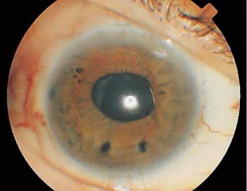
On clinical examination, retinoscopy identifies a red reflex that may be normal or slightly dull and irregular. Despite the ability to perform retinoscopy, the manifest refraction typically does not improve visual acuity. Slit lamp examination with side illumination demonstrates a thickened, irregular posterior capsule that may be slightly opacified or translucent (Fig. 1). Multiple vesicles in the capsule represent flattened Elschnig's pearls. Fluid in the vesicles is typically clear but may occasionally be turbid. Retroillumination may enhance the thickened, vesicular appearance of the posterior capsule. Examination of the retina is imperative to exclude other causes of decreased central vision that may occur after cataract surgery including cystoid macular edema, diabetic macular edema, or retinal detachment. Venous occlusive disease and age-related macular degeneration are additional causes of diminished visual acuity that commonly occur in this elderly population who undergo cataract surgery.

Fig. 1. Examination by diffuse (A) and side (B) illumination demonstrates a thickened, opacified posterior capsule.

PROCEDURES

The Nd:YAG laser is a slit-lamp-mounted system with a double spot helium-neon aiming beam that merges to one beam with fine focusing. The eye is anesthetized topically and a contact lens maybe placed on the cornea if magnification of the posterior capsule is desired. The pupil may be pharmacologically dilated or remain undilated, depending on the preference of the treating ophthalmologist. Apraclonidine hydrochloride 1% is administered before laser treatment to prevent postoperative intraocular pressure (IOP) elevations. With the patient secured in the slit-lamp-mounted laser, the red helium-neon beam is focused on the posterior capsule so that energy is not transferred to the IOL or anterior hyaloid face. When properly focused, the two aiming beams merge into one. An initial laser power setting of 1.0 mJ or less is used to commence treatment. When Nd:YAG energy is successfully delivered to the posterior capsule, a break appears that enlarges along the lines of tension. Occasionally gas bubbles form on the capsule, which either clear spontaneously or can be dislodged by gently tapping on the contact lens. If the initial power setting fails to create an opening through the capsule, the energy level is gradually increased until laser disruption of the capsule occurs. Attempting to create a single opening in the capsule without increasing the energy level is ineffective and acts only to increase the total energy delivered to the eye. It is rarely necessary to increase the Nd:YAG laser power above 2.5 mJ.

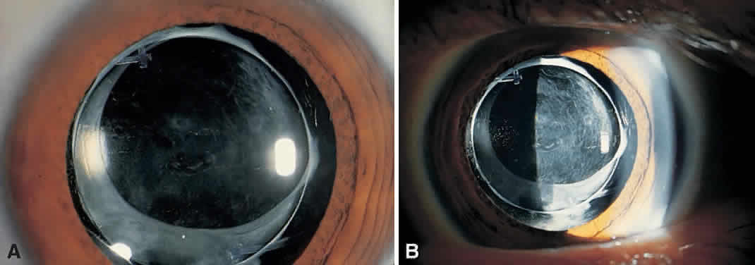
Multiple techniques exist to create an opening in the posterior capsule. These include cruciate, circular, or inverted U-shaped patterns. With the pupil in an undilated state, single shots are placed in the visual axis. Regardless of the technique, the goal is to create an opening in the posterior capsule that is slightly larger than the resting pupil (Fig. 2). Typically, this will be 3 to 4 mm and require 30 to 40 spots of laser to the posterior capsule. An effective way to minimize the number of laser applications is to treat the areas of capsule under tension. The openings created expand from the tensile forces of the opacified capsular bag. In a thickened opacified capsule, pieces of the capsule may be released into the anterior vitreous. These pieces inferiorly migrate out of the visual axis within 1 week of treatment without inducing any visual disturbances.

Fig. 2. Opening in the posterior capsule from a Nd:YAG laser following cataract surgery.

Most YAG capsulotomies are uncomplicated; an opening in the capsule is successfully created in 98% of cases.11 Visual acuity improves almost immediately following treatment. The eye may be treated before or after the procedure with apraclonidine 1% to blunt a potential rise in IOP, which is measured 20 to 60 minutes later. Persistent elevation of IOP occurs infrequently and is typically treated with an ocular hypotensive agent and close observation. Corticosteroid eye drops three or four times daily (e.g., fluorometholone acetate or prednisolone acetate 1%) for 4 to 7 days is a management option used by some ophthalmologists. Generally, patients experience only minimal symptoms, if any, after this procedure. Follow-up typically occurs in 1 week, unless the IOP is significantly elevated on post-YAG testing. Long-term follow-up is required in eyes with persistently elevated IOP.12

COMPLICATIONS

Although Nd:YAG laser posterior capsulotomy is a relatively straightforward procedure, complications may be encountered. The most common intraoperative complication reported in a cohort of 2110 patients followed by the Food and Drug Administration (FDA) was damage to the IOL.11 Accurate focusing of the helium-neon beam on the posterior capsule prevents the occurrence of this event. However, postoperative contraction of the capsular bag can move it into very close proximity to the posterior surface of the IOL, leading to this complication. Other factors that increase the likelihood of IOL damage include increased thickness of the PCO membrane and the total laser power used. Despite the occasional creation of laser-induced pits in the IOL, these typically do not cause visual disturbances. Rupture of the anterior hyaloid face was the second most frequently reported complication occurring in 19% of the patients observed during the FDA study. Less frequently encountered intraoperative complications were bleeding in 1.0%, corneal edema in 0.3%, and iris damage in 0.4% of cases.11

Elevation of IOP after Nd:YAG capsulotomy is the major postoperative complication encountered that requires treatment.11,13–15 The cause of this elevation is not certain but may be related to the release of lens epithelial cells into the anterior chamber. Variables such as the total energy delivered, number of laser pulses, size of the capsulot-omy, iris bleeding, and the amount of inflammation do not appear to affect IOP.14–16 However, some authors dispute this and assert that higher IOP after Nd:YAG capsulotomy is associated with larger capsulotomies and increased energy.12,13 In an FDA cohort of 213 patients following a protocol of measuring postprocedure IOP at selected intervals, 39% of patients had an increase of 5 mmHg or more in IOP 2 to 6 hours after laser capsulot-omy. About 28% experienced an increase in absolute IOP of more than 30 mm Hg. None of the eyes received prophylactic ocular hypotensive agents in the perioperative period.11 The maximum elevation of IOP occurred between 1.5 and 4 hours after laser treatment. Of these eyes, 60% returned to an IOP of less than 22 mmHg by 24 hours with 90% of eyes achieving normalization of IOP by 1 week.11 Results by Slomovic and Parrish15 mirrored the FDA's findings. Thus, 64% of eyes developed a maximum IOP rise by the second postoperative hour with 41% of eyes developing an IOP greater than 30 mm Hg. The results of the study of Slomovic and Parrish indicated that glaucomatous eyes, which were either medically or surgically controlled prior to capsulotomy, had a lower mean IOP rise after laser capsulotomy compared with eyes that had not been prophylactically treated.

With the discovery that pretreatment may blunt the postprocedure elevation in IOP, a prospective multicentered trial was initiated to evaluate the effect of apraclonidine applied in the perioperative period to treated eyes versus placebo. Apraclonidine 1% was administered 1 hour before and immediately after performing YAG laser capsulotomy.17 Placebo-treated eyes experienced a maximum IOP elevation of 4.4 mm Hg 3 hours after treatment. The IOP in apraclonidine-treated eyes decreased 2.8 mm Hg from baseline. As a result of the findings, eyes are now typically treated with apraclonidine or brimonidine when performing YAG laser capsulotomy.

Ge and colleagues12 evaluated long-term IOP fluctuations in eyes followed for a median of 1.5 years after YAG posterior capsulotomy. Eligible patients were pseudophakic bilaterally with one eye receiving laser capsulotomy and the other eye serving as a control. All eyes received apraclonidine before and after treatment. An elevated IOP mea-sured at 1 hour postprocedure was a significant risk factor for chronically increased IOP. This indicates that patients with elevated IOP after YAG laser capsulotomy despite pretreatment with ocular hypotensive agents should be monitored for persistent IOP elevation.

Other less common complications include retinal detachment and cystoid macular edema. In one recent evaluation of 196 eyes examined at 1 month after laser capsulotomy, only one eye sustained a new retinal break that was not demonstrated before treatment.18 Steinert and associates16 reviewed 879 Nd:YAG laser posterior capsulotomies over a 3-year period. Cystoid macular edema developed in 1.23% and retinal detachment occurred in 0.89%. Three of eight patients developed retinal detachment more than 1 year after laser capsulotomy. The number of laser pulses and amount of total energy delivered to the eye were not identified as risk factors. The FDA report of 2110 patients registered similar numbers.11 Cystoid macular edema occurred in 1.2% of the study eyes with retinal detachment in 0.5%. Less common complications included vitritis, iritis, and retinal hemorrhage in less than 0.6% of patients.11 Case reports of delayed posterior dislocation of silicone plate-haptic lenses have been described in the literature.19

Back to Top
PREVENTION OF POSTERIOR CAPSULAR OPACIFICATION
Regardless of recent advances in cataract extraction, lens epithelial cells remaining in the capsule proliferate and will cause eventual opacification. Tissue culture studies have demonstrated that a monolayer of lens epithelial cells completely cover the lens capsule within days of surgery.20,21 Manual aspiration, capsule polishing, ultrasound aspiration, cryocoagulation, and osmolysis have been unsuccessfully employed to decrease the incidence of PCO.22,23 Results of pharmacologically targeting these cells with monoclonal antibodies, infusion of agents into the anterior chamber at the time of cataract surgery, or immunotoxin-coated IOLs have been mixed.2–5,24–26 Although inhibition of lens epithelial cells can be observed, toxic side effects to the ciliary body, cornea epithelium, and iris limit the in vivo usefulness. This has led to the investigation of other variables that may limit posterior capsular opacification.

Hollick and associates evaluated the effect of capsulorrhexis size on PCO.27 A small capsulorrhexis with the anterior capsule covering the optic was performed in one group of patients and compared with a second group with a larger capsulorrhexis that extended past the edge of the IOL optic. Both groups were implanted with 5.5-mm optic polymethylmethacrylate (PMMA) lenses. The presence of a large capsulorrhexis was associated with a significant increase in posterior capsular wrinkling and opacification. Eyes in which the capsulorrhexis did not extend past the edge of the optic had a better visual acuity at 1 year.

Recent attention has focused on the material and design of the IOL. Inhibition of PCO involves the presence of the IOL in the capsule, obliteration of the space between the posterior side of the optic and the capsule, and the adhesiveness of the IOL.28 Nishi compared 225 eyes implanted with a PCIOL with 379 eyes that remained aphakic.23 The incidence of PCO was significantly less in the eyes implanted with a PCIOL suggesting that an IOL has suppressive effects on the migration of lens epithelial cells toward the center of the capsule.

Hollick and associates concentrated on the IOL material and randomized 93 patients to receive either PMMA, silicone, or hydrogel lenses.29 At 2 years after surgery, hydrogel IOLs induced the greatest mean percentage area of posterior capsular opacification followed by PMMA and then silicone lenses. YAG laser capsulotomy was performed in 28% of eyes with hydrogel lenses and 14% of eyes with PMMA lenses at 2 years. No eyes randomized to silicone lens implantation required YAG laser capsulotomy. Polyacrylic lenses have demonstrated even lower rates of PCO.21,30 The presence of lens epithelial cells was significantly lower with the polyacrylic lens compared with PMMA and silicone lenses. Regression of epithelial cells was observed in 83% of the acrylic lenses with regression occurring in less than 20% of eyes with the PMMA and silicone lenses.21 YAG posterior capsulotomy was not required in any of the eyes implanted with the acrylic lenses.30

The design of the polyacrylic IOL appears to decrease PCO. Polyacrylic lenses have a tacky surface that create adhesion between the IOL and lens capsule. This ablates the space posterior to the IOL causing compression of lens epithelial cells and preventing further migration of lens cells behind the IOL. This has been called the “no space, no cell” theory. Hollick and colleagues suggested that the adhesion formed with a polyacrylic lens takes a few weeks to form during which some lens epithelial cell migration my occur.21 This would explain the regression of PCO that is observed in the poly-acrylic IOL.

PCIOLs designed to hold the capsule away from the lens surface to facilitate YAG capsulotomy provide less inhibition to lens epithelial cell migration. Posterior-convex or biconvex lenses with edged optics bring the capsule into contact with the posterior surface of the optic inhibiting cell migration and PCO. The edged optic design also forms a barrier against the remaining lens cells. The use of posterior-convex and biconvex optics reduced the rate of YAG laser capsulotomy after cataract surgery to 6.5% after 5 years compared with a rate of 40.0% at 5 years with a convex-plano optic.31

Further research has investigated the design of IOL haptics in preventing lens epithelial cell migration. In vivo animal studies where a sharp, discontinuous bend in the posterior capsule was created with a capsule-bending haptic or a capsule-bending ring significantly inhibited lens epithelial cell migration. Lens epithelial cells accumulated at the equator of the capsule around the haptic ring with less PCO.28

Back to Top
OTHER USES OF THE Nd:YAG LASER IN PSEUDOPHAKIA
The photodisruptive effect of the Nd:YAG is also useful in laser peripheral iridotomy where a full-thickness hole is created through the iris. With an anterior chamber IOL, pupillary block may occur if a surgical iridectomy is not performed or does not extend full-thickness through the iris (Fig. 3). Laser iridotomy with the Nd:YAG laser efficiently creates a full-thickness peripheral iris hole to break the pupillary block.

Fig. 3. Two laser iridotomies are seen peripheral to the inferior edge of the optic. The Nd:YAG laser iridotomy was performed emergently when the patient experienced pupillary block 2 weeks after cataract surgery.

Vitreous strands may adhere to the corneoscleral wound after cataract surgery complicated by rupture of the posterior capsule and anterior hyaloid face. On examination, the vitreous strands are visible and there is often peaking of the pupil. Vision may be diminished from the development of cystoid macular edema secondary to vitreous traction (Irvine-Gass syndrome). Lysis of these transparent strands may be performed with the Nd:YAG laser to release the vitreous traction and resolve the cystoid macular edema. This procedure becomes more difficult with time as vitreous fibers that are chronically incarcerated in the wound become encased in corneal endothelial cells making the strands thicker. Multiple sessions may be required to cut the vitreous strands.

Back to Top
CONCLUSION
The Nd:YAG laser has become an invaluable tool for ophthalmologists after cataract surgery. It provides an efficient, safe, noninvasive technique to treat PCOs that occur after cataract surgery. Although it is important to be aware of the complications related to Nd:YAG laser use, these are relatively infrequent and typically are not severe.
Back to Top
REFERENCES

1. Apple DJ, Solomon KD, Tetz MR et al. Posterior capsular opacification. Surv Ophthalmol 1992;37:73

2. Duncan G, Wormstone IM, Liu CS et al. Thapsigargin coated intraocular lenses inhibit human lens cell growth. Nat Med 1997;3:1026

3. Tetz MR, Ries WR, Lucas MS et al. Inhibition of posterior capsule opacification by an intraocular lens bound sustained drug delivery system: An experimental animal study and review of the literature. J Cataract Refract Surg 1996;22:1070

4. Nishi O, Nishi K, Yamada Y et al. Effect of indomethacin coated posterior chamber intraocular lenses on postoperative inflammation and posterior capsule opacification. J Cataract Refract Surg 1995;21:574

5. Clark DS, Munsell MF, Emery JM. Mathematical model to predict the need for neodymium:YAG capsulotomy based on posterior capsule opacification rate. J Cataract Refract Surg 1998;24:1621

6. Steinert RF, Puliafito CA. Posterior capsulotomy and pupillary membranectomy. In: The Nd:YAG Laser in Ophthalmology. Philadelphia: WB Saunders, 1985:76–77

7. Kraff MC, Sanders DR, Jampol LM et al. Effect of primary capsulotomy with extracapsular surgery on the incidence of pseudophakic cystoid macular edema. Am J Ophthalmol 1984;98;166

8. History and principles of ophthalmic lasers. In: Noyori K, Shimizu K, Trokel S, eds. Ophthalmic Laser Therapy. Tokyo: Igaku-Shoin, 1992:1–9

9. Kolder HE. YAG laser capsulotomy. In: Weingeist TA, Sneed SR, eds. Laser Surgery in Ophthalmology: Practical Applications. Norwalk, CT: Appleton & Lange, 1992:167–174

10. Marcantonio JM, Vrensen GFJM. Cell biology of posterior capsular opacification. Eye 1999;13:484

11. Stark JW, Worthen D, Holladay JT et al. Neodymium:YAG lasers: An FDA report. Ophthalmology 1985;92:209

12. Ge J, Wand M, Chiang R et al. Long-term effect of Nd:YAG laser posterior capsulotomy on intraocular pressure. Arch Ophthalmol 2000;118:1334

13. Channell MM, Beckman H. Intraocular pressure changes after neodymium-YAG laser posterior capsulotomy. Arch Ophthalmol 1984;102:1024

14. Flohr MJ, Robin AL, Kelley JS. Early complications following Q-switched neodymium:YAG laser posterior capsulot-omy. Ophthalmology 1985;92:360

15. Slomovic AR, Parrish RK. Acute elevations of intraocular pressure following Nd:YAG laser posterior capsulotomy. Ophthalmology 1985;92:973

16. Steinert RF, Puliafito CA, Kumar SR et al. Cystoid macular edema, retinal detachment, and glaucoma after Nd:YAG laser posterior capsulotomy. Am J Ophthalmol 1991;112:373

17. Pollack IP, Brown RH, Crandall AS et al. Effectiveness of apraclonidine in preventing the rise in intraocular pressure after neodymium:YAG posterior capsulotomy. Trans Am Ophthalmol Soc 1988;86:461

18. Ranta P, Tommila P, Immonen I et al. Retinal breaks before and after neodymium:YAG posterior capsulotomy. J Cataract Refract Surg 2000;26:1190

19. Petersen AM, Bluth LL, Campion M. Delayed posterior dislocation of silicone plate-haptic lenses after neodymium:YAG capsulotomy. J Cataract Refract Surg 2000;26:1827

20. Spalton DJ. Posterior capsular opacification after cataract surgery. Eye 1999;13:489

21. Hollick EJ, Spalton DJ, Ursell PG et al. Lens epithelial cell regression on the posterior capsule with different intraocular lens materials. Br J Ophthalmol 1998;82:1182

22. Nishi O. Update/review: Posterior capsule opacification. J Cataract Refract Surg 1999;25:106

23. Nishi O. Incidence of posterior capsule opacification with and without posterior chamber intraocular lenses. J Cataract Refract Surg 1986;12:519

24. Liu CSC, Duncan G, Wormstone IM et al. Strategies to prevent lens cell growth causing posterior capsule opacification: An in vitro study. Invest Ophthalmol Vis Sci 1996;37:S758

25. Emery J, Clark DS, Munsell M et al. Inhibition of posterior capsule opacification with an immunotoxin specific for lens epithelial cells: Eighteen month results of a phase I/II clinical study. Invest Ophthalmol Vis Sci 1996;37:S758

26. Behar-Cohen F, David T, D'Hermies F et al. In vivo inhibition of lens regrowth by fibroblast growth factor 2-saporin. Invest Ophthalmol Vis Sci 1995;36:2434

27. Hollick EJ, Spalton DJ, Meacock WR. The effect of capsulorrhexis size on posterior capsular opacification: one-year results of a randomized prospective trial. Am J Ophthalmol 1999;128:271

28. Nishi O, Nishi K, Mano C et al. The inhibition of lens epithelial cell migration by a discontinuous capsular bend created by a band-shaped circular loop or a capsule bending ring. Ophthalmic Surg Lasers 1998;29:119

29. Hollick EJ, Spalton DJ, Ursell PG et al. Posterior capsular opacification with hydrogel, polymethylmethacrylate, andsilicone lenses: two-year results of a randomized prospective trial. Am J Ophthalmol 2000;129:577

30. Hollick EJ, Spalton DJ, Ursell PG et al. The effect of polymethylmethacrylate, silicone, and polyacrylic intraocular lenses on posterior capsular opacification 3 years after cataract surgery. Ophthalmology 1999;106:49

31. Born CP, Ryan DK. Effect of intraocular lens optic design on posterior capsular opacification. J Cataract Refract Surg 1990;16:188

Back to Top