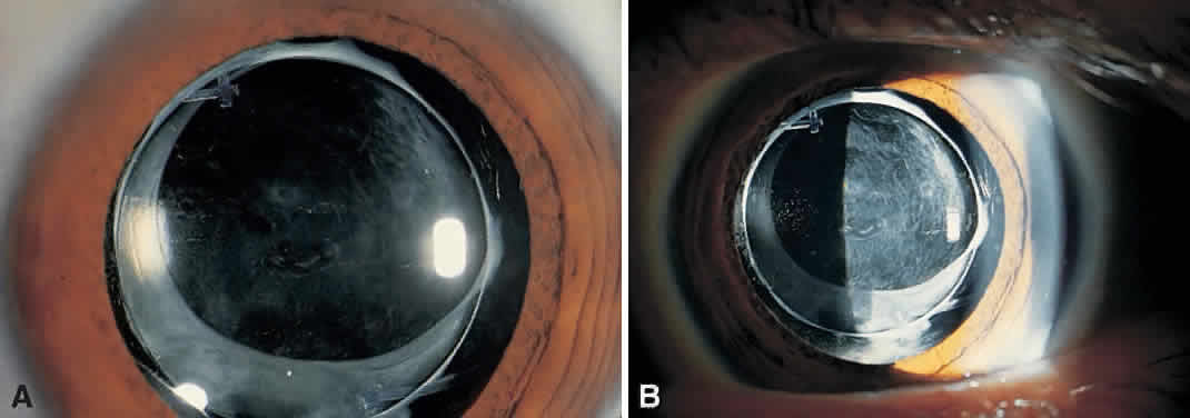
Fig. 1.
Examination by diffuse (
A
) and side (
B
) illumination demonstrates a thickened, opacified posterior capsule.