Chapter 8
Secondary Intraocular Lens Implantation
NEIL J. FRIEDMAN, TIMOTHY T. KHATER and DOUGLAS D. KOCH
Main Menu   Table Of Contents

Search

PREOPERATIVE SCREENING AND SELECTION OF INTRAOCULAR LENS TYPE
POSTERIOR CHAMBER INTRAOCULAR LENSES
ANTERIOR CHAMBER INTRAOCULAR LENSES
SURGICAL TECHNIQUE
ANTERIOR CHAMBER INTRAOCULAR LENS IMPLANTATION
ENDOCAPSULAR POSTERIOR CHAMBER LENS
SULCUS POSTERIOR CHAMBER LENS (UNSUTURED)
IRIS-SUTURED SULCUS POSTERIOR CHAMBER INTRAOCULAR LENS
TRANSSCLERALLY SULCUS–SUTURED POSTERIOR CHAMBER INTRAOCULAR LENS
RESULTS
CONCLUSION
REFERENCES

The role of secondary intraocular lens (IOL) implantation has evolved with advances in cataract surgery. Traditionally, most patients who required secondary IOL implantation were either elderly aphakes who had previously undergone intracapsular cataract extraction, or aphakes who had lensectomy during early childhood for congenital cataracts. Individuals also may be left aphakic after complicated cataract surgery, lensectomy after trauma, or removal of a dislocated crystalline lens. For these patients, secondary implantation of an IOL generally is recommended when traditional spectacle or contact lens correction of aphakia is unsuccessful. For bilateral aphakia corrected with aphakic spectacles, surgery is indicated when the patient cannot readily cope with the optical distortions produced by the glasses. For the unilateral aphake, spectacle correction usually is intolerable because of the large amount of anisometropia. Contact lenses can reduce the aberrations and aniseikonia produced by aphakic spectacles. However, many patients are unable to wear contact lenses because of an inability to handle or care for the contact lens, difficulty in fitting the lens, discomfort, contact lens-related complications such as giant papillary conjunctivitis or poor motivation for proper use.

For patients unable to use these devices, various surgical procedures have been investigated, including secondary IOL implantation, epikeratophakia,1 and intracorneal implants.2 The major concern with all of these approaches is the risk of vision-threatening complications. Epikeratophakia and corneal inlays produced disappointing results because of irregular corneal surface changes and poor refractive predictability.3–6

Early results of secondary IOL implantation were mixed. Several reports documented excellent outcomes after secondary posterior chamber intraocular lens (PC IOL) implantation in patients with intact posterior capsules.7 Good short-term results were initially reported with early closed-loop anterior chamber intraocular lenses (AC IOLs), but unfortunately, numerous complications, including uveitis, glaucoma, hyphema, cystoid macular edema, endothelial cell loss, and corneal decompensation (Fig. 1), later developed in many eyes receiving lenses of this type.7–32 The newer, open-loop, one-piece flexible design (i.e., Kelman multiflex style) has eliminated nearly all of these complications, and, as a result, has gained growing acceptance.33–37 As an alternative option for eyes without sufficient capsular support, various lenses and surgical techniques have been developed for implanting iris-supported (e.g., lobster-claw lens)38–44 and sulcus-sutured posterior chamber lenses.

Fig. 1. Corneal decompensation after implantation of closed-loop anterior chamber intraocular lens.

Because of advances in primary cataract surgery, surgical aphakia is becoming an increasingly rare condition, and secondary intraocular lens insertion is now most commonly performed as part of an IOL exchange procedure. This is indicated for mechanical, inflammatory, or optical IOL-related complications.45 In addition, secondary IOLs are sometimes implanted in combination with other procedures, such as pars plana lensectomy for the treatment of a dislocated crystalline lens, penetrating keratoplasty, or trabeculectomy.

For patients with adequate posterior capsular support, we believe that implantation of a PC IOL into the capsular bag or ciliary sulcus most often is the preferred approach. However, debate continues regarding which of several methods of secondary IOL implantation is superior for eyes lacking sufficient capsular support. Because a clear-cut answer does not exist, we compare the advantages and disadvantages, surgical techniques, and results of studies of IOL implantation with each of these methods.

Back to Top
PREOPERATIVE SCREENING AND SELECTION OF INTRAOCULAR LENS TYPE
The health of the eye is critical in deciding which patients are candidates and, in some instances, which type of lens is to be implanted. Conditions such as corneal endothelial abnormalities, glaucoma, and diabetes require thorough investigation before the insertion of any type of lens. Cystoid macular edema (CME) is generally a contraindication for secondary IOL implantation, unless the CME is caused by a problematic IOL and the problematic IOL is to be repositioned or exchanged.

The peer-reviewed literature suggests that otherwise healthy eyes without capsular support can receive an anterior chamber, iris-supported, or sutured posterior chamber lens with equally satisfactory results.33,46–49 The surgeon's experience and comfort with each procedure usually determines which one is chosen; however, some surgeons believe that patients younger than 50 years of age generally should not receive an AC IOL. This age limit is arbitrary and varies among surgeons. However, the long-term biostability of the polypropylene sutures required for sutured PC IOL implantation is uncertain.50

During the preoperative evaluation, it is important to perform meticulous slit-lamp biomicroscopy, funduscopy, and, where indicated, gonioscopy to evaluate any ocular pathology that might affect surgical planning. In addition, aphakes are commonly chronic contact lens users, and an extended period of contact lens disuse is indicated for optimal refractive results. We currently recommend discontinuing contact lenses for at least 1 to 2 weeks for soft contact lens wearers, and 2 to 4 weeks or longer for rigid gas permeable contact lens (RGP) wearers. A more conservative approach is to discontinue lens wear 1 month for each decade of RGP wear. Corneal stability is then verified by serial topography, keratometry, and refraction. Factors to assess include the following:

Best spectacle-corrected visual acuity
Corneal astigmatism
Corneal endothelial status
Anterior chamber (depth, cell, and flare)
Anterior chamber angle (peripheral anterior synechiae, recession, and neovascularization)
Iris and pupil (size, reactivity, peripheral iridectomies, and anterior and posterior synechiae)
Posterior capsule (adequacy of support and clarity)
Presence of vitreous in anterior segment
Macula (cystoid macular edema, macular degeneration)
Peripheral retina

In some cases, specialized testing such as corneal pachymetry, specular micrography, optical coherence tomography, and fluorescein angiography may be indicated.

The timing of secondary IOL surgery also must be considered. In one study,51 the risk for CME was increased if secondary IOL implantation was performed within 1 year of intracapsular cataract extraction; unfortunately, there are no comparable data on the timing of surgery after extracapsular procedures.

The status of the patient's fellow eye is obviously important. Does it have good vision? Does it have any sight-threatening conditions? Either of these factors might make one more cautious in performing intraocular surgery in the fellow eye. What is the refractive error of the fellow eye?

Furthermore, the patient's overall health may contribute to the surgical decision. Because implanting an anterior chamber lens generally is faster than suturing an IOL into the sulcus, an AC IOL may be chosen for a patient who may not be able to tolerate a lengthy procedure. Similarly, an anticoagulated patient may be at greater risk for intraocular hemorrhage from needle passes through the iris or ciliary sulcus and sclera; therefore, a safer approach might be to insert an AC IOL.

Finally, the technique of choice is often determined at the time of surgery and is often a matter of clinical judgment. Intraoperative findings that alter the surgical approach include the adequacy of zonular support, posterior iridocapsular adhesions, status of the anterior and posterior capsular remnants, and the presence of lenticular pieces that were not visible during the preoperative examination. The surgeon should be well versed in multiple techniques, in case one or more techniques are surgically contraindicated.

Back to Top
POSTERIOR CHAMBER INTRAOCULAR LENSES
Clearly, if the preoperative eye has adequate capsular support, the preferred site for secondary IOL implantation is the capsular bag or the ciliary sulcus. We believe that the optimal lens for secure sulcus fixation is a large optic (6.5 mm or more), one-piece polymethyl methacrylate (PMMA) IOL with an overall diameter of 13.5 to 14 mm. However, a foldable IOL with a 6-mm optic and 13-mm overall length allows insertion through a smaller wound and is acceptable if the IOL is stable. Unfortunately, many patients requiring a secondary IOL lack adequate zonular or capsular support for either of these procedures. Sulcus implantation may be contraindicated in patients with a history of pigment dispersion from previous posterior chamber lens implantation or with an excessively large sulcus that might not permit firm haptic fixation. Options in these patients might include suture fixation of the posterior chamber lens to the sclera or iris, an iris-supported lens, or an anterior chamber IOL. In rare instances, if the capsular bag is intact and can be reopened but lacks adequate zonular support, a capsular tension ring (CTR) with or without fixation sutures can be inserted to enable implantation of an unsutured posterior chamber lens.52–56

Various techniques of posterior chamber IOL fixation have been tried in patients who were poor candidates for an anterior chamber lens and who lacked sufficient capsular support. There has been a recent resurgence of interest in the use of iris-suture lenses. Advantages of this approach include: (a) Avoidance of the risks associated with needle passes through the sclera and tend to spare the anterior chamber angle,39–41,44 (b) ability to use a foldable IOL and small incision, and (c) greater ease compared to suturing a lens into the ciliary sulcus. Fixation to the peripheral iris is most commonly accomplished with a modified McCannel retrievable suture, although a number of closed and open-sky techniques have been described.57–61 Complications reported with this method of IOL fixation include pigment dispersion, uveitis, glaucoma, pupillary block, iris/pupil distortion, peripheral anterior synechiae (PAS), hyphema, CME, and retinal detachment (RD).62,63 However, comparative studies suggest that the results with iris-fixation match or exceed those with anterior chamber or scleral-sutured IOLs.

A more popular method of suture fixation has been transscleral suturing. A variety of techniques and lens modifications have been reported for transsclerally suturing PC IOLs. Gess64 first described scleral fixation of one haptic of a posterior chamber lens. In 1986, Malbran and colleagues65 described an open-sky technique for sutured PC IOLs, and in 1988, Cowden and Hu66 reported secondary PC lens implantation with scleral fixation of both haptics through scleral stab incisions.

Advantages of transsclerally sutured IOL fixation include the elimination of corneal and angle trauma associated with anterior chamber lenses, decreased risk of pupillary block and secondary glaucoma, and little or no IOL contact with the iris, thereby decreasing the risk of iritis, pigment dispersion, and CME.9,13,24,25,29,67–70 The sulcus location most closely approximates the normal anatomic position of the crystalline lens, and this method of PC IOL fixation minimizes the risk of pseudophakodonesis.20 In addition, transsclerally sulcus–sutured posterior chamber IOLs theoretically can be used in any age group, including children,71–73 because the lenses are more likely than anterior chamber lenses to accommodate to the growing eye; however, long-term results are unknown.

Important drawbacks to this type of lens fixation need to be considered. Compared with AC IOL insertion and iris-sutured PC IOLs, the procedure is technically more difficult, requiring longer surgical time and a thorough anterior vitrectomy, both of which might increase the risk of intraoperative and postoperative complications. In addition, it is difficult to precisely and symmetrically fixate both haptics within the ciliary sulcus.74–76 The anatomy of this space, which averages 11.0 ± 0.37 mm in diameter77 and is located approximately 0.83 mm and 0.46 mm posterior to the surgical limbus in the vertical and horizontal meridians,78 respectively, may be altered in long-standing aphakia.75 In Pavlin's ultrasound biomicroscopy study of 34 transsclerally fixated PC IOLs, 13 IOLs were in the ciliary sulcus, 8 were posterior, and 13 were anterior.74 Manabe also used the ultrasound biomicroscope to identify haptic placement and found only 32 of 86 haptics sutured at the sulcus; there were 29 at the ciliary processes, 25 posterior to the pars plicata, and 41 with vitreous incarceration.76 Additionally, in a clinical study comparing complications of AC IOL versus transsclerally fixated PC IOL implantation, Bellucci showed that six of 32 PC IOLs were in the sulcus, 24 were in the pars plana, and 2 were in the iris root.75 We suspect that in many instances one or both haptics may not be properly positioned in the ciliary sulcus, but this is not noted clinically because of the excellent stabilization provided by the sutures.

Despite its anatomic advantages, PC IOL implantation into the ciliary sulcus is associated with complications, primarily because of haptic contact with uveal tissue and the need for haptic fibrosis to ensure long-term stability. Even without suture fixation, sulcus placement of a lens implant carries the risks of lens decentration (Fig. 2), pigment dispersion, uveitis, recurrent hemorrhage (Fig. 3), ciliary body erosion,79–84 and, in one reported case, occlusion of the major arterial circle of the iris (located in the ciliary body), with devastating results.70 Transscleral fixation introduces additional risks caused by needle penetration of uveal and scleral tissue, abnormal positioning of the haptics, and external suture exposure; these risks include lens tilt and decentration (Fig. 4), lens subluxation, episcleritis, corneal decompensation, hypotony, PAS formation, secondary glaucoma, hyphema, vitreous hemorrhage, suprachoroidal hemorrhage, choroidal effusion, CME, RD, external suture erosion (Fig. 5), and endophthalmitis.43,47,85–101

Fig. 2. Inferior subluxation (sunset syndrome) of a sulcus-fixated posterior chamber intraocular lens.

Fig. 3. Recurrent iritis and hyphema associated with a loose posterior chamber intraocular lens in the ciliary sulcus. Note fine dusting of red blood cells on endothelial surface.

Fig. 4. Mild inferior decentration of a transsclerally sutured intraocular lens; the fixation sutures were not placed 180 degrees apart.

Fig. 5. A. External suture erosion and exposure after placement of a transsclerally fixated posterior chamber intraocular lens. B. Note fluorescein pooling around exposed suture ends.

A variety of modifications of surgical technique and lens design intended to reduce some of these risks have been described.39,47,63,86,93,94,102–119 Goals have included limiting the time the eye is open (to reduce the risk of intraoperative complications), decreasing the number of needle passes through the sclera (to reduce the risk of intraocular bleeding), burying the suture knots (to reduce the risk of suture erosion and subsequent endophthalmitis), using direct visualization (endoscopic or mirror needle holder to ensure more accurate placement of the haptics in the sulcus), and, perhaps most importantly, suturing with stronger material and double fixation (9-0 Prolene or 8-0 Gore-Tex to reduce the risk of suture breakage and lens subluxation).102,120–123

Anand and Bowman124 first described using scleral flaps to cover and protect the suture knots. However, Solomon98 found a 73% rate of suture erosion through scleral flaps, suggesting that this approach delays but does not prevent this complication.46,111 Friedberg125 created scleral grooves in which the knots were secured and then protected. Later, Bucci and coworkers126 showed that covering the knot with corneal tissue prevents erosion. Lewis85,120 subsequently reported methods for burying the suture knots in the sclera to avoid erosion through sclera and conjunctiva.

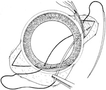
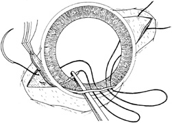
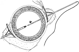
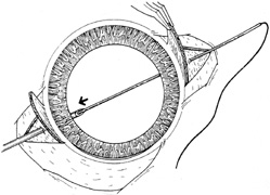
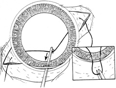
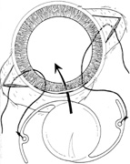
Numerous variations for making the suture passes also have been explored. These include either an ab externo or ab interno approach, single or dual passes, and direct passage or capture with a 25- to 27-gauge hollow needle (Figs. 6 and 7). The ab externo approach uses an outside to inside suture pass that allows more precise needle placement and reduces the duration of hypotony.146 Ab interno techniques are performed by passing the suture needle from the inside to the outside of the eye, and have the advantage of being faster. Different IOL designs (i.e., with or without eyelets, one-piece or foldable) and suture needles (i.e., long or short, straight or curved) exist for different techniques.

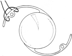
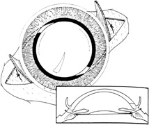
Fig. 6. Technique for the ab interno approach. A. First the long needles are passed under the iris, aiming for the inferior ciliary sulcus. Two needle passes are made for each haptic if four-point fixation is desired. Needles exit under previously dissected scleral flaps. B. A second pair of short needle passes is made under the superior iris for the suture to be tied to the second haptic. C. Girth hitch can be used to attach the polypropylene suture loop to the intraocular lens (IOL) haptic. This technique is more rapid than tying the suture to the haptic. Alternately, the suture can be attached to the IOL haptics before the transscleral needle passes, but the surgeon must avoid tangling the long sutures. D. After exiting the eye under the previously dissected scleral flaps, the sutures are tied securing the IOL into position. Appropriate suture tension is important to avoid lens decentration. The inset shows the cross-sectional view of the eye with the IOL correctly positioned in the ciliary sulcus. (From Steinert RF, Arkin MS:. Secondary intraocular lenses. In: Steinert RF, ed. Cataract Surgery: Techniques, Complications, and Management,. 2nd ed. Philadelphia: :Saunders, 2004:433, with permission from Elsevier.)

Fig. 7. Technique for the ab externo approach. A. The long, straight solid needle is passed through the sclera (usually under partial-thickness scleral flaps) approximately 0.75 mm posterior to the limbus. Inside the eye, the needle should exit at the ciliary sulcus. A second hollow needle is passed from the opposite side of the eye. A pair of sutures can be used if four-point fixation is desired. B. Solid needle is “docked” inside the tip of the hollow needle, which has been passed through ciliary sulcus on the opposite side. After docking, the pair of needles are withdrawn together from the eye, with the solid needle inside the hollow needle. C. A hook is used to pull the suture out through a superior limbal wound so that it can be tied to the intraocular lens (IOL). D. Suture is cut, and each end is tied to a haptic of the IOL. After the IOL is placed into position, the scleral sutures must be anchored to the sclera. Either a “blind pass” in the sclera is made so that the suture is tied to itself, or the transscleral suture is tied to a second suture that has been tied to the sclera with a short partial-thickness pass within the bed of the scleral flap. (From Steinert RF, Arkin MS:. Secondary intraocular lenses. In: Steinert RF, ed. Cataract Surgery: Techniques, Complications, and Management,. 2nd ed. Philadelphia: Saunders, 2004:434, with permission from Elsevier.)

Despite its widespread use, we are unaware of any long-term studies of the outcomes of transsclerally sulcus–fixated PC IOLs. Recent reports of suture breakage raise the specter of an unacceptably high risk of late dislocation. This may be avoidable with the use of stronger, more durable suture materials (to minimize hydrolysis, breakage, and erosion) and more accurate placement of the haptics (to achieve fixation via fibrosis in the sulcus). Despite its obvious advantages and many reports of excellent short-term results, this technique will require follow-up of 10 or more years to fully ascertain its safety and stability.

Back to Top
ANTERIOR CHAMBER INTRAOCULAR LENSES
Anterior chamber IOLs have been in clinical use for more than 40 years, and a remarkable number of design and material combinations have been investigated.127 The main advantages of an AC IOL for secondary implantation are the relative ease, speed, and minimally traumatic nature of the surgical procedure, which may reduce intraoperative complications. In addition, in patients with vitreous that extends to or just through the pupil, insertion of an AC IOL may prevent the need for an anterior vitrectomy, which would be required for PC IOL insertion, particularly if suture fixated.

Older anterior chamber lens designs have a problematic track record, however. The original rigid designs and second-generation, flexible, closed-loop haptic designs caused many complications, including erosion into the angle; pain; corneal edema (see Fig. 1); iris/pupil distortion; PAS; uveitis, glaucoma, and hyphema (UGH) syndrome; CME; RD; and endophthalmitis.7–32 Therefore, these designs have since been replaced by lenses with flexible, open-loop haptics (i.e., Kelman multiflex style) that have proved to be safer and have produced better long-term visual outcomes.33–37

However, if used incorrectly, the multiflex AC IOL designs also may cause some of the complications associated with the older AC IOL designs (Figs. 8 and 9).128 Despite the flexibility of the multiflex haptics, lens sizing is critical for safe AC IOL implantation. Many surgeons use the rule of measuring the horizontal “white-to-white plus one” millimeter to determine overall AC IOL diameter. Although valuable as a starting point, this is only an approximation, and lens size can be assessed only after implantation. Indeed, a recent study suggests that the horizontal white-to-white measurement is not correlated with AC diameter.129 A lens that is too large or implanted in the iris root may cause ocular discomfort, angle erosion, iris distortion (Fig. 10), PAS, and iritis. A lens that is too small will not fixate and may either “propeller” in the eye or rock back and forth like an upside-down pendulum, predisposing the patient to progressive endothelial cell loss, uveitis, hyphema, and CME.

Fig. 8. Corneal decompensation in a patient with a malpositioned Kelman multiflex-style intraocular lens. The superior haptic of the intraocular lens rested on peripheral anterior synechiae and was in contact with the corneal endothelium.

Fig. 9. Recurrent uveitis (A) and intraocular lens deposits (B) in a patient with a Kelman multiflex-style intraocular lens that is in contact with large peripheral anterior synechiae.

Fig. 10. Malpositioned Kelman multiflex-style anterior chamber intraocular lens and peaked pupil associated with haptic placement in the iris root. A. Direct view. B. Gonioscopic view.

Anterior chamber lenses should be used cautiously in eyes with poorly controlled glaucoma, PAS greater than 2 clock hours, compromised angles, shallow chambers, or insufficient iris support (i.e., large or multiple iridectomies, trauma, or aniridia). The safety of AC IOLs in eyes with marginal corneal endothelium (endothelial cell count less than 1,000/mm2) is not firmly established. A well-positioned multiflex lens does not seem to predispose to progressive endothelial cell loss, and during 10 years of follow-up on a small number of eyes that have multiflex IOLs, we have seen no evidence of ongoing endothelial cell loss.

In elderly patients with normal anterior segments, AC IOL implantation produces results comparable to sutured PC IOLs.33,46–49 What is the youngest age at which a patient should be implanted with these lenses? Unfortunately, there are no large long-term studies of the results of the flexible, open-loop lenses. However, they may be acceptable for an eye of any age that meets the criteria for implantation and is fully grown. Indeed, variants of these designs are being implanted with good results in phakic eyes of young adults for correction of myopia.130,131

Some experts advocate the use of the newer iris-fixated models, such as the Worst-Fechner iris-claw lens.43 These lenses have a greater than 10-year history of use in the Netherlands, and a newer version of this design (Verisyse, Advanced Medical Optics, Santa Ana, CA) has received Food and Drug Administration (FDA) approval for use as a phakic IOL for the correction of high myopia. Concerns with this approach derive from reports of low-grade corneal endothelial cell loss132 and breakdown of the blood–aqueous barrier133 in studies of these lenses in phakic individuals. However, complications were minimal in FDA studies of phakic version of this lens, and it is likely that results in aphakic eyes will match or exceed this because of the greater anterior chamber depth. Nevertheless, careful studies with long-term follow up are required to determine their potential role in the correction of aphakia.38,42

Back to Top
SURGICAL TECHNIQUE
Regardless of the type of lens to be inserted, the initial surgical steps are the same for each procedure. Depending on the lens type and optic diameter, the incision is 6 to 7.5 mm in length for one-piece PMMA lenses and ≤3.5 mm for foldable IOLs. For larger incisions, we prefer a limbal or scleral incision for most eyes because of the more rapid healing and reduced long-term, surgically induced astigmatism; however, a temporal corneal incision is used for eyes with superior filtering blebs or against-the-rule astigmatism in excess of 1.5 D. When feasible, the wound should be centered on the steep corneal meridian in order to use the natural flattening along the meridian of the incision to reduce pre-existing astigmatism. In most eyes, however, a temporal approach is selected because it provides better exposure and usually avoids the site of the old surgical incision. If the patient has with-the-rule astigmatism and a temporal incision is used, the astigmatism may be simultaneously treated with limbal or corneal relaxing incisions. However, with the popularity of the temporal approach for cataract surgery, secondary implant incisions may need to be placed supertemporally or superiorly to avoid any previous wounds.

All eyes receive a paracentesis incision in the peripheral cornea. For limbal or scleral incisions, a conjunctival peritomy is performed, hemostasis is achieved with light bipolar cautery, and a scleral tunnel incision is fashioned in the usual manner, as for cataract surgery.134 Corneal incisions are prepared by grooving at 0.35 to 0.60 mm depth and entering the chamber with a steel or diamond microkeratome. Our preferred technique for implanting each type of secondary IOL is described in detail later in this chapter.

Back to Top
ANTERIOR CHAMBER INTRAOCULAR LENS IMPLANTATION
There are four essential steps in the proper insertion of an AC IOL:
  1. An appropriately sized multiflex-style IOL must be selected. This is estimated by the “white-to-white plus one” measurement of the horizontal limbus. Although most eyes accept a “medium”-sized lens, the range of anterior segment sizes can be dramatic. Unfortunately, this usually requires the immediate availability of AC IOLs of two different sizes.
  2. The need for an anterior vitrectomy must be determined. One is almost always required if there is vitreous anterior to the plane of the iris. The approach may be anterior through the scleral tunnel incision or pars plana through a separate sclerotomy 3 to 3.5 mm posterior to the limbus; we prefer the latter method, because it permits more thorough removal of the anterior vitreous and minimizes vitreous traction. With either method, however, it is important to separate the infusion from the cutting/aspiration instrument to avoid hydrating the vitreous during the procedure. This can be achieved with a 22- to 24-gauge anterior chamber maintainer or a 24-gauge flexible angiocatheter inserted through the paracentesis site. Typical machine settings are cutting rate of 600 cuts per minute and vacuum of 100 mm Hg. After an adequate vitrectomy is performed, a rapidly acting miotic agent is injected intracamerally (i.e., acetylcholine [Miochol]).
  3. If not already present, a peripheral iridectomy is made to eliminate the risk of pupillary block (Fig. 11). After a viscoelastic agent is injected and the wound is enlarged internally to its full extent, the peripheral iris is grasped with a toothed forceps and cut with scissors. Performing a peripheral iridectomy can be challenging in the presence of the internal corneal valve incision; therefore, if difficulty is encountered, a microvitreoretinal blade or sharp Vannas scissors may be used to create one or two stab openings (iridotomies) in the peripheral iris. The patency of the iridectomy or iridotomy can be confirmed by passing a blunt cannula or hook through it. It is preferable to place the peripheral iridectomy superiorly, where eyelid coverage eliminates the risk of inducing monocular diplopia.
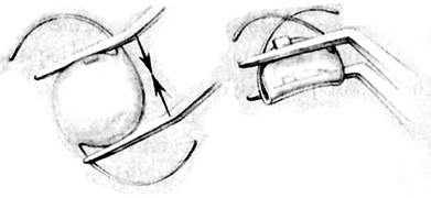
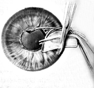
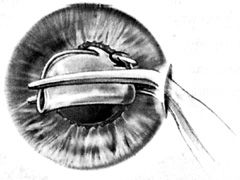
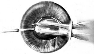
  4. After the anterior chamber is filled with a viscoelastic, the IOL is grasped with smooth Bechert or McPherson forceps, and the leading haptic is gently placed in the angle without deforming the pupil (Fig. 12), usually with the aid of a Sheets glide. The trailing haptic is positioned in a similar fashion, with either the forceps or a microhook. After inserting the lens, the lens is rotated as necessary to avoid haptic contact with any pre-existing PAS or the iridectomy (to prevent haptic migration through it). This is accomplished by gently retracting the distal haptic, rotating the lens, and then releasing the haptic. This process is repeated as necessary for either haptic until the desired lens position is achieved. A similar maneuver is used to seat the implant correctly in the angle (Fig. 13). With a microhook, the surgeon grasps one of the haptics, compresses it, elevates the lens slightly (Fig. 14), and then releases the haptic. This process, when repeated for the other haptic, ensures that the lens haptics are not implanted into peripheral iris (Fig. 15).
  5. The appropriateness of the lens size can be confirmed only after the IOL has been implanted. An undersized lens will either not center or will move excessively with only minimal contact with an instrument. If there is no resistance to rotation, then it is likely that the lens is too small. However, an oversized lens is difficult to insert and requires excessive stretching or gaping of the wound. It also may create intractable ovaling of the iris or of the entire cornea itself after it has been inserted.

Fig. 11. Pupillary block after anterior chamber intraocular lens implantation and anterior vitrectomy without a peripheral iridectomy.

Fig. 12. Anterior chamber lens insertion proceeds by placing the appropriately sized anterior chamber intraocular lens (see text) through a scleral flap incision (temporal in this case). A Sheets glide may be used to facilitate intraocular lens insertion.

Fig. 13. Proper anterior chamber intraocular lens insertion requires that all four footplates be positioned in the anterior chamber angle. Frequently, the haptics can become malpositioned in the iris root.

Fig. 14. A hook is used to pull the malpositioned haptic centrally while lifting the lens toward the cornea. This maneuver releases the captured footplates from the iris root.

Fig. 15. The anterior chamber intraocular lens is released and allowed to slide into its appropriate anatomic position.

After proper IOL positioning is ensured (Fig. 16), the wound is sutured, the viscoelastic is manually removed and replaced with balanced salt solution, and the conjunctiva is closed. When a vitrectomy is performed, we also inject subconjunctival corticosteroids and antibiotics.

Fig. 16. The peripheral iridectomy is typically made superiorly, where it is covered by the upper eyelid. The haptics are rotated as necessary to prevent footplate capture in the iridectomy.

Back to Top
ENDOCAPSULAR POSTERIOR CHAMBER LENS
Ideally, the posterior chamber intraocular lens should be fixated in the capsular bag. If sufficient capsular support exists, in-the-bag placement often can be achieved, even in eyes with open posterior capsules. This is accomplished by reopening the capsular bag and inserting a one-piece PMMA lens or, if possible, a foldable lens through a small, clear corneal incision.

First, if necessary, a pars plana anterior vitrectomy is performed using the technique described above. Next, the capsular anatomy is inspected, both visually and tactilely, to assess its integrity. This is initially accomplished by retracting the iris 360 degrees and determining the extent of intact capsule and zonules. A viscoelastic then is injected to gently tease open the fused leaflets of the anterior and posterior capsule. Much of this dissection can be performed passively, because the capillary action of the viscoelastic filling the bag usually separates the capsular surfaces. If resistance is encountered, careful manipulation with a spatula or microhook may be necessary.

In order to gain sufficient access to all areas of the capsule, multiple, strategically placed paracenteses must be created. Care must be taken in areas of fibrosis and strong adhesions to avoid tearing the capsule or disinserting the zonules. If the capsule cannot be safely reopened, it is best to desist and place a large foldable or one-piece PMMA IOL in the ciliary sulcus (see following section). Once the lens is positioned within the capsular bag, its centration and stability should be assessed by carefully decentering it and observing its rebound to the correct location.

Back to Top
SULCUS POSTERIOR CHAMBER LENS (UNSUTURED)
If in-the-bag placement is not possible, but sufficient capsular support remains, it is usually possible to place an unsutured PC IOL in the ciliary sulcus. As previously described, a vitrectomy should be performed when indicated, and the capsular remnants should be thoroughly inspected before lens insertion. A viscoelastic substance is injected between the iris and anterior capsule; any residual adhesions are sharply lysed with scissors or bluntly dissected with the cannula or a spatula, taking care not to tear the posterior capsule or disinsert the zonules.

A foldable or solid, one-piece, large-diameter lens now can be inserted into the sulcus. If there is a large posterior capsular opening, with foldable lenses it is sometimes safest to allow the lens to open in the anterior chamber with the haptics above the iris plane, and then sequentially place each haptic in the sulcus with a microhook. For solid, one-piece lenses, the leading haptic is directly placed into the sulcus (again, a Sheets glide may be helpful), and the trailing haptic is pronated into position with forceps while a hook inserted through the paracentesis simultaneously guides the lens optic posteriorly or retracts the iris.

Lens centration and stability are assessed as outlined earlier. It is important to remember that the IOL power should be reduced by 0.5 to 1.0 D for sulcus-positioned versus endocapsular PC IOLs. If the optic of the sulcus lens is captured posteriorly through an intact capsulorrhexis, we reduce the IOL power by only 0.5 D.

Back to Top
IRIS-SUTURED SULCUS POSTERIOR CHAMBER INTRAOCULAR LENS
Modified McCannel sutures are a convenient and efficient method of securing a sulcus PC IOL to the iris when capsular support is deficient.57 Aa soon as a lens is placed into the ciliary sulcus as outlined above, an intracameral miotic agent (Miochol) is administered so that the IOL optic can be captured through the pupillary space. Iris capture of the optic stabilizes the lens and enables better visualization of the haptics, which are tented up against the posterior iris surface. This is easily accomplished by positioning a microhook underneath the optic and gently lifting it anteriorly.

For eyes with minimal or no capsular remnants, iris capture can be safely achieved in a single maneuver with a foldable acrylic lens.60,61 An AcrySof lens is folded in the “bucket-handle” or “mustache” configuration (across the 3 to 9 o'clock meridian), inserted into the anterior chamber so that the optic is above the iris and the haptics are directed below the pupil, and unfolded over a spatula (inserted through a paracentesis incision opposite the wound) to prevent the optic from slipping into the posterior chamber (Fig. 17).

Fig. 17. The intraocular lens (IOL) is folded using a moustache fold. The haptics are placed within the sulcus, and the optic is captured above the plane of the iris. A Barraquer sweep is passed through the paracentesis and placed beneath the optic as the IOL is unfolded. (Reprinted from Stutzman RD, Stark WJ:. Surgical technique for suture fixation of an acrylic intraocular lens in the absence of capsular support. J Cataract Refract Surg 29:1660, 2003, with permission from ASCRS & ESCRS.).

When the IOL is in the correct position, two McCannel style sutures are used to fixate the haptics to the peripheral iris. A 9-0 or 10-0 polypropylene (Prolene) suture on a curved needle (Ethicon CIF-4 or CTC-6) is passed perpendicular to the haptic from a peripheral corneal site (with or without a paracentesis) into the anterior chamber, down through the iris, underneath the haptic, then back up through the iris, and finally through the peripheral cornea, where it is withdrawn from the eye (Fig. 18). Maneuvering the needle inside the eye sometimes proves challenging. If a needle exit paracentesis is used, a cannula can be placed through this paracentesis, and can aid in retrieving the needle by docking it. The suture tail is then cut off, leaving a 30-mm piece of polypropylene.

Fig. 18. Passage of the 10-0 Prolene suture to fixate the haptic. (Reprinted from Stutzman RD, Stark WJ:. Surgical technique for suture fixation of an acrylic intraocular lens in the absence of capsular support. J Cataract Refract Surg 29:1660, 2003, with permission from ASCRS & ESCRS.)

After the suture has been positioned, a new stab incision is placed in peripheral cornea over the entry points of the suture into the iris, and the two loops of the suture are withdrawn with a Kuglen or Sinskey hook and tied with a triple throw (Figs. 19 and 20). The knot is secured, trimmed, and then reposited into the eye. Alternatively, a closed-chamber suture tying technique can be employed to minimize traction on the iris and haptics.58 The other haptic is sewn to the iris in a similar fashion. The optic then is repositioned behind the iris by gently prolapsing it posteriorly with a microhook (Fig. 21).

Fig. 19. Retrieval of the suture through a paracentesis overlying the haptic. (Reprinted from Stutzman RD, Stark WJ:. Surgical technique for suture fixation of an acrylic intraocular lens in the absence of capsular support. J Cataract Refract Surg 29:1660, 2003, with permission from ASCRS & ESCRS.)

Fig. 20. The suture is tied externally. (Reprinted from Stutzman RD, Stark WJ:. Surgical technique for suture fixation of an acrylic intraocular lens in the absence of capsular support. J Cataract Refract Surg 29:1660, 2003, with permission from ASCRS & ESCRS.)

Fig. 21. Appearance of an iris-sutured posterior chamber IOL after the optic has been prolapsed into the posterior chamber The blue suture knots are visible on the iris at the 3- and 9-o'clock positions. Reprinted from Condon GP. Simplified small-incision peripheral iris fixation of AcrySof intraocular lens in the absence of capsular support. J Cataract Refract Surg 29:1666, 2003, with permission from ASCRS & ESCRS.)

Back to Top
TRANSSCLERALLY SULCUS–SUTURED POSTERIOR CHAMBER INTRAOCULAR LENS
We prefer to use a modification of Lane's technique,85 in which one knot is buried and the other is placed in the posterior lip of the incision beneath the thick scleral flap of the wound (Fig. 22). An IOL with a large optic diameter and haptic eyelets, such as the Alcon CZ70BD or Storz P366UV, is recommended, along with 10-0 Prolene suture on a long curved needle (Ethicon CTC-6L).

Fig. 22. Cross-sectional view showing placement of sutures for scleral fixation of a posterior chamber intraocular lens. Note that the double-armed sutures make full thickness bites, but the single-armed suture exits through the bed of the scleral wound.

An anterior vitrectomy, preferably through the pars plana (see earlier) is required to eliminate vitreous contact with the posterior iris surface and sulcus-suturing site. It is important to visually inspect any capsular remnants and iris adhesions by gently retracting the iris 360 degrees with a microhook. Any posterior iris attachments should be sharply dissected with scissors or lysed bluntly with a cannula, spatula, or hook.

The lens is implanted through a scleral tunnel incision that has two important characteristics: (a) it should extend 1.5 to 2 mm posterior to the limbus to permit subsequent passage of the needle in the bed of the tunnel and provide coverage of the knot, and (b) the anterior flap should be about 75% of scleral thickness to prevent late erosion of the underlying knot. For fixation of the distal haptic, the site exactly 180 degrees opposite the wound is identified; bare sclera is exposed by creating a small limbal peritomy and applying light focal cautery. Although the oblique meridians are probably the best orientation to avoid vessels and nerves, most published series describe using the vertical or horizontal meridians for fixation. However, it may be preferable to avoid the 3 and 9 o'clock positions to prevent direct compression of the long ciliary nerves; in our experience this can occasionally produce increased pain in the early postoperative period.

Traditionally, the recommended suture material has been 10-0 polypropylene, for which a variety of needles is available (Ethicon CTC-6L, STC-6, CIF-4, CS-140-6, or Alcon PC-7). Our preference is the Ethicon CTC-6L (a 14-mm spatulated needle), which is the longest available needle; it is thin but not flimsy, and has a gentle curve that is well suited for passage across the anterior chamber under the distal iris. Because of uncertainty regarding long-term stability we recently have converted to using 9-0 polypropylene, which is available by special order on the Ethicon CTC-6L needle (or routinely available on short needles). One needle of the double-armed suture is placed through the eyelet of the distal haptic, passed through the wound and pupil, walked under the iris into the ciliary sulcus, and pushed through the previously prepared scleral bed approximately 0.5 mm (in the horizontal meridian) or 1 mm (in the vertical meridian) posterior to the limbus (Fig. 23). 78 The second arm of this suture is passed in an identical manner so that it exits about 2 mm lateral to the first needle pass (Fig. 24). As the needle is retrieved, it is “wiggled” from side to side while the cutting portion of the needle is still intrascleral to allow a slight enlargement of the suture tract; this facilitates the eventual burying of the knot in the sclera. In eyes with large corneal diameters, even this long needle may be too short to span the eye, and straightening the mid-portion of the needle shaft provides extra length that enhances atraumatic passage across the eye.

Fig. 23. One end of a double-armed 10-0 polypropylene suture (Ethicon CTC-6L) is passed through the distal eyelet of the intraocular lens. The needle is walked under the iris, into the ciliary sulcus, and exits through the previously exposed scleral bed.

Fig. 24. The second arm of this suture is passed in an identical manner so that it exits about 2 mm lateral to the first needle pass.

Next, for fixation of the proximal haptic, the needle of a single-armed CTC-6L suture is passed through a paracentesis located about 150 degrees away from the center of the wound. This needle is directed through the pupil, under the iris, and out through the posterior scleral lip of the wound 180 degrees away from the midpoint of the previous suture (Fig. 25). A radial keratotomy marker may be used to more accurately place the suture passes 180 degrees apart. The free end of this suture is retrieved from the pupillary space and brought through the wound with a microhook. Then it is securely tied to the inner aspect of the eyelet in the proximal lens haptic, ensuring that it is not entangled with the two previously placed distal sutures. Care is taken to position the sutures symmetrically on either the upper or lower side of the haptics, thus minimizing torquing and tilting of the lens optic.

Fig. 25. The needle of a single-armed CTC-6L suture is passed through a paracentesis located about 150 degrees away from the center of the wound, through the pupil, under the iris, and out through the posterior scleral lip of the wound 180 degrees away from the midpoint of the previous suture passes.

All sutures are gently pulled to remove excessive slack, and the lens optic is grasped with insertion forceps and eased into the eye, using gentle traction on the distal sutures to guide the leading haptic into the sulcus (Fig. 26). The trailing or proximal haptic is then tucked under the iris by inserting a Sinskey hook into the eyelet; the haptic is gently flexed and pushed posteriorly while pulling on the suture exiting in the bed of the flap to eliminate slack. The sutures are gently pulled until taut and lens position is assessed. If the lens is not well centered or the pupil becomes oval, the surgeon should inspect for the presence of one of several problems, including entanglement of sutures, penetration of the iris with a suture, or malposition of a suture in relation to the haptic.

Fig. 26. The free end of this suture is retrieved from the pupillary space and brought through the wound with a microhook. It is then securely tied to the eyelet in the proximal haptic of the lens, ensuring that it is not entangled with any of the previously placed distal sutures. Care is taken to loop the double-armed suture symmetrically through the eyelets, thus minimizing torquing and tilting of the lens optic.

The sutures are then secured. For the leading haptic, this is accomplished by cutting off the needles and tying the suture ends together, cutting the suture close to the knot, and burying the knot by rotating it into the sclera. We prefer to use a Dangel (slip) knot135 because it is more compact and thus easier to bury than the conventional surgeon's knot. The proximal suture, which is still attached to its needle, is pulled up, and a partial-thickness scleral bite is taken in the posterior bed of the incision. The suture is tied to itself and then trimmed. This knot is covered by sclera and thus effectively buried when the wound is closed in a watertight fashion (Fig. 27). The conjunctiva is reapposed and, depending on the surgeon's preference, subconjunctival corticosteroids and antibiotics may be injected.

Fig. 27. All sutures are gently pulled to remove excessive slack, and gentle traction on the distal sutures helps to guide the lens as insertion forceps are used to carefully place the leading haptic in the sulcus. The trailing or proximal haptic then is tucked under the iris with the assistance of tying forceps and a hook. The sutures then are secured. For the leading haptic, this is accomplished by cutting off the needles and tying the suture ends together, cutting the suture close to the knot, and burying the knot by rotating it into the sclera. The proximal suture, which is still attached to its needle, is pulled up, and a partial-thickness scleral bite is taken in the posterior bed of the incision. The suture is tied to itself and then trimmed. This knot is covered by sclera and thus effectively buried when the wound is closed in a watertight fashion with 10-0 nylon.

Back to Top
RESULTS
Many studies have evaluated visual results of secondary anterior chamber lenses or sulcus-sutured lenses, but few studies have directly compared the two procedures. Key factors in assessing the outcomes of such surgeries are the percent of patients achieving useful vision, the percent of those with a loss of best spectacle-corrected vision (BSCVA), and the incidence and types of complications.

In the 1980s, a number of authors reported their experiences with secondary anterior chamber lens implants. In a review of the pooled literature, including 2,142 cases, Lindstrom and Harris32 found the incidence of various complications to be as great as 19.8% for CME, 10.8% for hyphema, 8% for secondary glaucoma, 5.4% for corneal edema, 3.3% for uveitis, and 1.6% for retinal detachment. In their personal experience with 153 cases, the incidences of the same sequelae were 4.6%, 0.7%, 3.9%, 2%, 2.6%, and 1.3%, respectively, with 81.6% of patients attaining visual acuity of 20/40 or better and 8.5% losing two or more lines of BSCVA. Depending on the study, as many as 94% of eyes achieved better than 20/40 vision, and the loss of more than two lines of BSCVA ranged from 3% to 10%.32,33,136–138 However, these reports antedated the availability of multiflex-style anterior chamber IOLs and sutured posterior chamber IOLs.

The roles of vitreous loss and vitrectomy and their relationships to the development of CME and RD during the implantation of secondary AC IOLs is controversial. Cozean138 suggested that there is a correlation between CME and disruption of the vitreous. Shammas and Milkie51 also noted this relationship and stated that the incidence of CME increases if the secondary IOL implantation is performed within 1 year of intracapsular cataract extraction, the preoperative fluorescein angiogram is positive for CME, or there is vitreous loss. Kraff and colleagues139 reported that vitrectomy may increase the risk of RD. Wong and colleagues7 also suggested that vitrectomy might increase the chance of RD (their rate was 4%), but might lower the risk of CME, which they found to be 3%. Not surprisingly, this study also found a higher incidence of these problems in eyes with closed-loop anterior chamber as opposed to sulcus-placed posterior chamber lenses. Conversely, Lyle and Jin30 reported similar incidences of CME (6%) when comparing eyes that received an AC IOL or PC IOL (68% had scleral suture fixation), but the rate of RD was higher in the latter group (0.8% for AC IOL versus 3.5% for PC IOL). Posterior vitreous detachment and vitreous liquefaction occurs in most aphakic eyes within 1 year postoperatively;140 this may suggest an explanation for the decreased RD rate when secondary IOL insertion is performed after 1 year.42,43

In studies of sulcus-sutured PC IOLs, results are comparable to contemporary studies of AC IOLs (as noted earlier). In Gess' original description,64 92% of the 303 eyes achieved better than 20/40 visual acuity. Loss of more than two lines of BSCVA has been reported to occur in 2.4% to 6.2% of eyes.102,120,141 The most common complication is suture erosion (see Fig. 5),85,98,120 which in Solomon's series occurred in 73% of cases, even with the use of scleral flaps. We have not seen any suture erosion when the polypropylene knots are either rotated and buried into sclera or covered by a thick scleral flap in the entrance wound, as described earlier.

If suture erosion does occur, this problem can be treated in several ways, including cutting the free ends flush on the knot, cauterizing the knot, revising the scleral flap, or using a scleral patch graft.98 A number of cases have been reported in which the sutures were removed, either to treat suture erosion or inadvertently.98,141,142 As a general rule, this is not recommended as lens decentration or dislocation can result, because there may be little or no fibrosis of the lens haptic to uveal tissue.111 According to the literature, only in rare cases have transsclerally sulcus–sutured lenses remained stable after suture removal.141,142 If the eroding suture is obviously loose and there is no pseudophakodonesis, it may be safe to cut the suture without producing lens dislocation. In each instance, patients are warned of risks and alternative options, instructed not to rub or bump their eyes, and advised of the symptoms of lens dislocation.

Glaucoma and RD have been described in as many as 19% and 5.4% of eyes receiving sutured PC IOLs, respectively.143 The reported incidence of new CME has ranged from 0%98 to 18% in patients receiving a transsclerally sulcus-sutured PC IOL.120 Price and Wellemeyer found hyphema and/or vitreous hemorrhage in 13% of eyes, although Bleckmann and Kaczmarek144 reported hyphema in 16.7% and vitreous hemorrhage in 25% of patients in their series. Other studies, however, state much lower incidences of intraocular bleeding, such as Helal's group,102 which had a 4.9% rate of hyphema.

Lens tilt and decentration are potential complications that may occur in as many as 10% of cases with transsclerally sulcus–sutured PC IOLs (see Fig. 4).98 Lens tilt occurs because of the torque created by asymmetric suture placement on the IOL. Teichmann87 studied the combinations of suture configurations and found that only four were torque free. He recommended looping the sutures symmetrically through the opposing eyelets. Theoretically, tilt also can be eliminated with radial suture placement, but this is anatomically undesirable, because one of the sutures will exit through the ciliary body. In Sharpe's study of children,71 an IOL tilt of 15 degrees in one case produced 6.5 D of cylinder.

A decentered sutured PC IOL may result from a number of factors. These include asymmetric attachment of the sutures to the haptics; failure to place the needles through the sclera 180 degrees apart; and suture loosening, breakage, or slippage on the haptics. Therefore, to prevent lens malposition, it is important to use an IOL with eyelets on the haptics, symmetrically secure the sutures to the eyelet, and place the needle passes an equal distance from the limbus and exactly 180 degrees apart. Fortunately, small degrees of lens tilt (less than 10 degrees) and decentration (less than 2 mm) are asymptomatic and clinically insignificant.88,144

The majority of studies that have directly compared AC and sutured PC IOLs evaluated secondary lens implantation at the time of penetrating keratoplasty. Davis and colleagues46 found no significant difference in outcomes between anterior chamber, iris-sutured, or transsclerally sutured posterior chamber IOLs. Schein116 noted that although the complications were similar among patients randomized to one of these three implantation techniques, the risk of CME was significantly less for the iris-sutured group than for either the AC or scleral-sutured PC IOL groups, and a greater percentage of those with PC IOLs achieved visual acuities of 20/40. Likewise, in a series by Brunette,145 better outcomes occurred in the PC IOL eyes, specifically, visual acuity was improved or maintained in 91% of patients with a PC IOL as opposed to 79% of those with an AC IOL. However, Evereklioglu and coworkers48 demonstrated no significant difference in postoperative BSCVA in a study of aphakias receiving an AC or PC secondary implant. Similarly, a report by the American Academy of Ophthalmology,49 which reviewed clinically relevant studies of IOL implantation in the absence of capsular support published between 1980 and 2001, found that there were no significant differences in visual outcome or complication rates among open-loop AC IOLs, scleral-sutured PC IOLs, and iris-sutured PC IOLs.

Back to Top
CONCLUSION
Secondary intraocular lens implantation is an effective method of correcting aphakia. When there is insufficient capsular support for IOL placement in the capsular bag or unsutured in the ciliary sulcus, collective experience indicates excellent results from both modern anterior chamber lenses and sutured sulcus lenses. These procedures are safe and effective; however, as with any type of surgery, case selection and meticulous surgical technique are critical and can strongly influence the success of the procedure. Long-term results as well as randomized studies are still needed to determine which procedure is better in eyes that could equally accommodate either type of lens.
Back to Top
REFERENCES

1. Kaufman HE: The correction of aphakia. XXXVI Edward Jackson Memorial Lecture. Am J Ophthalmol 89:1, 1980

2. Barraquer JI: Modification of refraction by means of intracorneal inclusions. Int Ophthalmol Clin 6:53, 1966

3. McDonnell PJ, Dietze T, Durrie DS, Schanzlin DJ: Surgical correction of aphakia: Intraocular lens implantation and epikeratophakia. J Refract Surg 3:209, 1987

4. Lane SL, Lindstrom RL, Cameron JD, et al: Polysulfone corneal lenses. J Cataract Refract Surg 12:50, 1986

5. Horgan SE, Fraser SG, Choyce DP, Alexander WL: Twelve year follow-up of unfenestrated polysulfone intracorneal lenses in human sighted eyes. J Cataract Refract Surg 22:1045, 1996

6. Steinert RF, Storie B, Smith P, et al: Hydrogel intracorneal lenses in aphakic eyes. Arch Ophthalmol 114:135, 1996

7. Wong SK, Koch DD, Emery JM: Secondary intraocular lens implantation. J Cataract Refract Surg 13:17, 1987

8. Ellington FT: Complications with Choyce Mark VIII and Mark IV anterior chamber implants: Uveitis, glaucoma, hyphema. Am Intraocular Implant Soc J 3:199, 1977

9. Apple DJ, Mamalis N, Lotfield K, et al: Complications of intraocular lenses: A historical and histopathological review. Surv Ophthalmol 29:1, 1984

10. Kraff MC, Sanders DR, Lieberman HL: Monitoring for continuing endothelial cell loss with cataract extraction and intraocular lens implantation. Ophthalmology 98:30, 1982

11. Sugar A, Meyer RF, Heidemann D, et al: Specular microscopic followup of corneal grafts for pseudophakic bullous keratopathy. Ophthalmology 92:325, 1985

12. Busin M, Arffa RO, McDonald MB, et al: Intraocular lens removal during penetrating keratoplasty for pseudophakic bullous keratopathy. Ophthalmology 94:505, 1987

13. Smith PW, Wong SK, Stark WJ, et al: Complications of semiflexible closed-loop anterior chamber intraocular lenses. Arch Ophthalmol 105:52, 1987

14. Speaker MG, Lugo M, Laibson PR, et al: Penetrating keratoplasty for pseudophakic bullous keratopathy: Management of intraocular lens. Ophthalmology 95:1260, 1988

15. Cohen EJ, Brady SE, Leavitt K, et al: Pseudophakic bullous keratopathy. Am J Ophthalmol 106:264, 1988

16. Waring GO: The 50-year epidemic of pseudophakic corneal edema. Arch Ophthalmol 107:657, 1989

17. Insler S, Kook MS, Kaufmann HE: Penetrating keratoplasty for pseudophakic bullous keratopathy associated with semi-flexible, close-loop anterior chamber intraocular lenses. Am J Ophthalmol 107:252, 1989

18. Koenig SB, McDermott ML, Hyndluk RA: Penetrating keratoplasty and intraocular lens exchange for pseudophakic bullous keratopathy associated with a closed-loop anterior chamber intraocular lens. Am J Ophthalmol 108:43, 1989

19. Kornmehl EW, Steinert RF, Odrich MG, et al: Penetrating keratoplasty for pseudophakic bullous keratopathy associated with closed-loop anterior chamber intraocular lenses. Ophthalmology 97:407, 1990

20. Binkhorst CD: Corneal and retinal complications after cataract extraction: The mechanical aspect of endophthalmodonesis. Ophthalmology 87:609, 1980

21. Drews RC: Intermittent touch syndrome. Arch Ophthalmol 110:1440, 1982

22. Stark WJ, Worthen DM, Holladay JT, et al: The FDA report on intraocular lenses. Ophthalmology 90:311, 1983

23. McDonnell PJ, Green WR, Maumenee AE, et al: Pathology of intraocular lenses in 33 eyes examined postmortem. Ophthalmology 90:386, 1983

24. Champion R, McDonnell PJ, Green WR: Intraocular lenses: Histopathologic characteristics of a large series of autopsy eyes. Surv Ophthalmol 30:1, 1985

25. Apple DJ, Olson RJ: Closed-loop anterior chamber lenses. Arch Ophthalmol 105:52, 1987

26. Apple DJ, Brems RN, Park RB, et al: Anterior chamber lenses. Part I: Complications and pathology and a review of design. J Cataract Refract Surg 13:157, 1987

27. Lorusso V, Moramarco A, Pacella E, Balacco-Gabrieli C: Intraocular lens complications. Ann Ophthalmol 22:377, 1990

28. Champion R, Green WR: Intraocular lenses: A histopathologic study of eyes, ocular tissue and intraocular lenses obtained surgically. Ophthalmology 92:1628, 1985

29. Maynor RCJ: Five cases of severe anterior chamber lens implant complications. Am Intraocular Implant Soc J 10:223, 1984

30. Lyle WA, Jin JC: Secondary intraocular lens implantation: Anterior chamber vs. posterior chamber lenses. Ophthal Surg 24:375, 1993

31. Price FWJ, Whitson WE, Collins K, Johns S: Changing trends in explanted intraocular lenses: A single center study. J Cataract Refract Surg 18:470, 1992

32. Lindstrom RL, Harris WS: Secondary anterior chamber lens implantation. CLAO J 10:133, 1984

33. Weene LE: Flexible open-loop anterior chamber intraocular lens implants. Ophthalmology 100:1636, 1993

34. Waring GO, Stulting RD, Street D, et al: Penetrating keratoplasty for pseudophakic corneal edema with exchange of intraocular lenses. Arch Ophthalmol 105:58, 1987

35. Lim ES, Apple DJ, Tsai JC, et al: An analysis of flexible anterior chamber lenses with special reference to the normalized rate of lens explantation. Ophthalmology 98:243, 1991

36. Solomon KD, Apple DJ, Mamalis N, et al: Complications of intraocular lenses with special reference to an analysis of 2500 explanted intraocular lenses (IOLs). Eur J Implant Refract Surg 3:195, 1991

37. Auffarth GU, Wesendahl , TA, Brown SJ, Apple DJ: Are there acceptable anterior chamber intraocular lenses for clinical use in the 1990s? An analysis of 4104 explanted anterior chamber intraocular lenses. Ophthalmology 101:1913, 1994

38. Perez-Santonja JJ, Iradier MT, Sanz-Iglesias L, et al: Endothelial changes in phakic eyes with anterior chamber intraocular lenses to correct high myopia. J Cataract Refract Surg 22:1017, 1996

39. Soong HK, Musch DC, Kowal V, et al: Implantation of posterior chamber intraocular lenses in the absence of lens capsule during penetrating keratoplasty. Arch Ophthalmol 107:660, 1989

40. Drews RC: Posterior chamber lens implantation during keratoplasty without posterior lens capsule support. Cornea 6:38, 1987

41. Koch DD: New optic hole configuration for iris fixation of posterior chamber lenses. Arch Ophthalmol 106:163, 1988

42. Menezo JL, Cisneros AL, Cervera M, et al: Iris claw phakic lens: Intermediate and long-term corneal endothelial changes. Eur J Implant Ref Surg 6:195, 1994

43. Menezo JL, Martinez MC, Cisneros AL: Iris-fixated Worst claw versus sulcus-fixated posterior chamber lenses in the absence of capsular support. J Cataract Refract Surg 22:1476, 1996

44. Wong SK, Stark WJ, Gottsch JD, et al: Use of posterior chamber lenses in pseudophakic bullous keratopathy. Arch Ophthalmol 105:856, 1987

45. Carlson AN, Stewart WC, Tso PC: Intraocular lens complications requiring removal or exchange. Surv Ophthalmol 42:417, 1998

46. Davis RM, Best D, Gilbert GE: Comparison of intraocular lens fixation techniques performed during keratoplasty. Am J Ophthalmol 111:743, 1991

47. Lindquist TD, Agapitos PJ, Lindstrom RL, et al: Transscleral fixation of posterior chamber intraocular lenses in the absence of capsular support. Ophthalmic Surg 20:769, 1989

48. Evereklioglu C, Er H, Bekir NA, et al: Comparison of secondary implantation of flexible open-loop anterior chamber and scleral-fixated posterior chamber intraocular lenses. J Cataract Refract Surg 29:30, 2003

49. Wagoner MD, Cox TA, Ariyasu RG, et al: Intraocular lens implantation in the absence of capsular support, a report by the American Academy of Ophthalmology. Ophthalmology 110:840, 2003

50. Assia EI, Nemet A, Sach D: Bilateral spontaneous subluxation of scleral-fixated intraocular lenses. J Cataract Refract Surg 28:2214, 2002

51. Shammas HJ, Milkie CF: Cystoid macular edema following secondary lens implantation. Am Intraocular Implant Soc J 7:40, 1981

52. Hara T, Hara T, Yamada Y: “Equator ring” for maintenance of the completely circular contour of the capsular bag equator after cataract removal. Ophthalmic Surg 22:358, 1991

53. Cionni RJ, Osher RH: Management of zonular dialysis with the endocapsular ring. J Cataract Refract Surg 21:245, 1995

54. Cionni RJ, Osher RH: Management of profound zonular dialysis or weakness with a new endocapsular ring designed for scleral fixation. J Cataract Refract Surg 24:1299, 1998

55. Lam DS, Young AL, Leung AT, et al: Scleral fixation of a capsular tension ring for severe ectopia lentis. J Cataract Refract Surg 26:609, 2000

56. Cionni RJ, Osher RH, Marques DM, et al: Modified capsular tension ring for patients with congenital loss of zonular support. J Cataract Refract Surg 29:1668, 2003

57. McCannel MA: A retrievable suture idea for anterior uveal problems. Ophthalmic Surg 7:98, 1976

58. Siepser SB: The closed chamber slipping suture technique for iris repair. Ann Ophthalmol 26:71, 1994

59. Zeh WG, Price FW: Iris fixation of posterior chamber intraocular lenses. J Cataract Refract Surg 26:1028, 2000

60. Stutzman RD, Stark WJ: Surgical technique for suture fixation of an acrylic intraocular lens in the absence of capsule support. J Cataract Refract Surg 29:1658, 2003

61. Condon GP: Simplified small-incision peripheral iris fixation of an AcrySof intraocular lens in the absence of capsule support. J Cataract Refract Surg 29:1663, 2003

62. Chu MW, Font RL, Koch DD: Visual results and complications following posterior iris-fixated posterior chamber lenses at penetrating keratoplasty. Ophthalmic Surg 23:608, 1992

63. Stark WJ, Goodman G, Goodman D, Gottsch J: Posterior chamber intraocular lens implantation in the absence of posterior capsular support. Ophthalmic Surg 19:240, 1988

64. Gess LA: Scleral fixation for intraocular lenses. Am Intraocular Implant Soc J 9:453, 1983

65. Malbran ES, Malbran EJ, Negir I: Lens guide suture for transport and fixation in secondary IOL implantation after intracapsular extraction. In Ophthalmol 9:161, 1986

66. Cowden JW, Hu BV: A new surgical technique for posterior chamber lens fixation during penetrating keratoplasty in the absence of capsular or zonular support. Cornea 7:231, 1988

67. McGill J, Liakos G: Complications of anterior chamber intraocular lenses and their effect on the endothelium. Trans Ophthalmol Soc UK 104:273, 1985

68. Moses L: Complications of rigid anterior chamber implants. Ophthalmology 91:819, 1984

69. Passo MS, Van Buskirk EM: Pupillary block with flexible anterior chamber intraocular lenses. Am J Ophthalmol 99:603, 1985

70. Apple DJ, Reidy JJ, Googe JM, et al: A comparison of ciliary sulcus and capsular bag fixation of posterior chamber intraocular lenses. Am Intraocular Implant Soc J 11:44, 1985

71. Sharpe MR, Biglan AW, Gerontis CC: Scleral fixation of posterior chamber intraocular lenses in children. Ophthalmic Surg Lasers 27:337, 1996

72. Buckley EG: Scleral fixated (sutured) posterior chamber intraocular lens implantation in children. J AAPOS 3:289, 1999

73. Mittelviefhaus H, Mittelviefhaus K, Gerling J: Transscleral suture fixation of posterior chamber intraocular lenses in children under 3 years. Graefes Arch Clin Exp Ophthalmol 238:143, 2000

74. Pavlin CJ, Rootman D, Arshinoff S, et al: Determination of haptic position of transsclerally fixated posterior chamber intraocular lenses by ultrasound biomicroscopy. J Cataract Refract Surg 19:573, 1993

75. Bellucci R, Marchini G, Morselli S, et al: Scleral fixation re-examined by ultrasound biomicroscopy. Eur J Implant Refract Surg 7:326, 1995

76. Manabe S, Oh H, Amino K, et al: Ultrasound biomicroscopic analysis of posterior chamber intraocular lenses with transscleral sulcus suture. Ophthalmology 107:2172, 2000

77. Campbell D, Davis R, Ferguson J: Ciliary sulcus anatomical dimensions. Invest Ophthalmol Vis Sci 29(suppl):34, 29 (abstract)

78. Duffey RJ, Holland EJ, Agapitos PJ, Lindstrom RL: Anatomic study of transsclerally sutured intraocular lens implantation. Am J Ophthalmol 108:300, 1989

79. Woodhams JT, Lester JC: Pigmentary dispersion glaucoma secondary to posterior chamber intraocular lenses. Ann Ophthalmol 16:852, 1984

80. Smith JP: Pigmentary open-angle glaucoma secondary to posterior chamber intraocular lens implantation and erosion of the iris pigment epithelium. Am Intraocular Implant Soc J 11:174, 1985

81. Huber C: The gray iris syndrome: An iatrogenic form of pigmentary glaucoma. Arch Ophthalmol 102:397, 1984

82. Samples JR, Van Buskirk EM: Pigmentary glaucoma associated with posterior chamber intraocular lenses. Am J Ophthalmol 100:385, 1985

83. Masket S: Pseudophakic posterior iris chafing syndrome. J Cataract Refract Surg 12:252, 1986

84. Lieppman ME: Intermittent visual “white out”: A new intraocular lens complication. Ophthalmology 89:109, 1982

85. Lane SS, Lubniewski AJ, Holland EJ: Transsclerally sutured posterior chamber lenses: Improved lens designs and techniques to maximize lens stability and minimize suture erosion. Semin Ophthalmol 7:245, 1992

86. Heidemann DG, Dunn SP: Visual results and complications of transsclerally sutured intraocular lenses in penetrating keratoplasty. Ophthalmic Surg 21:609, 1990

87. Teichmann KD, Teichmann IA: The torque and tilt gamble. J Cataract Refract Surg 23:413, 1997

88. Sivak JG, Kreuzer RO, Hildebrand T: Intraocular lenses, tilt and astigmatism. Ophthalmic Res 17:54, 1985

89. Durak A, Oner HF, Kocak N, Kaynak S: Tilt and decentration after primary and secondary transsclerally sutured posterior chamber intraocular lens implantation. J Cataract Refract Surg 27:227, 2001

90. Leo RJ, Palmer DJ: Episcleritis and secondary glaucoma after transscleral fixation of a posterior chamber intraocular lens. Arch Ophthalmol 109:617, 1991

91. Small EA, Solomon JM, Prince AM: Hypotonus cyclodialysis cleft following suture fixation of a posterior chamber intraocular lens. Ophthalmic Surg 25:107, 1994

92. Lane SS, Lindquist TD, Spigelman AV: Scleral fixation complications. Invest Ophthalmol Vis Sci 29(suppl):34, 1988

93. Stark WJ, Gottsch JD, Goodman DF, et al: Posterior chamber intraocular lens implantation in the absence of capsular support. Arch Ophthalmol 107:1078, 1989

94. Johnson SM: Results of exchanging anterior chamber lenses with sulcus-fixated posterior chamber IOLs without capsular support in penetrating keratoplasty. Ophthalmic Surg 20:465, 1989

95. Price FW, Whitson WE: Suprachoroidal hemorrhage after placement of a scleral-fixated lens. J Cataract Refract Surg 16:514, 1990

96. Kershner RM: Simple method of transscleral fixation of a posterior chamber intraocular lens in the absence of the lens capsule. J Refract Corneal Surg 10:647, 1994

97. Kay MD, Epstein RJ, Torczynski E: Histopathology of acute intraoperative suprachoroidal hemorrhage associated with transscleral intraocular lens fixation. J Cataract Refract Surg 19:83, 1993

98. Solomon K, Gussler JR, Gussler C, Van Meter WS: Incidence of management of complications of transsclerally sutured posterior chamber lenses. J Cataract Refract Surg 19:488, 1993

99. Heilskov T, Joondeph BC, Olsen KR, Blankenship GW: Late endophthalmitis after transscleral fixation of a posterior chamber intraocular lens. Arch Ophthalmol 107:1427, 1989

100. Schechter RJ: Suture-wick endophthalmitis with sutured posterior chamber intraocular lenses. J Cataract Refract Surg 16:755, 1990

101. Epstein E: Suture problems. J Cataract Refract Surg 16:116, 1989

102. Helal M, El Sayyad F, Elsherif Z, et al: Transscleral fixation of posterior chamber intraocular lenses in the absence of capsular support. J Cataract Refract Surg 22:347, 1996

103. Girard IJ: PC IOL implantation in the absence of posterior capsular support. Ophthalmic Surg 19:680, 1988

104. Spigelman AV, Lindstrom RL, Nichols BD, et al: Implantation of a posterior chamber lens without capsular support during penetrating keratoplasty or as a secondary lens implant. Ophthalmic Surg 19:396, 1988

105. Shin DH, Hu BV, Hong Y, et al: Posterior chamber lens implantation in the absence of posterior capsular support. Ophthalmic Surg 19:606, 1988

106. Shin DH: Implantation of a posterior chamber lens without capsular support during penetrating keratoplasty or as a secondary lens. Ophthalmic Surg 19:755, 1988

107. Pannu JS: A new suturing technique for ciliary sulcus fixation in the absence of posterior capsule. Ophthalmic Surg 19:751, 1989

108. Malbran ES, Malbran E, Aranguren AN: Scleral fixated intraocular lenses. Arch Ophthalmol 106:1347, 1988

109. Smiddy WE: Dislocated posterior chamber intraocular lens. Arch Ophthalmol 107:1678, 1989

110. Smiddy WE, Sawusch MR, O'Brien TP, et al: Articles: Implantation of scleral-fixated posterior chamber intraocular lenses. J Cataract Refract Surg 16:691, 1990

111. Lubniewski AJ, Holland EJ, Van Meter WS, et al: Histologic study of eyes with transsclerally sutured posterior chamber intraocular lenses. Am J Ophthalmol 110:237, 1990

112. Koplin RS, Colquhoun J: Surgical technique for managing a subluxed posterior chamber lens implant. J Cataract Refract Surg 14:685, 1988

113. McDonnell PJ, Champion R, Green WR: Location and composition of haptics of posterior chamber intraocular lenses: Histopathologic study of postmortem eyes. Ophthalmology 94:136, 1987

114. Mannarino AP, Hannush SB: A new technique for transscleral fixation of a posterior chamber intraocular lens in the absence of capsular support during penetrating keratoplasty. J Refract Corneal Surg 6:353, 1990

115. Hu BV, Shin DH, Gibbs KA, Hong YJ: Implantation of posterior chamber lens in the absence of capsular and zonular support. Arch Ophthalmol 106:416, 1988

116. Schein OD, Kenyon KR, Steinert RF, et al: A randomized trial of intraocular lens fixation techniques with penetrating keratoplasty. Ophthalmology 100:1437, 1993

117. Mittelviefhaus H, Wiek J: A refined technique of transscleral suture fixation of posterior chamber lenses developed for cases of complicated cataract surgery with vitreous loss. Ophthalmic Surg 24:698, 1993

118. Lewis JS: Ab externo sulcus fixation. Ophthalmic Surg 22:692, 1991

119. Bergren RL: Four-point fixation technique for sutured posterior chamber intraocular lenses. Arch Ophthalmol 112:1485, 1994

120. Lewis JS: Sulcus fixation without flaps. Ophthalmology 100:1346, 1993

121. Leon CS, Leon JA: Microendoscopic ocular surgery: A new intraoperative, diagnostic and therapeutic strategy. Part I: Endoscopic equipment/methodology applied to cataract surgery with intraocular lens implantation. J Cataract Refract Surg 17:568, 1991

122. Tsai JC, Rowsey JJ, Fouraker BD, et al: Use of a mirror needle holder with transsclerally sutured posterior chamber intraocular lenses. Ophthalmic Surg Lasers 27:720, 1996

123. Kora Y, Fukado Y, Yaguchi S: Sulcus fixation of posterior chamber lenses by transscleral sutures. J Cataract Refract Surg 17:636, 1991

124. Anand R, Bowman RW: Simplified technique for suturing dislocated posterior chamber intraocular lens to the ciliary sulcus. Arch Ophthalmol 108:1205, 1990

125. Friedberg MA, Berler DK: Scleral fixation of posterior chamber intraocular lenses combined with vitrectomy. Ophthalmic Surg 23:18, 1992

126. Bucci FAJ, Holland EJ, Lindstrom RL: Corneal autografts for external knots in transsclerally sutured posterior chamber lenses. Am J Ophthalmol 112:353, 1991

127. Choyce DP: The latest facts and figures on anterior chamber lens implants. Contact Intraoc Lens Med J 2:60, 1976

128. Drolsum L: Long-term follow-up of secondary flexible, open-loop, anterior chamber intraocular lenses. J Cataract Refract Surg 29:498, 2003

129. Werner L, Izak AM, Pandey SK, et al: Correlation between different measurements within the eye relative to phakic intraocular lens implantation. J Cataract Refract Surg 30:1982, 2004.

130. Allemann N, Chamon W, Tanaka HM, et al: Myopic angle-supported intraocular lenses: two-year follow-up. Ophthalmology 107:1549, 2000

131. de Souza RF, Forseto A, Nose R, et al: Anterior chamber intraocular lens for high myopia: five year results. J Cataract Refract Surg 27:1248, 2001

132. Perez-Santonja JJ, Iradier MT, Sanz-Iglesias L, et al: Endothelial changes in phakic eyes with anterior chamber intraocular lenses to correct high myopia. J Cataract Refract Surg 22:1017, 1996

133. Perez-Santonja JJ, Iradier MT, Benitez del Castillo JM, et al: Chronic subclinical inflammation in phakic eyes with intraocular lenses to correct myopia. J Cataract Refract Surg 22:183, 1996

134. Friedman NJ, Koch DD: Scleral tunnel incisions: Principles and methods. In Ophthalmic Surgery: Principles and Practice, 3rd ed. Philadelphia: Saunders, 2002:51

135. Dangel M, Keates R: The adjustable slide knot: An alternative technique. Ophthalmic Surg 11:843, 1980

136. Kraff MC, Sanders DR, Lieberman HL, Kraff J: Secondary intraocular lens implantation. Ophthalmology 90:324, 1983

137. Shammas HJ, Milkie CF: Secondary implantation of anterior chamber lenses. Am Intraocular Implant Soc J 9:313, 1983

138. Cozean CHJ: A longer view of secondary intraocular lens implantation with special emphasis on the role of the vitreous. Am Intraocular Implant Soc J 6:361, 1980

139. Kraff MC, Lieberman HL, Sanders DR: Secondary intraocular lens implantation: Rigid/semi-rigid versus flexible lenses. J Cataract Refract Surg 13:21, 1987

140. Heller MD, Straatsma BR, Foos RY: Detachment of the posterior vitreous in phakic and aphakic eyes. Mod Probl Ophthalmol 10:23, 1972

141. McCluskey P, Harrisberg B: Long-term results using scleral-fixated posterior chamber intraocular lenses. J Cataract Refract Surg 20:34, 1994

142. Maus M, Sivalingam E: Alternative method for sulcus fixation of posterior chamber lenses in the absence of capsular support. Ophthalmic Surg 20:476, 1989

143. Price FWJ, Wellemeyer M: Transscleral fixation of posterior chamber intraocular lenses. J Cataract Refract Surg 21:567, 1995

144. Bleckmann H, Kaczmarek U: Functional results of posterior chamber lens implantation with scleral fixation. J Cataract Refract Surg 20:321, 1994

145. Brunette I, Stulting RD, Rinne JR, et al: Penetrating keratoplasty with anterior or posterior chamber intraocular lens implantation. Arch Ophthalmol 112:1311, 1994

146. Steinert RF: Cataract Surgery: Techniques, Complications and Management, 2nd ed. Philadelphia: Saunders, 2004:433.

Back to Top