|
|
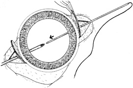
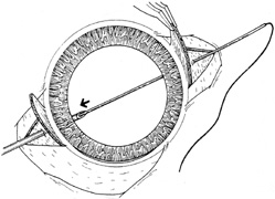
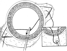
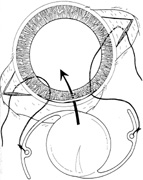
| Fig. 7. Technique for the ab externo approach. A. The long, straight solid needle is passed through the sclera (usually under partial-thickness scleral flaps) approximately 0.75 mm posterior to the limbus. Inside the eye, the needle should exit at the ciliary sulcus. A second hollow needle is passed from the opposite side of the eye. A pair of sutures can be used if four-point fixation is desired. B. Solid needle is “docked” inside the tip of the hollow needle, which has been passed through ciliary sulcus on the opposite side. After docking, the pair of needles are withdrawn together from the eye, with the solid needle inside the hollow needle. C. A hook is used to pull the suture out through a superior limbal wound so that it can be tied to the intraocular lens (IOL). D. Suture is cut, and each end is tied to a haptic of the IOL. After the IOL is placed into position, the scleral sutures must be anchored to the sclera. Either a “blind pass” in the sclera is made so that the suture is tied to itself, or the transscleral suture is tied to a second suture that has been tied to the sclera with a short partial-thickness pass within the bed of the scleral flap. (From Steinert RF, Arkin MS:. Secondary intraocular lenses. In: Steinert RF, ed. Cataract Surgery: Techniques, Complications, and Management,. 2nd ed. Philadelphia: Saunders, 2004:434, with permission from Elsevier.) |