Chapter 1
Anesthesia for Eye Surgery
SCOTT GREENBAUM
Main Menu   Table Of Contents

Search

HISTORICAL PERSPECTIVES
ANATOMIC PERSPECTIVES
GENERAL ANESTHESIA
RETROBULBAR ANESTHESIA
PERIBULBAR ANESTHESIA
PARABULBAR (SUB-TENON'S) ANESTHESIA
TOPICAL ANESTHESIA
FACIAL NERVE BLOCKS
COMPLICATIONS OF OCULAR ANESTHESIA
WHICH BLOCK FOR WHICH CATARACT SURGERY?
WHICH BLOCK FOR CORNEAL SURGERY?
ANESTHESIA FOR REFRACTIVE SURGERY
ANESTHESIA FOR GLAUCOMA SURGERY
ANESTHESIA FOR STRABISMUS SURGERY
ANESTHESIA FOR VITREORETINAL SURGERY
ANESTHESIA FOR OCULOPLASTIC SURGERY
REFERENCES

HISTORICAL PERSPECTIVES
The history of anesthesia for eye surgery dates back 2,500 years. The earliest authentic writings on the subject were those of Sus'ruta, the ancient Indian surgeon who first described couching—the depression of the cataract into the vitreous—around 600 BC.1 He outlined the use of inhalational anesthesia for this method, and also described aseptic technique. Later, Egyptian and Assyrian surgeons used carotid compression to produce transient cerebral ischemia, under which couching was performed. A Spanish alchemist in the thirteenth century described a mixture of sulfuric acid and alcohol, which he called “sweet vitriol”2; in 1730, this substance was renamed either. Faraday reported on its accidentally discovered anesthetic effect in 1818. The use of other general anesthetic agents, such as carbonic acid gas, chloroform, and nitrous oxide was described in the late eighteenth and the nineteenth centuries.1

The eighteenth century was also a time of discovery for more modern techniques of cataract surgery. In 1748, Daviel published a report describing a corneal incision that started at the inferior limbus and continued nasally and temporally for approximately 240 degrees. He next performed an anterior capsulotomy and delivered the lens with a curette or spatula, depending on its density; therefore this, the first planned cataract extraction, was done in an extracapsular fashion.3 In 1753, Samuel Sharp described a planned intracapsular extraction, using a single knife to make the corneal section. It took more than a century for these techniques to become standard practice, during which time couching procedures continued to be performed using soporific drugs and psychological control of the patient.2

In 1865, Albrecht von Graefe described a scleral incision made with a single passage of a knife, followed by the creation of an iridectomy. For this procedure he preferred the use of general anesthetics, especially chloroform.4 He noted that there were potential dangers if the patient strained while awakening from anesthesia, and this may have been one of his main incentives for reducing the size of Daviel's corneal flap incision.

The next great advance in the evolution of anesthesia had its origins in 1855, when Gaedicke isolated the alkaloid of the coca plant. In 1860, Nieman noted its anesthetic effect on his tongue, and named it cocaine. Carl Koller further described its use as a local anesthetic, and in 1884 Knapp5 and Turnbull6 both reported on the use of cocaine in eye surgery. Knapp described a technique for cataract removal under topical anesthesia using frequent administration of cocaine drops. He also mentioned retrobulbar injection of cocaine for enucleation (the hypodermic needle had been developed in 1853 by Alexander Wood).7 Turnbull introduced another anesthetic technique for enucleation, using topical and sub-Tenon's cocaine.

An appreciation of the systemic and local toxicity of cocaine soon followed; episodes of syncope, excessive stimulation, hallucinations, and even death made it a far from perfect anesthetic. In addition, its corneal epithelial toxicity and drying effect, as well as its prolonged hypesthesia, led to cases of exposure keratopathy and ulceration.

In 1904, the next advancement in ophthalmic anesthesia began with Einhorn's discovery of procaine hydrochloride, which could be used for infiltration, instillation, and nerve block anesthesia without the toxic effects of cocaine.2 Procaine had no inherent vasoconstrictive effects, was rapidly absorbed, and had a short duration of action. Therefore, epinephrine was added to slow absorption and thus hasten, intensify, and lengthen the anesthetic effect. This addition, however, introduced the cardiovascular side effects of sympathetic stimulation in susceptible individuals.

A decade later, Van Lint was the first to describe a method for blocking the orbicularis muscle of the eye to prevent blepharospasm during cataract surgery.8 He injected a combination of procaine and epinephrine near the lateral orbital rim, blocking the terminal branches of the facial nerve and innervating the orbicularis. He advocated waiting 30 to 60 minutes before surgery, thus allowing the block to have its full effect. This block was easy to perform and had a good rate of success. Its localized effect was an advantage; however, eyelid edema, bruising, and bleeding were its main drawbacks.

O'Brien originated a method for a more proximal facial nerve block in 1929.9 He injected procaine anterior to the tragus of the ear, over the condyloid process of the mandible. He used 2 mL of anesthetic and advised waiting 5 minutes before operating. Although the block was found to be safe, with a reduced risk of bleeding and intravascular injection, the anatomical variability of the zygomatic, mandibular, and buccal branches of the facial nerve reduced the certainty of a complete block.

In 1943, Lofgren and Lundquist synthesized lidocaine, and 3 years later Lofgren reported on its anesthetic properties.10 It was found to have a faster onset, better diffusion, and longer duration than procaine. In addition, its early proponents believed that lidocaine was less toxic.11 However, more toxicity was observed in higher concentrations. Circulatory depression was also associated with lidocaine, and respiratory depression was noted with procaine.12

In 1949, Atkinson13 reported on the use of hyaluronidase to increase the diffusion of procaine. His method originated in the observation that aqueous testicular extract increased the spread of vaccinia virus.14 This extract was later identified as hyaluronidase.15 Atkinson found that the addition of 6 turbidity-reducing units per milliliter to a solution of 2% procaine with epinephrine provided more profound anesthesia, without reducing the duration of action.

In 1953, Atkinson proposed another approach to blocking the facial nerve. He introduced his needle through an intradermal wheal at the inferior edge of the zygomatic bone, “A little posterior to the lateral margin of the orbit.”16 He infiltrated anesthetic along the zygomatic arch, which he thought was the ideal site to block the upper branches of the facial nerve, sparing those innervating the lips and lower facial muscles. The benefits of this method included the avoidance of lid edema and injection into major vessels and nerves. As with O'Brien's technique, however, its main drawback was its unpredictability because of anatomic variability.

A decade later, Nadbath and Rehman17 introduced the most proximal variation on the facial nerve block. They injected anesthetic behind the mandibular ramus to block the main trunk of the facial nerve, where it exited the stylomastoid foramen. Although this approach was the least subject to anatomic variation, its site was a veritable minefield of major blood vessels and cranial nerves. In addition, access to the dura surrounding the spinal cord was reported when this method was used in certain frail patients, with resultant respiratory depression and paralysis.18 More commonly, hoarseness, pooling of secretions, and dysphagia were noted when the glossopharyngeal, vagus, and spinal accessory nerves were blocked along with the facial nerve.19

The literature of the next two decades is filled with case reports of complications of retrobulbar anesthesia, the most serious being blindness and death.20–32 Alternatives to standard intraconal retrobulbar anesthesia were employed as well. In 1985 and again the following year, Davis reported on the use of peribulbar anesthesia.33,34 He credited Kelman for its introduction in the mid-1970s, Davis's technique involved three injections: two given anteriorly, into and just beneath the upper and lower orbicularis muscle, and one posteriorly, along the floor of the orbit near the equator of the globe. Although Davis cited 1,600 cases without complication,34 it took only a year from the date of his publication for the first reports of globe penetration to be published.35,36

Transconjunctival retrobulbar anesthesia was also introduced in the mid-1980s.37 Gills37 advocated this method to reduce the potential for globe perforation. It was also intended to avoid the need for a separate facial nerve block.

In 1990, Hanson and colleagues38 described a modification of Turnbull's sub-Tenon's technique. Although sub-Tenon's anesthesia had not been forgotten in the intervening 106 years, it had been relegated to a mostly ancillary position. Atkinson16 advocated the technique as a supplement to retrobulbar anesthesia in patients with preoperative inflammation. The author observed Dr. Arnold Turtz, at the Manhattan Eye, Ear and Throat Hospital, perform sub-Tenon's with a blunt, metal cannula to improve the anesthetic effect of a retrobulbar block and to provide postoperative anesthesia to patients who had received general anesthesia.

In 1950, Kirby2 reported his preference for sub-Tenon's over retrobulbar anesthesia, because of the frequent complications of retrobulbar hemorrhage and proptosis with the latter technique. He routinely used a needle to inject a combination of procaine, pontocaine, and potassium sulfate before surgery. Forty years later, Yanoff39 described an ocular perforation with the anterior sub-Tenon's injection. In 1992, the author reported on the elimination of the needle in sub-Tenon's anesthesia through the use of a flexible, blunt, polyethylene cannula.40 In the same year, Stevens41 described the use of a blunt, metal cannula in delivering sub-Tenon's anesthesia without a needle, a method similar to that of Turtz.

The 1990s also heralded the resurgence of topical anesthesia. Although Koller had introduced topical cocaine in 1884, its toxicity limited its popularity. In 1956, Atkinson42 had reported using tetracaine (pontocaine) for topical ocular anesthesia. In 1965, Thorson and co-workers43 had described a topical proparacaine anesthetic in strabismus surgery. In 1986, Shimizu44 had demonstrated the use of topical cocaine (3%) in performing clear corneal cataract surgery. In 1992, Fichman45 presented a method for performing topical anesthesia with tetracaine, and began popularizing its use in the United States. This was also the year that the author and Stevens separately described their use of blunt cannula subtenons anesthesia for cataract surgery through an anterior or parabulbar, and posterior approach, respectively.40,41

In an effort to address the inadequacies of topical anesthesia Gills and others proposed the addition of intracameral unpreserved lidocaine in the late 1990s.128,129 These reports were followed by randomized controlled studies refuting their claims of additional anesthetic effect.130,131

Many surgeons who lecture and write on topical anesthetic techniques use the term “vocal local” to emphasize the importance of constant communication with the patient during the procedure. In his discussion of papers presented at the 16th annual session of the American Academy of Ophthalmology and Otolaryngology in Chicago in 1955, Sadove remarked, “I want to emphasize that a few carefully chosen words are as potent as any sedation ... vocal is about as good as local.”46

It is easy to see that the rediscovery and modification of previously described techniques has been the driving force in the evolution of ocular anesthesia since 1884.

Back to Top
ANATOMIC PERSPECTIVES
Understanding how to provide the most effective ocular anesthesia in the safest manner requires an anatomic knowledge of the nerves, blood vessels, muscles, and bony landmarks of the orbit, face, and globe. Although this anatomy is unique to any given patient, some basic rules apply that can be of great help.

The first consideration in providing anesthesia for cataract surgery or any other purpose is understanding the goals of the block. Which parts of the eye does one intend to leave without sensation? Which muscles does one wish to paralyze? Once these questions are addressed, the next task is to understand the cranial neuroanatomy.

The details of trigeminal, oculomotor, and orbital anatomy are beyond the scope of this chapter. The reader is referred to Ocular Anesthesia (WB Saunders, 1997) and Atlas of Clinical and Surgical Orbital Anatomy (WB Saunders, 1994). A brief summary follows.

The trigeminal nerve carries the sensory innervation of the eye and adnexa in three divisions: ophthalmic, maxillary, and mandibular. Except for a portion of the sensory input from the lower lid that is carried by the maxillary division, the sensory fibers of the eye and adnexa are found in the ophthalmic division. This division in turn has three components: frontal, lacrimal, and nasociliary, as shown in Figure 1. The frontal nerve usually branches into two more divisions: the supraorbital, which carries sensation from the conjunctiva and skin of the central two-thirds of the upperlid; and the supratrochlear, which carries sensory fibers from the medial third of the upper lid. The lacrimal nerve carries sensory input from the skin and conjunctiva of the lateral aspect of the upper lid.47 The nasociliary nerve carries sensory fibers from the cornea, iris, ciliary body, perilimbal bulbar conjunctiva, and optic nerve sheath; these fibers proceed through its long ciliary branches and sensory root to the ciliary ganglion. The infratrochlear branch of the nasociliary nerve carries sensory input from the medial canthus, medial portion of lower lid skin and conjunctiva, caruncle, lacrimal sac, and canaliculi.

Fig. 1. Sensory nerves of the orbit. Note that the path of the nasociliary branch of the ophthalmic division of the trigeminal nerve runs through the intraconal space, but those of the frontal and lacrimal branches do not.

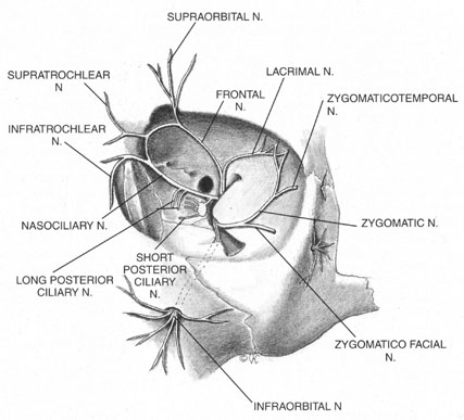
Once it is known which branches are responsible for carrying sensory input from which structures, an approach can be planned that has a reasonable chance of blocking the targeted area. Because, for example, the nasociliary nerve carries fibers that pass through the intraconal space, a standard intraconal retrobulbar block may provide excellent intraocular and partial surface anesthesia. It could not be expected, however, to effectively block the conjunctiva of the upper or lower lids or the lateral aspect of the globe. Because the frontal and lacrimal branches enter the orbit through the superior fissure, above the annulus of Zinn, and the maxillary division enters the orbit through the infraorbital foramen, below the annulus, an intraconal approach probably would not effectively block the structures these branches innervate. If an intraconal retrobulbar block is the only one administered before surgery, patients can be expected to feel irrigating solutions being dropped on the conjunctiva, away from the limbus; they also will be aware of the lid speculum and any manipulation of the lateral surface of the globe. They will probably attempt to close the eye in response to these stimuli. This is the basis for the traditional facial–retrobulbar block combination.

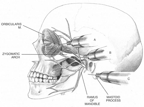
As has been mentioned, numerous facial blocks have been devised to prevent patients from squeezing the eye shut during surgery. None of these blocks keeps patients from wanting to close the eye—only from succeeding. Because the facial block also involves added discomfort, many anesthesiologists and surgeons provide intravenous sedation along with it. Thus, limiting the initial block to the intraconal retrobulbar space also limits its potential benefits, requiring two supplemental procedures. Anatomy determines effect.

The anterior blocks—peribulbar, parabulbar/sub-Tenon's, and topical—reduce or eliminate the need for a separate facial block by providing better surface anesthesia than the retrobulbar block. Although the first two also provide intraocular anesthesia through their effect on the nasociliary branch, topical blocks provide only surface anesthesia, so that intraocular sensations, such as stretching of the zonules during filling of the anterior chamber, may be felt throughout surgery. Some authors advocate the addition of a subconjunctival injection to add more anesthetic effect to a topical block.48 Recently, intraocular injections of local anesthetics have been popularized as an adjunct to topical anesthesia. This technique is discussed later in this chapter.

The motor supply of the superior, medial, and inferior rectus, the inferior oblique, and the levator palpebrae superioris is carried by the oculomotor nerve (Fig. 2). It also carries proprioceptive input from these muscles and parasympathetic fibers to the ciliary ganglion. As the oculomotor nerve enters the orbit through the superior orbital fissure, it splits into two divisions, superior and inferior. The superior division is smaller; it courses forward in the superolateral portion of the intraconal space, and turns medially toward the lateral aspect of the superior rectus muscle, where it divides into a network of small branches.49 The innervation of all extraocular muscles is multifocal, with nerve fibers extending distally and proximally between the muscle fibers, before ending at myoneural junctions.47 Some branches innervate the superior rectus, and others pass through it to enter the levator muscle through its inferior surface.

Fig. 2. Motor nerves of the orbit. Note the multifocal innervation of the extraocular muscles.

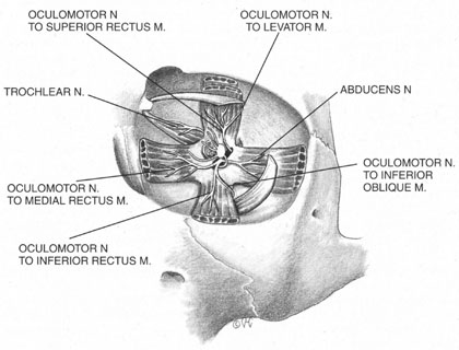
The inferior division of the oculomotor nerve splits into at least three trunks within the intraconal space, and these in turn divide into eight to ten branches as they course forward, lateral to the optic nerve. The medial rectus is innervated by branches that run from beneath the optic nerve into the muscle, beginning at its posterior third. The inferior rectus muscle is similarly penetrated at its posterior conal surface by branches of the inferior division of the oculomotor nerve. The inferior oblique muscle is innervated by a branch that initially contains parasympathetic fibers; these fibers originate in the Edinger-Westphal nucleus and enter the ciliary ganglion inferolateral to the optic nerve. The remainder of this branch then breaks up into smaller fascicles, which penetrate the inferior oblique at its posterolateral aspect.47

The trochlear nerve supplies motor fibers to the superior oblique muscle. It enters the orbit through the superior oblique fissure above the annulus of Zinn, along with the frontal and lacrimal branches of the ophthalmic division of the trigeminal nerve. It crosses the superior rectus origin above the levator and enters the superolateral surface of the superior oblique muscle.47

The abducens nerve enters the orbit through the superior orbital fissure, along with the oculomotor nerve. They are sometimes divided by a dense septum connecting the superior rectus origin to the superior rectus sheath.47 The abducens nerve enters the lateral rectus sheath just anterior to the annulus of Zinn, and first enters the lateral rectus muscle at the medial aspect of the junction of its posterior and medial thirds.47,50

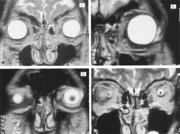
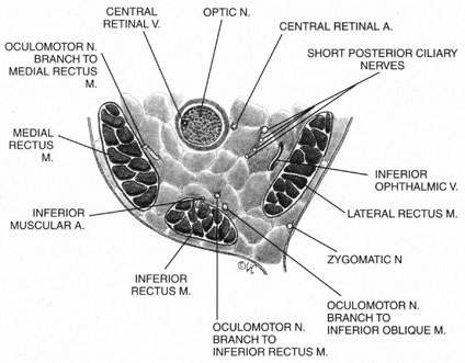
The ciliary ganglion is an irregular structure, measuring 1 mm by 2 mm, that lies just temporal to the optic nerve (Figs. 3 and 4), 7 to 10 mm from the orbital apex.47,51 In it the presynaptic parasympathetic fibers from the Edinger-Westphal nucleus synapse with the postsynaptic fibers that form the short ciliary nerves. Most of these fibers innervate the ciliary muscle, and the remaining 3% to 5% supply the iris sphincter. The ganglion also contains sensory branches of the nasociliary nerve and sympathetic fibers en route to the choroidal vasculature.

Fig. 3. Posterior orbit. Note the proximity of the optic nerve to the oculomotor branches in this region.

Fig. 4. Connective tissue planes of the midorbit.

When attempting to provide akinesia, it should be kept in mind that the motor nerves enter the rectus muscles at the junction of their posterior and medial thirds, or more anteriorly.52 It is also important to remember that these fibers run both distally and proximally between the muscle fibers before they end at the myoneural junctions. A motor block may therefore be achieved at many points along their path. The oculomotor divisions may be blocked in the posterior orbit before their insertion into the rectus muscles, but it is necessary to keep in mind the proximity of the optic nerve, the ophthalmic vein, and the anterior muscular branches to the oculomotor branches in this region (see Fig. 3). Because the nerve and artery supplying the inferior oblique muscle insert more anteriorly, they are more vulnerable to needle trauma from peribulbar or retrobulbar blocks delivered along the floor of the orbit.48 The superior oblique muscle is innervated and receives its blood supply in the posterior orbit. Because it is relatively immobile in the superotemporal orbit, it is possible to injure this muscle with blocks in this area.

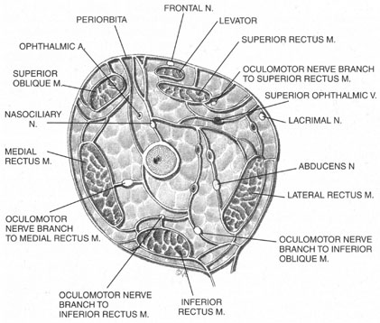
Blocks delivered in the anterior or middle orbit depend on diffusion of the anesthetic agent into either the posterior orbit, to the origin of the nerve branches, or into the muscles themselves at the point where distal branches insert into myoneural junctions. The first process is dependent on the anatomy of the connective tissue planes that subdivide the orbit into compartments (see Fig. 4). This architecture varies among patients. The classic teaching of a single intermuscular septum that connects the rectus muscles and divides the orbit into an intraconal and an extraconal space is overly simplified. Histologic examination of the orbit reveals an arrangement of roughly parallel and partially broken septa of various thicknesses, with and without fenestrations.48 This anatomic variability, therefore, accounts for the variability in akinesia seen with orbital blocks.

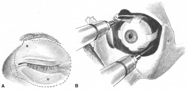
Atkinson16 discussed supplementing incomplete akinesia by injecting 0.5 to 1.0 mL of anesthetic solution 3 cm back along a rectus muscle, if it was still active after retrobulbar block. He grasped a horizontal rectus muscle with forceps and rotated the eye away from the needle, or placed a muscle hook under the eyelid and separated it from a vertical rectus muscle before injection.16 Because the rectus muscles measure 40 to 42 mm in length without their tendons, which in turn may vary from 3.7 mm for the medial rectus to 8.8 mm for the lateral, Atkinson's injection was administered 14 to 21 mm anterior to the origin of the muscle. This would place it anterior to the insertion of the oculomotor branch innervating the muscle. Because akinesia can be achieved with this technique, the anesthetic must be diffused within the muscle, blocking distal branches of the oculomotor divisions at their most distal myoneural junctions. However, these injections may produce myotoxicity with prolonged muscle paresis.53,54

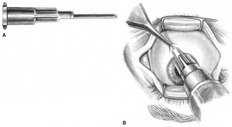
Because permanent extraocular muscle damage is more a product of intramuscular or intraneural injection than of the concentration of anesthetic injected,48,55 a less traumatic delivery of anesthetic can be used around the extraocular muscles to achieve akinesia without risking either myotoxicity or needle trauma to the adjacent optic nerve and orbital vessels. Using the sub-Tenon's space for this delivery allows for such atraumatic akinesia.

Back to Top
GENERAL ANESTHESIA
Although a discussion of the specific techniques for induction of general anesthesia is beyond the scope of this chapter, it is important to consider the indications, contraindications, ophthalmic effects, and complications of this procedure. In 1955, Atkinson16 wrote that the indications for general anesthesia in eye surgery included operations on young children, on adults undergoing extensive orbital surgery, and on those who objected to local anesthesia. General anesthesia also may be considered in patients unable to cooperate, such as the mentally retarded or those with general movement disorders, nystagmus, an inability to lie flat, excessive anxiety, or claustrophobia.

An inability to communicate with the patient may contribute to the decision to choose general anesthesia, but an interpreter can often be employed to instruct a patient preoperatively or even intraoperatively, thus facilitating surgery under local anesthesia. It is also advisable for the surgeon to learn a few crucial phrases in the patient's language. It can be helpful to have an interpreter write down translations of “look up,” “look down,” “don't move,” “open the eye,” and “close the eye,” and tape them to the wall of the operating room or to the microscope. Patients with hearing impairment should be encouraged to wear their hearing aids.

Although bleeding disorders have in the past been considered a relative indication for general rather than local anesthesia, newer techniques such as sub-Tenon's/parabulbar and topical anesthesia avoid the risks of retrobulbar hemorrhage and thus eliminate the advantage of general anesthesia in these cases.

In the 1950s, when intracapsular cataract surgery was the state of the art, general anesthesia was thought to be helpful in the prevention of vitreous loss through “relief of abnormal muscle tone due to fear of pain.”2 Studies of the era compared a 7% loss of vitreous under local anesthesia to a 3% loss under a general anesthesia.56 Stress was constantly placed on the importance of a stay suture to close the eye quickly should the patient lighten under general anesthesia and put a strain on the endotracheal tube. Cataract surgery currently is executed in a more controlled manner, within a closed system. The occasional intracapsular extraction is still performed, of course, as in the case of a subluxed lens with a large zonular dehiscence. In these cases, the lessons of the past should not be forgotten.

In 1980, Badear and co-workers57 published a study demonstrating that elderly patients with a history of myocardial infarction had a significantly higher risk of developing a second infarction with general anesthesia than with local anesthesia. However, a study conducted 3 years later by Lang58 at the Massachusetts Eye and Ear Infirmary demonstrated a low rate of morbidity and mortality with both techniques. In reviewing nearly 15,000 cases between 1977 and 1979, Lang found that they were evenly divided between the two types of anesthesia, and that there were only two postoperative deaths, also equally divided between the two groups. The only two myocardial infarctions occurred in the local group; both were in patients who had a history of infarction more than 6 months before surgery. In comparing the two groups, however, it must be kept in mind that these patients were not randomly selected, and that the patients who were given local anesthesia had a significantly higher average age. What this study does highlight is the overall safety of eye surgery when the appropriate anesthetic technique is chosen for a given patient. Again, the choice of anesthetic should be based on factors other than the type of surgery being performed. As early as 1974, Lynch and co-workers59 compared the rate of vitreous loss and iris prolapse during cataract extractions performed under local to the rate of those performed under general anesthesia and found no significant difference.

The broad range of contraindications to general anesthesia is a chapter in itself, but those that are of special interest in ophthalmology are mentioned here. Up to 90% of patients with myotonic dystrophy, for example, develop cataracts in their early years.60 The myotonia and muscular dystrophy seen in these patients increase the risks of general anesthesia.61 These patients may develop significant bradycardia with a prolonged P-R interval that can be unresponsive to atropine. They also may experience respiratory complications caused by their decreased vital and maximum breathing capacities, resulting in prolonged postoperative respiratory depression. In addition, they experience an increased risk of aspiration caused by delayed gastric emptying and abnormal swallowing. All of these factors make local anesthesia the preferred method in these patients.

Care also must be taken when considering general anesthesia in patients with Marfan's syndrome. These patients have a high prevalence of cardiovascular abnormalities, such as incompetent or prolapsed valves, arrhythmias, and aortic aneurysms that may dissect with an elevation in blood pressure.62 In addition, pulmonary anomalies such as bronchogenic cysts, abnormal lobulations, and emphysematous changes can lead to spontaneous pneumothorax. The risk of tension pneumothorax increases during controlled breathing.63 For all these reasons, local anesthesia may be safer for such individuals.

When deciding whether to use general anesthesia, the patient's total physical condition must be taken into account, as must any medications the patient may be taking that could interact with the anesthetic. If possible, smoking should be discontinued. A history of significant prostatic enlargement should be followed up by a urologic consultation before surgery. Patients who are difficult to intubate, such as those with cervical spondylosis, should be identified and carefully evaluated. Previous reactions to general anesthesia should be investigated, as should a family history of unexplained perioperative death or malignant hyperthermia.

Although the incidence of malignant hyperthermia is cited as 1 in 15,000 anesthetic events in children and 1 in 50,000 in adults,64 the true incidence varies with the condition and the anesthesia used. A Danish study estimated an incidence of 1 in 5,000, if episodes of fever, unexplained tachycardia, and masseter muscle rigidity were included in cases where succinylcholine and an inhalation agent were used.65 The specific diagnosis and treatment of malignant hyperthermia is beyond the scope of this chapter, but it is important to remember that with current treatment the mortality rate is approximately 10%. Therefore, in adult patients who may be susceptible to this condition, local anesthesia is obviously preferable to general anesthesia.

When general anesthesia is required in pediatric cases, a history of susceptibility to malignant hyperthermia triggers a cascade of precautionary steps. These include intravenous dantrolene, continuous body temperature monitoring, and the use of drugs considered to be relatively safe for this population, such as narcotics, benzodiazepines, barbiturates, and nondepolarizing muscle relaxants. Careful postoperative monitoring of these patients should be continued for at least 8 hours.

A thorough review of all medications the patient is taking must be made before eye surgery, especially when it is to be performed under general anesthesia. Guidelines for preanesthetic management include the cessation of diuretics, which could cause urinary retention in patients with unsuspected prostatic enlargement, and the discontinuation of those psychotropic drugs that might interact with general anesthetics, such as monoamine oxidase (MAO) inhibitors, which intensify and prolong the anesthetic effect. However, vasodilators and antiarrhythmic agents should be continued through the day of surgery. Their elimination could lead to rebound hypertension and tachycardia, conditions that, along with congestive heart failure and unexplained anemia, represent the major cardiovascular contraindications to general anesthesia.

Because people with diabetes are overrepresented among patients undergoing nearly every type of eye surgery, the perioperative management of insulin and oral hypoglycemics deserves special attention here. General anesthesia and surgery cause a major disruption of the daily routines of a diabetic patient, and the patient's treatment regimen must be altered accordingly. Type 1 diabetics tend to be more affected by changes in therapy than insulin-dependent, type 2 diabetics, so these juvenile-onset patients should have surgery scheduled as early in the day as feasible.64 Both groups should be given one-third to one-half of their standard morning dose of intermediate- or long-acting insulin. The blood glucose level should be checked before insulin administration and at hourly intervals throughout the perioperative period, and any hyperglycemia should be treated with small amounts of short-acting insulin. Hypoglycemia should be treated with a continuous intravenous infusion of 5% dextrose; the rate of administration should be guided by blood sugar levels.

Oral hypoglycemics should be discontinued on the day of surgery. Patients taking them should have a fasting blood sugar level in the morning, and a continuous intravenous infusion of dextrose as guided by frequently monitored blood sugar levels. Once the patient is able to resume a regular diet, which with most eye surgeries should be relatively soon after the procedure, the normal insulin or oral therapy is resumed.

In patients with diabetes, the type of anesthesia used does not determine the morbidity and mortality of the surgery. Rather, the preexisting status of the diabetes, specifically the presence or absence of retinopathy, nephropathy, and neuropathy, as well as peripheral vascular and cardiovascular disease is predictive of the surgical risk. In a study of diabetics undergoing nonocular procedures, the overall risk of complications was 15%.66 Those with peripheral vascular disease had the greatest risk (35%), and patients with none of the conditions mentioned had a complication risk of approximately 5%.

Ophthalmic medications used in the treatment and preparation of patients undergoing eye surgery also may interact with general anesthetic agents. Epinephrine, for example, may be used on a long-term basis by glaucoma patients for the reduction of intraocular pressure. It also may be used intraoperatively in cataract surgery for its mydriatic effect; in patients who are inadequately dilated, an infusion of epinephrine into the anterior chamber can help to enlarge the pupil enough so that surgery may be performed without the aid of iridectomy, sphincterotomy, or iris hooks. Epinephrine is also routinely added to the fluid used for intraocular irrigation during cataract surgery.

But what are the implications of its use, and how much is safe? The major concern is the interaction between epinephrine and halogenated hydrocarbon anesthetics, which can potentiate ventricular fibrillation. This effect may be especially dramatic with epinephrine and cyclopropane, and this combination surely should be avoided. However, epinephrine can be safely used with halothane, the maximal safe dose having been reported as 68 μg/kg, infused into the anterior chamber.67

The rate of administration is also important. It has been estimated that 10 mL of a 1:100,000 solution of epinephrine may be safely administered in 10 minutes in the presence of halothane; as much as 30 mL per hour may be administered in a healthy 70-kg patient.68 Complications such as hypertension, syncope, headache, diaphoresis, tachycardia, and extrasystoles have been reported in patients using topical epinephrine.69,70 Because as little as 0.5 mg may trigger these effects, and because one drop of a 2% solution contains this amount, it may be advisable to substitute another glaucoma treatment in patients scheduled for general anesthesia.

A glaucoma treatment that is less commonly used but that may be seen in the setting of aphakia is echothiophate, a long-acting anticholinesterase. If a patient has received this drug in the 4 weeks before surgery, anesthetic agents that are metabolized by plasma pseudocholinesterase should be avoided, or at least significantly restricted.71 These include succinylcholine, cocaine, procaine, and chloroprocaine. The use of succinylcholine in this setting can lead to prolonged apnea.

Acetazolamide is a carbonic anhydrase inhibitor used in the long-term treatment of glaucoma, and in the short-term treatment and prophylaxis of postoperative intraocular pressure elevation. Both ocular and renal carbonic anhydrase activity are inhibited by this drug, so that bicarbonate, water, sodium, and potassium are wasted with its use. Patients undergoing long-term acetazolamide therapy are often hypokalemic and hyponatremic, a combination that can potentiate significant arrhythmias under general anesthesia. Because these electrolyte imbalances are worsened by hepatic or renal failure, acetazolamide should be avoided in such cases. It also should be avoided in patients with chronic obstructive pulmonary disease who have a tendency to retain carbon dioxide and therefore to become acidotic. Short-term treatment of intraocular pressure with intravenous acetazolamide has become a popular technique. The drug begins to have an effect in 5 minutes and peaks by 30 minutes. It should be noted that this is a sulfonamide derivative, and so carries a risk, however rare, of anaphylaxis, Stevens-Johnson syndrome, erythema multiform, and aplastic anemia.

General anesthesia is more likely to cause adverse systemic effects than local or ocular complications. Those ocular problems that do occur usually are not serious. They include corneal abrasion, chemical keratitis, hemorrhagic retinopathy, and, rarely, retinal ischemia.

The incidence of corneal abrasion from general anesthesia has been reported to be as high as 44%.72 But simple precautions, such as instilling a bland ointment or taping closed the lids of the unoperative eye, may prevent surface trauma produced by the surgical drape, anesthetic mask, or exposure. Decreased tear production under general anesthesia, proptosis, and poor Bell's phenomenon may worsen corneal exposure, requiring eyelid suturing in some susceptible patients. Another potential danger to the cornea is the inadvertent trauma by the anesthesiologist during induction.73 Reusable masks also may cause chemical injury to the cornea because of the liquid disinfectants used on them.74 Disposable masks or those that can be autoclaved are simple solutions to this problem.

Postoperative vomiting, difficult extubation, and straining on the endotracheal tube may lead to the formation of retinal hemorrhages, a condition dubbed Valsalva retinopathy.75 This is usually benign, because its source is the venous circulation and the location is intraretinal. These hemorrhages clear without interrupting vision unless they are located in the macula.

Retinal ischemia is by far the most serious adverse effect of general anesthesia. It can be caused by an oversized anesthetic mask, inadvertent pressure exerted by an assistant, or the surgeon's leaning on the unoperative eye. In the case of posterior segment surgery, it can also result from expansion of intraocular gases in the presence of high concentrations of nitrous oxide.76 Fortunately, this condition is rare, and can be readily prevented if kept in mind during surgery.

Back to Top
RETROBULBAR ANESTHESIA
In 1985, 76% of the members of the American Society of Cataract and Refractive Surgeons, responding to an annual survey, indicated their preference for retrobulbar anesthesia with a facial nerve block for cataract surgery.77 Another 16% preferred retrobulbar without the facial block. Thus 92% of these surgeons selected retrobulbar blocks as their procedure of choice a little less than two decades ago. The same survey published in 2004 listed 9% as preferring retrobulbar plus facial block and 11% as choosing retrobulbar alone, a decline from 92% to 20% overall.132

The technique that most respondents still preferred varies from Atkinson's42 classic description published in 1955, but not substantially. He wrote:

Good results will be obtained and orbital hemorrhage rarely occurs if the following directions are observed: An intradermal wheal is made. A 3.5-cm 23-gauge needle with a rounded point is introduced through this wheal at the inferior temporal margin of the orbit and the skin is moved upward with the needle until the point just clears the orbital margin. About 0.5 cc of the anesthesia solution is now injected so that pain is not experienced when the orbital septum is pierced. Incidentally, the septum can be pierced more easily close to the orbital margin and is pierced more easily if the needle is rotated producing a boring effect. A pause of a few moments allows the anesthesia to work, during which time the patient is directed to look upward and away from the site of injection. This is done to move the inferior oblique muscle and fascia between the lateral and inferior rectus muscles forward and upward, out of the way. The needle is directed straight back close to the floor of the orbit, until the point is beyond the globe and fascia before directing it upward toward the apex of the orbit for a depth of 2.5 to 3.5 cm depending on the size of the orbit. When the needle has reached the proper depth, one should aspirate before injecting the anesthetic solution in order to determine whether or not the needle has entered a vessel. However, in this location, it would be most unlikely unless there is an abnormally large vessel. The anesthetic solution is then slowly injected, the amount depending on the size of the orbit, the operation that is to be done and whether or not hyaluronidase has been added to the anesthetic solution. When hyaluronidase is not used, 1.5 mL (of anesthetic) is considered a safe amount for intraocular operations. At least 5 minutes should elapse before the operation is begun ... more effective results are obtained for cataract extractions and larger injections may be given if hyaluronidase is added to the anesthetic solution. It may be safely injected until there is a noticeable proptosis, which usually occurs after injecting 2 to 3 cc in the average orbit. The hyaluronidase causes the solution to diffuse rapidly and the proptosis quickly subsides. Pressure over the eye, combined with moving it in the orbit for at least 5 minutes, produces still greater diffusion of the anesthetic solution, enhances anesthesia and akinesia, and increases hypotony. Each is an important defense against vitreous loss and other complications.

The biggest change in this technique as used today is the direction of gaze the patient is asked to assume during the block. This came about because of a 1981 study conducted by Unsold and associates, in which computed tomographic images of retrobulbar needle placement in a cadaver, using two different techniques, were studied.78 On one side of the cadaver, the eye was sutured into the position described by Atkinson. Next, a 3.5-cm 25-gauge needle was introduced just above the orbital rim, and was directed superomedially toward the optic canal. On the other side, the eye was sutured into a position of inferotemporal gaze, and the needle was directed in a more inferior direction. In the first case, computed tomography (CT) demonstrated that the needle tip was extremely close to the optic nerve, having crossed underneath it and stopped medially in front of the optic canal. By having the patient look upward and inward, the anesthesiologist would have caused the optic nerve to rotate downward and outward, into or at least very near the path of the needle.

These scans also revealed the close proximity of the ophthalmic artery and superior ophthalmic vein to the needle. Even the inferior oblique muscle, which Atkinson made a point of describing as being moved away from the injection by the upward and inward direction of the gaze, was found to be displaced anteriorly, temporally, and inferiorly. This brought it into the path of the needle, near its lateral border. Interestingly, the first report of inferior oblique trauma in three patients who underwent retrobulbar anesthesia and in one who underwent peribulbar anesthesia has recently been published.55

In the second set of scans, performed with the eye directed downward and outward, the needle position was above the inferior rectus muscle, and anterior to the inferior aspect of the superior orbital fissure. The needle was within the muscle cone but away from the optic nerve, the superior ophthalmic vein, and the ophthalmic artery. The inferior oblique muscle was found to rotate posteriorly, medially, and superiorly, away from the needle path.

Unsold78 concluded that most of the complications of the traditional retrobulbar technique could be explained by direct trauma to the optic nerve or the orbital vessels. He also postulated that in the traditional position, the dural sheath of the optic nerve was on stretch, and was therefore more easily penetrated, allowing better access to the subarachnoid space, the central retinal artery, and the nerve itself. Similarly, the contraction of the inferior oblique muscle with an upward and inward gaze made it a target more susceptible to puncture. Unsold's observations provided an impetus for many surgeons and anesthesiologists to change their technique, so that more retrobulbar blocks are currently performed with the patient in primary gaze.

In 1987, Nicoll evaluated standard retrobulbar anesthesia performed both with an upward and inward gaze, and with a gaze looking straight ahead or “minimally upward.”79 Out of a total of 6,000 patients who received retrobulbar anesthesia, 16 developed symptoms suggestive of central nervous system spread. These symptoms included blindness of the contralateral eye, drowsiness, abnormal shivering, vomiting, respiratory depression, hemiplegia, aphasia, convulsions, unconsciousness, and cardiac arrest. The combined incidence of these symptoms in both groups was 1 in 375. The incidence in the 2,000 patients who looked upward and inward was 1 in 333, and in the group who looked straight ahead or slightly upward, it was 1 in 400. This difference is not statistically significant.

Unsold's findings were, after all, based on the study of a single cadaver, and might have overemphasized the importance of gaze direction. One eye was directed “downward and outward,” and a 35-mm needle was introduced in a direction described as “slightly more” inferior to the “traditional technique.” In Nicoll's study, one group of patients was looking straight ahead or slightly upward while a 38-mm needle was introduced by way of “a standard inferotemporal approach.” The difference in needle length and angle of introduction may explain the difference between the theoretical advantage of Unsold's findings and the disappointing reality of Nicoll's.

Katsev51 may explain the disagreement. In 120 human orbits, he measured the distance between the retrobulbar needle site (the junction between the middle and lateral third of the intraorbital rim), and the optic foramen. He found that 11% of optic nerves could be perforated by a 38-mm needle, the length used in Nicoll's study. The needle may have been so long that it overcame the advantage of rotating the optic nerve toward or away from the point. Unsold did not report the size of the orbit he studied; regardless,no two skulls or orbits are identical. Comparing Unsold's findings to Katsev's, it could be concluded that in any individual, the shorter the needle and the more it is rotated away from the area of the optic nerve, the less likely a complication will occur. Katsev suggested using a 31-mm needle. Unsold's rotation of the eye downward and outward toward the injection site might not be objectionable to a cadaver, but certainly might be to a patient. Having the patient assume the primary position of gaze and using a 31-mm needle may, therefore, represent the safest combination for retrobulbar anesthesia.

These studies have led to alternative retrobulbar approaches. In an effort to avoid the globe and the optic nerve, many experts on injection anesthesia have advocated multineedle techniques.48 The type of needle preferred by most is a sharp disposable one that can be introduced with less patient discomfort, thus requiring less intravenous sedation. It is considered no more likely to produce scleral, optic nerve, or blood vessel perforation than a blunt needle.80

Hustead's multiinjection technique includes an inferotemporal injection of 0.5 mL of a mixture of two parts 0.75% bupivacaine and one part 2% mepivacaine, with epinephrine and hyaluronidase. It is administered with a 1-inch 30-gauge needle introduced to a depth of 20 mm. The same needle is partly withdrawn and redirected toward the intraorbital foramen. Next, 1.25 mL of anesthetic is injected anterior to the orbital septum, to prepare for the lid block. The needle is withdrawn completely. After 30 seconds, a ¼-inch 27-gauge needle is introduced along the same inferotemporal route beneath the lateral rectus muscle to a point ⅝ mm behind the globe (presumably the anesthesiologist checks the axial length before injecting). The 4 mL of anesthetic is slowly injected. The needle is again partially withdrawn and introduced into the preseptal space near the infraorbital foramen, to serve as a partial lid block. Ocular compression is performed, and ocular mobility is checked after 5 minutes. A medial periconal injection is performed with a 30-gauge needle directed superiorly or inferiorly, depending on which muscles are predominantly mobile. In 5% of cases, a fifth injection is given superotemporally, to yield complete akinesia. Hustead reports no incidence of hemorrhage in his last 6,000 cases.48

Hamilton also begins with a 30-gauge needle, introducing it transconjunctivally in the inferotemporal position, after administering topical anesthetic. Next, 1 mL of 2% mepivacaine with epinephrine and hyaluronidase is injected behind the inferior tarsal plate 10 mm from the conjunctival surface. This injection is given to anesthetize the path of the next one, which is given with a 27-gauge needle previously bent to an angle of 10 degrees for half of its 31-mm length. The bevel of the needle is on the concave side. This needle is introduced transconjunctivally, with the patient in primary gaze. The site is the farthest lateral extension of the inferior fornix, just behind the inferior tarsal plate. The target site is the sagittal plane of the lateral limbus, which requires a medial tilt of the needle pathway. The 10-degree bend mimics the rise in the orbital floor. Once the needle has been inserted to a depth of 25 to 31 mm, approximately 3 to 4 mL of anesthetic is injected for 2 minutes. Ocular compression for 10 minutes follows this injection, and if any motility remains, a medial periconal injection is performed using a 30-gauge needle, as in Hustead's technique.

Two other modifications in the retrobulbar technique have been made by Thornton and Straus. Each has changed the design of the needle to avoid globe perforation.

The 25-gauge Thornton needle was designed without cutting edges; it has a sharp smooth point and a 15-degree bevel.48 Although its design is intended to “run truer through tissue,” it has been reported to cause undesirable globe rotation in about a third of cases because of septal adherence to the needle shaft.48

Straus designed a curved needle with a 20-mm radius, a 25-mm chord length, and a 10-mm straight distal portion, creating a 30-degree angle in reference to the needle hub.81 This radius of curvature was designed to exceed that of the average globe. The angle of the distal segment was intended to parallel that of the optic nerve and the globe. Although Straus reports no globe or optic nerve trauma in 10,000 cases, and only one retrobulbar hemorrhage, similarly curved needles have failed to eliminate these complications in the past.48

The popularity of retrobulbar anesthesia has continued to decline in the past two decades but it is still a frequently used technique. It actually is not one technique, but a family of single or multiple injections ending up somewhere behind the eye. Many alterations have been proposed to reduce the incidence of complications. This proliferation of modifications in the technique Atkinson described more than 40 years ago, and the continued reports of complications, suggest that the ideal retrobulbar method is still in evolution. Finally, regardless of which modification is used, the importance of monitored anesthesia care attending the patient undergoing local techniques for cataract surgery cannot be overemphasized, as in more than one third of such cases intervention by anesthetic personnel is required for the safe conclusion of the operation.133

Back to Top
PERIBULBAR ANESTHESIA
In 1986, Davis and Mandel82 reported their use of peribulbar anesthesia. According to their article, the technique was actually introduced by Kelman in the mid-1970s but was not published at that time. Davis and Mandel make one injection, a finger's width medial to the lateral canthus, through the lower lid, just above the inferior orbital rim. There, 0.5 mL of lidocaine is injected subcutaneously, and a second 0.5 mL is injected into the orbicularis muscle. Next, 1 mL of anesthetic is introduced beneath the orbicularis “into the anterior orbit,” and 2 mL is introduced through the upper lid in the same manner, beginning just below the supraorbital notch. This is followed by 1 minute of ocular compression. In their original paper the authors did not describe the needle used for the first two injections, but in a later report48 they mention the use of a ½-inch 27-gauge needle.

A more posterior injection is made next, using a 23-gauge 1 ¼-inch (31 mm) blunt retrobulbar needle; 4 mL of 0.75% bupivacaine mixed with 4 mL of 1% lidocaine and hyaluronidase is administered. An inferior injection is given 15 mm medial to the lateral canthus, just above the inferior orbital rim, and 1 mL of anesthetic is injected just beneath the orbicularis muscle. “The needle is then advanced along the inferior orbit to the equator of the globe.”82 (This is evidently an assumed depth, because the authors do not mention measuring or adjusting for the patient's axial length before injecting.) Next, 1 mL of anesthetic mixture is delivered in this region. “The barrel of the syringe is angled over the malar eminence, and the needle is advanced in a superior and medial direction to the full depth of the needle.” Another 1 to 2 mL is injected. These injections are repeated beneath the supraorbital notch anteriorly and “at the superonasal equator of the globe.” The third stage of this superior posterior injection is delivered “to the superior orbital fissure,” where another 1 mL of anesthetic is injected (Fig. 5). Because the distance from the superior orbital rim to the superior orbital fissure is really 4.5 to 5 cm,47 the 31-mm needle is actually falling far short of this target.

Fig. 5. The sites and delivery of Davis and Mandel's classic peribulbar block.

Davis and Mandel always aspirate before injecting, and describe the volumes delivered as approximate and dependent on the dimensions of the eye and orbit. After a total volume of 12 to 14 mL of anesthetic mixture is given through the four injections described, the eyelids may be tense from the filling of the orbital volume. Next, 8 minutes of ocular compression ensue, and the completeness of the block is evaluated. With practice, the authors describe achieving complete akinesia in 90% of cases. In the remaining 10%, an additional 3 to 4 mL are injected at a site determined by the muscles not completely blocked. The anesthetic effect is described as taking place 10 to 12 minutes from the time of injection. No further facial block is required, in part because of the multiple injections into and just below the orbicularis muscle, which are in themselves distal facial nerve blocks.

Davis and Mandel described an excellent record of safety in their first 1,600 cases. They conclude that this technique “eliminates the need for standard retrobulbar injections.” However, it is possible to wonder how much the two most posterior injections they describe differ from “standard” retrobulbar injections. Although the peribulbar injection is not intended to enter the muscle cone, the delivery of anesthetic through a needle introduced 31-mm deep into the orbit, starting at the inferior orbital rim and advancing superomedially, more than casually resembles the retrobulbar anesthetic technique, as does the superior injection made anterior to the superior orbital fissure. Davis and Mandel48 later modified their technique, using a blunt 23- or 25-gauge, 7/8-inch needle, instead of the 1 ¼-inch needle previously described. The later report also substituted an anesthetic mixture of two-thirds 0.75% bupivacaine and one-third 1% lidocaine without epinephrine but with hyaluronidase. This represents a longer-acting mixture than the original, which contained half lidocaine and half bupivacaine.

The shortening of the needle for more anterior delivery of anesthetic was a response by the authors to reports of optic nerve trauma, globe perforation, and respiratory depression after peribulbar anesthesia—complications in common with the retrobulbar technique. Indeed, the site of injection chosen by Davis and Mandel was nearly identical to the one Braun83 described in 1918, and Pitkin84 later abandoned because of similar adverse effects. It is clear that deep orbital injections carry with them vision- and life-threatening risks, no matter what their direction.

A study in 1988 by Weiss and Deichman85 comparing the efficacy of retrobulbar with peribulbar or periocular injections for cataract surgery demonstrated that, as with their complications, the therapeutic effects of the techniques were similar. Although only 79 patients were included in the study, it was performed in a randomized, masked, prospective fashion. In both groups, 5 mL of a half-and-half mixture of 2% lidocaine and 0.75% bupivacaine with epinephrine and hyaluronidase was used. A single injection was performed with the patient in primary gaze. Interestingly, the only difference in technique between the two groups was the length of the needle used.

In the retrobulbar group, a 25-gauge 38-mm needle was inserted “almost to the hub,” starting at the juncture of the lateral third and medial two-thirds of the inferior orbital rim and “aiming toward an imaginary posterior extension of the visual axis.” In the periocular group, the same site and angle of administration were used, with a 25-gauge 16-mm needle. No facial block was given to either group, but 10 minutes of ocular compression was administered in both. At this point the surgeon evaluated lid and globe akinesia as well as the depth of anesthesia. If the globe was not totally akinetic or anesthetized, a supplemental block of 0.5 mL of anesthetic was injected in the supratrochlear region. No intravenous sedation was administered.

There was no significant difference between the two groups in the degree of lid or globe akinesia, anesthesia, or patient-reported sensation. There also was no significant difference in the number of patients who required a supplemental block; 21% of the retrobulbar and 28% of the periocular patients received one. The only significant difference was the greater degree of chemosis seen with the periocular patients.

Weiss and Deichman speculate that a reduction in the needle length and the volume of anesthetic made their particular technique safer and no less efficacious than the standard retrobulbar method or the multi-injection peribulbar procedure (actually, in 28% of cases their periocular technique did include more than one injection). However, their sample size was too small to conclude this with great certainty. Surely, shortening the needle at least changes the type of complications seen after injection anesthesia.

Another “anterior” peribulbar technique was described by Bloomberg86 in 1986 and reviewed 5 years later.87 A 27-gauge 18- to 24-mm needle was used to inject 8 to 10 mL of a half-and-half mixture of 2% mepivacaine (Carbocaine) and 0.75% bupivacaine, to which hyaluronidase and sodium bicarbonate (0.2 mL per 10 mL of solution) were added. It is possible that the pH adjustment of the anesthetic sped the onset of akinesia.88,89 If the longer needle was used, it was advanced only for three-quarters of its length. The site of injection was the standard juncture of the lateral third and medial two-thirds of the infraorbital rim, and the needle was directed toward the floor of the orbit, away from the globe. Ocular compression was an important adjunct to this technique; Bloomberg used a Honan balloon set at 30 to 40 mm of mercury for 12 to 20 minutes.

Bloomberg reported a 10% to 50% rate of supplemental blocks for residual movement. These blocks were made with an 18-mm needle in the quadrant of the still active muscle. The needle was placed tangential to the globe, and was directed approximately 10 degrees toward the optical axis, unlike the original injection.

In their latest study of peribulbar anesthesia, Davis and Mandel90 evaluated a number of prospectively collected peribulbar blocks, performed mostly for cataract surgery, at 12 different facilities between 1988 and 1992. A total of 16,224 cases were studied. These blocks were performed in at least 12 different ways by ophthalmologists, anesthesiologists, and nurse anesthetists. Most included intravenous sedation. All involved 10 to 60 minutes of ocular compression and an inferotemporal injection site. Two centers performed 2 primary injections, and 10 performed only the inferotemporal one, with a supplemental injection given when necessary.

Davis and Mandel's technique has continued to evolve since their first publication. Their current block begins with a 2-mL injection of warmed (98°F) 1% lidocaine in a 4:1 to 9:1 mixture, with a balanced salt solution. This injection is given with a 27-gauge 12-mm needle into and through the orbicularis muscle and into the anterior orbit inferotemporally. Its purpose, according to the authors, is to anesthetize the lids in preparation for the second injection. This is given with a 23- to 26-gauge 18- to 24-mm needle in the same inferotemporal location. A 10-mL syringe is filled with 6 mL of 0.75% bupivacaine, 3 mL of 1% lidocaine, and 1 mL of hyaluronidase. Between 4 and 10 mL are injected, presumably in the manner previously described, with the eye in primary position. Intravenous sedation with 6 to 10 mL of midazolam is delivered “as needed.”

Next, 10 to 25 minutes of ocular compression are applied. After 8 to 10 minutes, akinesia is checked and a superonasal or inferonasal injection of the same mixture is given as needed. In 9% of the 1200 cases using this technique, a supplemental block was required. In another 2% an “additional nerve block,” presumably facial, was required.

Davis and Mandel reported that almost all of their patients had nearly complete akinesia, and that even in the cases where it was incomplete, no positive vitreous pressure was noted. The amount of movement remaining did not interfere with surgery. Approximately three quarters of their cases were performed by phacoemulsification and one quarter by extracapsular extraction.

The other centers included in this study reported similar results. The highest rate of supplemental blocks reported was 11%, from a center that already began with two blocks. Despite these multiple injections, however, the rates of orbital hemorrhage, globe perforation, and central nervous system involvement were low. The study did not evaluate the incidence of extraocular muscle trauma or ptosis, nor was there a protocol for examining patients postoperatively. For example, it is unclear how many patients were dilated for postoperative peripheral retinal evaluation.

Another drawback of this study was that it did not evaluate the degree of analgesia patients experienced, except in a small subgroup of 200 subjects. Of these patients, 140 remembered having the injection. The authors do not mention how many of the 200 had intravenous sedation, nor why they waited until the day after surgery to question their patients. Of those who remembered the block, one-third had mild to severe discomfort, but two-thirds had no discomfort. If it is assumed that the discomfort mentioned was caused by the block (which would explain why the 60 patients who did not remember the injection were excluded from evaluation) and not the surgery, there is no mention of the effectiveness of the peribulbar technique in providing anesthesia in this study. One recent proposal to improve the level of anesthesia and analgesia of the peribulbar block is to substitute carticaine for lidocaine.134

Although Davis and Mandel concede that peribulbar technique is not “one hundred percent safe,” they do demonstrate that it can be given relatively safely and effectively in large numbers by a variety of providers. It is mainly because of their work that the percentage of cataract operations performed by members of the American Society of Cataract and Refractive Surgeons (ASCRS) under peribulbar anesthetic grew in the late 1980s and early 1990s. In 1985, 4% of surgeons who responded to the ASCRS survey used peribulbar; in 1993, 35% used this technique.77 In 2003, however, only 17% of respondents used peribulbar anesthesia.132 The popularity of peribulbar anesthesia for cataract surgery seems to have suffered because of the rise of topical anesthesia. Still, as surgeons, anesthesiologists, and nurse anesthetists continue to use shorter needles directed away from the globe, it is unclear which if any complications increase in incidence. This point is addressed later in this chapter.

Back to Top
PARABULBAR (SUB-TENON'S) ANESTHESIA

Orbital dissections have shown that Tenon's capsule is the anterior extension of the visceral layer of dura investing the optic nerve.48 Therefore, the sub-Tenon's space is continuous with the subdural space and is, in effect, an anatomic pathway from the limbus to the retrobulbar space. Within this space lie the sensory and motor nerves of the globe, as well as the four rectus muscles themselves; they penetrate Tenon's capsule behind the equator of the globe. These muscles are invested posteriorly with an extensive network of connective tissue septa that fuse with Tenon's capsule approximately 2 mm from the corneal limbus.47 Because conjunctiva fuses with Tenon's capsule in this same area, the sub-Tenon's space can be accessed easily through a snip of the scissors made 2 to 3 mm behind the limbus. From this site, therefore, anesthetic, antibiotic, antiinflammatory, antiangiogenesis, and other solutions can be delivered along the globe, around the rectus muscles to the nerves innervating them, and into the retrobulbar space around the optic nerve. Figure 6 demonstrates the passage of 2 mL of anesthetic from the limbus, around the lateral rectus muscle, to the optic nerve sheath. (This demonstration can be reproduced by performing magnetic resonance imaging [MRI] immediately after performing parabulbar anesthesia because any liquid lights up on the T2-weighted scan; no contact material is required.)

Fig. 6. T2-weighted magnetic resonance imaging (MRI) study of 2 mL of anesthetic passing from the limbus, around the rectus muscles, into the posterior orbit, and finally highlighting the subdural space, contiguous with the sub-Tenon's space, surrounding the optic nerve.

Credit for the anatomical appreciation of the sub-Tenon's space goes to O'Ferrall and Bonnet.91 Credit for using it to deliver anesthetic should go to Turnbull,6 who dropped 4% cocaine into a cut made through conjunctiva and Tenon's capsule before an enucleation. Credit for bringing the technique back to the attention of the ophthalmic community belongs to Mein and co-workers,38,92 who published two papers in 1990 demonstrating that vitreoretinal and cataract surgery could be performed successfully under sub-Tenon's anesthesia. Their work and that of Smith93 and Redmond94 in Great Britain, and Furata and associates95 in Japan have led to three methods of sub-Tenon's anesthesia.

Parabulbar anesthesia40 is the method in which a sub-Tenon's infusion of anesthetic mixture is administered from the site previously described, 2 to 3 mm posterior to the limbus, through a specially designed, flexible cannula. The site was chosen for its anatomic advantages. A single cut is all that is required to access bare sclera and the sub-Tenon's space. Cauterizing the space before incision, a suggestion made by Hugh Williams (personal communication), a British cataract surgeon, subsequent to publication of this technique, has been extremely helpful in limiting both subconjunctival hemorrhage and the unintended extension of the incision. This cautery has also improved the ease of accessing the sub-Tenon's space, as the gentle application of the bipolar cautery, barely touching but not pressing down on the conjunctival surface, causes the fusion of Tenon's capsule and conjunctiva, lifting Tenon's away from the sclera.

Although the incision can be made anywhere along the globe, more posterior placement requires dissection through three separate layers of tissue; these are conjunctiva, Tenon's capsule, and intermuscular septum. This procedure is not unduly difficult for the practiced ophthalmic surgeon, but it is not one that an anesthesiologist or nurse anesthetist would find as easy to learn as the single anterior snip. Once the sub-Tenon's space is identified, a polyethylene cannula with a flat bottom, expanded proximal hub, and distal opening along its bottom surface (the Greenbaum anesthesia cannula, shown in Fig. 7A) is introduced through the incision so that the cauterized opening in conjunctiva and Tenon's capsule fits snugly over its hub. This requires that the opening be approximately 0.5 mm in diameter. It is easy to create such an incision with a Vannas scissors under a drop of topical anesthetic. The scissors should not be opened more than halfway.

Fig. 7. A: Greenbaum anesthesia cannula. Its flexibility, length, and proximal hub allow for posterior sub-Tenon's dissection of infused fluids. B: Proper placement of the Greenbaum anesthesia cannula into the sub-Tenon's space. Note the use of the proximal hub as a stopper.

The infusion is not begun until there is a tight seal between the cannula hub and the fused conjunctiva/Tenon's (see Fig. 7B). If the original incision is too large to allow this seal, the tissue is folded tautly over the cannula with a fine-toothed forceps. Between 1.0 and 1.5 mL of anesthetic solution is infused quickly to create a pressure head through the distal opening of the cannula of sufficient force to dissect hydraulically along the globe, around the rectus muscles, past the ciliary ganglion, and finally to the optic nerve itself. Because the cannula is 10 mm in length from hub to tip, and is inserted 2 to 3 mm posterior to the limbus, the opening in the bottom of the cannula sits posterior to the equator of the globe. Fluid tracks along the path of least resistance. By sealing the opening created for its passage, conforming to the sclera, and sealing the tunnel in Tenon's capsule created by its passage, the cannula discourages anterior extension of the infused fluid and encourages posterior dissection to the only unblocked opening in Tenon's capsule, the one surrounding the optic nerve. Although some fluid does in fact extend anteriorly around the cannula, the posterior excursion of anesthetic fluid extends to the junction of the optic nerve and the globe. The speed of infusion must be brisk. The intended effect is a gush of fluid, not the slowly expanding puddle that would be created by a needle or by a conventional metal cannula. The opening in the 15-gauge half-round Greenbaum anesthesia cannula is D-shaped and approximately 2 mm in diameter. Because of the flat bottom and half-round shape, the flow characteristics are those of an 18-gauge cannula. Rate of flow is determined by the pressure head and the facility of flow. Therefore the equation is F = (P1 − P2)C, where F is the rate of flow, P1 − P2 is the pressure head created by the syringe, and C is the facility of flow.96 Because the facility of a tube is proportional to its diameter, the cannula and its opening were designed to be of maximal size, yet clinically useful and not excessively bulky.

The amount of anesthetic infused was determined by gradually reducing the volume from an initial 5 mL and observing the amount of akinesia and anesthesia obtained. Reduction to the current volume of 1 to 1.5 mL has not led to a diminution of clinical effect. A 3-mL syringe is used to infuse the anesthetic because it is easy to handle in a sterile fashion under the surgical microscope, where the block is most often delivered at the beginning of surgery. Filling the syringe with 3 mL of anesthetic mixture leaves an additional 1.5 to 2 mL of solution ready should additional anesthetic be required, as in cases with leakage around the hub. But we have not required a second injection in our last 2 years of experience with this technique. The amount of force that can be generated by quickly depressing the plunger is about 400 mm Hg.48 The volume of anesthetic infused has, therefore, been minimized to allow this pressure head to dissect back hydraulically to the retrobulbar space without creating a tight orbit.

The particular mixture we use is 1.0 mL each of 0.75% bupivacaine and 2% or 4% lidocaine (depending on the maximum percentage available at the time). Approximately 1.25 mL of the mixture is infused. No significant reduction in effect has been noted with the use of 2% lidocaine. Given the small amount used and the high margin of safety, it seems desirable to maximize potency to ensure greatest patient comfort as well as adequate akinesia. Other surgeons and anesthetists have successfully used lidocaine alone in various percentages; etidocaine also has been used successfully. The use of bupivacaine is intended to increase the duration of anesthesia. Indeed, we use the parabulbar technique at the end of pediatric cataract surgery performed under general anesthesia to infuse 1.5 mL of 0.75% bupivacaine alone. This practice has led to a high degree of comfort and satisfaction among children and their parents during the night and day after surgery.

For surgeons who desire to leave the patient unpatched and able to see after surgery, an infusion of 0.5 mL of 2% to 4% lidocaine, alone or with bupivacaine (depending on the expected operating time), is suggested. This technique also has been used successfully to perform panretinal photocoagulation: the patient is able to see throughout, and is extremely comfortable, even when deeper penetrating wavelengths such as krypton are used. With our anesthetic mixture and volume, a significant reduction in optic nerve function is noted in all cases. This is desirable because patients are protected from the discomfort that often occurs under topical anesthetic because of the strong light from the operating microscope.

Griffiths and co-workers97 performed a study of the effect of optic nerve blockade under retrobulbar anesthesia. The authors enrolled 50 patients undergoing anterior segment surgery. They injected a mixture of 2% lidocaine and 0.75% bupivacaine with hyaluronidase in a retrobulbar fashion. Only 10% of their patients had no light perception after injection, and 8% had light perception vision. Forty-two percent had hand-motion and 40% had finger-counting vision. The degree of visual effect did not correlate with the degree of akinesia. These values are not significantly different from those seen with parabulbar anesthesia, clinically confirming the retrobulbar spread of the anesthetic.

The effect of parabulbar anesthesia is immediate. There is no need to pause before operating. The technique can be performed either in a holding area or in the operating room under the microscope. Unlike retrobulbar or peribulbar techniques, parabulbar anesthesia requires no ocular compression to reduce orbital pressure. This freedom, coupled with its immediate onset, makes it the most efficient of the anesthetic methods for eye surgery. A recent prospective study comparing retrobulbar, peribulbar, and sub-Tenon's anesthesia found no significant rise in intraocular pressures with sub-Tenon's or retrobulbar, but a significant (5 mm of mercury) rise after peribulbar technique.98 This study supports the safe elimination of ocular compression after parabulbar anesthesia and has recently been confirmed by a second independent study.

In contrast to its immediate anesthesia, the akinetic effect of parabulbar technique takes 4 to 5 minutes to develop. This discrepancy is probably caused by the larger caliber and myelination of the motor axons. The mechanism of akinesia is most likely on a local muscular level. The four rectus muscles penetrate posterior Tenon's capsule behind the equator of the globe.47 They are innervated on their scleral side by branches of the third and sixth cranial nerves. Therefore, these muscles and the distal twigs innervating them are individually bathed in anesthetic mixture, in the same way that Atkinson supplemented the akinesia of his retrobulbar blocks by injecting anesthetic directly into muscle that was still active. By infusing anesthetic around the muscle from its scleral side, the parabulbar technique provides akinesia in the only way possible without using a needle. By the time the surgeon enters the eye, the akinesia is adequate for intraoperative manipulation, such as capsulorhexis, phacoemulsification, and cortical aspiration.

If a small amount of movement persists after an adequate period of time, an inferior traction suture is suggested to stabilize the eye. This suture is placed tangential to the 6-o'clock limbus through clear cornea at one-half corneal depth, using a 6-0 silk suture on a corneal needle. For cases performed from the temporal position, this suture is placed at the 3 or 9 o'clock limbus, and is clamped over a drape retractor nasally. This suture is left long, and when it is clamped to a drape retractor, it can be used to direct the eye away from the site of incision, facilitating dissection. We strongly prefer this technique to superior rectus suturing because it is associated with far less postoperative ptosis. No superior suture is necessary for any additional degree of infraduction because this one can be tightened, loosened, or reclamped as needed. When used, it is usually released immediately after scleral dissection, before capsulorhexis is performed. It is helpful to have patients cooperate in moving the eye during placement of the lid speculum, having them look away from the limb of the speculum to avoid corneal epithelial trauma.

While this technique is being learned, additional anesthesia can be infused safely through the same opening created for the initial block, or through a newly created one, to supplement the akinetic or anesthetic effects of the parabulbar anesthesia. Unique to sub-Tenon's anesthesia is the fact that the original block and any supplementation can be performed without the additional risk of globe or optic nerve perforation, retrobulbar hemorrhage, extraocular muscle trauma, respiratory depression, or corneal epithelial toxicity (which can interfere with the surgeon's view of the posterior capsule). The fact that adequate levels of akinesia can be obtained without these risks, and in most cases without supplementation, is also unique to this technique. The increased margin of safety provided by akinesia and the patient's increased level of comfort is another desirable result of parabulbar anesthesia.

As Turnbull discovered more than a century ago, sub-Tenon's anesthesia provides an even greater level of sensory block than retrobulbar anesthesia. The explanation for his finding lies in the blockage of the sensory origins of all three branches of the ophthalmic division of the trigeminal nerve. Retrobulbar anesthesia blocks the nasociliary branch, providing excellent intraocular and corneal anesthesia, yet it misses the bulbar and palpebral conjunctiva. Topical anesthesia provides excellent surface anesthesia, but does not block intraocular sensations such as those felt during iris manipulation or stretching of the zonules when the anterior chamber is filled. Intraocular anesthestic can be added, but the safety of this practice is yet to be established. By infusing anesthetic directly beneath Tenon's capsule and the conjunctiva, and posteriorly to the ciliary ganglion, parabulbar anesthesia provides the greatest amount of sensory block. For this reason, we do not advise using a facial. Patients do not feel the surgery, nor are they bothered by the operating microscope light; therefore, they do not want to squeeze the eye closed. Facial blocks are given with other techniques to prevent patients from squeezing in response to noxious stimuli. Presumably, they can still feel the stimuli, but are prevented from responding. With parabulbar anesthesia, there is no response because there are no perceived stimuli.

After surgery, sub-Tenon's infusions of any steroids or antibiotics the surgeon chooses can be safely performed with the same cannula and the same opening. This avoids the risk of intraocular injection and resulting retinal toxicity. Anesthetic can be added in the same way for postoperative analgesia, a practice especially advisable in pediatric surgeries performed under general anesthesia. Because the pharmacokinetics of anesthetic agents do not change whether they are injected through a sharp needle or infused through a blunt cannula, the use of bupivacaine in this setting allows for long-acting postoperative analgesia.

It is important to make the initial incision in a quadrant of the globe away from the rectus muscles, to avoid toxicity from infusion of these agents, as well as from the anesthetic. The inferior quadrants are ergonomically easier to access when operating superiorly, and the superior quadrants may be more accessible from the temporal approach. We generally suggest crossing over the cornea with the dominant hand to create the opening. If cosmesis is of primary importance, the opening in Tenon's capsule should be done superiorly, where it can be hidden by the upper lid. Currently, more than 40,000 surgeries have been performed with this parabulbar technique, with no report of vision-threatening complications.

In 1992, almost simultaneously with the publication of the parabulbar technique, a report on another sub-Tenon's block was published by Stevens.41 In this technique, a curved metal cannula is inserted through an incision created approximately 5 mm from the limbus. At this location, three levels of dissection are required to access the sub-Tenon's space. There, 3 to 3.5 mL of a mixture of 2% lignocaine and 0.5% bupivacaine is infused. The first 1 mL is delivered just posterior to the equator, and the remainder is administered after moving the cannula 15 to 20 mm back. Stevens chose the inferonasal quadrant to avoid damage to the temporal vortex vein with his metal cannula. A waiting period of 15 minutes is observed for the anesthetic to take effect. In approximately half of the cases reported, a facial block was administered and “less than complete” akinesia was noted. In 16% of patients, a supplemental infusion of 1.5 mL of anesthetic was given, with an additional 5-minute waiting period.

During the past few years, a third sub-Tenon's anesthetic technique has been developed in Japan by Fukasaku and Marron.99 The first of their “pinpoint” methods involved the injection of 1.5 mL of 2% lidocaine beneath Tenon's capsule with a 27-gauge needle, after instillation of 4% lidocaine drops. The second involved the placement of a sponge saturated with 4% lidocaine in the superior fornix. Both of these methods were abandoned by the authors because of an “unacceptable incidence of pain.” The third technique, recently published, involves the placement of a curved metal cannula through an incision in conjunctiva, Tenon's capsule, and the intramuscular septum, 8 to 12 mm posterior to the limbus in the superotemporal quadrant. The cannula is advanced approximately 20 mm, and 1 mL of 2% lidocaine is infused. Fukasaku reported rapid, complete anesthesia with no akinesia. He compared the comfort level of patients who were blocked with either retrobulbar, sub-Tenon's, or topical anesthesia, and found that those who received the cannula-delivered sub-Tenon's block were the most comfortable. The lack of akinesia with this technique serves as further clinical confirmation that the mechanism of motor blockade with parabulbar anesthesia is on the local level, because individual muscles are blocked just posterior to the equator as they penetrate Tenon's capsule, a site missed by Fukasaku's long cannula.

More recently, McNeela and Kumar136 have published their work in providing sub-Tenon's anesthesia via a 16-guage metal cannula. The 6mm shaft was introduced 5 mm from the limbus. The technique includes the use of both epinephrine and hyaluronidase and seems to be safe and effective.

Some anesthesiologists seem hesitant to adopt the sub-Tenon's technique because it requires an incision in conjunctiva and Tenon's capsule. The globe itself is not of uniform size or shape. Highly myopic globes are not just longer than average, but wider as well. Anterior and posterior staphylomas provide unexpected obstacles, with an increased risk of needle trauma because of their increased dimensions. Anesthesiologists using retrobulbar anesthesia should realize that highly myopic globes have a higher incidence of perforation. Creating a single snip through a fused, cauterized layer of connective tissue, just posterior to the limbus, for the delivery of a parabulbar block is clearly safer. Indeed, two vitreoretinal surgeons writing on the complications of cataract surgery noted that “the major advantage of this technique is its safety.”100 They recommended that surgeons using retrobulbar or peribulbar anesthesia “consider changing to this simple and safe technique.”

The safety of sub-Tenon's anesthesia apparently depends on the length of the cannula. A recent study demonstrates that three commercially available cannulas, manufactured specifically to deliver posterior sub-Tenon's anesthesia through a site of entry 3-5mm posterior to the limbus, can injure the optic nerve.137 The motivation for the study was a case of optic neuropathy secondary to optic nerve trauma from a 23-mm metal cannula used to deliver anesthesia prior to cataract surgery. A second report of central retinal artery occlusion after posterior sub-Tenon's injection with long metal cannulas further begs the question: why perform a safe technique with a blunt needle analog?138 With two short cannulas available for anterior sub-Tenon's or parabulabar anesthesia, a method for which the worst complication to date has been chemosis, the passage of long cannulas more deeply into the sub-Tenon's space increases the risk to benefit ratio of anesthesia intended to achieve the same goal.

Back to Top
TOPICAL ANESTHESIA
Although topical anesthesia for cataract surgery is not a new technique, it is undergoing a resurgence with the increased popularity of clear corneal phacoemulsification combined with foldable intraocular lenses. A recent survey of ASCRS members indicates that 61% of respondents employ topical anesthesia,132 but it seems that even a larger percentage of lecturers giving talks on cataract surgery do so. It is clearly a technique with both advantages and drawbacks. In 1986, Shimizu44 reported on his use of topical 3% cocaine for clear corneal cataract surgery. In 1992, Fichman presented his experience with cataract surgery under topical anesthesia at the annual ASCRS symposium. Many variations have been described by others since then.

Fichman's technique begins immediately before the surgical scrub. He uses 0.5% tetracaine drops, which he does not give in the preoperative period, to avoid epithelial toxicity.101 After the draping of the patient and placement of a lid speculum, two additional drops of tetracaine are administered. Fichman then waits for 30 seconds and assesses the anesthetic effect by questioning the patient while manipulating the conjunctiva. Additional drops are given until complete surface anesthesia is obtained. Fichman and all other authors writing on this subject emphasize the importance of constant verbal communication with the patient before, during, and after surgery. Every sensation that the patient is likely to experience is explained before performing the step that elicits it. Paul Arnold, in a lecture on peribulbar anesthesia given in 1994, reported that during his experience with topical anesthesia he was “exhausted by the amount of psychotherapy” he was doing. Fichman refers to this as “constant coaching.”

Fichman does not use a bridle suture, but depends on the patient's ability to fixate on the operating microscope light and to look away from the incision site when he is beginning the operation. He notes that it is important to “release the patient from any previous instructions”101 to avoid operating on a confused patient with a moving eye.

He reports that although patients may initially be photophobic, they eventually are able to accept the intensity of the operating microscope light, which he sets to the lowest illumination at the beginning of surgery (he provides no guidelines for increasing the intensity throughout surgery, however). He also states that only one patient was unable to tolerate the light.

Fichman reports frequent complaints during phacoemulsification when the eye is inflated. When patients do complain, he releases the foot pedal immediately and lowers the bottle “so that the globe is barely inflated with the foot pedal still in position one (irrigating only).” He then gradually elevates the bottle to its desired height. He repeats this step each time he enters the eye for these patients. He also advises that “a small dose of IV sedation a minute before lens injection may be advisable so as to lesson ciliary-body irritation.” He has at times needed to inject “a small amount of topical anesthetic directly into the anterior chamber,” noting that his “preferred anesthetic” is nonpreserved tetracaine. This is a technique currently being used by Gills and others, who infuse nonpreserved lidocaine.

For surgeries longer than 15 to 20 minutes, Fichman adds an additional 1 or 2 drops of tetracaine (presumably on an open eye). He reports that this additional dose has worked well in cases lasting up to 45 minutes.

In order to avoid the pain of ciliary spasm experienced by patients who undergo phacoemulsification under topical anesthesia, Fichman no longer uses intraocular miotics. Instead he has reduced the amount of epinephrine he adds to the irrigating solution (presumably lessening mydriasis during surgery), and instills carbachol drops postoperatively. In cases in which he believes an intraocular miotic is necessary, he provides additional intravenous sedation.

Fichman avoids subconjunctival injections at the end of the operation in order to prevent discomfort and “cosmetic deformity.” Instead, he employs a collagen shield that has been presoaked in an antibiotic-steroid combination. He also prescribes a narcotic analgesic, but reports that “most” patients fail to take it. He cites the elimination of needle trauma, plus improved cosmesis and better vision in the first 24 hours after surgery, as reasons for using topical anesthesia.

Although there is no arguing with the first justification, the latter two require further comment. In exchange for an extremely short-term (1 day) improvement in the patient's “quality of life,” the surgeons performing phacoemulsification under topical anesthesia must modify and perhaps compromise their surgical technique in some patients. They must reduce the intensity of the microscope light and, presumably, the clarity of their view, at the beginning of the procedure. They must lower the irrigating bottle and adjust it repeatedly, reducing and changing the depth of the anterior chamber, and hence the space in which to operate, as well as the distance between the corneal endothelium and the phacoemulsification probe. They reduce the quantity of epinephrine in the irrigating solution and, in turn, the amount of mydriasis during surgery. They use intraocular anesthetics or sometimes agents intended for topical use, subjecting the eye to the potential risk of corneal, endothelial, and retinal damage (see section on complications). In other words, they must be willing to put the iris, posterior capsule, cornea, and retina at increased risk, with all the long-term ramifications of this potential trauma. All this is necessary to provide most patients with a whiter eye, without a patch, for one additional day of their lives. Is this goal worth the additional risk? This is a question each of us must consider and answer.

The use of other agents for topical anesthesia during phacoemulsification also has been reported. Williamson uses lidocaine, noting its reduced corneal epithelial toxicity, increased duration of action, and improved depth of anesthetic effect in comparison with tetracaine.101,102 He begins with tetracaine preoperatively, before instilling the dilating drops. He cautions the nursing staff to advise patients about keeping their eyes closed before surgery, thus avoiding exposure and drying of the cornea. The preoperative dose of lidocaine administered is two sets of four drops of topical 4% lidocaine, 8.5 to 10 minutes before entering the operating room. Williamson advises “rigid patient selection” for these procedures, and suggests that patients be well dilated to avoid the pain inherent in iris manipulation.101

Before operating, Williamson administers four more drops of lidocaine, allowing additional time for dilation of the pupil, if required. Alternatively, he adds a subconjunctival injection of 1% lidocaine to his topical regimen in these cases. Williamson also states that peribulbar needles and anesthetic, as well as intravenous sedation, should be available in the operating room if needed. He cautions that oversedation must be avoided in patients undergoing topical anesthesia because their cooperation is required. He advises “mild sedation” for the pain experienced during filling of the anterior chamber or iris manipulation, advice similar to that given by Fichman for tetracaine-blocked patients. He says that subconjunctival anesthesia should be added if extracapsular cataract surgery is performed, or if a superior rectus suture is used in cases where a scleral tunnel is dissected. He uses a disposable contact lens and antibiotic-steroid drops postoperatively, taping the eye closed for 30 minutes after surgery.

Williamson warns that the threat to reimbursement for anesthesia services during cataract surgery makes the use of topical anesthesia “almost imperative.”101 However, who is to administer intravenous sedation during filling of the anterior chamber, chafing of the iris, or, in the odd case, when a peribulbar block is used to supplement the topical anesthesia? Who is to monitor the patient after its administration? More importantly, is Williamson aware of the other anesthetic techniques that have eliminated the risks inherent in the use of needles without increasing the use of intravenous sedation? He, too, cites the value of “instantaneous visual rehabilitation.”

In his description of the use of topical lidocaine for phacoemulsification, Grabow cautions that patients lose clear view of the operating microscope light during the procedure, and that two instruments can be used to stabilize the eye and provide fixation.101 He also advises 10-mg doses of intravenous Diprivan for patients who are uncomfortable during the procedure. He notes that topically blocked patients require more intraoperative intravenous support than those who are needle blocked, again calling into question Williamson's use of topical anesthesia without an anesthesiologist. Grabow cautions against topical anesthesia in patients who are hard of hearing, dysphasic, speak a language foreign to the surgeon, or who cannot be relied on to follow the surgeon's commands. He also cautions against using this technique with those who cannot fixate well, specifically those with strabismus, nystagmus, macular scars, or very dense cataracts.

Despite all the limitations, warnings, and possible need for supplemental blocks inherent in the use of topical anesthesia, which are raised by even its biggest proponents, cataract surgery can be successfully performed with this technique in most circumstances. There is no shortage of moderate cataracts in patients who dilate well, speak the surgeon's native tongue, are able to comprehend and follow directions, and have a high threshold of discomfort and anxiety. But even in these cases, things can go wrong.

Someone once said that the only way to avoid complications is not to operate; even in a perfect situation, unforeseen and unforeseeable problems do occur. Because experts in topical anesthesia advise against its use as the only block for extracapsular extraction, or for cases that run longer than 45 minutes, there is a question about their protocol for dealing with intraocular complications. Broken capsules, dislocating nuclei, prolapsing vitreous, and edematous corneas are to some small degree risks of phacoemulsification that never can be totally eliminated; the challenge of managing them while simultaneously providing additional anesthesia is a daunting one. Operating under a level of anesthesia that is sufficient for the quick, perfect phacoemulsification, but inadequate for a longer, more complicated case, the surgeon must, at the very moment when attention is most needed to manage an evolving problem in the eye, turn attention away from the often downwardly advancing series of events to the provision of additional anesthesia.

If the surgeon chooses to provide additional anesthesia by dropping more topical anesthetic on an open eye or, as Fichman advises, by infusing inside the eye an anesthetic with efficacy that has not been shown in controlled trials to be superior to topical alone130,131 what is accomplished? Perhaps the use of lidocaine gel, a technique which seems to be more convenient and effective than drops alone139 would be a better, although slower, supplement. The surgeon may opt to close the eye, or to remove the instruments and allow a self-sealing incision to seal, and then administer peribulbar or retrobulbar anesthesia. But this subjects the patient to the needle-inherent risks that topical anesthesia was meant to avoid, and to an increase in orbital pressure when the eye may be most vulnerable to it.

When Williamson uses the peribulbar needles that he advises be kept available in the operating room during topical anesthesia, the question arises as to how much additional intravenous sedation he asks his anesthesiologist to provide, and how he decompresses the eye to avoid further complicating a situation in which a nucleus may be subluxating or vitreous may be prolapsing. These circumstances are rare, but physicians must anticipate rare circumstances and plan for them. The first rule of ethics taught in medical school is primum no nocere, “first do no harm.” Although the intention of avoiding needle trauma is admirable, doing so in a way that provides “the minimum daily requirement” of anesthesia may be trading one potential complication for another, possibly worse, complication. This practice would obviously be more palatable if there were no alternative.

Experts on topical anesthesia advise using “subconjunctival” injections of anesthetic when a superior bridle suture is required, extracapsular cataract extraction is to be performed, a scleral tunnel is to be dissected, or the pupil is poorly dilated. However, it is difficult to understand why patients should be subjected to the risk of anterior eye wall perforation, as was reported by Yanoff39 in 1990, if the intention is to completely eliminate the risk of needle trauma. Why not completely eliminate needles while providing a level of anesthesia more than adequate for performing all forms of cataract surgery, with or without complications—that is, with parabulbar or sub-Tenon's anesthesia?

In deciding whether or not to use topical anesthesia, surgeons might first investigate their intent, and then honestly evaluate their surgical history. If the intent is complete elimination of needle trauma or intravenous sedation, another technique obviously should be employed. Surgeons having a significant rate of complications or conversion to extracapsular cataract extraction should likewise consider using more complete anesthesia.

If, however, the motivation is to speed up operating time or to eliminate postoperative patching for the first 18 to 24 hours, it is important to note that parabulbar anesthesia takes the same or less time, can be performed without patching, and requires no supplementation in the case of complication. However, topical anesthesia for cataract surgery has become the preferred technique for the majority of ophthalmic surgeons in the United States who have overcome the potential pitfalls. Each surgeon must decide which technique is most comfortable for the surgeon and which serves the patient's best interest.

Back to Top
FACIAL NERVE BLOCKS

Van Lint's was the first account of the facial block:8 “Introduce the needle 1 cm back of the intersection of a horizontal line extending from the lowest part of the inferior margin of the orbit and a vertical line from the most temporal part of the lateral margin of the orbit, about the center of the zygoma (Fig. 8A). The needle is introduced as far as the bone, and directed inward and slightly downward into the deep tissues just below the orbital margin. The injection is given as the needle is withdrawn. Through the same opening in the skin, the needle is again inserted as far as the bone, and directed upward and inward near the orbital margin close to the bone.” In 1914, Van Lint recognized that he could inject anesthetic at the trunk of the facial nerve, but he believed that blocking other branches of the nerve was undesirable.

Fig. 8. The three major approaches to blocking the facial nerve. A: Van Lint block. B: O'Brien block. C: Nadbath-Rehman block.

O'Brien's description of a more proximal facial nerve block was published 15 years later (see Fig. 8B). He wrote, “The point of injection is just anterior to the tragus of the ear, below the posterior portion of the zygomatic process, and directly over the condyloid process of the mandible. Going straight inward with a short needle, one strikes the bony condyloid process at a depth of about 1 cm. As soon as this bone is felt with the needle, I begin injecting the 2% solution of procaine hydrochloride and, gradually withdrawing, inject about 2 (mL) of the solution.”9 Spaeth recommends increasing the volume to 7 mL, with an additional 5 mL injected after redirecting the needle toward the lateral canthus, putting his technique somewhere between the O'Brien and van Lint blocks.103 However, facial paralysis has been reported with this method.

Atkinson16 performed a block that was more proximal than Van Lint's but more distal than O'Brien's:

[A] different site for injection has been chosen. It is along the inferior edge of the zygomatic bone and upward across the zygomatic arch toward the top of the ear. A 23-gauge 3.5-cm needle with rounded point, the same needle as is used for retrobulbar injections, may be used. It is introduced through an intradermal wheal at the interior edge of the zygomatic bone, a little posterior to a vertical line drawn from the lateral margin of the orbit. The injection is made close to the bone and the anesthetic solution is injected as the needle advances. The index finger of the free hand is placed over the arch of the zygoma just anterior to the ear and over the superficial temporal vessels. This is done to prevent the vessels from being pierced by the needle and to indicate the direction the needle should follow. About 3 mL of the anesthetic solution are injected. Firm pressure exerted over the site of injection provides a more rapid and complete block.

Nadbath's proximal facial nerve block, described in 1963,17 results in complete hemifacial akinesia (see Fig. 8C) by blocking the main trunk of the nerve as it exits the stylomastoid foramen. The needle is introduced behind the posterior border of the ramus of the mandible, in front of the mastoid process. It is helpful to have patients open the mouth widely to palpate this space before injection. The injection should be given with a short, 25-gauge disposable needle, advanced in an anterocephalad direction. It is crucial to aspirate before injecting 3 to 5 mL of anesthetic to avoid intravascular injection into one of the many blood vessels in this region.

The main disadvantage all facial blocks have in common is pain. They usually require pretreatment with a short-acting intravenous sedative such as methohexital (Brevital)—which is, in effect, a block for a block. The need for a facial block is greatest with retrobulbar anesthesia, moderate with peribulbar, and nonexistent with parabulbar or sub-Tenon's techniques. This is because of the combination of excellent surface as well as intraocular anesthesia with the latter two techniques, and the middling quality of surface anesthesia with the first two. By providing both surface and intraocular anesthesia through the parabulbar block, the surgeon or anesthesiologist is eliminating the patient's desire to squeeze the eyes closed. A facial block only eliminates the ability to do so.

The number of surgeons using retrobulbar anesthesia with a facial block has decreased from 76% in 1985 to 40% in 1993.77 Although most of these converts have switched to peribulbar blocks, some have chosen to continue doing retrobulbar anesthesia without the facial counterpart. Most eye surgery is performed on the elderly population, who are more likely to have concurrent medical problems and be undergoing long-term medical therapy. Thus the attempt to reduce or eliminate the use of facial blocks with their required intravenous sedation is highly desirable. Many ophthalmic surgeons clearly agree with this, and are changing their anesthetic method of choice accordingly. Williamson's prediction of a future without anesthetic support for cataract surgery may prove to be true; witness the decreasing numbers of assistant surgeons today, as well as a recent attempt in Wisconsin to eliminate reimbursement for anesthetic personnel during most routine cataract operations. If Williamson's prediction is correct, methods that require no facial block or intravenous sedation are the most desirable. This might prove to be a motivation for those who have not yet considered sub-Tenon's techniques. Because Grabow reports an increased use of intraoperative intravenous sedation with his conversion to topical anesthesia,101 elimination of intravenous sedation should not be a motivation for adopting that technique.

Some forms of eye surgery clearly require more lid akinesia than others. The more the eye is opened, the greater the risk of expulsive choroidal hemorrhage, iris prolapse, vitreous loss, and posterior capsular rupture from increased vitreous pressure.100 In performing intracapsular surgery under retrobulbar anesthesia, the need for excellent lid akinesia is crucial, because patients will want to squeeze the eye whenever they feel manipulation of the conjunctiva away from the limbus, because of the incomplete sensory block inherent in retrobulbar anesthesia. Because intracapsular surgery involves the use of many sutures for closure, this conjunctival stimulation is extremely significant. An incomplete block should be supplemented before beginning the operation. However, if intracapsular surgery is performed under parabulbar anesthesia, it is our experience that no facial block is required because the patient feels no surface sensation and has no stimulus to squeeze the eye. Intracapsular surgery performed under peribulbar block should be evaluated on a case-by-case basis as to the need for supplemental facial blocks. In such a case, the need for prolonged and complete orbital decompression is paramount. No author has advised doing intracapsular surgery (or, in fact, anything but phacoemulsification) under topical anesthesia.

Nucleus expression extracapsular cataract surgery is a wastebasket term for many techniques. Some surgeons maintain a relatively closed system during the procedure, with many sutures, preplaced and tied, after nucleus expression. Some place a couple of sutures and begin irrigating and aspirating the cortex with a large probe, keeping the incision relatively open. The nucleus can be prolapsed with the pressure of a muscle hook or with an irrigating vectus, needle, or lens spoon. The same guidelines that apply to intracapsular surgery are obviously more applicable to extracapsular techniques performed in a less controlled system, and less applicable to those performed in a more controlled, closed manner. Certainly retrobulbar or peribulbar blocks can be successfully employed without additional facial anesthesia in most such cases. Parabulbar blocks have been used extensively, by expert and novice surgeons alike, without a problem. One article on topical anesthesia advises the addition of a subconjunctival (sub-Tenon's?) injection when extracapsular surgery is attempted under this technique.101

Perhaps the most important benefit of phacoemulsification is that it can be performed in an almost completely closed and controlled environment. To some degree, this fact turns the existence of positive vitreous pressure into an asset rather than a detriment. A moderate amount of pressure helps to prolapse nuclear quadrants after the nucleus is divided in the nuclear cracking or chopping process. In the original three-step nuclear prolapsing method popularized by Maloney, vitreous pressure was almost required. That technique was actually more difficult in the rare patient who had previously undergone trans pars plana vitrectomy. The need for lid akinesia is clearly less pressing in phacoemulsification, and any of the four techniques of ocular anesthesia can be successfully used during this procedure. The desirability of lid akinesia with other forms of eye surgery is covered later in this chapter.

Guidelines for the use of ocular decompression parallel those for the use of facial blocks. Large volume peribulbar blocks require the most complete decompression; smaller volume blocks are less dependent on it. Parabulbar anesthesia produces a film of anesthetic surrounding the globe and extraocular muscles. This sub-Tenon's distribution does nothing to increase the orbital pressure. If a large volume of anesthetic is used, as has previously been described with older sub-Tenon's techniques,92 ocular compression should be utilized, but such a practice is not advised. By providing akinesia, parabulbar anesthesia further prevents a rise in ocular pressure by lessening the increase seen with muscle contraction. It is this last point that limits the use of topical anesthesia to phacoemulsification, wound repairs, and extraocular surgery, because these procedures are not hindered by the rise in pressure inherent in extraocular muscle contraction, which is associated with topical anesthesia. A preoperative attempt could be made to decompress the eye undergoing surgery under topical anesthesia, but the pressure in the decompression device should be kept low (20 to 25 mm Hg) to avoid vagal stimulation.48 This same technique can be used after administration of parabulbar anesthesia to reduce any anterior chemosis if the surgeon believes there is a need for it (we do not).

Back to Top
COMPLICATIONS OF OCULAR ANESTHESIA
With all of the advances made in eye surgery during the past two decades, it might be supposed that the patient's and surgeon's tolerance for anesthetic-related complications would be diminished. Yet only a small percentage of surgeons can currently offer their patients a 100% guarantee that there will be no risk of needle-related trauma. There are manifold dangers inherent in any use of a needle, whether it is directed behind, around, in front of, or alongside the eye; these include globe perforation, optic nerve trauma, extraocular muscle trauma, retrobulbar hemorrhage with possible subsequent retinal vein or arterial occlusion, and intrathecal anesthesia with possible respiratory depression and death. Whether the needle is blunt or sharp, and whether its length is short, medium, or long, the risk of these events can be completely eliminated only by using parabulbar/sub-Tenon's (with a blunt cannula) or purely topical (not topical plus subconjunctival injection) anesthesia. It is for this reason that Stewart and Lambrou100 recommended “that surgeons using retrobulbar or peribulbar anesthesia consider changing to this simple and safe technique (parabulbar or posterior sub-Tenon's anesthesia).”

The literature is filled with reports of adverse effects experienced with retrobulbar, peribulbar, and subconjunctival needle-delivered anesthesia. The risk of globe perforation has been estimated to be anywhere from 1:1,000104 to 1:4,200.36 The first estimate cited referred to patients undergoing retinal detachment surgery, and the second to those undergoing cataract surgery, given peribulbar injections by a single anesthesiologist (presumably a very experienced one). If the average value falls somewhere in between, and if approximately 1.5 million patients undergo needle-delivered anesthesia in the United States each year, then approximately 750 Americans per year, or 2 per day, are potentially blinded as part of their elective and hopefully vision-enhancing surgery.

If a patient has an eye that is 26-mm long or longer, it has been estimated that the risk of globe perforation with retrobulbar or peribulbar anesthesia increases to 1:140.32 This makes sense, because longer eyes are also wider and more prone to anterior and posterior staphylomas. They are, in effect, better targets. In fact, this complication has been reported with anterior, subconjunctival injections as well,39 perhaps for the same reason. In a recent ultrasound study of retrobulbar blocks, 14 of 25 needles were found to be indenting the globe, with none of them farther than 3.3 mm from it.105 It is surprising that more eyes are not perforated. It may be that some cases go unreported or are subclinical and not noted postoperatively.

We do know that there is a small but real risk of retinal detachment after any form of cataract surgery, even with an intact posterior capsule. Although most cases are caused by vitreous detachment from increased hydration, some may be a product of unsuspected peripheral retinal tears stemming from globe perforation. This is made more plausible by the fact that all surgeons in the previously noted ultrasound study erroneously thought that their needle was at least 5 mm from the globe.

The risks of optic nerve trauma and subdural anesthetic injections are anatomically connected. In a study of 6,000 consecutive patients undergoing ophthalmic surgery under retrobulbar anesthesia, the risk of central nervous system spread was 1:375.79 Although some of the subsequent effects were minor, such as shivering or drowsiness, half were life-threatening, including apnea, respiratory depression, convulsions, and cardiopulmonary arrest. Other complications included blindness of the contralateral eye, hemiplegia, and unconsciousness. It is certainly possible that many cases of “postsurgical optic atrophy” are actually related to unrecognized needle trauma. If there is any doubt that respiratory arrest after retrobulbar injection can be caused by a direct spread of anesthetic intrathecally, it should be allayed by a report demonstrating recovery of anesthetic from the cerebral spinal fluid after a retrobulbar block.106

The risk of retrobulbar hemorrhage from needle-delivered anesthesia before surgery is reported as being between 1:1,000 and 1:60.107 This complication has been reported both with retrobulbar and peribulbar anesthesia.108 Most of these hemorrhages are not vision-threatening, although one related case of blindness has been reported after a peribulbar block.107 Many cases of hemorrhage, however, require postponement of surgery, and all are disconcerting to patient and surgeon alike. Recent cases of central retinal artery occlusion following peribulbar anesthesia with140 and without141 globe penetration have been described. Even more devastating, a case of ophthalmic artery occlusion, causing necrosis of the eyelids and sclera, after retrobulbar anesthesia has been seen in India.142 One case of hyaluronidase allergy following a peribulbar injection, simulating a delayed orbital hemorrhage or cellulitis, has recently been reported.143

Extraocular muscle trauma, with subsequent diplopia, is currently being recognized as one result of the use of local anesthetic needles. Inferior rectus trauma with subsequent paresis or contracture has been reported after both retrobulbar and peribulbar anesthesia.109–114 This complication may have become more common recently because of the increased popularity of peribulbar blocks. In an effort to avoid the globe, surgeons may be more likely to hit the muscles surrounding it. A recent report of inferior oblique muscle trauma in four patients who underwent retrobulbar or peribulbar blocks noted a contracture of the muscle in three and paresis in one.55 It should be noted that the authors of this report advise adopting sub-Tenon's anesthesia as a way of avoiding diplopia. Certainly, the use of topical anesthesia, without needle supplementation, is another effective way of reducing postoperative diplopia.144

Drug-related complications may be seen with both needle-delivered and topical anesthesia. One case of hyaluronidase allergy after retrobulbar anesthesia was found to simulate expulsive choroidal hemorrhage. The patient required a second procedure for intraocular lens implantation because of a rapid increase in orbital pressure.115 Because hyaluronidase is not used with parabulbar or topical anesthesia, this risk is not a concern for surgeons employing these techniques. This is a rare complication, but the use of an agent that is both expensive and potentially dangerous should be of concern to all those utilizing hyaluronidase to improve the effectiveness of retrobulbar and peribulbar blocks. Recently, ovine and human recombinant versions of the traditional bovine-derived enzyme are being studied in an effort to improve its margin of safety.

Topical anesthesia may, however, subject the patient to other drug-related risks. As previously noted, Fichman advocated the use of intraocular tetracaine101 during topical anesthesia, for periods when the patient becomes uncomfortable. Even when a topical agent is not delivered directly into the eye, some cases of unintended intraocular anesthesia are probably caused by surgeons dropping topical agents on an open eye. There is one report of inadvertent anterior chamber tetracaine injection causing a semidilated, atonic pupil and bullous keratopathy that did not resolve.116

In any case, it would be hard to justify a complication directly caused by topical anesthesia, a method intended to increase safety. A corroborating report on the complications of intraocular lidocaine injection found similarly dilated, atonic pupils, as well as retinal toxicity; one patient experienced a permanent visual field defect.117 Reports of temporary loss of vision after rupture of the posterior capsule from diffusion of intraocular lidocaine back to the retina, and a poster presented by Kim and associates from Emory University at the 1996 American Academy of Ophthalmology meeting demonstrating temporary corneal endothelial edema from intraocular lidocaine infusion, indicate that the widespread use of intraocular lidocaine may not be without risk. Because everything we do in medicine is decided on the risk/benefit ratio, it is incumbent on the proponents of this method to perform a well controlled, double blind, random clinical trial demonstrating its added effect over topically applied lidocaine drops or gel. Should it become impossible to demonstrate such a result, as has here to for been the case, perhaps the technique should no longer be advocated.

In his editorial “Avoiding Complications from Local Anesthesia,” Lichter speculated on ways “to avoid iatrogenic injury that can turn a hoped-for happy visual result into a nightmare.”118 As we have seen, there is a way to do this. In the entire literature there has been just three reports of real complications with posterior sub-Tenon's anesthesia. One was the creation of a tight orbit with a 6-mL infusion.92 The other two have been previously cited. The world's experience with parabulbar or anterior sub-Tenon's anesthesia has been totally devoid of complications. As Stewart and Lambrou100 postulate, this may in part be because of the reduction to 1.5 mL of anesthesia infused. To date, there have been more than 100,000 parabulbar blocks given worldwide. Not every one can be expected to have worked perfectly, but no complications have been reported other than subconjunctival hemorrhage or chemosis. It seems that this technique is the method for which Lichter was searching.

Back to Top
WHICH BLOCK FOR WHICH CATARACT SURGERY?
Although phacoemulsification is fast becoming the method by which most surgeons perform cataract surgery, there probably never will be only one way for all surgeons to perform all operations on all patients. In a perfect future we may perhaps be able to inject an enzyme through the capsule, dissolving the lens. It could be aspirated through a pipette, followed by the injection of a liquid intraocular lens that would harden to a semisoft consistency, retaining the ability to change shape during accommodation. Even so, there will probably always be a number of cases requiring phacoemulsification, irrigational aspiration, and even intracapsular surgery. In choosing anesthesia for today and tomorrow, two variables must be kept in mind. The larger the incision (in other words, the more open the surgical environment), and the earlier the technique is on a particular surgeon's “learning curve,” the more desirable is akinesia. Anesthesia and analgesia, however, are desirable regardless of the technique or the surgeon's experience.

To provide akinesia for an intracapsular extraction, therefore, it may be better to use general anesthesia or a larger volume of parabulbar anesthetic (2 to 3 mL at one or two sites). These added precautions would certainly not be advisable for phacoemulsification. A prospective study of parabulbar anesthesia, presented at the 1995 meeting of the Association of Research in Vision and Ophthalmology, demonstrated the excellent safety and more than adequate efficacy of a surgeon's early experience with parabulbar anesthesia in 53 consecutive anterior segment cases.119 Markoff presented a second, larger prospective study at the 1997 ASCRS meeting, which surpassed his earlier findings.

With the future promising better and less traumatic techniques for extracting the lens and rehabilitating vision, we must continue to develop better and less traumatic ways of providing anesthesia to assist in accomplishing this important task in the safest and most comfortable manner. Perhaps parabulbar anesthesia, though currently “the road less traveled,”120 is a route worthy of continued pursuit in the coming years.

Back to Top
WHICH BLOCK FOR CORNEAL SURGERY?
From the standpoint of anesthesia, corneal surgery can be divided into penetrating and nonpenetrating procedures. Control of extraocular muscle function and intraocular pressure is much more important in penetrating surgery. Akinesia, however, may be an aid to nonpenetrating surgery as well. All procedures demand and are greatly enhanced by a high degree of patient comfort and a low level of patient anxiety.

Topical anesthesia is used for external corneal procedures, such as foreign body removal, diagnostic scraping, most keratorefractive procedures, anterior stromal puncture, and corneal suturing. Extensive suturing of corneal or corneoscleral lacerations is performed better with parabulbar or general anesthesia. Because proparacaine is thought to be the least bacteriostatic of the topical agents, it is used when scraping for bacterial cultures or performing a biopsy.121

Penetrating keratoplasty is the least forgiving of all ophthalmic procedures with regard to anesthesia. During corneal transplantation, there is a significant period of time in which the eye is wide open. Small movements interfere with prolonged suturing, and a minor degree of lid function can lead to extrusion of the intraocular contents. If a regional block is used, its success should be ensured before entering the anterior chamber. This is the only situation in which a facial block should be administered along with a parabulbar block. If a peribulbar block is given, a waiting period of at least 15 minutes should be allowed before entering the eye.121 Because sub-Tenon's and retrobulbar anesthesia do not raise the intraocular pressure,83,135 such a waiting period may not be necessary with these techniques.

Penetrating keratoplasty is not the setting for trying an anesthetic technique with which the surgeon is not quite familiar. The use of general anesthesia, especially in settings in which the patient may not be cooperative, should be given consideration. If used, it is important to paralyze the patient to reduce the risk of straining on the intubation tube while the eye is open.

Back to Top
ANESTHESIA FOR REFRACTIVE SURGERY
Although topical anesthesia is the mainstay for refractive surgery, supplemental oral sedation is often added.122 A 5-mg dose of diazepam may be helpful in photorefractive keratoplasties. One should be aware of the risks of oversedation, including loss of patient cooperation and upward rotation of the eye. Incisional techniques often require the use of 10 mg of diazepam; however, in keratomileusis, depending on the patient's weight, as much as 20 mg can be given.

There are patients from whom cooperation is questionable. Perhaps these individuals should be deemed poor candidates for refractive surgery. However, the need for patient cooperation is lessened with the addition of some degree of akinesia. If the eye moves significantly during an excimer procedure and the surgeon is not able to stop in time, the desired change is diminished, and an undesired irregular astigmatism can be created. Although the additional needle-inherent risks seem excessive for refractive surgery, a low potency parabulbar technique may be helpful. The use of 0.5 to 1 mL of 1% lidocaine creates excellent anesthesia while providing incomplete akinesia. In most cases, the patient is able to fixate on the target light but has blunted range of motion. For intraocular refractive procedures, now and in the future, such as clear lens extraction and the placement of phakic lenses, the use of topical or parabulbar anesthesia is suggested. Needles have no place in this setting.

Back to Top
ANESTHESIA FOR GLAUCOMA SURGERY
Given the risks of increased intraocular pressure, retrobulbar hemorrhage, and ocular perforation with peribulbar anesthesia, more anterior anesthetic techniques have been advised for use before filtering surgery. Anterior, needle-delivered subconjunctival anesthesia has been advocated.123 To further enhance the safety of the block, parabulbar anesthesia may be used. The ½-mm opening created to access the sub-Tenon's space should be placed 180 degrees from the sclerostomy site. If one desires, it can be closed; some surgeons do this routinely after cataract surgery. While subconjunctival injections have been shown to leasd to morethin walled blebs following trabeculectomy, sub-Tenon's anesthesia has not been shown to do so or to lessen the success rate of filtering surgery in any way.145
Back to Top
ANESTHESIA FOR STRABISMUS SURGERY
Although strabismus surgery is mainly a pediatric subspecialty using general anesthesia, surgery on adults and the occasional older child can be performed under local anesthesia. In evaluating children for general anesthesia, the highest consideration must be given to the risk of malignant hyperthermia. Family history, blepharoptosis, and strabismus are all related to a higher risk of this dreaded condition. Precautions that may be taken include pretreatment with dantrolene; purging of the anesthesia equipment with oxygen; the avoidance of halothane; and the use of nondepolarizing muscle relaxants, fentanyl, and nitros oxide.124

Traditionally, mask anesthesia using desflurane, fluroxene, or halothane is followed by intubation with an RAE endotracheal tube. Because succinylcholine and other depolarizing agents interfere with forced ductions,124 nondepolarizing agents, such as pancuronium are advised. Recent advances include the use of the laryngeal mask for shorter procedures and propofol for intravenous general anesthesia.

The movement away from general anesthesia for older patients has been hastened by an increased frequency of outpatient surgery and use of adjustable sutures, and improvements in local anesthetic techniques. Although the rapid recovery from propofol makes it an ideal agent for suture adjustment, parabulbar anesthesia, using a low potency agent such as 1% lidocaine, allows for adjustment on the operating table, without delay. No recovery is required. With the risk of extraocular muscle trauma from retrobulbar and peribulbar needle use always present, sometimes resulting in the need for strabismus surgery, these techniques seem ironically unsuited for this indication.

Back to Top
ANESTHESIA FOR VITREORETINAL SURGERY
Scleral buckling surgery, with its often extensive manipulation of the extraocular muscles and placement of sometimes large silicone explants, has been traditionally performed under general anesthesia. The trend toward more outpatient surgery, coupled with improvements in local anesthetic techniques, have led to an increase in buckling under local anesthesia. Both needle and blunt cannula techniques have been used successfully.125 Given the wide open access to the sub-Tenon's space during this procedure, blunt cannula-delivered supplementation of general anesthesia has been used for decades. Knowing this, it seems ironic that a surgeon would use a needle and risk further retinal damage to deliver anesthesia to aid in repairing retinal damage.

Pneumatic retinopexy and panretinal photocoagulation, using deeper penetrating wavelengths, are ideal procedures for local anesthesia. One percent or 2% lidocaine is usually used. If a parabulbar block is given and motility is desired to aid in monitoring fixation, 0.75 mL of 1% lidocaine is sufficient.

Pars plana vitrectomy surgery has experienced the same movement toward local anesthesia. Many of the techniques for injection or infusion of anesthesia into the subconjunctival or sub-Tenon's space used in cataract surgery were first developed and used for vitrectomies. If general anesthesia is to be used and the patient requires intraocular gas tamponade, nitrous oxide should be turned off at least 15 minutes before gas injection in order to prevent a rapid elevation in intraocular gas volume.126 A study of 276 consecutive patients undergoing posterior segment surgery under parabulbar anesthesia demonstrated that one infusion through this technique provides safe and effective anesthesia for three hours of operating time.146

Back to Top
ANESTHESIA FOR OCULOPLASTIC SURGERY
The reduction of pain and anxiety, common to anesthesia for all previously discussed subspecialties, is joined by optimal postoperative cosmesis as major goals of anesthesia for oculoplastic surgery. This surgery can be divided into three categories: adnexal, lacrimal, and orbital.127

The need for a reduction of intraoperative bleeding during surgery on the highly vascular ocular adnexal, lacrimal, and orbital structures necessitates a cessation of all agents that impair platelet function at least 10 days before elective surgery. More urgent orbital surgeries performed within these 10 days should include the placement of a drain left intact until drainage has ceased for at least a day.127

Most adnexal/lid surgery is performed under local anesthesia, usually employing 1% or 2% lidocaine, with epinephrine in a 1:100,000 dilution added for hemostasis in appropriate cases, and 0.75% bupivacaine added when postoperative analgesia is desired, or for longer procedures. As with pediatric cataract surgery, local anesthesia is an excellent supplement to general, improving hemostasis and adding postoperative analgesia. Lid blocks are commonly given after administration of a short-acting sedative, typically a benzodiazepine. Midazolam (Versed) is gaining in popularity because of its extremely short, 1- to 12-hour half-life.

Although lacrimal surgery generally takes longer to perform than does adnexal, local anesthesia remains a viable choice in adult patients. General anesthesia is used in children. In addition to inhalational general anesthesia, commonly employing enflurane, halothane, and nitrous oxide, excellent levels of general anesthesia can be achieved with slow, continuous infusion of propofol. The advantage of propofol is in its fast recovery time, making it a good option for general anesthesia in adult lacrimal surgery. If local anesthesia is used, the supratrochlear, supraorbital, infratrochlear, and infraorbital branches of the trigeminal nerve, the nasal mucosa, and the area of the intended skin incision must be blocked. These multiple injections, each containing epinephrine, result in the best hemostasis for lacrimal surgery.127

Although some highly experienced orbital surgeons perform orbitotomies with bone removal under local anesthesia with intravenous sedation, most of these procedures are accomplished using general anesthesia. Enucleations are also usually performed under general anesthesia, partly for psychological reasons. A local block also should be employed to supplement postoperative analgesia and to reduce the need for general.

Back to Top
REFERENCES

1. Duke-Elder WS: Textbook of Ophthalmology. Vol. 3. London: Kimpton, 1940

2. Kirby DB: Surgery of Cataract. Philadelphia: JB Lippincott, 1950

3. Hubbell A: Jacques Daviel and the beginning of the modern operation of extraction of cataract. JAMA July 26, 1902

4. Von Graefe A: On linear extraction. Br Med J 1:379, 1867

5. Knapp H: On cocaine and its use in ophthalmic surgery. Arch Ophthalmol 13:402, 1884

6. Turnbull CS: Editorial. Med Surg Rep 29:628, 1884

7. Traquair HM: Anesthesia in ophthalmic surgery. Trans Ophthalmol Soc UK 58:697, 1938

8. van Lint A: Paralysie palbeprae temporaire provoquee dans l'operation de la cataracte. Ann Ocul 151:420, 1914

9. O'Brian CS: Akinesis during cataract extraction. Arch Ophthalmol 1:447, 1929

10. Atkinson WS: Anesthesia in Ophthalmology. Springfield, IL: Charles C Thomas, 1955

11. Adriani J: Local and regional anesthesia for minor surgery. Surg Clin North Am 31:1507, 1951

12. DeJong RH: Local Anesthetics. Springfield, IL: Charles C Thomas, 1977

13. Atkinson WS: Use of hyaluronidase with local anesthesia in ophthalmology: A preliminary report. Arch Ophthalmol 42:628, 1940

14. Duran Reynals F: The effect of extracts of certain organs from normal and immunized animals on the infecting power of the vaccine virus. J Exp Med 50:327, 1929

15. Chain E, Duthie ES: Identity of hyaluronidase and spreading factor. Br J Exp Pathol 21:324, 1940

16. Atkinson WS: Akinesia of the orbicularis. Am J Ophthalmol 36:1255, 1953

17. Nadbath RP, Rehman I: Facial nerve block. Am J Ophthalmol 55:143, 1963

18. Cofer HF: Cord paralysis after Nadbath facial nerve block. Arch Ophthalmol 104:337, 1986

19. Wilson CA, Ruiz RS: Respiratory obstruction following the Nadbath facial nerve block. Arch Ophthalmol 103:1454, 1985

20. Ramsay RC, Knobloch WH: Ocular perforation following retrobulbar anesthesia for retinal detachment surgery. Am J Ophthalmol 86:61, 1978

21. Krausher MF, Seelenfreund MH, Freilich DB: Central retinal artery closure during orbital hemorrhage from retrobulbar injection. Trans Am Acad Ophthalmol Otolaryngol 78:OP65, 1974

22. Meyers EF, Ramirez RC, Boniuk I: Grand mal seizures after retrobulbar block. Arch Ophthalmol 96:847, 1978

23. Beltranena HP, Vega MJ, Garcia JJ, et al: Complications of retrobulbar marcaine injection. J Clin Neuro Ophthalmol 2:159, 1982

24. Sullivan KL, Brown GC, Forman AR, et al: Retrobulbar anesthesia and retinal vascular obstruction. Ophthalmology 90:373, 1983

25. Brookshire GL, Gleitsmann KY, Schenk EC: Life-threatening complication of retrobulbar block: A hypothesis. Ophthalmology 93:1476, 1986

26. Antoszyk AN, Buckley EG: Contralateral decreased visual acuity and extraocular muscle palsies following retrobulbar anesthesia. Ophthalmology 93:462, 1986

27. Friedberg HL, Kline OR Jr: Contralateral amaurosis after retrobulbar injection. Am J Ophthalmol 101:688, 1986

28. Paulter SE, Grizzard WS, Thompson LN, et al: Blindness from retrobulbar injection into the optic nerve. Ophthalmic Surg 17:334, 1986

29. Javitt JC, Addiego R, Friedberg HL, et al: Brainstem anesthesia after retrobulbar block. Ophthalmology 94:718, 1987

30. Wittpenn JR, Rapoza P, Sternberg P Jr, et al: Respiratory arrest following retrobulbar anesthesia. Ophthalmology 93:867, 1986

31. Morgan CM, Schatz H, Vine AK, et al: Ocular complications associated with retrobulbar injections. Ophthalmology 95:660, 1988

32. Duker JS, Belmont JB, Benson WE, et al: Inadvertent globe perforation during retrobulbar and peribulbar anesthesia. Ophthalmology 98:519, 1991

33. Davis DB II: Retrobulbar and facial nerve block? No; Peribulbar? Yes [Letter]. Opthalmic Surg 16:604, 1985

34. Davis DB II, Mandel MR: Posterior peribulbar anesthesia: An alternative to retrobulbar anesthesia. J Cat Refract Surg 12:182, 1986

35. Berg P, Kroll P, Kuchie HJ: Iatrogenic perforations in periand retrobulbar injections. Klin Monatsbl Augenheilkd 189:170, 1986

36. Kimble JA, Morris RE, Witherspoon CD, et al: Globe perforation from peribulbar injection. Arch Ophthalmol 105:749, 1987

37. Gills JP, Loyd TL: A technique of retrobulbar block with paralysis of the orbicularis oculi. Am Intraocular Implant Soc J 9:339, 1983

38. Hansen EA, Mein CE, Mazzoli R: Ocular anesthesia for cataract surgery: A direct sub-Tenon's approach. Ophthalmic Surg 21:696, 1990

39. Yanoff M, Redovan EG: Anterior eyewall perforation during subconjunctival cataract block. Ophthalmic Surg 21:262, 1990

40. Greenbaum S: Parabulbar anesthesia. Am J Ophthalmol 114:776, 1992

41. Stevens JD: A new local anaesthesia technique for cataract extraction by one quadrant sub-Tenon's infiltration. Br J Ophthalmol 76:670, 1992

42. Atkinson WS: Observations on anesthesia for ocular surgery. Trans Am Acad Ophthalmol Otolaryngol 60:377, 1956

43. Thorson J, Jampolsky A, Scott A: Topical anesthesia for strabismus surgery. Trans Am Acad Ophthalmol Otolaryngol 70:968, 1966

44. Shimizu K: Surgery from Japan. Audiovis J Cat Implant Surg , Vol 2, 1986

45. Fichman RA, Hoffman J: Anesthesia for cataract surgery and its complications. Curr Opin Ophthalmol 5:21, 1994

46. Sadove MS: Discussions of papers on ocular anesthesia. Trans Am Acad Ophthalmol Otolaryngol 60:396, 1956

47. Dutton JJ: Atlas of Clinical and Surgical Orbital Anatomy. Philadelphia: WB Saunders, 1994

48. Gills JP, Hustead RF, Sanders DR, eds: Ophthalmic Anesthesia. Thorofare, NJ: Slack, 1993

49. Sacks JG: Peripheral innervation of extraocular muscles. Am J Ophthalmol 95:520, 1983

50. Nathan H, Ouaknine G, Kosary IZ: The abducens nerve: Anatomical variations in its course. J Neurosurg 41:561, 1979

51. Katsev DA, Drews RC, Rose BT: An anatomic study of retrobulbar needle path length. Ophthalmology 96:1221, 1989

52. von Nordon GK: Burian-von Nordon's Binocular Vision and Ocular Motility: Theory and Management of Strabismus. St. Louis: CV Mosby, 1985

53. Carlson BM, Emerick S, Komorowski TE, et al: Extraocular muscle regeneration in primates: Local anesthesia-induced lesions. Ophthalmology 99:582, 1992

54. Rainin EA, Carlson BM: Postoperative diplopia and ptosis: A clinical hypothesis on the myotoxicity of local anesthetics. Arch Ophthalmol 103:1337, 1985

55. Hunter DG, Lam GC, Guyton DL: Inferior oblique muscle injury from local anesthesia for cataract surgery. Ophthalmology 102:501, 1995

56. Barbour CM, Birge HL, Lammert TK, et al: Intraocular surgery under general anesthesia: A study of the problems from 1950 to 1954. Trans Am Acad Ophthalmol Otolaryngol 54:381, 1956

57. Becker CL, Tinker JH, Robertson DM: Myocardial reinfarction following local anesthesia for ophthalmic surgery. Anesth Analg 59:257, 1980

58. Lang DW: Morbidity and mortality in ophthalmology. In Bruce RA, McGoldrick KE, Oppenheimer P, eds. Anesthesia for Ophthalmology. Birmingham: Aesculapius Publishing, 1982:195

59. Lynch S, Wolf GL, Berlin :: General anesthesia for cataract surgery: A comparative review of 2217 consecutive cases. Anesth Analg 53:909, 1974

60. Luntz MH: Clinical types of cataracts. In Duane TD, ed. Clinical Ophthalmology. Philadelphia: JB Lippincott, 1988:14

61. Aldridge LM: Anaesthetic problems in myotonic dystrophy: A case report and review of the Aberdeen experience comprising 48 general anaesthetics in a further 16 patients. Br J Anaesth 57:1119, 1985

62. Wells DG, Podolakin W: Anesthesia and Marfan's syndrome: Case report. Can J Anesth 34:311, 1987

63. Mostafa SM: Anaesthesia for Ophthalmic Surgery. Oxford: Oxford University Press, 1991

64. McGoldrick KE: Anesthesia for Ophthalmic and Otolaryngologic Surgery. Philadelphia: WB Saunders, 1992

65. Ording H: Incidence of malignant hyperthermia in Denmark. Anesth Analg 64:700, 1985

66. MacKenzie CR, Charlson ME: Assessment of perioperative risk in the patient with diabetes mellitus. Surg Gynecol Obstet 167:293, 1988

67. Smith RB, Douglas H, Petruscak J, et al: Safety of intraocular adrenaline with halothane anaesthesia. Br J Anaesth 44:1314, 1972

68. Ritchie JM, Greene NM: Local anesthetics. In Gilman AG, Goodman LS, Gilman A (eds.): The Pharmacologic Basis of Therapeutics. New York: Macmillan, 1980

69. Ballin N, Becker B, Goldman ML: Systemic effects of epinephrine applied topically to the eye. Invest Ophthalmol 5:125, 1966

70. Lansche RK: Systemic reactions to topical epinephrine and phenylephrine. Am J Ophthalmol 49:95, 1966

71. DeRoeth A, Dettbar WD, Rosenberg P: Effect of phospholine iodide on blood cholinesterase levels. Am J Ophthalmol 59:586, 1963

72. Batra YK, Bali M: Corneal abrasions during general anesthesia. Anesth Analg 56:363, 1977

73. Watson WJ, Moran RL: Corneal abrasion during induction. Anesthesiology 66:440, 1987

74. Durkan W, Fleming N: Potential eye damage from reusable masks. Anesthesiology 67:444, 1987

75. DeVoe AG, Norton BWD,: Valsalva hemorrhagic retinopathy: Discussion. Trans Am Ophthalmol Soc 70:307, 1972

76. Stinson TW, Donlan JV: Interaction of SF6 and air with nitrous oxide. Anesthesiology 51:S16, 1979

77. Leaming DV: Practice styles and preferences of ASCRS members: 1993 survey. J Cat Refract Surg 20:459, 1994

78. Unsold R, Stanley J, DeGroot J: The CT-topography of retrobulbar anesthesia. Graefes Arch Klin Ophthalmol 217:125, 1981

79. Nicoll JMV, Acharya PA, Ahlen K, et al: Central nervous system complications after 6000 retrobulbar blocks. Anesth Analg 66:1298, 1987

80. Grizzard WS, Kirk N, Pavan PR et al: Perforating ocular injuries caused by anesthesia personnel. Ophthalmology 98:1011, 1991

81. Straus JG: A new retrobulbar needle and injection technique. Ophthalmol Surg 19:134, 1988

82. Davis DB, Mandel MR: Posterior peribulbar anesthesia: An alternative to retrobulbar anesthesia. J Cat Refract Surg 12:182, 1986

83. Braun H, Harris ML, eds: Local Anesthesia. Philadelphia: Lea and Febiger, 1924

84. Pitkin GP, Southworth JL, Hingson RA, eds: Conduction Anesthesia. Philadelphia: JB Lippincott, 1946:336

85. Weiss JL, Deichman CB: A comparison of retrobulbar and periocular anesthesia for cataract surgery. Arch Ophthalmol 107:96, 1989

86. Bloomberg L: Administration of periocular anesthesia. J Cat Refract Surg 12:677, 1986

87. Bloomberg L: Anterior peribulbar anesthesia: Five years experience. J Cat Refract Surg 17:508, 1991

88. Galindo A: pH adjusted local anesthetics: Clinical experience. Reg Anesth 8:35, 1983

89. Zahl K, Jordan A, McGrosty J, et al: The use of pH-adjusted bupivacaine/hyaluronidase for peribulbar anesthesia. Anesthesiology 72:230, 1990

90. Davis DB, Mandel MR: Efficacy and complication rate of 16,224 consecutive peribulbar blocks. J Cat Refract Surg 20:327, 1994

91. Snyder C: An operation designated “the extirpation of the eye.” Arch Ophthalmol 74:429, 1965

92. Mein CE, Woodcock MG: Local anesthesia for vitreoretinal surgery. Retina 10:47, 1990

93. Smith R: Cataract extraction without retrobulbar anaesthetic injection. Br J Ophthalmol 74:205, 1990

94. Redmond RM, Dallas NL: Extracapsular cataract extraction under local anaesthesia without retrobulbar injection. Br J Ophthalmol 74:203, 1990

95. Furata M, Toriumi T, Kashiwagi K, et al: Limbal anesthesia for cataract surgery. Ophthalmic Surg 21:22, 1990

96. Moses RA: Adler's Physiology of the Eye: Clinical Application. St Louis: CV Mosby, 1981:228

97. Griffiths JD, Pillai S, Lustbader JM: The effect of retrobulbar anesthesia on optic nerve function [abstract]. Invest Ophthalmol Vis Sci 35:1544, 1994

98. Stevens J, Giubilei M, Lanigan L, Hykin P: Sub-Tenon, retrobulbar and peribulbar local anaesthesia: The effect upon intraocular pressure. Eur J Implant Refract Surg 5:25, 1993

99. Fukasaku H, Marron JA: Pinpoint anesthesia: A new approach to local ocular anesthesia. J Cat Refract Surg 20:468, 1994

100. Stewart M, Lambrou F: The management of preoperative, intraoperative, and postoperative complications of cataract surgery from the perspective of vitreoretinal surgeons. In: Advances in Clinical Ophthalmology, Vol. 1. St. Louis: CV Mosby, 1994:63

101. Fine IH, Fichman RA, Grabow HR: Clear-Corneal Cataract Surgery and Topical Anesthesia. Thorofare, NJ: Slack, 1993:114

102. Marr WG, Wood R, Senterfit L, et al: Effect of topical anesthesia on regeneration of corneal epithelium. Am J Ophthalmol 43:606, 1957

103. Speeth GL: A new method to achieve complete akinesia of the facial muscles of the eyelids. Ophthalmic Surg 7:105, 1976

104. Cibis PA: General discussion: Opening remarks. In Shepens CL, Regan CDJ, eds. Controversial Aspects of the Management of Retinal Detachments. Boston: Little, Brown, 1965:222

105. Birch AA, Evans M, Redembo E: The ultrasonic localization of retrobulbar needles during retrobulbar block. Ophthalmology 102:824, 1995

106. Kobet KA: Cerebral spinal fluid recovery of lidocaine and bupivacaine following respiratory arrest subsequent to retrobulbar block. Ophthalmic Surg 18:11, 1987

107. Peterson WC: Yanoff M. Complications of local ocular anesthesia. Int Ophthalmol Clin 32:23, 1992

108. Puustjarvi T, Purhonen S: Permanent blindness following retrobulbar hemorrhage after peribulbar anesthesia for cataract surgery. Ophthalmic Surg 23:450, 1992

109. de Faber J-THN, von Noorden GK: Inferior rectus muscle palsy after retrobulbar anesthesia for cataract surgery. Am J Ophthalmol 112:209, 1991

110. Grimmett MR, Lambert SR: Superior rectus muscle overaction after cataract extraction. Am J Ophthalmol 114:72, 1992

111. Easwein MB, von Noorden GK: Paresis of a vertical rectus muscle after cataract extraction. Am J Ophthalmol 116:424, 1993

112. Ong-Tone L, Pearce WG: Inferior rectus muscle restriction after retrobulbar anesthesia for cataract extraction. Can J Ophthalmol 24:162, 1989

113. Hamed LM, Mancuso A: Inferior rectus muscle contracture syndrome after retrobulbar anesthesia. Ophthalmology 98:1506, 1991

114. Hamilton SM, Elsas FJ, Dawson TL: A cluster of patients with inferior rectus restriction following local anesthesia for cataract surgery. J Pediatr Ophthalmol Strabismus 30:288, 1993

115. Minning CA: Hyaluronidase allergy simulating expulsive choroidal hemorrhage [Report]. Arch Ophthalmol 112:585, 1994

116. Fraunfelder FT: Drug-Induced Ocular Side Effects and Drug Interactions. Philadelphia: Lea & Febiger, 1982:486

117. Lincoff H, Zweifach P, Brodie S, et al: Intraocular injection of lidocaine. Ophthalmology 92:1587, 1985

118. Lichter PR: Avoiding complications from local anesthesia (editorial). Ophthalmology 95:565, 1988

119. Markoff DD: Parabulbar anesthesia for cataract surgery [abstract]. Invest Ophthalmol Vis Sci 36:S809, 1995

120. Frost R: The road not taken. In Lathem EC, ed. The Poetry of Robert Frost. New York: Henry Holt, 1979:105

121. Ingraham HJ, Donnenfeld ED: Anesthesia for corneal surgery. In Greenbaum S, ed. Ocular Anesthesia. Philadelphia: WB Saunders, 1997:81

122. Kliger CH, Maloney RK: Anesthesia for refractive surgery. In Greenbaum S, ed. Ocular Anesthesia. Philadelphia: WB Saunders, 1997:105

123. Pinhas DJ, Liebmann JM, Ritch R: Anesthesia for glaucoma surgery. In Greenbaum S, ed. Ocular Anesthesia. Philadelphia: WB Saunders, 1997:118

124. Richards R, Lipsky SN: Anesthesia for eye muscle surgery. In Greenbaum S, ed. Ocular Anesthesia. Philadelphia: WB Saunders, 1997:129

125. Chang B, Fisher YL: Anesthesia in vitreoretinal surgery. In Greenbaum S, ed. Ocular Anesthesia. Philadelphia: WB Saunders, 1997:178

126. Stinson TW, Donlon JV: Interaction of intraocular air and sulfur hexafluoride with nitrous oxide: A computer simulation. Anesthesiology 56:385, 1982

127. Biesman BS, Hornblass A: Anesthesia for oculoplastic surgery. In Greenbaum S, ed. Ocular Anesthesia. Philadelphia: WB Saunders, 1997:197

128. Gills JP, Cherchio M, Raanan MG: Unpreserved lidocaine to control discomfort during cataract surgery using topical anesthesia. J Cataract Refract Surg 23:545, 1997

129. Crandall AS, Zabriskie NA, Patel BCK, et al: A comparison of patient comfort during cataract surgery with topical anesthesia versus topical anesthesia and intracameral lidocaine. Ophthalmology 106:60, 1999

130. Pang MP, Fujimoto DK, Wilkens LR: Pain, photophobia, and retinal and optic nerve function after phacoemulsification with intracameral lidocaine. Ophthalmology 108:2018, 2001

131. Boulton JE, Lopatatzidis A, Luck J, et al: A randomized controlled trial on intracameral lidocaine during phacoemulsification under topical anesthesia. Ophthalmology 107:68, 2000

132. Leaming DV: Practice styles and preferences of ASCRS members-2003 survey. J Cataract Refract Surg 30:892, 2004

133. Rosenfeld SI, Litinsky SM, Snyder DA, et al: Effectiveness of monitored anesthesia care in cataract sugery. Ophthalmology 106:1256, 1999

134. Fathi AA, Soliman MM: Carticaine versus lidocaine for peribulbar anesthesia in cataract surgery. J Cataract Refract Surg 28:513, 2002

135. Vallance JH, Patton N, Ferguson A, et al: Effect of the Honan intraocular pressure reducer in sub-Tenon's anesthesia. J Cataract Refract Surg 30:433, 2004

136. McNeela BJ, Kumar CM: Sub-Tenon's block with an ultrashort cannula. J Cataract Refract Surg 30:858, 2004

137. Kim SK, Andreoli CM, Rizzo JF, et al: Optic neuropathy secondary to sub-Tenon anesthetic injection in cataract surgery. Arch Ophthalmol 121:907, 2003

138. Feibel RM, Guyton DL: Transient central artery occlusion after posterior sub-Tenon's anesthesia. J Cataract Refract Surg 29:1821, 2003

139. Barducci A, Lofoco G, Perdicaro S, et al: Lidocaine 2% gel versus lidocaine 4% unpreserved drops for topical anesthesia in cataract surgery. A randomized controlled trial. Ophthalmology 110:144, 2003

140. Lake D, Mearza A, Ionides , A: Consequence of perforation during peribulbar anesthesia in an only eye. J Cataract Refeact Surg 29:2234, 2003

141. Vinerovsky A, Rath EZ, Rehany U, et al: Central retinal artery occlusion after peribulbar anesthesia. J Cataract Refract Surg 30:913, 2004

142. Sharma A, Gupta A, Bandyopadhyay S, et al: Necrosis of the eyelids and sclera after retrobulbar anesthesia. J Cataract Refract Surg 29:842, 2003

143. Quhill F, Bowling B, Packard RB: Hyaluronidase allergy after peribulbar anesthesia with orbital inflammation. J Cataract Refract Surg 30:916, 2004

144. Yanguela J, Gomez-Arnau JI, Martin-Rodrigo JC, et al: Diplopia after cataract surgery. Comparative results after topical or regional injection anesthesia. Ophthalmology 111:686, 2004

145. Edmunds B, Bunce CV, Thompson JR, et al: Factors associated with success in first-time trabeculectomy for patients at low risk of failure with chronic open-angle glaucoma. Ophthalmology 111:97, 2004

146. Li HK, Abouleish A, Grady J, et al: Sub-Tenon's injection for local anesthesia in posterior segment surgery. Ophthalmology 107:41, 2000

Back to Top