|
|
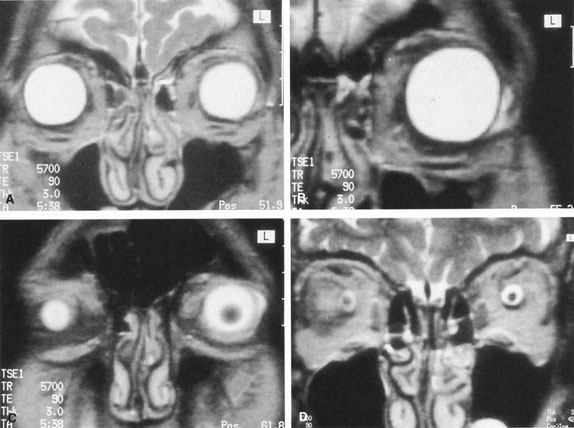
| Fig. 6. T2-weighted magnetic resonance imaging (MRI) study of 2 mL of anesthetic passing from the limbus, around the rectus muscles, into the posterior orbit, and finally highlighting the subdural space, contiguous with the sub-Tenon's space, surrounding the optic nerve. |