Chapter 83
Management of the Anophthalmic Socket and Techniques of Enucleation, Evisceration, and Exenteration: Surgical Procedures and Management of Complications
DAVID B. SOLL
Main Menu   Table Of Contents

Search

HISTORICAL DEVELOPMENT
ENUCLEATION AND EVISCERATION IMPLANTS--TECHNOLOGICAL ADVANCES
ANATOMIC CONSIDERATIONS
ENUCLEATION SURGICAL TECHNIQUES
MANAGEMENT OF COMPLICATIONS OF THE ANOPHTHALMIC SOCKET
EXENTERATION
CONGENITAL ANOPHTHALMIA
CONCLUSION
REFERENCES

HISTORICAL DEVELOPMENT
The objectives of enucleation and evisceration surgery using present state-of-the-art techniques should be obtaining good cosmesis as well as eliminating severe global pathology. Exenteration surgery is performed for malignancies confined to the orbit that spread by local extension; this is considered a radical life-saving operation.

By definition, enucleation is the removal of the entire globe. Indications for enucleation include:

  1. Intraocular malignancy
  2. Penetrating ocular wounds, especially involving the ciliary body, with irreversible loss of vision that might lead to sympathetic ophthalmia
  3. A blind, painful eye
  4. An eye that is blind and painless but markedly disfigured

Enucleation, rather than evisceration, is mandatory when intraocular malignancies not treatable by other methods are present or when sympathetic ophthalmia is likely to develop.

With present-day surgical techniques, many traumatic penetrating ocular injuries can be successfully managed with salvage of the globe and retention of useful vision. Thus, there are fewer enucleations performed in this category than there were previously.

Evisceration, by definition, is the removal of the contents of the globe, leaving the scleral shell and, in some instances, the cornea also. Because of improved enucleation surgical techniques, and also because there is a remote possibility of the development of sympathetic ophthalmia, as well as the presence of an undetected intraocular malignancy, performing an evisceration has become less popular.

The Egyptians and Sumerians were knowledgeable about the construction of artificial eyes using shells, gemstones, and other materials, as evidenced in many of the statues and decorative arts dating back to as early as 2500 BC. The first documented description of an enucleation appeared in 1555 by Lange, a German physician. In 1583, Bartisch, a Saxon, demonstrated removal of an eye by passing a thick suture through the globe, exerting forward traction, and then cutting the eye loose.1 It was not until 1885 that implants were used in orbital surgery. The first implant inserted was a glass sphere placed in an eviscerated globe by Mules.2 Glass spheres are now frequently called Mules spheres or implants.

Frost in 1887 was the first to describe the use of an intraorbital implant in enucleation surgery.3 The implant used in this instance was also a hollow, glass sphere.

The concept of replacing the contents of a globe or the entire globe in orbital surgery became widely accepted, and orbital implants were then widely used in both evisceration and enucleation surgery.

Back to Top
ENUCLEATION AND EVISCERATION IMPLANTS--TECHNOLOGICAL ADVANCES
Biotechnology has contributed to the great progress during the past decade in the development of implant materials that are compatible with orbital tissue and, in some cases, actually become integrated as part of the body. Glass, polymethylmethacrylate (PMMA), and silicone have been successfully used for many years; however, none of these materials encourage vascular ingrowth, and all are subject to orbital displacement. The use of hydroxyapatite for orbital surgery is a relatively new development.4 This material actually becomes an integral part of the body. Blood vessels and fibrous tissue invade the implant; thus, it is possible to couple the intraorbital hydroxyapatite implant with a plastic PMMA prosthesis. In the past, synthetic nonvascularized integrated intraocular implants were doomed to failure because of infection followed by socket contraction.

The anophthalmic socket has characteristics significantly different from those of a normal orbit. Immediately upon removal of the globe, multiple, irreversible orbital phenomena take place, the handling of which affects the long-term socket result and appearance. There are multiple changes in the anophthalmic orbit. The levator muscle complex essentially drops away from the superior orbital roof. The relation of the fornices to the eyelids and levator muscle complex is also immediately changed. All of these items affect the postoperative appearance. The correction of a postoperative problem depends on understanding the mechanism of its occurrence.5 Frequently, there is atrophy of orbital fat and upper eyelid ptosis along with a sulcus deformity that may produce an enophthalmic appearance. Ocularists attempt to correct this by enlarging the prosthesis and making a superior bulge to the prosthesis to eliminate the ptosis and to camouflage the superior sulcus defect.

The eye is what is known as a primary organizer. In patients who are born with congenital unilateral or bilateral anophthalmos, the bony orbit and surrounding structures do not develop normally.6 Until recently, the only advised treatment was expansion with progressively larger implants. This is still the initial procedure of choice; however, as will be discussed later, we now have the advantage of using fat grafting and craniofacial surgical procedures, as well as expandable types of implants.

Problems associated with congenital anophthalmic orbital deformities are harder to solve than those associated with enucleation or evisceration surgery in a previously well-developed orbit.

Back to Top
ANATOMIC CONSIDERATIONS
Respect for and understanding of anophthalmic orbital anatomy are essential for obtaining good results. Although the orbital structures are divided into compartments, all compartments are connected by the orbital septa, as described by Koornneef.7 The globe is surrounded by a layer of fibroelastic tissue known as Tenon's fascia. This fascia also encapsulates the extraocular muscles; therefore, even if a muscle retracts into its Tenon's sheath, because of the septal attachments between muscle fibers and sheath, there still remains extraocular muscle function and movement of orbital tissue, the fornices, and thus the prosthesis. Tenon's fascia is thicker and adherent to the conjunctiva anterior to where the four rectus extraocular muscles exit from their Tenon's sheaths. Posterior to these extraocular muscle exits, Tenon's fascia is thinner and less strong. This is the posterior layer of Tenon's fascia.

During any enucleation procedure, as much normal tissue and orbital fat as possible should be preserved.

In attempting to achieve motility of the prosthesis, multiple prosthetic shapes and implant shapes have been devised, often without understanding the true mechanism of motility of a nonintegrated intraorbital implant.

In all instances of enucleation surgery, deep fornices are essential, because it is the fornices that contribute to and allow for motility of the prosthesis--even when a hydroxyapatite implant is used.

Back to Top
ENUCLEATION SURGICAL TECHNIQUES
Each situation must be individualized. Surgical techniques include the insertion of an intraorbital implant either within Tenon's fascia or posterior to Tenon's fascia within the muscle cone area. Variations include the use of sclera or fascia to encase the implant and the use of a nonvascularizing implant, such as silicone or PMMA, or the use of a vascularizing implant, such as hydroxyapatite.

In certain instances discussed later in this chapter, it is not desirable to insert a synthetic implant within the orbit. In these cases, a dermal fat graft may be used.

The nonvascularizing, vascularizing, and dermal fat graft implants are also used for secondary intraorbital implantation.

BASIC ENUCLEATION SURGICAL TECHNIQUE

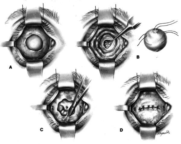
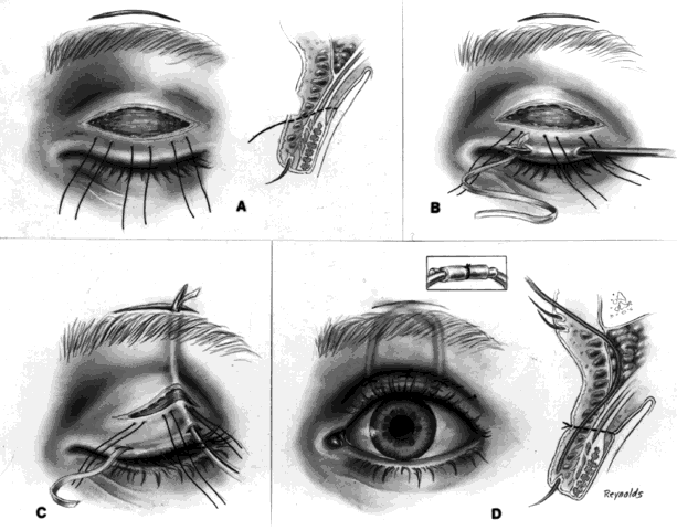
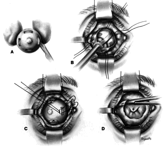
The basic enucleation technique, irrespective of the type of implant used, consists of removal of the diseased globe (Fig. 1).

Fig. 1. A. All four recti muscles are tagged. A superior fornix suture has been inserted. The superior and inferior oblique muscles are allowed to retract. In the event that a nonvascularizing implant, such as a silicone sphere, is used, only the medial and lateral recti muscles are tagged. B. The globe is pulled forward by use of previously placed traction sutures inserted through the insertional stumps of the medial and lateral recti muscles. A clamp is placed on the optic nerve. C. The posterior rent in Tenon's fascia is closed if a nonvascularizing implant is used.

Either general or local anesthesia may be used. General anesthesia is preferred because of the psychological implications of an enucleation. Preoperative in-depth discussion with the patient regarding what to expect postoperatively and what type of cosmetic result to expect is essential.

Regardless of whether general or local anesthesia is used, a 5-mL retrobulbar anesthetic injection should be given to eliminate the vagal reflex that can occur when traction is placed on the extraocular muscles or globe.

An eyelid speculum is inserted and a superior fornix double-armed 4-0 suture is hung by a hemostat. This aids in retracting the levator muscle complex and helps prevent injury to these structures.

Using a 30-gauge needle, an anesthetic solution or a balanced salt solution is injected subconjunctivally for 360°. This allows the surgeon to carefully perform a 360° peritomy, keeping the dissection in the episcleral plane and extending it to the four extraocular rectus muscles.

Each rectus muscle is severed at its insertion, and the tendons are tagged with a 5-0 or 6-0 synthetic absorbable suture, such as Vicryl or Dexon. The oblique muscles are not tagged but rather are severed close to their insertions on the globe and allowed to retract.

Two 4-0 black silk sutures are placed through the medial and lateral rectus muscle stumps on the globe. These sutures are then used as traction sutures for elevating and proptosing the globe. At this time, as many fibrinous connections as possible between Tenon's fascia and the episclera should be severed. An Arruga or similar type of speculum is useful at this stage.

This next step prevents significant bleeding once the optic nerve is severed. An Arruga speculum or a comparable speculum is placed between the medial rectus muscle and the globe. The medial and lateral traction sutures pull up on the globe, and a curved hemostat is used to clamp the optic nerve and the surrounding arteries. This hemostat clamp is left in place for 30 to 60 seconds and then removed. Enucleation scissors are used to sever the optic nerve. The globe is now removed. Any fine, residual fibrinous strands are cut. There usually is some bleeding, and the socket may be packed with a 4 × 4 gauze pad soaked with balanced salt solution. Occasionally, an epinephrine pledget is required when placing pressure against the posterior orbital structures.

I do not like or advocate the use of a snare, because when a snare severs the optic nerve, orbital fat is often snared and removed also. In addition, use of a snare is somewhat of a blind procedure, and occasionally, especially in an eye with a staphyloma, the posterior portion of the globe may be severed.

The socket should now be clean and dry, and a small rent should be visible posteriorly where the optic nerve went through the posterior Tenon's fascia.

A decision is now made as to the type of implant to use and its placement (i.e., within Tenon's fascia or posterior to the posterior layer of Tenon's fascia, within the muscle cone). Tenon's fascia that is posterior to the exits of the four rectus muscles is known as posterior Tenon's fascia and, as described previously, is thinner than anterior Tenon's fascia, which lies between the exits of the extraocular muscles and the conjunctiva. In general, the decision is made preoperatively as to whether or not a dermal fat graft is indicated. The technique and the implant used determine the immediate and long-term postoperative results.

INSERTION OF A HYDROXYAPATITE IMPLANT WITHIN TENON'S FASCIA

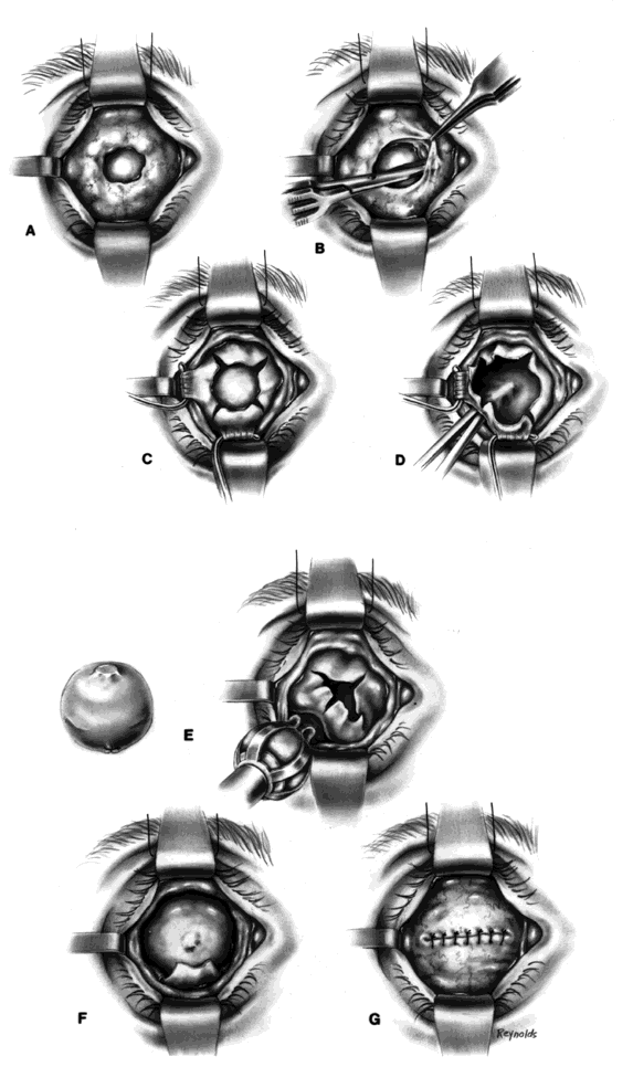
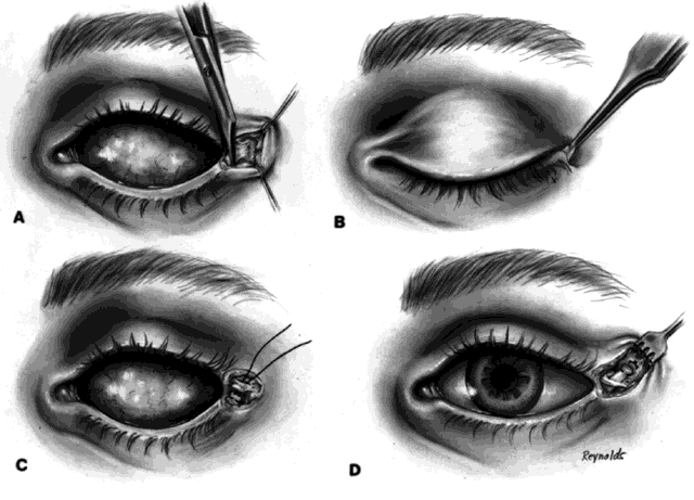
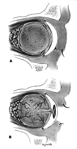
This technique is most useful when inserting a vascularizing implant, such as a hydroxyapatite implant (Fig. 2).8 Hydroxyapatite has a porous microarchitecture similar to human cancellous bone. Once implanted in an orbit with surrounding vascularized tissue, the implant readily fills with vascular tissue and becomes what might be termed as a living part of the orbit. This unique characteristic of a hydroxyapatite implant allows for the drilling of a hole through the anterior surface of the implant, once complete healing and vascularization of the implant have taken place. The walls of this drill hole become lined with conjunctival epithelium and allow for the insertion of a coupling peg, which aids in movement of the prosthesis. The surgeon evaluates the size of the hydroxyapatite implant to be used by first inserting sizing spheres. The properly sized sterile hydroxyapatite implant is wrapped in a preserved scleral shell. Autogenous temporalis fascia or fascia lata also may be used. The implant is wrapped to facilitate attachment of the extraocular muscles to the implant. Sclera preserved by freezing or by 95% alcohol preservation may be used. If preserved in 95% alcohol, the sclera should be washed thoroughly by three or more consecutive 2- to 3-hour washes with balanced salt solution. The scleral-wrapped implant may then be soaked in an antibiotic solution. The scleral envelope may not easily fit over the implant. If this is the case, then relaxing incisions must be made and resutured with either 5-0 Vicryl, Dexon, or Dacron sutures. The posterior portion of the implant should be exposed to facilitate vascularization.

Fig. 2. A. Windows are cut in the scleral covering of the hydroxyapatite implant. B. The implant is inserted within Tenon's fascia. C. The four recti muscles are sutured to the sclera, each in the region of a previously cut window. D. Tenon's fascia has been imbricated over the implant, and the conjunctiva is being undermined to the fornices.

In most instances, the size of the hydroxyapatite implant varies between 18 and 20 mm in diameter. When wrapped with sclera, the diameter of an implant increases by about 1.5 mm. An average eye 24 mm in axial length has a volume of 7.2 mL. The average prosthesis has a volume of 2.5 mL. Thus, to fill the volume replacement of a 24-mm globe, one would need an intraorbital implant volume of 4.7 mL (7.2 mL - 2.5 mL = 4.7 mL). Table 1 indicates that in this instance, a wrapped implant of 19.5 mm or an unwrapped implant of 21 mm would be required.


TABLE 1. Measurements for Proper Fit of a Hydroxyapatite Ocular Implant


Diameter of Enucleated GlobeVolume of Enucleated GlobeVolume of Artificial EyeNet Volume to be ReplacedSize of Implant if Not WrappedSize of Implant to Be Wrapped
(mm)(mL)(mL)(mL)(mm)(mm)
204.22.51.71513.5
214.92.52.416.515
225.62.53.11816.5
236.42.53.919.518
247.22.54.72119.5
258.22.55.72220.5
269.22.56.72322
(Courtesy of Arthur C. Perry, MD)


The enophthalmic appearance found in many anophthalmic orbits occurred as a result of volume deficiencies secondary to less volume of the implant and prosthesis as compared with the volume of the normal eye, and also because of some additional atrophy of orbital fat.

One may insert a bare hydroxyapatite implant by wrapping it in plastic strips and removing the strips once the implant is in position. It is difficult to place a bare hydroxyapatite implant because of spicules protruding from the surface of the implant, which tend to catch on tissue. If a bare implant is inserted, the rest of the technique is similar to the technique used for insertion of nonvascularizing implants. In this situation, the posterior rent in Tenon's fascia is sutured, and the sutures attached to the four extraocular muscle stumps are brought out through the conjunctival fornices because they cannot be attached directly to the bare hydroxyapatite implant.

If the implant is scleral-wrapped or fascial-wrapped, it is marked in four quadrants with a marking pen in areas where the four rectus muscles are to be attached. Windows in the sclera or fascia, approximately 4 mm wide and 7 to 8 mm long, are cut in the covering tissue, and the double-armed sutures previously attached to each extraocular muscle are brought out through the anterior scleral or fascial edge of the windows and tied. The implant is now firmly within Tenon's fascia with the posterior surface of the implant exposed to tissue within the muscle cone, because the rent in posterior Tenon's fascia, where the optic nerve passed through, is not sutured when the hydroxyapatite implant wrapped in sclera or fascia is used.

Vascularization of the implant occurs early on by blood vessels invading the hydroxyapatite through the posterior exposed areas and also through the windows through which the extraocular muscles were attached.

Some surgeons drill holes in the hydroxyapatite implant to facilitate vascularization; I have not found this to be necessary.

The conjunctiva is separated from the underlying anterior Tenon's fascia by injection of a balanced salt solution or an anesthetic solution between the two, with undermining to the medial, inferior, and lateral bony orbits, and superiorly as high as possible without affecting the levator muscle complex. This anterior layer of Tenon's fascia is imbricated and closed with 5-0 Vicryl or Dexon sutures. The conjunctiva is closed with either interrupted or 5-0 plain gut sutures. It is essential that the fornices be deep. A conformer is placed between the eyelids. The socket is dressed with an antibiotic solution, and a mild pressure bandage is applied for a period of 5 to 6 days. An artificial eye may be fit after 4 to 6 weeks.

After 4 to 6 months, the implant should be vascularized. This may be confirmed with a technetium 99m bone scan or with a magnetic resonance imaging (MRI) scan with contrast. The bone scan should show evidence of vascularization at a level between that seen in normal orbital bones and that seen in midfacial bones. If the patient is anxious for increased motility when the implant is determined to be fully vascularized (in my experience this occurs only in approximately 30% to 40% of patients), then a peg hole can be drilled in the implant.

A retrobulbar anesthetic injection is given. The center of the orbital implant should be drilled in a perpendicular manner with a 3-mm cutting blade to a depth of 10 to 12 mm. A flat-topped peg is then inserted. The artificial eye is next replaced in the socket and antibiotic drops are used for a period of 4 to 6 weeks, after which time the drill hole is usually completely lined with conjunctival epithelium. The flat peg is then replaced with a ball-type peg, and the ocularist burrs a corresponding socket in the appropriate part of the posterior portion of the prosthesis so that a coupling of the implant and prosthesis occurs. This then becomes a truly coupled implant and prosthesis (Fig. 3).

Fig. 3. A. Appearance immediately after insertion of hydroxyapatite implant--vascularization has not yet occurred. B. The hydroxyapatite implant becomes vascularized by ingrowth of blood vessels through the posterior defect in the scleral covering and the scleral windows, where the muscles are attached. Once complete vascularization has occurred, a hole is drilled through the anterior portion of the implant and a peg that is coupled to the implant is inserted. The channel becomes epithelialized.

This was never possible in the past because synthetic pegs attached to implants, exited through conjunctiva, and acted as a nidus for infection to enter the orbit. These early so-called integrated implants were disasters and were followed by the development of contracted sockets. This problem does not occur with hydroxyapatite implants because there is no exposure of the implant and the peg is in a cavity lined with conjunctival epithelium.

MANAGEMENT OF COMPLICATIONS RELATED TO HYDROXYAPATITE IMPLANTS

Because a hydroxyapatite implant becomes an integral part of the orbit by virtue of invasion of blood vessels and fibrous tissue within the implant, postoperative complications are handled in a specific manner. Exposure of the implant surface is not common, but it has occurred. It has been my experience that this problem cannot be satisfactorily managed by undermining and advancing conjunctiva and Tenon's fascia over the defect. If the defect is small and secondary to a poorly fitting prosthesis, and if the fit of the prosthesis is corrected, the defect may granulate in and re-epithelialize. As in all anophthalmic sockets, it is essential to have a properly fitting prosthesis. Most often, however, the exposure is great when the patient presents in the ophthalmologist's office. In this instance, my procedure of choice is to undermine conjunctiva and Tenon's fascia over the implant, making no attempt to significantly advance these tissues. A de-epithelialized dermal graft is placed over the exposed implant surface, the edges of the graft are tucked under the adjacent conjunctiva, and sutured with 6-0 Vicryl or Dexon sutures. The dermal graft should extend several millimeters under the adjacent Tenon's fascia and conjunctival tissue. The graft vascularizes and re-epithelializes with conjunctival epithelium. After 4 to 6 weeks, a properly fitting artificial eye is given to the patient. It is also possible to use this technique with autogenous fascia lata, temporalis fascia, or donor sclera.

Other problems unique to the hydroxyapatite implant are related to the peg. The shaft of the peg must be perpendicular to the apex of the socket and the implant. If the shaft is drilled at an angle, there is a great likelihood that extrusion of the peg will occur. This problem is less common but does occur, even in patients in whom the hole for the peg shaft has been drilled properly. When this occurs, the shaft hole becomes filled with fibrous tissue, which extrudes the shaft of the peg. Once the peg is out, the surface of the implant becomes resurfaced with conjunctiva and the patient may again be fitted with a standard artificial eye. If desired by the patient, the ophthalmologist may drill a repeat hole. Often, however, the patient is satisfied with the motility, and nothing further need be done.

A less common complication is the erosion of a spicule of hydroxyapatite through the conjunctiva at the start of or in the depths of the drill hole. The offending spicule should be burred off.

Because this problem has occurred even in properly drilled shafts, Perry and his co-workers have devised a plastic screw mechanism that is threaded into the implant at the time of drilling.8 This screw implant has a central shaft into which the peg is fit. Early results have indicated that this mechanism is more satisfactory than merely drilling the hole for the shaft of the peg directly into the implant.

INSERTION OF A NONVASCULARIZED IMPLANT

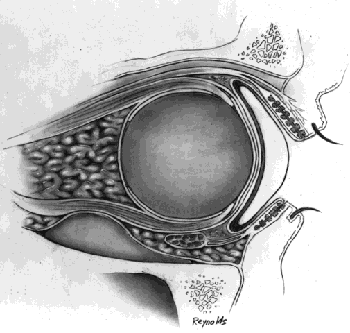
Nonvascularized implants such as polymethylmethacrylate or silicone (scleral- or fascial-wrapped, or without any wrapping) may be inserted within Tenon's fascia or posterior to all layers of Tenon's fascia within the muscle cone. If inserted within Tenon's fascia, the technique is as previously described; however, the posterior opening in Tenon's fascia where the optic nerve originally passed through should first be closed to prevent migration of the sphere. The extraocular muscles should never be imbricated over the anterior surface of the implant because this causes displacement and migration of the implant. Rather, they should be attached to fornix tissue by bringing the suture ends full-thickness out through the fornices and tying them over the conjunctival surface.

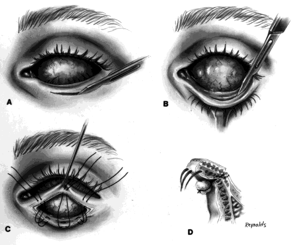
My preference when using these type of implants is to insert the nonvascularized scleral- or fascial-covered implant posterior to all layers of Tenon's fascia within the muscle cone. A larger implant can be used in this space, and there is little, if any, tendency for migration (Fig. 4).9,10

Fig. 4. A. Double-armed sutures are placed in the scleral covering of the implant in the areas where the medial and lateral recti would be if the implant were a small globe. A central, apical, double-armed suture also is placed. B. The implant sutures are passed through posterior Tenon's fascia and tied. The apical suture imbricates the lips of posterior Tenon's fascia. C. Conjunctiva is now undermined from anterior Tenon's fascia. The sutures tagging the horizontal recti muscles are passed through anterior Tenon's fascia and conjunctiva and tied in the fornices. D. Anterior Tenon's fascia is imbricated and conjunctiva is closed.

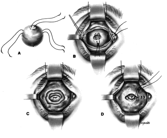
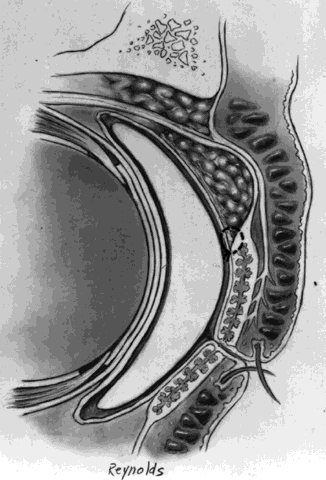
Ideally, the implant is wrapped in sclera or fascia, and three 5-0 or 6-0 double-armed Vicryl or Dexon sutures are attached to this scleral or fascial shell. Two sutures are placed where the medial and lateral rectus muscles would ordinarily be. These sutures are brought out through posterior Tenon's fascia medially and laterally and tied. A central, double-armed apical suture, passed through the scleral or fascial covering of the implant, is then used to imbricate posterior Tenon's fascia over the implant. Additional sutures may also be used to close any openings in posterior Tenon's fascia. These sutures virtually lock the implant in position. The superior and inferior rectus muscles are allowed to retract. They still continue to function. Pulling the superior rectus muscle forward can cause ptosis postoperatively because of its attachments to the levator muscle. Anterior Tenon's fascia is then separated from conjunctiva and undermined to the medial, inferior, and lateral orbital walls, and superiorly as high as possible without injuring the levator muscle complex. The double-armed sutures that were attached to the medial and lateral extraocular muscle tendons are now brought out full-thickness through anterior Tenon's fascia and conjunctiva and tied in the respective conjunctival fornices. Anterior Tenon's fascia is imbricated with use of 4-0 or 5-0 Vicryl or Dexon mattress sutures. The conjunctiva is closed with interrupted sutures or a running 6-0 plain gut suture.

It should be mentioned at this time that the undermining and separating of anterior Tenon's fascia and conjunctiva is essential to create deep fornices. It is the fornices that allow for movement of the prosthesis. For example, in a patient with a right-sided prosthesis, if the patient looks to the right, the lateral fornix deepens, the medial fornix shallows, and the prosthesis is literally pushed toward the right and drops into the space created by the deeper lateral fornix. The same type of situation occurs in whatever direction the patient looks. In the situation described, if the right lateral fornix were shallow, the prosthesis would be restricted in lateral motion.

Complications Associated With Nonvascularized Orbital Implants

Exposure, extrusion, and migration of the implants are complications related to these type of implants. If the exposure is small, undermining and advancing conjunctiva and Tenon's fascia may be all that is necessary for its correction. Late exposure is often due to an improperly fitting prosthesis, which causes pressure on the posterior socket wall. For large exposures, insertion of a scleral transplant or an autogenous de-epithelialized dermal transplant, as previously described, is usually satisfactory. If the exposure is very large and extrusion is imminent, it is usually best to remove the implant and insert a secondary implant within the muscle cone or to do a dermal fat graft.11–13

DERMAL FAT GRAFTS

There are situations in which placement of an intraorbital implant is not advisable but orbital bulk is necessary to fill the void left by the enucleated globe. This situation is not uncommon after extreme trauma when Tenon's fascia is severely lacerated. Also, this type of implant often does well in irradiated orbits where healing of tissue over synthetic implants may be a problem.

Surgical Technique

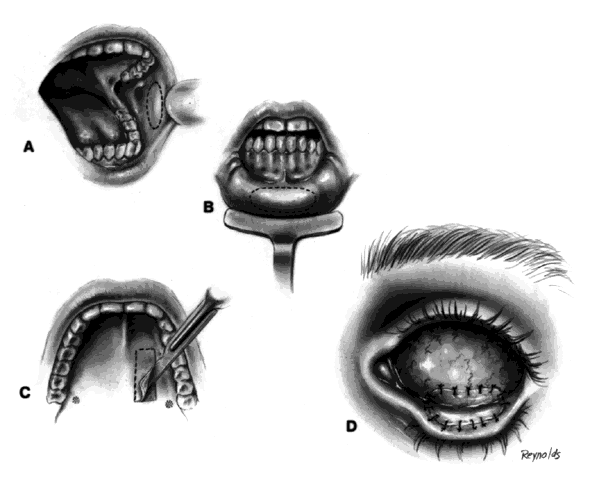
The globe is removed in the manner previously described. A de-epithelialized dermal fat graft is taken from the lateral buttock area; usually a graft 20 mm in diameter is taken. The donor site is closed with 2-0 Vicryl or Dexon sutures. If a 20/1000-in split-thickness skin flap was elevated first, this may be replaced with 6-0 Vicryl or Dexon sutures over the sutured donor site (Fig. 5).

Fig. 5. A. A 20/1000-inch split-thickness skin graft is elevated and a dermal fat graft obtained. B. The fatty portion of the graft is encased within the dermal shell. C. Dermal fat graft is inserted within Tenon's fascia and the four recti muscles are sutured to the dermal surface. D. Anterior Tenon's fascia is brought forward and also sutured to the dermal surface, as is conjunctiva.

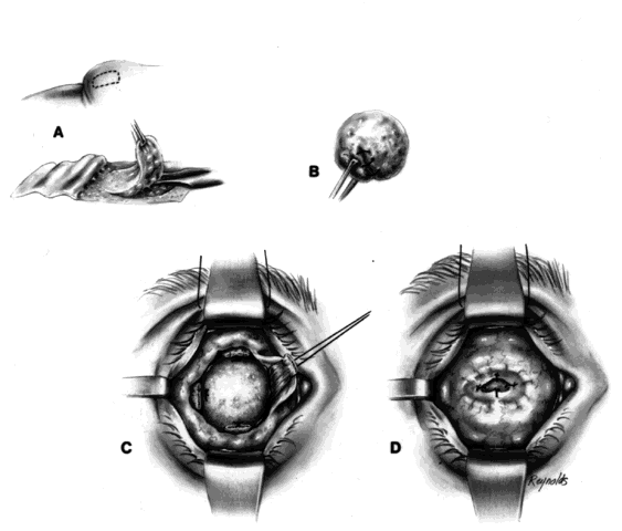
The de-epithelialized dermal graft is sutured into the orbit with the four extraocular muscles and Tenon's fascia sutured to the dermis. To decrease the amount of fat atrophy, it is advisable to encase the donor fat within a dermal shell, essentially making a baseball-type of implant. The sutures used to close dermal edges to dermal edges can be either 4-0 or 5-0 Vicryl or Dexon sutures. Conjunctival edges are sutured directly to the dermis. It is not necessary to completely cover the entire dermal graft with conjunctiva if there is a conjunctival deficiency. The central surface of the dermal graft will usually re-epithelialize if the defect is no larger than 5 to 6 mm.

It is my experience that if the extraocular muscles are not readily identifiable, as may occur in a secondary dermal fat implantation, the dermal fat graft can be sutured to Tenon's fascia. Movement of the prosthesis still occurs because of the connections of the extraocular muscles to the Tenon's fascial sheaths, as previously described in the section on Anatomic Considerations.

The socket is dressed with an antibiotic solution or ointment, and a conformer and patch are left in place for 5 days. After this time, an antibiotic ointment is applied daily for a period of 4 to 6 weeks. The patient now is ready for an artificial eye.

Complications of Dermal Fat Grafts

If the orbital circulation is poor, the central apical portion of the graft may necrose. In most instances, this area eventually granulates in. The only treatment necessary is the use of a topical antibiotic ointment to prevent infection. The defect usually granulates in without a problem.

If the orbital circulation is extremely poor, it may not be able to support the dermal fat graft, and necrosis of the entire graft is possible. The result is a contracted socket, the repair of which is discussed later.

If the orbital circulation is so poor that it does not even support a dermal fat graft, then the problem should be addressed first by use of a temporalis muscle transfer from the anterior portion of the temporalis muscle into the orbit through an osteotomy in the lateral orbital wall.

EVISCERATION

By definition, this is the removal of the contents of the globe, leaving the scleral shell. The surgical procedure may be performed with preservation or excision of the cornea.

Evisceration as a primary procedure is less popular today than it was in the past. The main indication for evisceration is an endophthalmitis, either acute with no response to therapy, or old with loss of any vision in the affected globe. Most of these cases are secondary to cataract surgery; however, endophthalmitis can be metastatic and can occur secondary to intraocular foreign bodies. It can also occur secondary to perforation of a very virulent corneal ulcer.

When an evisceration is performed for endophthalmitis secondary to cataract surgery, and a large limbal incision was originally used, it is best to excise the cornea, insert the secondary implant, imbricate the scleral edges, and cover the area with conjunctiva. This type of evisceration should be performed whenever there is compromise of limbal or corneal integrity. I frequently insert an antibiotic dusting powder within the scleral shell, which previously was wiped very clean with 70% alcohol and irrigated with balanced salt solution. The contents of the globe in this instance are scooped out with an evisceration spoon or a cyclodialysis spatula. If the cornea is to be preserved, an incision is made between the superior and lateral rectus muscles parallel with the limbus. The incision is carried down to the choroid. A cyclodialysis spatula is then used to break all connections between the choroid and the sclera and also to transsect the optic nerve fibers. The incision is then enlarged so that an intrascleral implant can be inserted. The scleral edges are imbricated and sutured with multiple 4-0 or 5-0 mattress sutures and the conjunctiva closed. The only variation is when a vascular type of implant such as hydroxyapatite is inserted within the scleral shell. In this instance, posterior windows of sclera should be excised to allow vascular tissue to invade the implant. If a vascularized implant is used, a peg can be drilled similar to the way the peg was drilled in the enucleated socket. If the cornea was preserved, it is not necessary to bring the conjunctiva over the corneal surface.

Traditionally, eviscerations have been thought to give better cosmetic results than enucleations. However, with current enucleation techniques, the results are very comparable.

Back to Top
MANAGEMENT OF COMPLICATIONS OF THE ANOPHTHALMIC SOCKET

EXTRUSION/EXPOSURE

Secondary insertion of an intraorbital implant is performed if an implant is extruding or has extruded, or if no implant was placed in the socket originally.

If there is any infection with an extruding implant, it should be controlled with topical and local antibiotic therapy. The extruding implant should be removed unless the defect over the implant is small (5 mm or less), in which case it may be possible to prevent further extrusion by insertion of a fascial or scleral patch, or a de-epithelialized dermal graft over the implant and under the conjunctiva.

Most often, the implant has to be removed and the surrounding capsule excised, preserving as much conjunctiva and normal orbital tissue as possible. If the extraocular muscles are easily found, they should be tagged; however, as previously mentioned, extensive dissection to find an extraocular muscle should not be performed because this causes unnecessary tissue damage. All layers of Tenon's fascia are centrally perforated and the fatty tissue of the muscle cone exposed. A fascial- or scleral-wrapped implant is inserted within the muscle cone. Four sutures attached to the implant are brought out through the fornices, with care being taken that the superior and inferior sutures are not too peripheral. Tenon's fascia is imbricated over the implant, and the conjunctiva is undermined and closed. Again, extensive conjunctival undermining is essential to have deep fornices. If the tissue is too friable for insertion of a vascularized or nonvascularized implant, a dermal fat graft can be used as a secondary implant (Fig. 6).

Fig. 6. A. Extruding synthetic enucleation implant. B. Implant has been removed and an opening is made through all layers of the posterior aspect of the orbit to expose the fatty tissue of the muscle cone. A scleral- or fascial-wrapped implant with preplaced sutures is placed within the fatty tissue of the muscle cone. C. The medial and lateral implant sutures are brought out through the fornices and tied. The apical suture imbricates Tenon's fascia. D. Conjunctiva is undermined and closed.

Exposure and extrusion of the implant are the most common complications associated with evisceration. If the exposure is small, it may be patched with a scleral, fascial, or dermal transplant. A smaller implant may also be inserted. If these measures are not effective and a repeat exposure occurs, or if the exposure was large, it is best to separate conjunctiva and Tenon's fascia from the scleral surface, make four relaxing incisions in the patient's sclera, sever the optic nerve, and remove the extruding implant. A spherical implant, which may be sclera or fascia covered, is inserted within the muscle cone behind the scleral shell. In adult-sized orbits, the implant is usually 16 to 18 mm in diameter. The scleral edges are everted and tucked under Tenon's fascia. The inverted scleral shell edges are covered with conjunctiva. This technique is an excellent method of insertion of a secondary implant in an eviscerated socket. It is especially useful if multiple extrusions have occurred or if extrusion of a large implant has occurred with scleral deficiency around the extruded area. The postoperative appearance and motility with this technique are usually excellent (Fig. 7).14

Fig. 7. A. Large exposure of evisceration implant. B. Conjunctiva and Tenon's fascia are separated from scleral surface. C. Relaxing incisions that extend to the level of the insertions of the recti muscles are made in four quadrants. D. The extruding implant has been removed, and the optic nerve is clamped and severed. E. With use of an introducer, the secondary implant is inserted into the muscle cone, posterior to the everted scleral shell. F. The implant is positioned in the muscle cone; three of the four scleral edges have already been tucked under Tenon's fascia. G. Conjunctiva and Tenon's fascia are closed.

SUPERIOR EYELID SULCUS

One of the most common difficulties associated with the anophthalmic orbit is the superior eyelid sulcus defect, often associated with ptosis. This defect is secondary to atrophy of the preaponeurotic fat layer. It can be improved by insertion of a subperiosteal floor implant with a large posterior surface. This gives additional mass to the orbit and pushes tissue upward and forward (Fig. 8).15

Fig. 8. Posterior volumetric increase is obtained by insertion of a subperiosteal implant. Elevation and forward displacement of the implant are attained, thereby ameliorating a superior eyelid sulcus defect.

The floor implant may be fashioned from Teflon, PMMA, or any one of the other available biocompatible synthetics. A blepharoplasty incision is made for the insertion of the implant. It is essential that the anterior lip of the implant not extend beyond the bony orbital rim. Wiring of the implant in place is often necessary. Additionally, it is not a good idea to close the periosteum because this can cause inferior eyelid retraction. If this technique is properly performed, the implant remains in position and the periosteum heals properly.

I frequently put an inferior eyelid traction suture in place at the conclusion of the procedure for several days to help prevent the problem of inferior eyelid retraction.

If room temperature vulcanizing (RTV) silicone is available, this can be injected subperiosteally through a lateral canthal incision. The periosteum is elevated and a pocket is created into which the RTV silicone is injected. The vulcanization time should be determined prior to injection of the material and should usually be timed to be 20 to 30 seconds. Care should be taken that no silicone extends anterior to the bony orbital rim.16

It is also possible to gain exposure to the orbital floor through an inferior conjunctival cul-de-sac approach. This eliminates the blepharoplasty incision. The technique involves performing a lateral canthoplasty, severing the inferior crux of the lateral canthal tendon, and reflecting the lower eyelid down. An incision is then made through conjunctiva and underlying tissue to the periosteum at the inferior orbital rim. Periosteum is elevated and the procedure completed as previously described. The lateral canthal tendon is then reapproximated, usually with nonabsorbable sutures, and the 0.5-mm skin incision is closed.

If the problem of the superior eyelid sulcus defect is small, a dermal fat graft placed on the inner surface of the eyelid, superior to the tarsus and subconjunctivally, can be a very effective surgical procedure in reducing the defect. It also facilitates closure of the eyelids (Fig. 9).

Fig. 9. A dermal fat graft placed subconjunctivally, immediately superior to the upper border of the tarsus, effectively corrects a superior eyelid sulcus defect.

PTOSIS

Ptosis can be corrected by modification of the prosthesis; however, if this interferes with eyelid closure, drying of the prosthesis occurs, and this is not satisfactory. Conservative levator aponeurosis advancement and/or sling operations are effective. Nothing should be done to compromise the superior fornix.

When the ocularist has done all that is possible to correct the ptosis without causing dryness of the prosthesis, and the ptosis remains, the ophthalmologist can perform several different types of ptosis procedures. If the ptosis is minimal (less than 2 mm) and there is adequate superior fornix tissue, then a tarsoconjunctival Müller's muscle resection can be performed. This procedure, however, must be coordinated with the ocularist, and the optimal prosthesis should be inserted prior to this procedure being performed. However, I am often reluctant to do any type of resection of normal fornix conjunctival tissue or of superior eyelid tarsus tissue in the anophthalmic socket. Many times these patients have disinsertions of the levator aponeurosis. This may be corrected by use of an external approach with reapproximation of the aponeurosis edge to the upper one third of the tarsus using three nonabsorbable half-thickness tarsus sutures. If no disinsertion is found, a conservative aponeurosis resection with advancement usually suffices. To maintain eversion of the eyelashes, several 6-0 Vicryl or similar absorbable sutures can be placed between orbicularis fibers close to the eyelid margin and the lower edge of the levator aponeurosis. It is not necessary to involve the conjunctiva in this type of surgery. Lastly, if the patient has very poor or no levator function, then a fascia lata or silicone sling procedure is used. The procedure is the same whether fascial strips or silicone rods are used.

Fascial Sling/Silicone Procedures

The area of the proposed eyelid crease is marked and an ellipse of skin excised. The amount of skin between the lash margin and the incision is usually 7 mm ± 1 mm. Next, full-thickness, double-armed, 4-0 black silk sutures are placed in the medial, middle, and lateral one third of the eyelid. Each arm of the suture is passed from just above the tarsal border full-thickness to the upper edge of the lower segment of eyelid skin (Fig. 10).

Fig. 10. A. A skin incision is made through the eyelid crease, and three double-armed 4-0 black silk sutures are passed full-thickness immediately above the superior edge of the tarsus, exiting through the upper edge of the lower section of the eyelid skin. A brow incision is made above the middle one third of the eyebrow. B. Incisions are made through the eyelid skin 1 mm above the lash margin, each incision being 2 to 3 mm in length. The medial incision is made at a position corresponding to the medial limbus of the eye in the primary position. The lateral incision is made 2 mm lateral to the lateral limbus. A straight needle is used to pass the silicone rod or fascial strip across the eyelid into the epitarsal space. C. A curved needle is used to pass the silicone or fascia to the brow incision. D. If a silicone rod is used, it is passed through a Watskie sleeve and the eyelid height adjusted. If fascia is used, the fascia is tied.

Incisions are next made to the level of epitarsal tissue 1 mm above the eyelid margin. Each incision is 2 to 3 mm in length. The medial incision is at the level of where the medial limbus would be if the patient had an eye in the primary position. The lateral incision is made 2 mm lateral to the position of the lateral limbus. Next, with use of a straight needle, either the silicone rod or fascial strip is passed through the epitarsal space above the eyelid margin. Using a curved needle, each end of the sling is then passed through the epitarsal tissue and the orbital septum into the preaponeurotic fat compartment; it is then passed through the orbital septum below the arcus marginalis and then out through an incision approximately 1 cm in length above the central portion of the eyebrow. If a silicone rod is used, each end is passed through a Watskie sleeve and the eyelid height is adjusted to a level 1 mm above the limbus of the prosthetic eye, which should be placed in the socket at this time. Nonabsorbable sutures secure the silicone ends in place. If fascia is used, it is passed in the same way, and the two ends are knotted in the brow incision. Nonabsorbable sutures are passed around the fascial ends to avoid slippage. The brow incision is closed with deep and superficial sutures. The eyelid crease incision is closed with either a running nonabsorbable suture or interrupted 6-0 plain gut sutures. It is not necessary to close the marginal incision. The previously placed 4-0 black silk sutures are tied. This maneuver keeps the skin edge above the lashes taut and tends to evert the lashes. In all ptosis procedures, it is important to wind up with some eversion of the eyelashes in order to give a pleasing cosmetic effect. As in all sling procedures, the patient has to learn forced eyelid closure using the orbicularis muscle. The 4-0 black silk full-thickness sutures are usually removed after 4 to 5 days. The eyelid crease sutures, if a nonabsorbable suture has been used, are removed after approximately 1 week (see Fig. 10).

LOWER EYELID PROBLEMS

All prostheses are supported by the lower eyelid. If the prosthesis is heavy or frequently removed, the medial and lateral canthal tendons stretch and the lower eyelid sags. The treatment is resection of the inferior portion of the lateral canthal tendon and repositioning of the lower eyelid to give better support to the prosthesis.

It is important to recognize that the sagging of the lower eyelid is secondary to the stretching of the medial and lateral canthal tendons rather than stretching of the tarsus. Resection of a tarsal segment causes the loss of normal eyelid tissue, adversely affects the person's appearance because of loss of eyelashes, and does not attack the basic problem.

The correct treatment of sagging of the lower eyelid is resection and reapproximation of the inferior crux of the lateral canthal tendon to the lateral orbital wall (Fig. 11). This is accomplished by first making a lateral canthotomy, resecting the excessive length of the stretched lateral canthal tendon, and, if necessary, creating a new tendon from the lateral 2 to 3 mm of tarsus. The reconstructed tendon is now passed through a slit in the remaining superior crux of the lateral canthal tendon and sutured to periosteum with a nonabsorbable suture. If the lower eyelid laxity is great, it may also be necessary to imbricate the inferior portion of the medial canthal tendon; however, this is usually not necessary.

Fig. 11. A. After a lateral canthotomy has been performed, the inferior crux of the stretched lateral canthal tendon is severed. B. The lateral canthal tendon is draped over the lateral orbital wall to determine the amount of lower eyelid tightening required. C. A stab incision is made through the superior portion of the lateral canthal tendon at its insertion. A double-armed suture is passed through the lateral extremity of the severed inferior portion of the lateral canthal tendon. The suture ends are brought through the previously made stab incision. D. The tightened lateral canthal tendon is sutured to periosteum.

CONTRACTED SOCKET

If the socket has chronic infection, contraction of the fornices and frequently entropion of the upper and lower eyelids occurs. Correction of the problem necessitates mucous membrane grafting and marginal rotation procedures for the correction of the entropion.

Cicatricial Entropion/Marginal Rotation Procedures

These operations are effective and should be performed only if the fornices are deep. One has to be careful not to disturb the blood supply to the rotated eyelid margin; therefore, the initial incision should be made 3 mm above or below the tarsal margin, depending on whether the upper or lower eyelid is being operated on. The incision should go through only the tarsus and should be parallel with the eyelid margin. Next, a skin incision is made 3 mm from the eyelid margin to the level of the orbicularis. The full-thickness incision is completed by slowly incising the orbicularis and evaluating the marginal tissue. The surgery is completed by suturing the severed tarsal edge of the marginal strip of tarsus to the adjacent skin edge, thus everting the margin (Fig. 12).

Fig. 12. A. A skin and muscle incision is made approximately 3 mm below the eyelid margin to the level of the tarsus. B. The eyelid is everted and a corresponding tarsal incision is made, thus creating a full-thickness eyelid incision. Extreme care should be taken not to interfere with the blood supply of the eyelid margin. C. Double-armed sutures are passed from the tarsal edge to the skin edge, thus connecting the skin margin to the tarsal edge. D. This effectively rotates the eyelid margin outward.

Mucous Membrane Grafting

Frequently, cicatricial entropion problems are also associated with contraction of the fornices. When this occurs, mucous membrane grafting of the fornices is necessary. Incisions are made in the contracted fornices, and tissue is undermined and spread until a conformer larger than necessary can easily be fit. Mucous membrane can be obtained from the mouth; buccal mucosa from the lower lip or side of the cheek is excellent for lining a socket. The mucosal graft should be at least 0.5 mm thick if taken with a mucotome, or it can be full-thickness if taken freehand. Thick grafts contract less than thin grafts. Another excellent source of mucous membrane is hard-palate mucosa. It is also possible to combine buccal mucosa and hard-palate mucosa. The grafts are sutured in place and a conformer is inserted to secure the grafts against the underlying vascular bed. The conformer should be left in place until the graft has healed, which usually occurs by 3 weeks. It is important that the graft be kept in apposition to the underlying blood supply during the healing phase because shifting of the graft can result in loss of all or part of the graft. There is always some tendency for shrinkage, and in a normal socket this usually amounts to 20% to 25%. If the socket is one in which there was a previous chemical injury, the conformer must be left in place for a much longer period of time and usually has to be sutured in place. In this instance, shrinkage may be as much as 50% or greater. It is also possible to combine mucous membrane and split-thickness skin in socket repair and reconstruction if not enough mucous membrane can be obtained (Fig. 13).

Fig. 13. Sources of soft mucosal grafts. A. Mucosal graft taken from inside the cheek. Care must be taken to avoid the parotid duct. B. Mucosa can easily be obtained from the lower lip. Care must be taken to avoid the vermilion border. C. Hard-palate graft. Care must be taken to avoid the area of the greater palatine foramen (stippled area) because severing of the anterior palatine artery can cause bleeding that is very difficult to control. D. Insertion of mucosal graft in inferior fornix.

Back to Top
EXENTERATION
This procedure is performed for the removal of all orbital contents. It is indicated for primary orbital malignancies extending behind the globe. In general, radical exenterations are not performed for malignant melanomas because these are spread by way of hematogenous routes.

Most exenterations are performed leaving several millimeters of eyelid skin that then drapes over the orbital margins. The procedure is relatively avascular because the dissection is carried out subperiosteally and the tissues coming through the superior and inferior orbital fissures and through the optic foramen are cauterized with electrocoagulation prior to their being cut. The exenterated socket may be lined with a split-thickness skin graft usually 0.015 to 0.018 of an inch thick. An alternative method is to allow the orbital defect to granulate in. I prefer the former because the patient becomes rehabilitated more rapidly.

The major problem occurring with exenteration is the creation of fistulous tracts during the surgical procedure. These often do not heal and constantly drain their contents into the exenteration cavity. Obviously, the best treatment is prevention by being gentle when doing the subperiosteal dissection, especially in the region of the ethmoidal plate. Sinus fistulous tracts cannot be treated by covering them with skin grafts. They can be treated by bringing vascularized pedicle flaps to the area, plugging the openings, and later severing the pedicles.

Various types of exenteration prostheses are available. Some are attached to eyeglass frames and some are manufactured so that they can adhere to adjacent tissue. Many patients, however, prefer a black patch.

Back to Top
CONGENITAL ANOPHTHALMIA
The treatment of this problem is very complex and requires coordination among ophthalmologist, ocularist, and parents. Occasionally, the service of a craniofacial surgeon may be required. Kennedy has shown, and it is well known, that in order for an orbit to develop properly, an eye must be present.6 The eye is thus known as a primary organizer. In virtually all cases of congenital anophthalmia there can be found at least a remnant of the optic vesicle. This is also a requirement for survival of the fetus. Without a globe, bony and soft-tissue development does not normally occur. Congenital anophthalmos may be unilateral or bilateral. The treatment is the same. Initially, progressively larger expansion-type conformers are placed within the socket, which most often is very tiny and underdeveloped. These progressively larger conformers help to expand the eyelids and encourage bony orbital development. With advances in biotechnology, expandable types of conformers have been developed that are sutured in place against the orbital rims. These conformers create pressure on the orbital structures and encourage bony growth. Work has also indicated that a dermal fat graft inserted in a congenitally anophthalmic orbit, when performed in an infant or very young child, is beneficial, and the fat graft actually grows and encourages bony growth.17 Most often, no matter how much lateral growth of the orbit has been achieved, there is always some anterior/posterior deficiency present. The orbit is expanded by using the progressively larger conformers or expandable implants. They also stretch and allow the eyelids and cul-de-sacs to grow and expand.

Treatment of the congenital anophthalmic orbit should be started within the first few weeks of life and should be continued until enough expansion is achieved to allow the satisfactory fitting of an artificial eye. In some cases, the amount of expansion is not satisfactory and, especially in unilateral cases, patients and family are not pleased with the asymmetry. The craniofacial surgeon has the option of further expanding the bony orbit with bone expansion and bone grafting techniques. Soft-tissue expansion then follows.

In congenital anophthalmos, not only are the eyelids, cul-de-sac, and bony orbit underdeveloped, but there is also underdevelopment of the levator muscle complex. Thus, compromises must be made when fitting the prosthesis to allow for a satisfactory cosmetic appearance. The patient's prosthesis should not appear dry secondary to an inability to adequately close the eyelids.

Back to Top
CONCLUSION
This chapter attempts to summarize the current thinking and procedures related to management of the anophthalmic socket. Enucleation, evisceration, exenteration, and procedures related to the congenital anophthalmic orbit are discussed. The chapter is not all-inclusive; however, the reader should be able to manage the majority of problems related to the various anophthalmic orbital -problems and should have a defined approach to the surgery or expansion procedures to be performed.
Back to Top
REFERENCES

1. Bartisch G: Ophthalmodouleia oder Augendienst. Dresden, 1583

2. Mules PH: Evisceration of the globe, with artificial vitreous. Trans Ophthalmol Soc UK 5:200, 1885

3. Frost WA: Cited in Lang W: On the insertion of artificial globes into Tenon's capsule after excising the eye. Trans Ophthalmol Soc UK 7:286, 1887 Ì

4. Perry AC: Perry motility implant. Presented at the combined meeting of the American Society of Ophthalmic Plastic and Reconstructive Surgery and the American Society of Ocularists, Dallas, October 25, 1987

5. Soll DB: Evolution and current concepts in the surgical treatment of the anophthalmic socket. Ophthal Plast Reconstr Surg 2(3):163, 1986

6. Kennedy RE: Growth retardation and volume determination of the anophthalmic orbit. Am J Ophthalmol Otolaryngol 56:42, 1952

7. Koornneef L: Spatial Aspects of Orbital Musculofibrous Tissues in Man. Amsterdam and Lisse, Swets and Zeitlinger, BV, 1977

8. Perry AC: Advances in enucleation. Ophthalmol Clin North Am 98:370, 1991

9. Soll DB: The anophthalmic socket. In Soll DB (ed): Management of Complications in Ophthalmic Plastic Surgery, pp 295–344. Birmingham, AL, Aesculapius Publishing, 1976

10. Soll DB: The anophthalmic socket. Ophthalmology 89:407, 1982

11. Frueh BR, Felker GV: Baseball implant. A method of secondary insertion of an intraorbital implant. Arch Ophthalmol 92:494, 1974

12. Smith B, Petrelli R: Dermis--fat graft as a movable implant within the muscle cone. Am J Ophthalmol 85:62, 1978

13. Bullock JD, Brickman KP: Dermis--fat graft in socket reconstruction: Theoretical and experimental considerations. Ophthalmology 91:204, 1984

14. Soll DB: Evisceration with eversion of the scleral shell and muscle cone positioning of the implant. Am J Ophthalmol 104:265, 1987

15. Soll DB: Correction of the superior lid sulcus with subperiosteal implants. Arch Ophthalmol 64:1088, 1971

16. Sergott TJ, Vistnes LM: Correction of enophthalmos and superior sulcus depression in the anophthalmic orbit: A long-term follow-up. Plast Reconstr Surg 79(3):331, 1987

17. Katowitz J: Personal communication, 1993

Back to Top