|
|
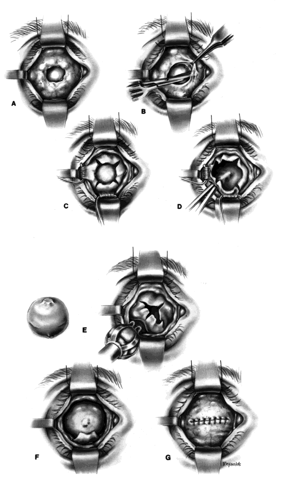
| Fig. 7. A. Large exposure of evisceration implant. B. Conjunctiva and Tenon's fascia are separated from scleral surface. C. Relaxing incisions that extend to the level of the insertions of the recti muscles are made in four quadrants. D. The extruding implant has been removed, and the optic nerve is clamped and severed. E. With use of an introducer, the secondary implant is inserted into the muscle cone, posterior to the everted scleral shell. F. The implant is positioned in the muscle cone; three of the four scleral edges have already been tucked under Tenon's fascia. G. Conjunctiva and Tenon's fascia are closed. |