Chapter 78
Ptosis Surgery
MALENA M. AMATO , BLYTHE MONHEIT and JOHN W. SHORE
Main Menu   Table Of Contents

Search

TYPES OF PTOSIS
EXAMINATION OF THE PTOSIS PATIENT
PTOSIS SURGERY
PROBLEMS IN PTOSIS SURGERY
REFERENCES

Blepharoptosis or ptosis refers to a congenital or acquired drooping of the upper eyelid.1 Ptosis can be classified by age of onset (e.g., congenital or acquired) or by etiology (e.g., aponeurotic, mechanical, neurologic, or traumatic). The age of the ptosis patient is an important factor in management especially in congenital ptosis, which may result in or contribute to the development of amblyopia. Amblyopia resulting from ptosis in an infant demands prompt diagnosis, monitoring, and treatment. Blepharoptosis may also be a cause of visual loss in the adult by obstruction of the superior visual field.2 Although a common condition encountered by the ophthalmologist, blepharoptosis still presents challenges in diagnosis and treatment. Proper classification of the type of ptosis is an essential guide to further workup, as well as to medical management or surgical correction.
Back to Top
TYPES OF PTOSIS
There are several types of ptosis that are classified in one of two general categories, congenital or acquired. Within these two groups, ptosis is broken down and subclassified by etiology (e.g. aponeurotic, neurogenic, myogenic, mechanical, and traumatic). In this chapter, we discuss congenital and acquired ptosis. We present historical and clinical clues to diagnosis, evaluation, and treatment.

CONGENITAL PTOSIS

Congenital ptosis is a condition present from birth, and, by history, the eyelid malposition has remained stable. Congenital ptosis is unilateral in 75% of cases. The remaining 25% are bilateral.3 In children with congenital ptosis, one must carefully asses refractive error and visual function to identify and treat amblyopia. Amblyopia occurs in 20% of congenital ptosis patients, only 4% of which is attributed to the ptotic eyelid occluding the pupil. Other causes of amblyopia-associated ptosis include anisometropia, high astigmatism, or strabismus.2 In all cases, early assessment and treatment of ptosis is necessary when the visual axis is partially or completely obstructed, because amblyopia and permanent visual loss are severe consequences. If the patient already demonstrates significant amblyopia, there is an extremely high rate of failure of ptosis surgery. Vision is required to motivate patients to learn to use their frontalis and forehead muscles to elevate the eyelids after ptosis surgery in patients with poor elevating power of the eyelids.

Congenital Myogenic Ptosis

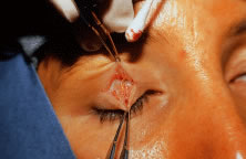
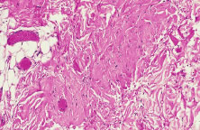
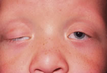
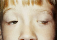
The most common type of congenital ptosis is myogenic ptosis. The pathophysiology involves dysgenesis in the levator palpebrae superioris (LPS) muscle. The skeletal muscle fibers of the LPS are replaced by fibroadipose tissue (Fig. 1). This causes decreased levator muscle function and ptosis. Both elevation and relaxation of the eyelid are impaired because of fibrosis within the muscle and the paucity of normal muscle tissue. Lagophthalmos on downgaze is the sine qui non of congenital ptosis and is caused by the fibrosis of the LPS, which restricts relaxation of the levator muscle. If the levator pulling power is markedly reduced or absent, the patient may also lack an eyelid crease (Fig. 2). In this situation, the abnormal aponeurotic fibers of the LPS do not insert onto the orbicularis and dermis. Lash ptosis may also be seen in myogenic congenital ptosis from decreased tone and loss of structural support.

Fig. 1. Histopathology of the levator palpebrae superioris of a patient with congenital ptosis demonstrates fibrovascular and fibroadipose infiltration.

Fig. 2. Patient with congenital ptosis. Note the lack of eyelid crease.

Superior rectus dysfunction is found in approximately 5% of congenital myogenic ptosis cases. The superior rectus muscle and LPS share similar embryologic origins. Here, abnormal fibrofatty tissue is seen in both muscles on biopsy.3 A concomitant maldevelopment of the superior rectus muscle seen in certain cases of congenital myogenic ptosis is called double elevator palsy or monocular elevation deficiency.2 In these patients, congenital myogenic ptosis is associated with poor Bell's phenomenon or vertical strabismus. Repair of congenital myogenic ptosis is difficult, because the pathophysiology involves an abnormal muscle, which can not be corrected surgically.

Congenital Aponeurotic Ptosis

Rarely, congenital ptosis may be aponeurotic in origin. Here, the ptosis is due to failure of the levator aponeurosis to insert at its normal position on the anterior tarsus. This may result from birth trauma, especially with forceps delivery. Usually there is good eyelid excursion yet an abnormally high eyelid crease.

Congenital Mechanical Ptosis

Mechanical ptosis can be a congenital abnormality resulting from a mass effect in the upper eyelid, brow, or orbit. Patients can present at birth with a hemangioma, dermoid, or plexiform neurofibroma, which are common causes of mechanical ptosis. Amblyopia can result from astigmatism induced from the mass effect on the globe or from obstruction of the visual axis resulting from the ptotic eyelid. Hemangiomas usually resolve spontaneously or respond to steroid injections.2 Removal of the offending dermoid will usually eliminate the associated mechanical ptosis. Ptosis associated with neurofibromatosis and plexiform neurofibromas is not easily corrected. The treatment is beyond the scope of the text.

Congenital Neurogenic Ptosis

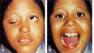
Congenital neurogenic ptosis, though rare, is caused by innervational defects during embryonic development. The Marcus Gunn (Jaw-Winking) ptosis (MGJWP) is the most common type of congenital neurogenic synkinetic ptosis. It is observed in 4% to 6% of patients with congenital ptosis. This eyelid malposition and synkinetic movement involves an abnormal innervation of a normal LPS muscle causing the unilaterally ptotic eyelid to wink with jaw movement (Fig. 3). Synkinesis occurs from aberrant connections between the motor branches of the fifth cranial nerve that normally supplies the pterygoid muscle or the muscles of mastication and the superior division of the oculomotor nerve (CN-3) that innervates the LPS and superior rectus muscles.4 The aberrant connections in the central nervous system can be bilateral, although only one side is symptomatic, demonstrating the wink.5 Clinically, these patients usually have a baseline ptosis on the affected side in primary gaze. The wink is induced by use of muscles of mastication (e.g., sucking, swallowing, chewing, or a lateral motion of the jaw to either side). The wink consists of a rapid elevation of the ptotic lid to a position higher than the unaffected side and then an equally rapid descent back to the ptotic state.6 In this situation, ipsilateral hypotropia or vertical strabismus is common because the superior rectus muscle may be involved. The ptosis is often corrected with surgery. Excision of the levator aponeurosis seems to alleviate some of the symptoms of the jaw-wink and synkinesis before ptosis surgery. Some children with the associated jaw-wink develop adaptive behavioral mechanisms and learn to avoid movements that reduce the wink reflex. This has caused some clinicians to state that MGJWP ptosis improves with age.

Fig. 3. Marcus-Gun jaw-winking ptosis (synkinesis linking fifth cranial nerve to third cranial nerve ptosis). A. Baseline congenital ptosis of the right eye. B. On jaw opening, the right lid is elevated. (Adapted from American Academy of Ophthalmology. Orbit, Eyelids and Lacrimal System; BCSC Section 7. San Francisco: American Academy of Ophthalmology, 2000-2001:Fig XII-15)

Congenital third nerve palsy is another type of neurogenic congenital ptosis. In these patients, blepharoptosis manifests along with deficits in adduction, elevation, and depression of the globe resulting from the innervation of the oculomotor nerve to the LPS muscle and rectus muscles. Mydriasis may be present depending on the severity of the paresis affecting the innervation to the pupillary dilator muscles.

Congenital Horner's syndrome is a rare neurogenic type of ptosis, which evidences a mild ptosis, miosis, anhidrosis, and a decreased pigmentation of the iris and areola on the affected side.7 The ptosis is due to decreased function of the sympathetically innervated Muller's muscle of the upper eyelid. Decreased sympathetic tone to the inferior tarsal muscle results in elevation of the lower eyelid, further narrowing the palpebral fissure on the involved side, the so-called reverse ptosis of the lower eyelid. Although the lesion may be anywhere on the sympathetic chain, congenital Horner's is most often the result of a second-order neuron involvement from brachial plexus injury at birth. The classification of the first (central), second (preganglionic), and third (postganglionic) order neuron lesions along the sympathetic chain are discussed later in the chapter.8

Blepharocanthal Syndrome

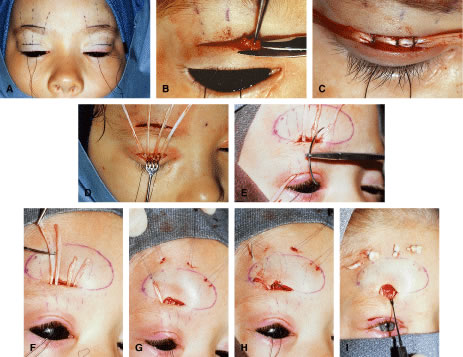
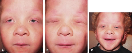
Another type of congenital ptosis that is inherited is the blepharocanthal syndrome. The inheritance is autosomal dominant and comprises about 5% of patients with congenital ptosis3 (Fig. 4).

Fig. 4. A patient with blepharocanthal syndrome. Note the presence of ptosis, horizontal shortening of the palpebral fissures, and epicanthus inversus.

The blepharocanthal syndrome includes two or more of the following features2,4:

  • Ptosis with poor levator muscle function and absence of a definitive eyelid
  • Epicanthus inversus—a medial canthal tendon fold most prominent in the lower eyelid and extending from the lower to the upper eyelid
  • Telecanthus—a widened intercanthal distance
  • Blepharophimosis—a redundancy and lengthening of the upper eyelid margin
  • Lower eyelid ectropion
  • Narrowing of the palpebral fissures

Associated findings with blepharocanthal syndrome include hypertelorism, poorly developed nasal bridge, hypoplasia of the superior orbital rims and the tarsal plate, and lop ears (upper third ear deformity of the helix causing overhanging of auricular cartilage).2 In addition, because of hypogonadism and specific hormonal deficiencies, females with this condition should be closely monitored for menstrual irregularity and infertility.9 The abnormalities of the female reproductive system, however, are hormonally related and are not sex linked. In cases of ptosis associated with coexisting congenital eyelid anomalies such as in blepharophimosis, eyelid reconstruction may be staged and ptosis repair is usually delayed until the other eyelid abnormalities are corrected. Ptosis correction at the same time of eyelid reconstruction may be necessary in the setting in which ptosis-induced amblyopia is threatening visual development of the patient.

ACQUIRED PTOSIS

Acquired ptosis is seen as a change from the patient's baseline. The incidence of acquired ptosis has increased in the recent years with heightened awareness among the patient population of diagnosis and treatment. Upper eyelid anatomy is crucial in understanding the pathogenesis of acquired ptosis. (See Chapter 72.) The levator complex of the upper eyelid includes the LPS muscle and the levator aponeurosis. Muller's muscle is sympathetically innervated muscle, which also plays a role in eyelid retraction. Ptosis of the upper eyelid can be due to a weakness of the elevating power of the eyelid retractors as a result of neuromyogenic, traumatic, or involutional insult. In addition, the levator aponeurosis is comprised of a group of collagenous sheets, or lamellae, which have attachments to the posterior orbicular fascia and the anterior tarsal surface. Among these lamellae, there may at one time be a mixture of attenuation, dehiscence, and disinsertion from their attachment sites, resulting in a ptotic eyelid.10 The various types of acquired ptosis (e.g., aponeurotic, neurogenic, myogenic, mechanical, and traumatic) are discussed.

Acquired Aponeurotic Ptosis

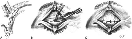
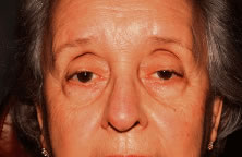
Acquired ptosis is most commonly aponeurotic in etiology. Involutional changes of aging, repetitive traction on the eyelid, or both result in a thinning of the levator aponeurosis or separation from its insertion site on the anterior tarsal surface. Though aponeurotic ptosis most often occurs in older adults, it can be seen in at any age. Younger patients may develop aponeurotic ptosis as a result of trauma, orbital or eyelid swelling, excessive eyelid rubbing, contact lens wear, pregnancy, blepharochalasis, prior ocular surgery, or chronic ocular inflammation.11 Cases of aponeurotic ptosis have even been seen in young children and infants. In these patients, the eyelid crease is usually elevated because of disinsertion from the tarsal plate with superior retraction of the levator aponeurosis (Fig. 5). The attached dermis and orbicularis move cephalad with the migration of the aponeurosis and lead to the formation or appearance of a superior sulcus deformity, a high eyelid crease, and fold and thinning of the upper eyelid.

Fig. 5. Patient with levator aponeurosis dehiscence. Note the raised eyelid creases bilaterally.

At times, the history may be elusive in which case a cause may not be determined. Patients with aponeurotic ptosis often complain of eyelid and brow fatigue. The ptosis is usually worse later in the day, and this finding should not be confused with those of myopathic ptosis [i.e., myasthenia gravis (MG)]. Patients often notice a superior visual field as a result of the lower upper eyelid position. Older patients often complain of difficulty reading because the ptosis is worse on downgaze. This can be attributed to the involutional changes of the eyelids associated with aging, including loss of tone of the levator muscle and Muller's muscle and atrophy of the orbital fat, leading to a mild degree of enophthalmos.3

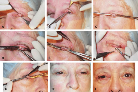
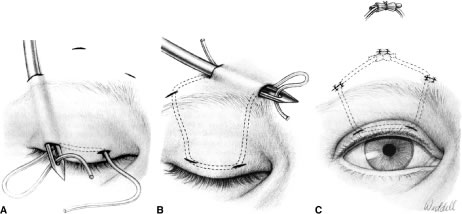
The LPS muscle function in aponeurotic ptosis is either normal or slightly reduced, as assessed by measuring in millimeters the amount of eyelid excursion from downgaze to upgaze.6 These measurements are helpful in distinguishing congenital from acquired ptosis (Table 1). LPS function in normal individuals measures 16 to 20 mm. This is reduced to 12 to 16 mm in acquired aponeurotic ptosis. Horizontal laxity in the upper eyelid is also associated with aponeurotic ptosis and can be attributed to weakness in the lateral canthal tendon or the supporting eyelid structures of the upper eyelid tarsal sling. Specifically, dehiscence of the medial aspect of Whitnall's ligament can occur, resulting in loss of medial support of the eyelid. The tarsal plate shifts laterally causing a more ptotic eyelid nasally and an abnormal eyelid contour. The lateral shift is usually associated with aging and should be recognized preoperatively, because this phenomenon may alter the surgical approach for ptosis repair. Lash ptosis can develop as a result of secondary ptosis from the hooding effect of redundant skin and soft tissue of the upper eyelid resting on the eyelashes. Lash ptosis may also be caused by aging changes in the upper eyelid, including decreased tone, loss of structural support from increasing laxity of the upper eyelid, and atrophy of the tarsus. Surgical repair in these patients with horizontal laxity is difficult and demands balance between horizontal tension of the eyelid and vertical pull of the levator aponeurosis. Correction of aponeurotic ptosis is usually by an external approach levator resection and is discussed later in this chapter.


Table 1. Comparison of Congenital Myogenic Ptosis to Acquired Aponeurotic Ptosis


 Congenital Myogenic PtosisAcquired Aponeurotic Ptosis
Palpebral fissure heightMild to severe ptosisMild to severe ptosis
Upper eyelid creaseWeak/absent crease in normal positionHigher than normal crease
Levator function on downgazeReduced eyelid lagNear normal eyelid droop

From American Academy of Ophthalmology. Orbit, Eyelids and Lacrimal System; BCSC Section 7. San Francisco: American Academy of Ophthalmology, 2000-2001:Fig XII-1.


Acquired Neurogenic Ptosis

Acquired neurogenic ptosis is usually caused by a third cranial nerve (oculomotor nerve) palsy or by disruption of sympathetic innervation to Muller's muscle. There are two classifications of third nerve palsies: vasculopathic, from diabetes, hypertension, and arteriosclerotic disease, and compressive, from a neoplasm or an aneurysm. Vasculopathic processes rarely affect the pupillary fibers of the third nerve and generally resolve spontaneously within 3 months. If the palsy has not resolved within 6 months or if the pupil is involved in the patient's initial presentation, further workup to rule out neoplasm or aneurysm must be performed.2 An oculomotor (CN-3) palsy rarely causes an isolated LPS muscle deficit, usually involving rectus muscles. Pupillary fibers are more likely to be involved with a compressive neuropathy. The classic presentation of a compressive third nerve palsy is unilateral, rapidly evolving ptosis, exotropia, hypotropia, and mydriasis. When the pupil is spared, one should suspect a vasculopathic cause.

A patient with an oculomotor nerve palsy may develop synkinetic ptosis as a result of aberrant regeneration and misdirection of third cranial nerve fibers. Although more often congenital, this condition may also develop during recovery from an oculomotor nerve paralysis. Synkinesis can cause elevation of the eyelid with the use of the extraocular muscles, as well as paradoxical pupillary dilatation or constriction.8 The ptotic eyelid may rise as the inferior rectus, medial rectus, or superior rectus muscle contracts. If aberrant regeneration of the nerve is observed in the examination of a patient without a known history of oculomotor nerve palsy, a workup is necessary to rule out a compressive neoplasm of the cavernous sinus or an aneurysm.

Horner's syndrome, an acquired neurogenic ptosis, is caused by a disruption of sympathetic innervation to eyelid retractor muscles, to pupillary dilator muscles, and to the sweat glands in the skin on the affected side. Horner's syndrome classically presents with unilateral ptosis, miosis, and anhidrosis. Paralysis of Muller's muscle in the upper eyelid results in mild ptosis measuring approximately 1 to 2 mm. The lower eyelid may be elevated approximately 1 mm because of paresis of the inferior tarsal muscle. This further narrows the palpebral fissure and may mislead the clinician to think that the patient manifests enophthalmos. Ptosis of the lower eyelid has been called reverse ptosis. The pupil on the involved side is constricted and dilates poorly in the dark as compared with the pupil of the unaffected eye. There also may be lack of sweating on the affected side of the face and neck, as well as depigmentation of the iris.8

Various lesions located along the course of the sympathetic chain cause Horner's syndrome. Tumors, inflammatory processes, aneurysms, and injuries are the most frequent causes of Horner's syndrome.3 The anatomic location of the lesion determines if it is situated along a first-order, second-order, or third-order neuron. The central or first-order neuron courses from the hypothalamus to the cervical portion of the spinal cord.12 Lesions in the brain, spinal cord, or brain stem may result in a first-order insult. The second-order, or preganglionic, neuron passes from the cervical portion of the spinal cord along the sympathetic chain through the upper thorax and neck to synapse in the superior cervical ganglion. Apical lung tumors (Pancoast tumors), metastasis, thoracic surgery, thoracic aortic aneurysms, or trauma to the brachial plexus are examples of second-order lesions that can cause disruption of the sympathetic chain. The third-order, or postganglionic, neuron travels with the sympathetic fibers along the internal carotid artery to the orbit. These lesions are most often of a vascular etiology, such as a carotid artery dissection. Third-order lesions also result from head and neck trauma, tumors, infections, and lesions of the cavernous sinus.13 Localization of the lesion along the sympathetic chain is essential in proper diagnosis and treatment of Horner's syndrome.

Topical pharmacologic testing is used to clinically localize the lesion causing the Horner's syndrome as first, second, or third order. Topical hydroxyamphetamine 1%, or Paredrine, is used to differentiate first and second order, or preganglionic lesions from a third order or postganglionic lesion.14 In a normal eye and in a preganglionic Horner's syndrome, the postganglionic neuron is intact and norepinephrine is present. The action of topical hydroxyamphetamine releases the norepinephrine from the presynaptic terminal into the myoneural junction of the pupillary dilator muscle fibers resulting in dilation of the pupil. In addition, hydroxyamphetamine acts similarly to stimulate Muller's muscle, which will raise the ptotic upper eyelid. A poor pupillary response or no improvement of ptosis usually indicates a third-order lesion, in which the postsynaptic terminal has degenerated and no norepinephrine is present.

Topical 4% to 10% cocaine is another pharmacologic test that localizes third-neuron Horner's syndrome by assessing pupillary response. Cocaine acts to block the reuptake of norepinephrine at the neuromuscular junction, which stimulates the pupillary dilator muscle. This blockade results in pupillary dilation in the normal eye or in a first- and second-order lesion where norepinephrine is present. Cocaine similarly activates Muller's muscle, consequently elevating the ptotic eyelid 1 to 2 mm. In a third-order neuron lesion, there is an absence of norepinephrine in the synaptic cleft and cocaine will have no effect. The typical response after the application of cocaine is a lack of pupillary dilation and failure of eyelid ptosis to resolve. However, addition of epinephrine or hydroxyamphetamine topically can dilate the pupil and elevate the ptotic eyelid in a third-order lesion.

Horner's syndrome is often associated with other neurologic signs, especially when caused by tumor, aneurysm, or inflammation. The etiology must be identified before initiating proper treatment. Surgery to correct eyelid ptosis is only considered once a thorough evaluation, proper diagnosis, and treatment has been implemented.

Myasthenia gravis (MG) is a B-cell mediated autoimmune disease and is a neurogenic acquired ptosis. MG is characterized by weakness of the affected muscles. Patients often initially present to an ophthalmologist with complaints of diplopia or ptosis or both, because the extraocular muscles, particularly the LPS, are often the first to be affected in new onset MG.3 In MG, antibodies to acetylcholine (ACH) receptors cause a functional blockade of the ACH receptors at the neuromuscular junction. The histopathology in MG also demonstrates a flattening and marked loss of the folds present in the postsynaptic membrane. In the normal membrane, these folds increase the surface area available for ACH-receptor binding.15 The reduced number and availability of receptors causes a decrease in the total binding of ACH in the synapses, reducing neural transmission and resulting in a weakness of the levator muscle and ptosis. The unbound ACH in the synaptic cleft is then degraded. Ptosis may be exacerbated by prolonged upgaze, which causes a saturation of the few remaining ACH receptors.

MG often initially presents as a neurogenic ptosis and must always be considered in the differential diagnosis of a patient presenting with an acquired ptosis. The eyelid ptosis often begins unilaterally but may variably affect the other eyelid. Other accompanying findings include dysphagia; hoarseness; dysarthria; dyspnea; weakness of the jaw, neck trunk, and arms; and fatigue with walking.12 Dysphagia and dyspnea can be life threatening. Patients often complain of minimal symptoms in the morning with progressive worsening of symptoms resulting from fatigue toward evening.3 Symptoms are exacerbated after exertion and improve with rest. Patients diagnosed with MG sometimes have associated autoimmune diseases. Five percent of patients with MG are also diagnosed with Grave's disease. Other patients with MG have been found to have hyperactivity of the thymus gland. Thymus hyperplasia is thought to play a role in the immune mechanism in MG.

Ocular myasthenia is myasthenia isolated to the periocular muscles, including the LPS, the orbicularis oculi, and the rectus muscles. Their symptoms are usually localized to include ptosis, diplopia, and fatigue of the periocular muscles. In this type of myasthenia, the ACH-receptor antibody normally found in myasthenic patients is often absent. This difference is theorized to represent the antigenic differences in the synaptic terminals of ocular muscles and other skeletal muscles. The extraocular muscles differ from other skeletal muscles in that they have multiple end plates for each muscle fiber and a myoneural junction that occurs within (hypolemmal) instead of on the muscle fiber. Ocular myasthenia may occasionally be seen with other autoimmune diseases, such as Hashimoto's disease, hyperthyroidism, multiple sclerosis, systemic lupus erythematosus, polymyositis, or hemolytic anemia.16

The classic diagnostic test for myasthenic ptosis is the Tensilon (edrophonium chloride) test. Tensilon is an antiacetylcholinesterase drug that is administered intravenously. Pharmacologically, it acts to overcome the antibody receptor blockade in MG and inhibits the breakdown of ACH. A positive test in a patient with myasthenia resolves the weakness of the affected muscle (e.g., LPS or rectus muscle) and results in resolution of symptoms (e.g., ptosis or diplopia). Patients must be forewarned of the potential yet short-acting adverse effects of the Tensilon test, which can be dramatic including fasciculations, nausea, lacrimation, salivation, flushing, abdominal cramping, bradycardia, and even respiratory arrest. Atropine sulfate (0.4 to 0.6 mg) is a muscarinic antagonist and must be available to administer in case of the adverse reactions of Tensilon. Atropine sulfate blocks the action of ACH by binding to postsynaptic muscarinic receptors. Some physicians treat prophylactically with atropine by injecting the patient with 0.6-mg intramuscular (IM) before administering Tensilon.

The Tensilon test can be administered in an office setting, provided that the clinician and ancillary help are comfortable and equipped to handle potential adverse reactions. Safer environments include the operating room (OR), ambulatory surgery center, or emergency room where resuscitation is easily performed. Whenever a Tensilon test is planned, the patient should be semireclined, and resuscitation equipment should be readily available. The patient's vital signs, including blood pressure and pulse, should be monitored throughout the procedure. Tensilon is initially administered as a test dose, 2 mg (0.2 cc) and is slowly injected intravenously through a butterfly needle. The patient is observed for 60 seconds for elevation of the ptotic eyelid or improvement of extraocular movements. If there is improvement of symptoms after 1 minute, it is a positive test, and, thus, the test is terminated. If no response occurs, the remaining 8 mg is slowly administered and the patient is observed for 1 to 5 minutes. This dose can be divided into two 4-mg doses, which seems to cause fewer adverse effects.12

Prostigmin (neostigmine methylsulfate) is another anticholinesterase drug that can be used as a pharmacologic diagnostic test for MG. Although the adverse reactions are similar to Tensilon, the more frequent side effects are less severe (e.g., salivation, fasciculations, gastrointestinal discomfort). This is useful in children and adults who may require a longer observation period than with the Tensilon test. Neostigmine is administered intramuscularly and is concurrently injected with atropine. The dosage formula is: (weight (kg) × adult dose) divided by 70. The adult dose typically measures 1.5 mg with 0.4 mg atropine. Resolution of ptosis or diplopia in a positive test is expected to occur over a 30- to 45-minute time period.

Other clinical tests for MG include the rest test and the ice pack test. The rest, or sleep test, is a clinical diagnostic test.17 This involves taking precise measurements of the patient's degree of ptosis on presentation, then allowing the patient to rest quietly with eyelids closed for up to 30 minutes. Immediately after the patient is aroused, the ptosis measurements are repeated. Improvement of the ptosis with rest is highly suggestive of MG, because the receptors not occupied by antibodies are free to bind newly released ACH.12 The ice pack test is a safe diagnostic clinical test for MG that is easily performed in the office. This entails placing an ice pack over the affected ptotic eye for 2 minutes and then reevaluating the patient for resolution of ptosis. Myasthenic weakness is known to improve with lowering of the temperature of skeletal muscle. Cold is thought to affect the neuromuscular junction by decreasing cholinesterase activity and by promoting efficiency of ACH by eliciting depolarizations at the end plate.18 Kubis and colleagues17 demonstrated that in 90% of patients with MG ptosis, there is an improvement of their ptosis with the ice test. Recent studies concur that the ice test is more effective than the rest test alone in diagnosing the ptosis of MG.19

Another diagnostic marker for MG is a serum assay for antiacetylcholine receptor antibodies (ACHR Ab test). Binding antibodies are usually measured, because they are detected in approximately 90% of patients with generalized MG and 70% of patients with ocular MG.12 There have, however, been false-positive reports with this test in cases of thymoma in patients without myasthenia. These include patients with Lambert-Eaton myasthenic syndrome and small cell lung cancer, as well as one third of the population older than 70 years.20 If the patient has not yet been diagnosed with MG or if the ophthalmologist is the first physician to suspect MG, the patient should be further evaluated by his or her internist for systemic involvement. Referral to a neurophthalmologist is usually appropriate for a patient suspected to have MG. Serologic screening tests for thyroid dysfunction and autoimmune diseases should be performed because there is a high association of MG with thyroidopathy and other autoimmune diseases.12 Radiographic studies looking for thymomas should also be performed on MG patients, because they are visible on computed tomography (CT) scan in 10% of patients with MG.

Patients with MG should undergo appropriate treatment with anticholinesterase medication (e.g., Mestinon and Prostigmin), corticosteroids, or other immunosuppressant agents under supervision of their internist or neurologist. In patients with enlargement of the thymus or a thymoma, a thymectomy is usually indicated. Some patients, especially younger individuals have gone into remission of the disease following thymectomy.3 The systemic manifestations of MG can be life threatening and must be managed before considering surgical treatment of blepharoptosis. Patients often demonstrate poor eyelid closure resulting from weakness of the involved orbicularis muscles, which may lead to problems at surgery. Patients must also be warned that the ptosis may fluctuate and correction of ptosis is difficult.

Acquired Myogenic Ptosis

Myogenic ptosis is a rare type of acquired of blepharoptosis and can be found in localized or diffuse muscular disease, such as the muscular dystrophies. The muscular dystrophies are distinguishable by their mode of inheritance and clinical features. Blepharoptosis is a clinical sign of certain muscular dystrophies, including myotonic dystrophy, chronic progressive ophthalmoplegia (CPEO), and oculopharyngeal muscular dystrophy.

Myotonic dystrophy is an autosomal dominant (chromosome 19) multisystem disorder with an onset usually in the second decade of life. Histologically, myotonic dystrophy is characterized by degenerative changes of skeletal muscle including disruption of myofilaments and sarcoplasmic reticulum, focal accumulation of mitochondria, and eventually atrophy and fibrosis of muscle.21 The distal musculature is usually the first to be affected. Myotonia is often the initial symptom, which results from delayed relaxation of skeletal muscle after contraction. Symptoms are exacerbated with cold, excitement, and fatigue. Patients often develop a characteristic myopathic facies, presenting with blepharoptosis, frontal balding, and wasting of the temporalis masseter muscle. Ocular findings include ptosis, blepharospasm, ophthalmoplegia (similar to CPEO), pigmentary retinopathy, “Christmas tree” cataracts, and miotic pupils.2 Associated systemic findings include low intelligence, insulin resistance, cardiac conduction abnormalities, and testicular and uterine atrophy. Electromyogram (EMG) is a diagnostic test demonstrating myotonic discharges.

Chronic progressive ophthalmoplegia (CPEO) is another muscular dystrophy with several variants, all of which result from mutations in mitochondrial DNA. Most cases are autosomal dominant and maternally inherited. Inherited CPEO has been linked to specific families of Italian and Finnish descent. One form of CPEO, however, has been found to occur as a sporadic mutation. On muscle biopsy, histopathologic examination reveals ragged red fibers, which represent accumulations of abnormal mitochondria within the myofibers.21 EMG findings can also be abnormal. Because the disorder is mitochondrial in origin, it tends to affect tissues with high oxidative needs: cardiac muscle, skeletal muscle, and the central nervous system.22

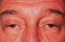
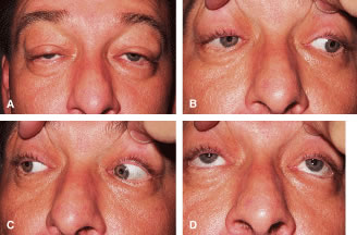
CPEO usually presents in the second to fourth decade often initially with bilateral progressive ptosis (Fig. 6A). In addition to LPS muscle involvement, patients also develop a weakness of other periocular muscles including the rectus muscles and the orbicularis oculi muscles (see Fig. 6B to D). Despite limited ductions from weakness of rectus muscles, binocular vision is usually preserved. Various types of CPEO are associated with systemic deficits such as retinal pigment degeneration,hyperacusia, dysphagia, ataxia, and proximal muscle weakness. The retinal pigment changes differ from retinitis pigmentosa in that they are mainly at the posterior pole with a mottled appearance of the pigment epithelium. Visual changes are mild if any.21 There is also a pediatric variant of CPEO, known as Kearns-Sayre syndrome, which includes cardiac conduction abnormalities (a significant surgical risk), retinal pigment degeneration, and bilateral ptosis.22

Fig. 6. A. Patient with chronic progressive ophthalmoplegia (CPEO) and ptosis. B, C, and D. Extraocular movements are limited in fields of gaze.

Oculopharyngeal muscular dystrophy is a disorder characterized by progressive atrophy and weakness of the hypopharyngeal and ocular muscles. The disorder results from a mutation of the PAB2 protein that binds to messenger ribonucleic acid (mRNA).21 It usually presents in the third to fourth decade with dysphagia or regurgitation of food resulting from lack of coordination of the pharyngeal muscles. The dysphagia can progress to intolerance of liquids and the patient is at risk from aspiration pneumonia and death. Surgical intervention such as cricopharyngostomy is sometimes necessary. The dysphagia usually precedes the ptosis by several years. The ptosis observed is bilateral, but usually asymmetric, and the extraocular muscles may also be involved.

Other rare causes of myogenic ptosis include Guillain-Barré syndrome or iatrogenic botulism. Injection of botulinum toxin for the treatment of rhytids or blepharospasm may result in a temporary ptosis usually lasting a few weeks.

Correction of acquired myogenic ptosis can be difficult, because most patients with myogenic ptosis have a progressive disease making the surgical outcome unpredictable. These patients are at increased risk for complications following ptosis surgery because of poor eye-protective mechanisms (EPMs) including inadequate blink, poor Bell's phenomenon, and consequently exposure keratitis. The surgical approach is most often based on the amount of levator muscle function present, and the patient is instructed on the potential variability following surgical correction. This is discussed in further detail later in the chapter.

Mechanical Acquired Ptosis

Mechanical acquired ptosis is seen with any abnormality that results in pulling down of the upper eyelid resulting in ptosis despite normal muscular function. This could include dermatochalasis, tumors of the lid or orbit, chalazion, postsurgical or traumatic edema, a lost contact lens in the superior fornix, or a scar.23 Eliminating the mass effect of the lesion usually alleviates mechanical ptosis.

Traumatic Acquired Ptosis

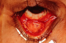
Traumatic acquired ptosis of the upper eyelid can result from blunt or sharp injury usually to the LPS muscle or the levator aponeurosis either by trauma or surgery. If the insult is traumatic, the underlying etiology may be myogenic, neurogenic, mechanical, cicatricial, aponeurotic, or even a combination of these. Exposure of orbital fat in the upper eyelid after injury typically demonstrates violation of the orbital septum. In this case, the upper eyelid should be explored for possible injury to the levator muscle and repaired at this time. Posttraumatic or postsurgical ptosis often results following orbital, ocular, and neurosurgical procedures. It is important to initially observe these patients without surgical intervention, because acute factors such as hemorrhage, neuropraxia, and edema are temporary. Spontaneous resolution or improvement often occurs in these patients over the course of 6 or more months. Surgical repair of patients with traumatic ptosis is highly variable based on the etiology of the injury and existing elevating power of the upper eyelid.

Pseudoptosis

Pseudoptosis should be differentiated from true blepharoptosis. Various conditions may make an upper eyelid appear low, including a hypertropia on the contralateral side, enophthalmos, microphthalmos, anophthalmos, blepharochalasis, phthisis bulbi, dermatochalasis, or a superior sulcus defect secondary to trauma or cicatrix. In addition, widening of the palpebral fissures on the contralateral side can give the appearance of pseudoptosis and may be due to eyelid retraction from Grave's disease, axial proptosis, congenital eyelid retraction, or high myopia.

Pseudoptosis is often manifested in aging patients as a result of redundancy and laxity of the skin and tissue of the upper eyelid and brow. In evaluating eyelid ptosis, it is important to distinguish the independently functioning units of the eyebrow, redundant eyelid skin, and the elevating power of the eyelid. In brow ptosis, recruitment of the frontalis muscle is needed to elevate the eyebrow and eyelid. Relaxation or fatigue of the forehead muscles that can be elicited during examination will manifest the brow ptosis and may induce mechanical ptosis of the eyelid. Dermatochalasis, a commonly seen condition, in which excess skin overhangs the eyelid margin, can also cause a mechanical pseudoptosis or a secondary ptosis. This “hooding” effect can be corrected during the examination by manual elevation of the redundant eyelid skin, allowing visualization of the true palpebral aperture. In brow ptosis and dermatochalasis, the levator muscle function is usually intact. Surgery to correct brow ptosis requires a brow lift, and correction of dermatochalasis requires blepharoplasty not ptosis surgery. In aging patients especially, involutional ptosis can occur in conjunction with brow ptosis or dermatochalasis, in which case ptosis surgery is indicated. Each factor plays an important role in the symptomatic ptosis patient and should be individually examined and recognized.

Back to Top
EXAMINATION OF THE PTOSIS PATIENT

HISTORY

Obtaining a meticulous history in a ptosis patient is crucial in determining the type of ptosis (i.e., congenital or acquired) and the planned correction. Determining the age of onset of ptosis is essential. In a child, prenatal and perinatal history may be helpful in determining whether the congenital ptosis has a developmental or a traumatic etiology (i.e., congenital Horner's syndrome or birth trauma). Often, children with congenital ptosis have lagophthalmos that clinically manifests in downgaze. The parents should be questioned as to whether the child sleeps with the eyelid(s) open. Other important features of ptosis include unilateral or bilateral involvement, progression or variability of the ptosis, any exacerbating factors (e.g., fatigue, prolonged upward gaze, chewing food), associated symptoms (diplopia, generalized muscular weakness), and a family history of ptosis.

In acquired ptosis, the clinician should be aware of prior eyelid, orbital, or head trauma; previous surgeries on the eyelid; and other preexisting ocular diseases. A history of diplopia, diurnal variability of either the eyelid level or of the diplopia, generalized fatigability, or family history of adult-onset ptosis may lead the clinician to suspect a myogenic etiology. In a patient with progressive or variable ptosis, a personal or family history of muscular disorders including myotonic dystrophy, chronic progressive external ophthalmoplegia, or any of the related myopathies should be obtained. New onset or progressive ptosis or diplopia suggests a neurogenic etiology, and the patient should be questioned about any recent trauma, history of vasculopathic disease or diabetes, or any associated neurologic deficits. Older adult patients may complain of difficulty reading and demonstrate increased ptosis in downgaze, which may be a good indication of acquired involutional ptosis.

It is important to obtain a history of asthenopic symptoms or of irritative ocular problems that may suggest a tear deficiency or exposure keratopathy. Blink adequacy and frequency, precorneal tear film, and corneal sensation should be known before considering correction of eyelid ptosis. Surgery for ptosis in patients with poor EPMs—including a history of dry eyes, seventh cranial nerve paralysis, or significant periocular muscle abnormalities such as severe thyroid ophthalmopathy, double elevator palsy, or CPEO—should be approached with caution.

Patients with ptosis should be questioned about signs or symptoms of associated systemic illnesses, including thyroid disease, MG, cranial nerve palsies, aneurysms, tumors, or Horner's syndrome. Other medical problems, including hypertension, bleeding dyscrasias, cardiorespiratory illnesses or diabetes should be known. The clinician should be aware of the patient's use of certain medications, such as aspirin, cardiac agents, psychoactive drugs, ophthalmic medications, and cholinesterase inhibitors for strabismus or glaucoma.

EXAMINATION

Initial observation of the patient can yield important data, such as unilateral or bilateral ptosis, use of the frontalis muscles by the patient to elevate the eyelids, chin lift, head tilt, lagophthalmos, and symmetry of the eyelids at primary gaze. Patients with severe ptosis instinctively assume a “chin up” head position to overcome their visual obstruction.10 Noting the position of the eyelid crease can help to differentiate aponeurotic (a higher crease) from congenital ptosis (an absent or faint crease). Careful observation may also reveal a mechanical or neurogenic cause of the eyelid ptosis. Certain elements of the examination of the ptosis patient are imperative, including measurements of eyelid position and the elevating power of the upper eyelid, as well as assessment of EPMs, visual acuity, visual field testing, and an ocular examination.

EPMs is a useful term pertaining to defense of the ocular surface (Table 2). These include tear production, blink frequency and adequacy, eyelid movement and position, ocular motility, Bell's phenomenon, corneal sensation, and the ocular surface. EPMs are assessed by clinical examination including observation, slit lamp examination, Shirmer's testing, fluorescein and rose Bengal staining, and tear breakup time. Elevating the eyelid in ptosis surgery in a patient with poor EPMs can exacerbate exposure keratopathy and should be recognized and treated preoperatively. Certain conditions are associated with patients with poor EPMs and are discussed later in the chapter.


Table 2. Eye-Protective Mechanisms

  1. Tear production
  2. Blink frequency and adequacy
  3. Eyelid movement and position
  4. Corneal sensation
  5. Ocular motility
  6. Bell's phenomenon

Adapted from McCord CD, Shore JW. Silicone rod frontalis suspension. Adv Ophthal Plast Reconstr Surg 1982;1:213.


Visual acuity and a manifest refraction should be obtained in both eyes. In children, retinoscopy should be performed, especially in congenital unilateral ptosis in which amblyopia is suspected.24 A thorough pupillary examination and assessment of extraocular muscle motility are important in the examination of the ptosis patient. Associated pupillary or motility dysfunction may indicate a neurogenic, myogenic, or traumatic etiology of the ptosis (e.g., MG, CPEO, Horner's syndrome, or aberrant regeneration of the oculomotor nerve after a third nerve palsy). A cover-uncover test should also be performed to elicit any strabismus.24 A slit lamp examination of the ocular surface and anterior segment, measurement of the intraocular pressure, and fundus examination are necessary components to a complete ocular examination. The fundus examination can lead to clues of CPEO or Kearns-Sayre disease, if retinal pigment changes are seen in a ptosis patient.

Visual fields should be obtained in ptosis patients. The amount of superior and peripheral field restriction is important is quantifying the severity of ptosis. The examiner should instruct the patient not to use the frontalis muscle to elevate the brow and eyelids during the test. Taping the upper eyelids and brows in patients with dermatochalasis may be necessary to alleviate the hooding effect of redundant upper eyelid tissue causing pseudoptosis. Comparing the visual field of the patient with the eyelid taped to without taping can demonstrate the amount of superior visual field obstruction from ptosis or pseudoptosis.

Clinicians have formulated essential measurements of eyelid position and elevating power of the upper eyelid in ptosis patients to aid in the diagnosis of the type of ptosis and in selecting the proper treatment modality. In both congenital and acquired ptosis patients, it is important to realize that the etiology of the poor elevating power of the eyelid can be multifactorial, including fatty or degenerative changes of the muscle, fibrosis, tethering, or insult to the nerves. Consequently, careful measurements may not always be consistent in determining the elevating power of the upper eyelid.

There are four standard clinical measurements in the ptosis examination:

  1. Palpebral fissure height: The palpebral fissure height is the widest distance measured between the upper and lower eyelid margins and reflects the amount of ptosis exhibited in primary gaze. Normally, this width is approximately 9.5 mm. This measurement is dependent on the position of the upper and lower eyelid.
  2. Margin reflex distance (MRD): The MRD is the most effective measurement in gauging upper eyelid ptosis. While the patient fixates on a distant target in primary gaze, the examiner holds a muscle light directly in front of the patient aiming it between the eyes. The distance from the pupillary light reflex to the upper eyelid margin of each eye is measured. This distance is normally 4 mm and is independent of lower eyelid position.2
  3. Upper eyelid crease and fold: The distance of the upper eyelid crease from the eyelid margin is measured when the patient looks down. It is approximately 8 mm in men and 9 to 10 mm in women. In congenital ptosis, the eyelid crease is often absent. In cases of levator aponeurosis dehiscence, the eyelid crease can be much higher. In the Asian eyelid, the eyelid crease and fold are variable. The eyelid fold in the Asian eyelid can be double, single, high, low, or absent. More often, the eyelid crease is often lower or absent in the Asian eyelid as compared with the Caucasian eyelid. (See Chapter 72.)
  4. Levator excursion: This is a measurement of levator muscle pulling power, the distance the levator elevates the upper lid from full downgaze to full upgaze. The levator excursion normally measures to be approximately 16 to 20 mm. In taking this measurement, it is important to ensure that the forehead and brows are not being recruited to elevate the eyelids. The examiner can manually hold the brows in a relaxed position to avoid use of the frontalis muscle. Measurement of the levator excursion has been used to differentiate acquired from congenital ptosis, although sometimes the etiology is not clearly defined. Acquired ptosis patients are commonly older adult patients with involutional ptosis. These patients demonstrate good levator muscle function, but the aponeurosis has simply become detached from the tarsus. A congenital ptosis patient usually has fair to poor levator muscle function. Restriction of both elevation and relaxation in these patients usually results in minimal levator excursion.

Other clinical measurements that are essential when examining ptosis patients are horizontal laxity and eyelid contour. Horizontal laxity of the eyelid is often seen in aging patients from attenuation and stretching of the eyelid structures of the tarsal sling and atrophic changes of the tarsus.25 Horizontal laxity can also affect the eyelid position as seen in involutional acquired ptosis. In aging patients, there is often a tarsal shift, which lateralizes the tarsus causing a medial ptosis and affecting eyelid contour. Eyelid notching from injury or prior surgery may also affect eyelid contour. These findings are important to acknowledge during the examination for proper surgical planning.

Clinicians have used a quantitative means to assess the degree of ptosis function. Table 3 compares the severity of eyelid ptosis with the amount of ptosis and the LPS excursion. Mild ptosis (1 to 2 mm) correlates with good LPS muscle function of at least 8 mm (15 mm is normal). In moderate ptosis of approximately 3 mm, LPS muscle function is fair, measuring 5 to 7 mm. A severe ptosis of greater than 4 mm indicates poor LPS muscle function of less than 4 mm. In congential ptosis, these measurements are not always an accurate gauge for measuring the degree of LPS muscle function primarily because of the fibrofatty degenerative changes of the levator muscle.


Table 3. Relation of Degree of Ptosis to Amount of Levator Palpebrae Superioris Function


Degree of PtosisAmount of PtosisLevator FunctionAmount of Levator Function
Mild1–2 mmGood8 mm or greater
Moderate3 mmFair5–7 mm
Severe4 mm or greaterPoor4 mm or less

Adapted from Iliff WJ. Ptosis surgery. In Tasman W, Jaeger E (eds): Duane's Foundations of Clinical Ophthalmology, vol 5. Philadelphia: Lippincott-Raven, 1992:6.


Other tests have been used as adjuncts to evaluate ptosis patients. One such test is called Iliff's sign. This involves everting the eyelid and asking the patient to look up. Failure of the eyelid return to its normal position when the patient looks up indicates poor LPS muscle function.6 Hering's law may be beneficial to assess in a ptosis patient in a preoperative setting. As the examiner manually elevates the ptotic eyelid, the normal eyelid may droop.2 This is known as Hering's law of equal innervation. Hering's law pertains to paired yolk muscles, such as the LPS, which is bilaterally innervated by the oculomotor nerve. The oculomotor nerve derives from the subnucleus of the oculomotor complex, which is a midline structure in the midbrain. A lesion to the oculomotor subnucleus is thought to result in bilateral ptosis. Similarly, elevation of an eyelid both manually, or after ptosis surgery, diminishes the innervation of the eyelids bilaterally and, thus, causes a contralateral eyelid droop.10 Patients with unilateral ptosis should be well informed of this phenomenon before surgery, and they may consequently require correction of the eyelid droop in the contralateral eyelid.

Pharmacologic testing to evaluate certain types of ptosis has also been used preoperatively. For example, topical 1/8% phenylephrine drops can be used to assess Muller's muscle response in patients with mild ptosis with suspected sympathetic nerve involvement. Topical 4% to 10% cocaine has been used to evaluate ptosis in patients with Horner's syndrome with a suspected third-neuron lesion. The Tensilon test is used to evaluate patients for MG. These and other pharmacologic tests used to evaluate particular ptosis patients are important in proper diagnosis and management and are described in further detail in this chapter.

Back to Top
PTOSIS SURGERY

PREPARATION FOR SURGERY

When considering surgery, a complete medical and social history of the patient should be obtained, including other medical problems, medication, allergies, or any bleeding diatheses. Blood thinning agents such as aspirin and Coumadin should be discontinued the week before surgery, if possible. A physical examination, especially of the cardiovascular system, should be performed before surgery. In an older adult patient or one with a cardiac history, an electrocardiogram should be performed. If the patient has a significant medical history, the patient should be evaluated by his or her internist before surgery. All ptosis patients should have photographic documentation both preoperatively and postoperatively.

The patient should sign an informed consent after the risks and benefits of surgery are discussed. Basic surgical risks of periocular surgery also pertain to the ptosis patient including hemorrhage, infection, wound dehiscence, scarring of the eyelid incisions, reoperation, and blindness. Certain postoperative problems occur more commonly in ptosis surgery, and the patient should be well informed and have realistic expectations before the surgery. These outcomes include overcorrection or undercorrection of ptosis with the possibility of readjustment, asymmetry in the eyelid crease or position, lagophthalmos, poor eyelid contour, ectropion, entropion, and Hering's law resulting in ptosis of the unaffected eyelid. Exposure keratopathy as a result of lagophthalmos or in a patient with poor EPMs can be a serious complication following ptosis surgery and must be promptly recognized and treated.

ANESTHESIA

A history of prior anesthesia and any anesthesia-associated complications in previous operations should be obtained. Most patients undergoing ptosis repair, except children, are administered local anesthesia to the upper eyelid with or without monitored anesthesia care. For local anesthesia, subcutaneous injection of 1% to 2% lidocaine (Xylocaine) with epinephrine 1:100,000 and hyaluronidase is administered to the upper eyelids. Usually about 1 cc is a sufficient amount of local anesthesia to infiltrate an upper eyelid and should not significantly distort the tissue. Some believe that local injection of lidocaine or of epinephrine may affect the elevating power of the eyelid and the desired eyelid position during surgery. An oral sedative [diazepam (Valium) 5.0 to 10 mg] can be administered to the patient 30 minutes before surgery, or intravenous sedation can be given by the anesthesiologist. Sedation of the patient continues to be controversial with the belief that patient participation in ptosis surgery is important intraoperatively to accurately assess eyelid contour, position, and symmetry. In this setting, patients need to be alert enough to sit upright and open and close the eyelids when instructed by the surgeon. Children or adults with certain medical problems may undergo general endotracheal anesthesia. Patients undergoing more extensive surgery, such as a frontalis sling and harvesting of the fascia lata usually undergo general anesthesia.

PTOSIS SURGERY: GOALS

There are several important goals to achieve in ptosis surgery: to create or enhance symmetry in the primary position, to elevate the upper eyelid to an appropriate level, to set the proper eyelid contour, to establish and maintain a proper eyelid crease and fold, and to preserve EPMs. There are many operations that can be performed to achieve these goals. The following six operations are described: the posterior approach tarsal-Muller's muscle resection, anterior levator aponeurosis resection, maximal anterior levator aponeurosis resection, frontalis suspension with fascia lata and silicone sling, and external tarsoaponeurectomy (ETA). The indications for the type of ptosis surgery, the procedure, and the advantages and disadvantages of each operation are discussed. Also, management of patients with poorly functioning EPMs are addressed. Poor EPMs are especially problematic in patients with a myogenic or neurogenic etiology of ptosis, such as CPEO, third cranial nerve palsy, or MG. Finally, common problems associated with ptosis surgery and methods of correction are also discussed.

Tarsal-Muller's Muscle Resection (Fasanella-Servat)26

This is an internal technique that is generally useful for patients with acquired ptosis and less often congenital ptosis in cases of moderate levator muscle function. Indications for this type of ptosis correction are (1) Horner's syndrome, (2) Muller's muscle weakness, (3) mild congenital ptosis (1 to 2 mm) with moderate LPS function, and (4) undercorrection after anterior approach levator surgery. Pharmacologic testing to assess the function of the sympathetically innervated Muller's muscle can be assessed preoperatively. A drop of 1/8% of phenylephrine is placed into the inferior cul-de-sac. If the eyelid elevates approximately 2 mm after 10 minutes, this indicates a hypersensitivity of Muller's muscle, which is attributed as a cause of the ptosis. The tarsal-Muller's muscle resection would be expected to achieve a result similar to the drop of phenylephrine. There are different degrees of resection, minimal to maximal, based on the degree of ptosis. Typically, patients with less than normal function may require 1.5 mm of resection per millimeter of correction desired.6 The maximal tarsal resection is up to 4 mm. Once again, several factors may contribute to the ptosis and exact measurements of the eyelid resection may not guarantee an optimal result. The benefits of the posterior approach ptosis correction are that it is efficient, it does not require any intraoperative adjusting, and there is no external skin incision. The main disadvantage is the inability to adjust the eyelid position and contour intraoperatively. If readjustment is needed postoperatively, this approach more difficult to correct than an external approach. Also, there is no control over the eyelid fold and crease such as with the anterior approach.

Procedure: (Fig. 7A to I) The upper eyelid is everted with forceps, exposing the tarsus (see Fig. 7A). Muller's muscle and conjunctiva are then grasped with the forceps and pulled down (Fig. 7B). Two fine curved hemostats are placed across the tarsus, Muller's muscle, and conjunctiva, carefully placing the hemostats at the measured amount of tarsus and Muller's muscle to be excised for the degree of ptosis (see Fig. 7C). The curved hemostats should be oriented and placed along the curvature of the eyelid to include medial and lateral aspects of the tarsus. Resecting only the central portion of the tarsus and Muller's muscle should be avoided because this may result in a centrally arched eyelid. Also, because of the lateral shift of the tarsus with aging, a medial droop may occur postoperatively if this is not recognized. If the tarsus is not present medially, it is necessary to place a curved hemostat across Muller's muscle. A suture of 6-0 Prolene is placed medially at the upper eyelid crease and passed full thickness through the eyelid to exit just inferior to the medially placed hemostat. The Prolene suture is carried across the inner eyelid beneath the hemostats in a continuous mattress fashion incorporating conjunctiva, tarsus, and Muller's muscle in each pass (Fig. 7D). The running suture is passed externally through skin at the eyelid crease at the lateral aspect of the upper eyelid (see Fig. 7E). The hemostats are removed and scissors are used to cut across the crushed tissue to remove the redundant tissue (see Fig. 7F). The procedure is repeated on the contralateral eyelid if bilateral ptosis correction is planned (see Fig. 7G). Ends of the Prolene suture exiting the lid crease laterally and medially are tied to each other overlying the skin and cut (see Fig. 7H and I). Patients are instructed to use antibiotic ointment three of four times a day the week after surgery. The suture does not usually produce corneal problems, but the patient may complain of temporary foreign body sensation. This can be relieved with lubrication. One week postoperatively, the sutures are removed by cutting and pulling out the continuous suture.

Fig. 7. Posterior approach: Fasanella Servat. A. The eyelid is everted and the tarsus is grasped. B. Muller's muscle and conjunctival are grasped with tarsus. C. Hemostats are placed across the tarsus, Muller's muscle, and conjunctiva 1 to 4 mm from the superior tarsal border depending on the amount of correction desired. D. Prolene 6-0 is passed from the medial aspect of the eyelid crease through full-thickness tarsus, Muller's muscle, and conjunctiva below the hemostats. Suture is carried across in a continuous mattress fashion until the lateral end is reached. E. Suture is passed out through the eyelid crease. F. The hemostats are removed and the tissue is excised across the crushed area. G. The procedure is repeated on the other eye if indicated. H. Eyelid position can be minimally adjusted by pulling on both ends of the suture. The medial and lateral ends of the suture are tied in a knot to each other. I. Sutures are trimmed and removed 1 week postoperatively.

External Approach—Anterior Levator Resection

This technique is appropriate in cases of moderate to severe ptosis with fair to poor LPS muscle function. Other indications for this approach are levator aponeurosis dehiscence from involutional ptosis, scarring or fibrosis of the levator muscle after traumatic ptosis, and moderate congenital ptosis (Fig. 8). It is also useful for the correction of asymmetric eyelid creases, because the location of the eyelid crease can be established at surgery.

Fig. 8. Patient with involutional ptosis.

In cases of mild ptosis, levator aponeurosis plication onto tarsus without resection was used at one time but has recently fallen out of favor because of a higher failure rate. A difficulty encountered with this technique was undercorrection of ptosis. The levator aponeurosis is merely advanced onto tarsus without creating a free edge or excising the redundant inferior portion of aponeurosis. This results in bunching at the superior border of the tarsus or slipping of the sutures that plicate the aponeurosis onto the tarsus. The technique of anterior levator resection is discussed.

The main advantage the anterior levator resection procedure is that patient cooperation is required during surgery to achieve a satisfactory result. The ability to adjust the eyelid contour during surgery by asking the patient to open and close his or her eyes helps to achieve the proper contour and symmetry. Also, the external skin incision and the technique of wound closure by incorporating pretarsal aponeurosis can be instrumental in reforming the eyelid crease. Another benefit to the external approach is the ability to perform a blepharoplasty simultaneously, which is commonly indicated in older adult patients demonstrating dermatochalasis and involutional ptosis.

Patients should be informed that a common outcome of ptosis surgery is undercorrection or overcorrection and should not be considered a complication. Undercorrection occurs more often than overcorrection and may require surgical readjustment. If early postoperative adjustment in indicated, the external wound provides easy access, with little distortion of tissue. Postoperative readjustment after ptosis surgery is discussed later in the chapter.

There are few disadvantages of an anterior approach to levator aponeurosis resection. First, the external skin incision may leave a scar. Second, tightening of the levator muscle may cause a lagophthalmos and result in exposure keratitis. Third, this procedure depends on the elevating power of the levator muscle and may not be sufficient in cases of severe ptosis or congenital ptosis with little or no levator muscle function.

Procedure: (Fig. 9A to N) The operation can be performed with a scalpel and scissors or with a CO2 laser. Corneal protective eye shields may be placed (see Fig. 9A). If the eyelid crease is absent or dehiscent, the desired crease is measured to match the normal side, or approximately 8 to 9 mm from the lash line. The skin incision is marked at the proposed eyelid crease or in a blepharoplasty type elliptical incision with the inferior horizontal incision at the height of the proposed eyelid crease (see Fig. 9B). Older patients often have concomitant dermatochalasis resulting in a mechanical ptosis along with involutional ptosis, in which case a blepharoplasty can be performed at the time of ptosis surgery. The skin is incised with a scalpel or laser and the skin and orbicularis is removed with blunt Wescott scissors and forceps or laser (see Fig. 9C and D). The orbital septum is opened across the entire aspect of the eyelid wound (see Fig. 9E). The fat pads are then carefully dissected free from the underlying aponeurosis and liposculpted (see Fig. 9F andG). Meticulous hemostasis is maintained with electrocautery, with care not to tug or pull on the fat pads that may induce orbital bleeding.

Fig. 9. A. Protective corneal shields are placed. B. Calipers are used to mark the eyelid for a blepharoplasty type incision. C. CO2 ultrapulse laser with setting of 6 W is used to incise the skin and orbicularis. D. The skin and muscle flap is excised. E. The orbital septum is opened across the entire aspect of the upper eyelid wound exposing preaponeurotic fat. F. Fat is liposculpted free from the aponeurosis. G. Fat is carefully excised. H. Small amounts of septal tissue and orbicularis muscle are trimmed. I. The levator aponeurosis is dissected free from the superior tarsus and underlying Muller's muscle with blunt and sharp dissection. J. The distal edge of the levator is trimmed. K. Interrupted sutures of 7-0 silk are placed onto the anterior tarsus. L. The levator aponeurosis distal edge is advanced and sutured to the superior tarsal border. M. Silk sutures (7-0) are placed sequentially to adjust the upper eyelid position and contour. Slipknots can be placed at this step, and the patient can sit up while sutures are adjusted for appropriate eyelid contour and height. N. The skin is closed with running sutures of 6-0 Prolene, and erythromycin ointment is placed over the wounds.

Attention is then turned to the levator aponeurosis advancement: The preaponeurotic fat is retracted posteriorly with a Desmarres retractor or resected if liposculption is desired, exposing the levator aponeurosis. If the aponeurosis is not dehiscent, dissection is carried down onto the anterior surface of the tarsus, exposing the upper half of the superior border of tarsus, and the aponeurosis is excised and freed at its insertion from tarsus. If dehiscence of the aponeurosis is found, the leading edge of the aponeurosis is already separated from the tarsus and posteriorly displaced, and the underlying Muller's muscle can be identified at the defect. Remaining septum and orbicularis is excised from the anterior surface of aponeurosis to reduce bulk when advancing the aponeurosis (see Fig. 9H). The aponeurosis is then carefully dissected free from the underlying Muller's muscle and continued towards Whitnall's ligament (see Fig. 9I). The lateral horns of the retinaculum of the aponeurosis are left intact. In congenital ptosis, the levator can be extremely attenuated or fibrofatty, and dissection is difficult.

The free edge of the aponeurosis is then advanced appropriately onto the tarsus for the degree of ptosis. The redundant distal end of levator aponeurosis is trimmed so that a clean edge can be securely reattached to the tarsus (see Fig. 9J). Three or four double armed or simple sutures of 7-0 silk are placed from the anterior tarsus medially, centrally, and laterally through the distal edge of levator aponeurosis at the desired position (see Fig. 9K and L). The sutures can be tied with a slipknot so that adjustment of the sutures is possible intraoperatively. The patient is raised upright and asked to look straight ahead to evaluate the eyelid position, and the eyelid contour and height can be adjusted. Several adjustments are sometimes necessary to achieve the desired eyelid position before the sutures are permanently tied and cut (see Fig. 9M). The redundant orbicularis can be excised from the wound edges for a better wound closure.

The horizontal eyelid wound is closed with 6-0 Prolene suture (see Fig. 9N). The eyelid crease can be reformed as interrupted skin sutures incorporate the pretarsal aponeurosis in the wound closure. The temporal wound can be closed with running Prolene suture incorporating skin edges only. After the wound is closed, the corneal protective shields are removed. Erythromycin ointment is then placed over the eyelid wounds. If one eyelid is repaired, that eye can be patched after ointment is instilled, and the patch can be removed the following day. In bilateral ptosis surgery in children or adults, the eyelids are left open, and the patients are instructed to use ice packs in the first 48 hours and antibiotic ointment on the suture line four times a day and in the cul-de-sac before bed for the following week.

Maximal Anterior Levator Resection

In patients with more severe ptosis, a greater levator muscle resection is indicated to achieve a satisfactory result. Maximal levator resection up to the level of Whitnall's ligament is used in cases of congenital ptosis or very poor levator resection. Up to 30 mm of levator resection has been described in severe unilateral ptosis.27 In children with severe unilateral ptosis, bilateral eyelid surgery is often recommended to improve the chances of achieving symmetry and to reduce the risk of Hering's law resulting in ptosis of the normal eyelid. Parents are usually reluctant to accept surgery on the normal eyelid, but they need to be well informed of these risks and that further eyelid surgery at a later date may ultimately be required to achieve an acceptable outcome. Patients with severe ptosis often lack an eyelid crease and reformation of the crease will be discussed.

A common outcome following maximal levator resection is lagophthalmos, which should be closely monitored. Lagophthalmos or incomplete eyelid closure places the patient at risk of developing exposure problems, especially keratitis and corneal ulceration. Hering's law may also play a role in causing the normal eyelid to drop once the ptotic eye is elevated.

Undercorrection is more common than overcorrection and may occur from the lack of structural support or poor function of the eyelid. Patients with severe ptosis typically have a fibrofatty and attenuated levator complex providing poor structural support causing the sutures to slip. Also, the lack of elevating power of the poorly functioning LPS muscle may contribute to undercorrection. Although a maximal levator resection is a simpler procedure, patients with insufficient elevating power may require a frontalis suspension to successfully correct ptosis.28

Procedure: The eyelid crease to be created is compared with the contralateral eyelid crease and marked where the incision is to be made. The skin and orbicularis muscle is incised with a scalpel. The orbital septum is opened across the wound. Dissection is carried down to the anterior superior border of tarsus where the levator aponeurosis is freed from the superior border of tarsus. It is important to properly identify the levator muscle and aponeurosis, because it is usually fibrotic, attenuated, and degenerated. The levator aponeurosis is freed from underlying Muller's muscle up to Whitnall's ligament, with care not to buttonhole the levator aponeurosis. The distal end of the levator aponeurosis is trimmed. The free edge is maximally advanced to the upper tarsal border and secured with sequential interrupted sutures of 7.0 silk.

Formation of an eyelid crease: In many instances of congenital ptosis, an eyelid crease must be altered or created. In milder cases, an anterior approach can be performed to reform the eyelid crease. Interrupted skin sutures of 6-0 Prolene can be used to incorporate fibers from the pretarsal aponeurosis in the wound closure. In more severe cases, full-thickness absorbable eyelid sutures may be indicated to reform the eyelid crease. Interrupted sutures of 6-0 chromic gut are passed in a double-armed fashion from the superior cul-de-sac through the conjunctiva above the tarsus and out through the skin on either side of the wound. The sutures are then tied for skin closure and to recreate the eyelid crease. Erythromycin ophthalmic ointment is placed on the suture line four times a day and in the cul-de-sac at night postoperatively for 1 week.

The absorbable skin sutures do not need to be removed postoperatively, but the patient should be seen approximately 5 days postoperatively to evaluate eyelid height, crease, contour, symmetry, and eye-protective mechanisms.

Frontalis Suspension

Frontalis suspension is useful in severe cases of congenital ptosis and in patients with severe acquired ptosis and poor LPS muscle function. Frontalis suspension is often recommended bilaterally, but most parents have difficulty operating on a normal eye in cases of unilateral ptosis. As a result, parents of children with congenital ptosis should be informed of the likely need for reoperation. Patients with severe ptosis often demonstrate minimal or absent Bell's phenomenon such as with ophthalmoplegia, third cranial nerve paralysis, or MG. Frontalis suspension may also be indicated for ptosis patients with severe ptosis and poorly functioning EPMs, as seen in patients with seventh nerve paralysis, CPEO, or in older individuals with decreased tear secretion. There are a number of available autogenous, homologous, and synthetic materials that have been used for frontalis suspension. These include autologous or banked fascia lata and alloplastic materials such as silicone rod, Superamid, Tutoplast, and Gore-Tex. The frontalis suspension using autogenous fascia lata with harvesting of the fascia lata and silicone rod frontalis sling is discussed. Although each material has its advantages and disadvantages, outcome and longevity of any material selected depend on good tarsal fixation and adequate forehead fixation at the frontalis level.

Frontalis Suspension, Fascia Lata

Autogenous fascia lata has been the preferred material for more permanent sling correction. In children and young adults with congenital ptosis, who demonstrate good extraocular muscle movements, the fascia lata suspension is preferred. Frontalis suspension is also appropriate in certain cases of myogenic ptosis with poor or absent LPS function. Fascia lata can be either banked or preserved allograft or autogenous fascia lata. The procedure described demonstrates the frontalis suspension using autogenous fascia lata.

The risks and benefits of using preserved fascia lata as compared with autogenous fascia lata are still controversial, and there are advantages and disadvantages of each material. Studies have demonstrated longer lasting results with autogenous fascia lata compared with banked fascia lata, showing an increased number of late failures with banked fascia.29 However, there is increased morbidity to the patient and prolonged OR time in harvesting the autogenous fascial lata from the leg. Also, the patient is limited by age and is usually 3 or 4 years of age before harvesting autogenous fascia lata is considered. The benefits of banked fascia lata is it is soft, pliable, and readily available from a local tissue bank. It does not carry the postoperative morbidity of a leg wound or the increased OR time it takes harvesting autogenous tissue. It can also be used in infants younger than 3 years. The question of disease transmission with banked tissue is always an issue. Although autogenous fascia lata is arguably longer lasting, those who have used banked fascia for frontalis suspension, however, emphasize that technique and placement is essential in achieving longer lasting results.

Procedure: (Figs. 10A to H and 11A to I) The patient is placed under general anesthesia, especially if an autogenous graft is obtained. The fascia lata is first harvested from the leg.

Fig. 10. A. The leg is bent at the knee to put the fascia lata on stretch. B. The lateral thigh is marked and a 2-in. incision is made 2.5 in. above the knee and carried down through skin, subcutaneous fat, and tissue to the glistening fascia. Skin rakes are used for exposure. C. Dissection is carried out towards the hip along the fascial plane. D. Two vertical cuts are made parallel to the fascial fibers, and a perpendicular incision is made at the distal end of the fascia to be obtained. A 4-0 silk suture is placed at the distal end of the fascia. E. A Crawford stripper is directed from the head of the fibula or lateral tibial condyle to the iliac crest. F. The Crawford stripper is passed along the tendon cutting the fascia with its sharp edges splitting it along its parallel fibers. G. The fascia is excised and the remaining fat and tissue are trimmed from the graft. H. Approximately 20 to 25 mm of fascia is obtained for the frontalis suspension.

Fig. 11. A. Corneal protective shields are placed. The planned incisions are marked at the proposed eyelid crease and above the eyebrow. B. The eyelid incision is carried out from skin down to the superior border of tarsus. Redundant orbicularis and tissue is cleaned off from the anterior surface of tarsus. C. Two strips of prepared fascia lata measuring approximately 5 mm by 7 cm are sutured to the upper tarsal border with interrupted sutures of 7-0 silk, one placed medially and one laterally. D. The superior brow incision is made. A forehead pocket anterior to the frontalis muscle is dissected above the brow. E. Each of the ends of the fascial strips is passed through the hub of a large curved needle and brought through the eyelid wound through orbital septum and exiting the superior brow wound. F and G. The fascial ends are trimmed and tagged with a double-armed Prolene suture. H. The suture is passed in a radial fashion from the brow incision out through the skin at the level of the hairline. I. The sutures are temporarily tied over bolsters, and the wounds are closed. Sutures are then adjusted and permanently tied over bolsters, then covered with Steri-strips.

The fascia lata to be obtained is put on stretch by slightly bending the patient's knee, turning the knee inward and the heel outward (see Fig. 10A). The leg is held into position with pillows and tape. The skin of the knee and lateral portion of the thigh is prepped and draped in a sterile fashion, exposing the thigh. A 2-in. horizontal incision is made with a scalpel 2.5 in. above the knee and dissection is carried down through skin, subcutaneous fat, and tissue to the fascia (see Fig. 10B). Fascia is seen as a glistening heavy tissue with fibers running parallel to the axis of the leg. Electrocautery is used to control bleeding. Skin rakes are used to expose the fascia. Adson skin forceps are used to pick up the fascia, and sharp and blunt dissection is carried towards the hip along the fascial plane (see Fig. 10C). A 1-cm full-thickness incision is made through fascia at the inferior end of the wound and perpendicular to the fascial fibers. Two vertical cuts, parallel to the fibers are made at the ends of the horizontal incision and extended towards the hip. A 4-0 black silk suture is tied in the distal end of fascia to aid in holding the fascia that is to be threaded into a Crawford stripper (see Fig. 10D). The Crawford stripper is passed up the tendon and directed along a line from the head of the fibula or lateral tibial condyle to the iliac crest2 (see Fig. 10E). The Crawford stripper cuts the fascia with its sharp edges, splitting the fascia along parallel fibers (see Fig. 10F). The stripper is passed along the tendon approximately 20 to 25 cm, and the fascia is excised. Harvesting the fascia lata can also be performed free hand with sharp and blunt dissection (see Fig. 10G). The subcutaneous tissue is closed with a 5-0 chromic catgut, and the skin is closed with a mattress suture of 4-0 silk, nylon, or Prolene or with skin staples. An elastic bandage is applied to the thigh for 2 days for hemostasis.

The harvested fascia strip is cleaned, removing fat and subcutaneous tissue, and placed on stretch on a board. Straight scissors are used to split the facial fibers longitudinally, resulting in two strips 3 to 4 mm wide and 20 to 25 mm long, enough for both eyelids (see Fig. 10H). Attention is turned towards the frontalis suspension.

Corneal protective lenses are placed (see Fig. 11A). The proposed eyelid crease is measured with calipers and marked with a marking pen; its highest point at the medial pupillary margin. Dissection is carried out with forceps and Wescott scissors down to the superior border of tarsus (see Fig. 11B). The orbital septum is not opened. Two strips of fascia lata measuring approximately 3 by 7 cm are used for repair. Each strip is sutured to the upper tarsal border with interrupted sutures of 7-0 silk, one placed medially and one laterally (see Fig. 11C). At this point the eyelids should be everted to ensure that no sutures protruded full thickness through tarsus to irritate the cornea. A supraciliary brow incision is marked and made with a scalpel (see Fig. 11D). With blunt and sharp dissection, a plane is developed between the frontalis muscle and the undersurface of the dermis superiorly to the level of the hairline. The fascial strips are then brought through the hub of the large curved needle and passed through the upper eyelid exiting the brow wound (see Fig. 11E). The four ends of the fascial strips are trimmed and tagged with double-armed 6-0 Prolene suture (see Fig. 11F and G). The sutures are passed through the superior aspect of the pocket and out through the skin at the level of the hairline (see Fig. 11H). These are placed in a radial fashion to sequentially control the eyelid position and contour. The four tagged fascial ends are temporarily tied over silicone bolsters, so that position and contour of the upper eyelids could be assessed (see Fig. 11I). Before the final tightening of the sutures, the upper eyelid crease is formed by incorporating epitarsal tissue in the skin closure or using full thickness 6-0 plain gut sutures as previously described. The sutures in the forehead are sequentially tied permanently leaving the eyelid in its proper anatomic position.

The brow incisions are closed with deep sutures of 6-0 Monocryl followed by skin closure of 6-0 plain gut or Prolene. The entire forehead wounds are steri-stripped with benzoin. Erythromycin ophthalmic ointment is instilled in the cul-de-sac of the eyelids and across the upper eyelid wounds.

The patient's activity should be limited for several days to avoid hemorrhage or rupturing of the sutures of the leg wound. The skin sutures or staples usually can be removed after 8 days. One week postoperatively, the bolsters are removed from the forehead and the eyelid function, contour, and lid height is evaluated (Fig. 12A). Patients learn to use the frontalis muscle to elevate and depress the brow and eyelids (see Fig. 12B and C).

Fig. 12. A. Ptosis repair 6-weeks postoperative. B. Adequate eyelid closure. C. Use of the frontalis muscle elevates the eyelids.

Frontalis Suspension, Silicone Rod

Alloplastic materials are useful for ptosis patients with a poorly functioning LPS muscle coupled with marginal EPMs. A silicone rod is an ideal suspensory material for children younger than 3 years with severe congenital ptosis and developmental delay or possible amblyopia to help achieve visual recovery.30 It has also been used to correct severe acquired ptosis associated with ophthalmoplegia, such as those with CPEO or third cranial nerve paralysis. It is a good reversible method for patients when temporary elevation of the eyelid is needed (i.e., infants with threatening amblyopia) and definite surgery is anticipated at a later date. Silicone rod allows an adequate approximation of the eyelids with forced closure, minimizing exposure keratopathy. Silicone is versatile and nonbiodegradable, and a connective tissue envelope develops around it. Readjustment of the eyelid position can be accomplished in the early postoperative stage, as well as months or years after the initial surgery.

An advantage to the silicone rod is the ease of adjustment to modify eyelid contour or position intraoperatively and postoperatively. There is also less scarring and the silicone rod is easily removable and reversible. A disadvantage is the longevity of the alloplastic material as compared with the autogenous fascia lata. Breakage, detachment, extrusion, or slipping of the silicone rod can occur, but is rare. Other complications include exposure problems and corneal ulceration from incomplete closure or lagophthalmos and infection of the forehead. If reoperation is necessary in a long-term setting, the material can be replaced.

Procedure: (Fig. 13A to C) Incisions are made at the upper eyelid crease and just above the brow. The skin and orbicularis muscle is dissected to expose the anterior tarsal surface. A 1-mm thick silicone rod is sutured to the anterior tarsal surface with 5-0 Mersilene or 6-0 silk sutures (see Fig. 13A). A curved Wright fascia needle can be used and is brought from the brow wound into the eyelid at the aponeurosis, then directed anteriorly to emerge at the level of the tarsus. Once the rod is tied to tarsus, the ends of the silicone rod are threaded one at a time through the needle, which is then passed from the eyelid out through the brow wound. Alternatively, a straight Visitek needle can be used. In this case, the silicone rod is attached to the ends of two Visitek needles, and once the silicone is sutured to tarsus, the rod is passed double armed on the needles from the eyelid wound and out the brow wound. To secure the rod medially and laterally in the forehead, a 5-0 Prolene suture is fixated to the deep frontalis muscle fascia and muscle. The ends of the silicone rods are brought out the upper middle brow incision (see Fig. 13B) and passed through a 5-mm long silicone tubing sleeve (see Fig. 13C). The rod ends are tightened while the eyelid is elevated to the desired eyelid position. Suture is tied around the sleeve to tighten the rod. The ends of the rod are cut, with 5 mm of excess rod remaining on either side for later adjustment if necessary.

Fig. 13. A. Incisions are made at the upper eyelid crease and just above the central part of the brow. Dissection is carried from the upper eyelid crease to tarsus. The silicone rod is sutured to the tarsus with 5-0 Merseline. A Wright fascia needle is brought from the brow into the eyelid at the aponeurosis. B. The silicone is threaded through the needle and passed in a pentagonal fashion first medially and laterally, then brought out the upper middle brow incision. C. The ends of the silicone rods are passed through a 5-mm long silicone-tubing sleeve with a 1.5-mm lumen. The rods can be tightened within the sleeve to achieve the desired lid level. Nonabsorbable suture is tied around the sleeve to secure the rods, leaving a tail of the silicone rod on either side. Absorbable sutures are used to close the skin. (From Beard C. Ptosis. 3rd ed. Philadelphia: CV Mosby, 1981:181, Fig B, C, D.)

The eyelid crease can be formed with full-thickness sutures or externally by including epitarsal tissue in the skin closure. The eyebrow incision is closed in two layers with 6-0 plain gut for deep sutures and 6-0 Prolene for skin closure. Lubricants with antibiotic ointment are applied to the wounds four times a day and in the cul-de-sac before bed.

PATIENTS WITH POOR EYE-PROTECTIVE MECHANISMS

In selected patients, ptosis is associated with other neurologic or musculoskeletal dysfunction and normal EPMs are compromised. There are several myogenic and neurogenic conditions associating ptosis with poor EPMs, and these are listed in Table 4.31


Table 4. Conditions Associated with Poor Eye-Protective Mechanisms

  1. Third cranial nerve palsy
  2. Myasthenia gravis
  3. Chronic progressive external ophthalmoplegia (CPEO)
  4. Seventh cranial nerve paresis associated with ptosis
  5. Neuroparalytic keratitis
  6. Orbital fibrosis syndrome
  7. Severe keratoconjunctivitis sicca
  8. Double elevator palsy with ptosis
  9. Floppy eyelid syndrome
  10. Muscular dystrophy

Adapted from McCord CD, Shore JW. Silicone rod frontalis suspension. Adv Ophthal Plast Reconstr Surg 1982;1:213–219.


Elevating the ptotic eyelid in these patients is often needed to clear the visual axis and eliminate neck extension or a head tilt, yet increased exposure of the ocular surface could be detrimental. In these patients, there is often loss of support in the lower eyelid resulting in improper closure, blink, and exposure problems. The degree of ptosis and associated muscle weakness along with the status of the ocular surface determines the surgical approach to ptosis repair.

In patients with neurogenic or myogenic ptosis, various types of surgical correction can be considered such as fascia lata suspension, internal or external levator resection, segmental or en bloc posterior lamellar resection, levator transposition, or ETA. In surgical correction of ptosis in these patients, it is crucial maintain EPMs by preserving blinking and the precorneal tear film and by avoiding overcorrection and lagophthalmos, which results in chronic exposure problems. A slight undercorrection of ptosis may be an acceptable outcome to avoid symptoms of exposure keratopathy.32 The ETA as described by Baylis33 is an accurate technique for ptosis correction in patients with poor EPMs.

External Tarsal Aponeurectomy

The ETA procedure provides a millimeter for millimeter correction of ptosis. The ETA not only elevates the upper eyelid but also provides a spacer or graft for the lower eyelid. In the lower eyelid, the retractors are recessed and the tarsoconjunctival composite graft is placed to elevate and support the lower eyelid. A similar composite graft of the levator aponeurosis alone that is resected from the upper eyelid can be used as a graft to support the lower eyelid (Fig. 14).

Fig. 14. Composite tarsal conjunctival graft to the lower eyelid in a patient with chronic progressive ophthalmoplegia (CPEO).

Procedure: (Fig. 15A to C) Corneal protective shields are placed. An incision is made at the eyelid crease and dissection is carried down to anterior tarsus. A rectangle is outlined including of the superior edge of tarsus and inferior aponeurosis with the millimeters of vertical correction intended as the amount of vertical height of the rectangle (see Fig. 15A). The block of the posterior lamella is excised full thickness with scissors (see Fig. 15B). Double armed sutures of 6-0 Vicryl or 7-0 silk are passed through the aponeurosis at the upper edge of the resection and brought partial thickness through tarsus inferiorly and tied in a horizontal mattress, forming a new eyelid height. Prolene 7-0 sutures can be used to reform the eyelid crease and close the wound (see Fig. 15C). As previously discussed, the resected rectangular tissue can be used as a composite graft to augment the posterior lamella of the lower eyelid, reducing the amount of corneal exposure after ptosis correction. An incision is made at the inferior tarsal border in the lower eyelid. The composite graft including tarsus, aponeurosis, and conjunctiva is placed at the wound at the inferior border of tarsus and sutured into place. A lateral canthoplasty may be performed to reestablish horizontal stability.

Fig. 15. A. An incision is made in the upper eyelid crease, and dissection is carried down to tarsus. A rectangle of upper tarsus, levator aponeurosis, Muller's muscle, and conjunctiva to be excised is outlined. B. Scissors is used to excise the ellipse of tissue that was scored with a scalpel. C. Silk (7-0) is passed through the distal edge of the levator aponeurosis and brought partial thickness through tarsus and tied in an interrupted fashion. Eyelid crease sutures are placed between skin edges and tied. (From Smith B, Della Rocca R, Nesi F. Ophthalmic Plastic and Reconstructive Surgery. Vol 1. Philadelphia: CV Mosby, 1987:Fig. 30-27, A, B, C.)

POSTOPERATIVE CARE

Postoperative care for most ptosis patients follows the standard care for eyelid surgery. The patient is prescribed oral pain medication and is encouraged to use ice packs and to elevate the head in the first 48 hours to alleviate postoperative discomfort and swelling. The patient is instructed to use antibiotic ointment in the eyelid cul-de-sac for lubrication and on the wound three to four times a day and every evening the week after surgery. The surgeon should be notified immediately for severe eye pain, visual loss, or signs of increased bleeding or bruising. The patient returns 5 to 7 days after surgery for suture removal and for evaluation of eyelid contour, function, and position. If lagophthalmos or risk of exposure keratopathy is suspected, the patient should be more closely monitored.

Back to Top
PROBLEMS IN PTOSIS SURGERY
Ptosis surgery is an imperfect science and patients should be well informed that the need for readjustment is a common outcome postoperatively. The key to successful repair is good contour and symmetry in the eyelid crease not only in eyelid position. In the first day or two after surgery, it is sometimes difficult to distinguish true asymmetry in eyelid crease, contour, or position because of stiffness and edema. Undercorrection is the most common unwanted outcome and is often adjusted in the early postoperative phase. Overcorrection and asymmetry in eyelid contour, crease, or position are other likely outcomes following ptosis surgery. Options for readjustment in ptosis surgery include close monitoring with watchful waiting and reevaluation of eyelid position after several weeks or months or early or late postoperative adjustment. Close monitoring postoperatively of EPMs is crucial in deciding the timing and necessity of readjustment.

Benefits to early postoperative adjustment occur within the first 5 to 7 days after surgery. Minor adjustments of the sutures can be achieved a few days to a few weeks following surgery, usually as an office procedure in adults. Children normally require a trip back to the OR and eyelid readjustment under general anesthesia. It is recommended to use the original incision and approach for early postoperative adjustment. At this time, the edema has subsided, the original wound opens with ease, and there is little bleeding because capillaries have sealed (Fig. 16). Little distortion of tissue in readjustment during the early postoperative phase makes it possible to adequately assess eyelid position, contour, or eyelid crease. Following readjustment, wound healing is not disrupted, and the patients demonstrate little postoperative edema. Interruption of both the anterior and posterior lamellae at this early stage may compromise the lymphatic drainage and blood supply of the eyelid, resulting in extended postoperative edema and congestion.

Fig. 16. Early postoperative ptosis revision within 1 week. The initial wound opens easily with little bleeding or distortion of tissues.

Undercorrection or overcorrection of a frontalis suspension can be managed at any time postoperatively by readjusting the silicone rods within the silicone sleeve through the brow incision. The ends of the rod can be loosened or tightened. Accurate adjustment can be obtained because the eyelid is not anesthetized. Rarely, the rod can detach from the tarsus. Fascia lata can also be adjusted through the brow incision, although not as easily as the silicone rod, because it lacks an adjustable sleeve.

Late postoperative adjustment of ptosis can be preferred in certain circumstances. In a posterior approach, early adjustment is often difficult, and waiting until the original wound has healed may be beneficial. The surgeon does not have to select the original approach for readjustment once the wound and lymphatic and blood supply has completely healed. A difficulty with late adjustment is the development of scarring and fibrosis that may distort the normal anatomy and function of the eyelid, making readjustment unpredictable.

UNDERCORRECTION

Undercorrection is the most common problem encountered in ptosis surgery, and can result from inadequate resection of the levator aponeurosis, slipping of the sutures, or underestimation of the elevating power of the LPS muscle. Options for undercorrection include early or late repeat of the levator surgery, readjustment of the sutures, or tarsoaponeurectomy-block resection as previously described in this chapter. The external tarsoaponeurectomy can lift the eyelid almost a millimeter for millimeter of desired correction and is a useful technique for readjustment in the early or late postoperative phase.

OVERCORRECTION

Overcorrection is more common in acquired ptosis in patients with fair to normal LPS muscle function and is rare in a congenital ptosis patient or in patients with poor LPS function. Observation in the early postoperative period is usually acceptable, because the eyelid tends to lower at least 1 mm in the postoperative phase after the edema subsides. The patient can be instructed to squeeze his or her eyelids shut and carefully massage the upper eyelid wound. However, the patient should be closely monitored for symptoms of exposure keratopathy. After 1 week, if there is definite overcorrection, especially with accompanying sequelae of exposure keratopathy, surgical correction should be considered.

For a patient with a recent levator resection resulting in overcorrection, recessing the levator aponeurosis to at least the superior border of tarsus may be sufficient. In this technique, the levator aponeurosis and Muller's muscle can be dissected and recessed from the superior tarsal border from an anterior or posterior approach. In severe cases of overcorrection, the lateral horns of the aponeurosis can be cut, or if more recession is needed, an autogenous graft such as buccal mucosa can be used as a spacer with the levator recession. Preserved sclera has also been used in the past as a spacer along with levator recession.

COMPLICATIONS IN PTOSIS SURGERY

Potential complications following ptosis surgery include bleeding, infection, scarring, notching of the eyelid margin, eyelash ptosis, loss of eyelashes, wound dehiscence, tarsal eversion, conjunctival prolapse, lagophthalmos, and exposure keratitis.32 Patients with congenital ptosis with lagophthalmos preoperatively or those with poor EPMs should be closely monitored for worsening of exposure symptoms postoperatively.

Lagophthalmos

This is usually a complication of patients with congenital ptosis, which is often evident before surgery, or in patients with traumatic ptosis. Lagophthalmos results from tethering or fibrosis of the poorly functioning LPS, and surgery may exacerbate the amount of lagophthalmos. If a patient underwent frontalis suspension, the sling can be adjusted as previously discussed, or following an anterior levator resection, the levator aponeurosis may be recessed.

Exposure Keratopathy

Incomplete closure after eyelid tightening may result in demise of the ocular surface, causing exposure keratitis and possible corneal ulceration or infection. It is imperative that this complication is recognized and treated early in the postoperative phase, which may occur primarily in the first postoperative week because of acute edema and while the muscles are regaining function in their new anatomic position. Patients with poor EPMs should also be closely followed, because they are more susceptible to develop complications postoperatively. Patients are encouraged to continue copious lubrication and to massage of the upper eyelid with eyelid closure exercises. If symptoms do not improve after 1 week, readjustment to recess the eyelid is indicated.

Ptosis surgery demands a combination of surgical intuition and good judgment.32 Meticulous history, clinical examination, and measurements should be a basis for decision for choosing the appropriate procedure for ptosis correction. Classification of ptosis as congenital or acquired is important in the proper management of these patients; however, often the etiology is ambiguous. If there is any question of the ptosis being acquired, it is prudent to see if the ptosis has remained stable for at least 6 months, as long as there is no risk of amblyopia, before proceeding with surgery. Understanding the etiology of the ptosis, as well as the degree of elevating power of the upper eyelid and which muscle is affected, are also important considerations in selecting the approach to ptosis surgery. A favorable surgical outcome after ptosis surgery is not solely dependent on eyelid height, but also on symmetry, eyelid crease, and maintenance of EPMs.

Back to Top
REFERENCES

1. Jones LT. The anatomy of the upper eyelid and its relation to ptosis surgery. Am J Ophthalmol 1954;57:943

2. Kersten R, Bartley G, Neuhaus R et al. Anatomy. In Orbit, Eyelids and Lacrimal System: Basic and Clinical Science Course, Section 7. San Francisco: American Academy of Ophthalmology, 1999

3. Beard C. Ptosis. 3rd ed. St Louis: Mosby, 1981

4. Shore JW, Leone C. The management of the ptosis patient I. Ophthalmol Surg 1985;16:667

5. Lyness RW, Collin JRO, Alexander RA et al. Histological appearances of the levator palpebrae superioris muscle in the Marcus Gunn phenomenon. Br J Ophthalmol 1988;72:104

6. Iliff WJ, Pacheco EM. Ptosis Surgery. In Tasman W, Jaeger E (eds): Duane's Foundations of Clinical Ophthalmology, vol. 5. Philadelphia: Lippincott-Raven, 1992

7. Woodruff G, Buncic JR, Morin JD. Horner's syndrome in children. J Pediatr Ophthalmol Strabismus 1988;25:40

8. Bienfang DC. Neuro-ophthalmology of the pupil and accommodation. In Albert DM, Jakobiec FA (eds). Principles and Practice of Ophthalmology. Philadelphia: WB Saunders, 1984

9. Zlotogora J, Sagi M, Cohen T. The blepharophimosis, ptosis, and epicanthus inversus syndrome: Delineation of two types. Am J Human Genet 1983;35:1020

10. Lyon D, Dortzbach R. Upper eyelid positions: Acquired ptosis. In Albert DM and Jakobiec FA (eds). Principles and Practice of Ophthalmology. Philadelphia: WB Saunders, 1984

11. Kersten RC, de Concih SC, Kulwin DR. Acquired ptosis in the young and middle aged adult population. Ophthalmology 1995;102:934

12. Hedges TR, Friedman DI, Horton JC et al. Basic Science and Clinical Course, Section 3. San Francisco: American Academy of Ophthalmology, 1999

13. Maloney WF, Young BR, Moyer NJ. Evaluation of the causes and accuracy of the pharmacologic location of Horner's syndrome. Am J Ophthalmol 1980;90:394

14. Cremer SA, Thompson HS, Dyre KB et al. Hydroxyamphetamine mydriasis in Horner's syndrome. Am J Ophthalmol 1990;110:71

15. Drachman DB. Myasthenia gravis. N Engl J Med 1978;298:136

16. Cogan DG. Myasthenia gravis. In Albert DM, Jakobiec FA (eds). Principles and Practice of Ophthalmology. Philadelphia: WB Saunders, 1984

17. Kubis KC, Danesh-Meyer HV, Savino PJ et al. Ice test versus the rest test in myasthenia gravis. J Ophthalmol 2000;107:1995

18. Hubbard JI. The effect of temperature change upon neuro transmitter release, facilitation, and post-tetanic potentiation. J Physiol 1971;216:591

19. Ertas M, Arc N, Kumra K et al. Ice test as a simple diagnostic aid for MG. Acta Neurol Scand 1994;89:227

20. Tindall RSA. Humoral immunity in myasthenia gravis: Biochemical characterization of acquired antireceptor antibodies and clinical correlations. Ann Neurol 1981;10:437

21. Borchert MS. Diseases of the ocular muscles. In Albert DM, Jakobiec FA (eds). Principles and Practice of Ophthalmology, Philadelphia; WB Saunders, 1984:2494

22. Pestronk A. Neuromuscular Disease Center. St Louis: Washington University, 2000. World Wide Web URL: http://www.neuro.wustl.edu/neuromuscular/

23. Epstein G, Putterman AM. Acquired blepharoptosis secondary to contact lens wear. Am J Ophthalmol 1981;91:634

24. Anderson RL, Baumgartner RA: Amblyopia in ptosis. Arch Ophthalmol 98:1068, 1980.

25. Bashour M, Harvey J. Causes of involutional ectropion and entropion-age related tarsal changes are the key. Ophthal Plast Reconstr Surg 2000;16:131

26. Fasanella, RM, Servat J. Levator resection for minimal ptosis: Another simplified operation. Arch Ophthalmol 1981;65:493

27. Epstein FA, Putterman AM. Super-maximum levator resection for severe unilateral congenital blepharoptosis. Ophthalmic Surg 1984;15:971

28. Foster JA, Katowitz. Embryology and anomalies of the eyelid, orbit, and lacrimal system. In Surgery of the Eyelid, Orbit and Lacrimal System. San Francisco: American Academy of Ophthalmology, 1993:113

29. Crawford JS. Repair of ptosis using frontalis muscle and fascia lata. Trans Am Acad Ophthalmol Otolaryngol 1956;60:672

30. Carter SR, Meecham W, Seiff SR. Silicone frontalis slings for the correction of blepharoptosis. Ophthalmology 1996;103:623

31. McCord CD, Shore JW. Silicone rod frontalis suspension. Adv Ophthal Plast Reconstruct Surg 1982;1:213

32. Shore JW, Leone C. The management of the ptosis patient II. Ophthalmol Surg 1985;16:720

33. Baylis HI, Shorr N. Anterior tarsectomy reoperation for upper eyelid blepharoptosis or contour abnormalities. Am J Ophthalmol 1977;84:67

Back to Top