|
|
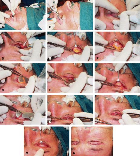
| Fig. 9. A. Protective corneal shields are placed. B. Calipers are used to mark the eyelid for a blepharoplasty type incision. C. CO2 ultrapulse laser with setting of 6 W is used to incise the skin and orbicularis. D. The skin and muscle flap is excised. E. The orbital septum is opened across the entire aspect of the upper eyelid wound exposing preaponeurotic fat. F. Fat is liposculpted free from the aponeurosis. G. Fat is carefully excised. H. Small amounts of septal tissue and orbicularis muscle are trimmed. I. The levator aponeurosis is dissected free from the superior tarsus and underlying Muller's muscle with blunt and sharp dissection. J. The distal edge of the levator is trimmed. K. Interrupted sutures of 7-0 silk are placed onto the anterior tarsus. L. The levator aponeurosis distal edge is advanced and sutured to the superior tarsal border. M. Silk sutures (7-0) are placed sequentially to adjust the upper eyelid position and contour. Slipknots can be placed at this step, and the patient can sit up while sutures are adjusted for appropriate eyelid contour and height. N. The skin is closed with running sutures of 6-0 Prolene, and erythromycin ointment is placed over the wounds. |