Chapter 75
The Evaluation and Management of Eyelid Trauma
JAMES W. KARESH
Main Menu   Table Of Contents

Search

ANATOMIC HIGHLIGHTS
PATIENT ASSESSMENT
GENERAL SURGICAL CONSIDERATIONS23–32
MANAGEMENT OF SPECIFIC INJURIES
SURGICAL MANAGEMENT OF SPECIFIC EYELID TISSUES23,25–27,29,51–54
MANAGEMENT OF SPECIFIC EYELID AND PERIORBITAL INJURIES
POSTOPERATIVE CARE
SCAR REVISION AND LATE POSTOPERATIVE INTERVENTIONS
REFERENCES

Injuries involving the eyelids, lacrimal drainage apparatus, and periorbital areas are common after either blunt or penetrating trauma. A detailed anatomic and functional knowledge of the ocular and periocular structures is needed for the ophthalmologist to manage such injuries. In this chapter the evaluation and management of lacerations and other injuries involving the eyelids, lacrimal drainage system, and periocular areas is discussed. The intent is to provide a firm basis for successful treatment of such injuries for both the beginning resident and general ophthalmologist. No attempt is made to exhaustively cover this broad subject. Rather the reader is referred to the various references at the end of the chapter for more detailed discussions of specific reparative techniques that may be useful to the more experienced surgeon.
Back to Top
ANATOMIC HIGHLIGHTS
An understanding of eyelid, lacrimal, and periorbital anatomy is essential for managing injuries to these structures (Fig. 1). The following description details those anatomic features important for managing eyelid trauma. More extensive discussions of anatomy are contained in the references at the end of this chapter.1–4

Fig. 1. Eyelid anatomy

SKIN

The upper eyelid skin is the thinnest in the body, with the lower eyelid skin being only slightly thicker. There is very minimal dermal tissue associated with eyelid skin; therefore, less substantial scarring occurs in this area when compared with the periorbital tissues. The periorbital, forehead, and cheek skin is considerably thicker, with a much more substantial dermal layer. The upper eyelid skin merges into the thicker forehead and glabellar skin at the superior orbital rim and eyebrow.

The major anatomic features of the upper eyelids include the eyelid crease and fold and the eyelashes. The normal crease is approximately 10 mm superior to the upper eyelid margin.5–8 There is some variation to this, with the eyelid crease in Asians being considerably lower in many individuals.1 The upper eyelid crease represents the point at which slips of the levator muscle interdigitate with the orbicularis muscle to insert into the undersurface of the skin. Recession of the eyelid crease associated with traumatic ptosis suggests an underlying dehiscence of the levator aponeurosis. A change in the eyelid crease can also occur after eyelid surgery or laceration repair where the aponeurotic interdigitations are disrupted.

The eyelid fold represents the point at which the looser preseptal skin overhangs the more firmly attached pretarsal skin when the eyelid is open. The full appearance of the preseptal area of the eyelid is due to the underlying fat. In Asians there is a lower eyelid fold due to both the lower attachments of the levator muscle and the lower attachment of the septum onto the levator allowing the anterior and inferior extent of the orbital fat to be at a lower level on the tarsus.1 Loss of upper eyelid fullness can be associated with orbital fractures or post-traumatic or inflammatory fat necrosis.9,10

The features of the lower eyelid are less distinct than the upper. Although there is a lower eyelid crease in most individuals it is not as distinct as that in the upper eyelid. However, the pretarsal skin is more firmly attached than the preseptal skin, and a crease may be seen 2 to 4 mm below the eyelid margin, especially in downgaze. Other creases occur in the lower eyelid and in the lateral temporal periorbital skin as aging occurs. Like the upper eyelid, the preseptal fullness to the lower eyelid is due to the presence of orbital fat. An asymmetric reduction in this fullness can be associated with orbital fractures and fat necrosis. The lower eyelid skin merges into the thick skin of the cheek at the inferior orbital rim. A malar fat pad can be found beneath the thick skin and muscle over the zygomatic bone as it forms the lateral aspect of the inferior orbital rim.

ORBICULARIS MUSCLE, MEDIAL CANTHAL TENDON, AND LATERAL CANTHAL TENDON

The next layer below the eyelid skin and dermis is the orbicularis muscle. This muscle is shaped like a sphincter with attachments medially forming the medial canthal tendon and laterally forming the lateral canthal raphe. The muscle extends to the eyelid margin where it forms the muscle of Riolan, which is seen as the gray line just anterior to the openings of the meibomian glands. Superiorly the muscle extends under the eyebrow. The facial nerve provides the innervation to this structure and can be damaged with large lacerations involving the lateral face.

Laterally, the fibers of the orbicularis muscle from the upper and lower eyelids interdigitate or become a raphe. A small tendon extends from this raphe and inserts onto the lateral orbital tubercle, which is located several millimeters inside the orbital rim. A small fat pad can be found in some individuals beneath this tendon.2 It is important to resuture the eyelids inside the orbital rim when repairing lateral canthal lacerations to prevent gaping between the eyelids and the globe.

The medial canthal ligament has two heads, a superficial head that inserts onto the anterior lacrimal crest and a deep one that inserts onto the posterior lacrimal crest.11 These two tendons surround the lacrimal sac, which sits in the lacrimal fossa. The deep or posterior limb of the tendon is formed by the deep heads of the pretarsal and preseptal portions of the orbicularis muscle. The anterior limb of the tendon is formed by the superficial heads of the preseptal and pretarsal orbicularis muscle. There is also a superior supporting branch of the anterior limb that inserts onto the frontal bone.11 The integrity of this structure and of the deep head of the tendon are important for preventing post-traumatic medial canthal dystopia. The upper and lower canaliculi and common canaliculus are directly below the superficial limb of the medial canthal tendon. These structures should be considered involved in all medial canthal injuries until probing and irrigation of the lacrimal system has been performed.

ORBITAL SEPTUM AND FAT

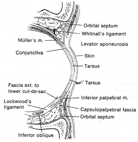
The orbital septum is an extension of the orbital rim periosteum. Superiorly this structure inserts onto the levator aponeurosis several millimeters above the level of the superior tarsal border. As already noted this can vary from individual to individual, with a much lower attachment being present in most Asians. In the lower eyelid the orbital septum attaches to the lower eyelid retractors approximately at the level of the inferior tarsal border. Behind the septum in the upper eyelid are two fat pads, one medially placed and one centrally placed. Laterally, the orbital portion of the lacrimal gland is present behind the septum. The fat pads are just anterior to the levator muscle and aponeurosis. In the lower eyelid there are three fat pads: medial, central, and lateral. They lie just anterior to the capsulopalpebral fascia, which is the analogue of the levator in the upper eyelid.

EYELID RETRACTORS

In the upper eyelid the eyelid retractors are Müller's (or the superior tarsal) muscle and the levator aponeurosis and muscle. In the lower eyelid they are the capsulopalpebral fascia and Müller's (or the inferior tarsal) muscle. The levator muscle originates just above the superior rectus muscle in the posterior orbit traveling anteriorly before changing direction at Whitnall's ligament and becoming an aponeurosis that ultimately inserts onto the anterior surface of the tarsus. Like the superior rectus muscle the superior division of the oculomotor nerve innervates the levator muscle. At Whitnall's ligament the aponeurosis fans out to form the so-called horns. The lateral horn separates the lacrimal gland into palpebral and orbital portions. Whitnall's ligament and the lateral horn of the aponeurosis attach onto the lateral orbital tubercle and contribute to the lateral canthal tendon. The aponeurosis lies behind the orbital septum and fat. It is only loosely connected to the orbicularis muscle anteriorly except for the slips of tissue that form the eyelid crease. This potential space between the aponeurosis and the orbicularis muscle is utilized in the anterior surgical approach to the eyelid as a way of avoiding damage to the aponeurosis. Identification of the orbital septum and fat is an important landmark during aponeurosis surgery or laceration repair.

Müller's muscle is a sympathetically innervated muscle immediately posterior to the levator aponeurosis. It is only loosely attached to the aponeurosis and can be easily dissected from it. Müller's muscle is 10 to 12 mm in height. It inserts onto the superior tarsal border and originates from the levator muscle. Posteriorly, conjunctiva is only minimally adherent to it. The peripheral palpebral arterial arcade travels on the anterior surface of Müller's muscle just superior to its insertion into the tarsus. This is an important landmark for identifying the muscle.

In the lower eyelid the capsulopalpebral fascia and inferior tarsal border are usually considered as one structure. Anteriorly there is a potential space between the orbicularis muscle and the eyelid retractors that can be utilized during lower eyelid surgery as an avascular plane for dissection. Posteriorly, the conjunctiva can be relatively easily dissected from this structure. Dehiscence of the lower eyelid retractors from the tarsus is thought in some cases to be associated with involutional entropion. Scarring between the septum and the retractors commonly occurs after orbital fracture repair, resulting in lower eyelid retraction.

TARSUS

The tarsus is a dense connective tissue structure that forms the basic support for the eyelid. There are about 25 meibomian glands contained in the upper eyelid tarsus and 20 in the lower. The tarsus is 1 mm thick and 26 to 28 mm long. The height of the upper eyelid tarsus is about 10 mm and that of the lower eyelid is 3.5 to 5 mm.12 Müller's muscle is attached to the superior tarsal border of the upper eyelid and the inferior tarsal border of the lower eyelid. The tarsus ends at the eyelid margin. The levator aponeurosis attaches to the inferior two thirds of the anterior surface of the tarsus. There is a potential space between the superior one third of the tarsus and the aponeurosis. The capsulopalpebral fascia inserts onto the anterior surface of the lower eyelid tarsus and Müller's muscle onto the inferior border of this structure. In both the upper and lower eyelids the marginal artery travels between the orbicularis muscle and the tarsus 3 to 4 mm from the eyelid margin.

CONJUNCTIVA

The conjunctiva lines the posterior surface of the eyelids reflecting upon itself at the superior and inferior fornices to become the bulbar conjunctiva covering the globe. The palpebral conjunctiva is firmly attached at the eyelid margin and to the tarsus. It is less firmly attached to Müller's muscle and can be fairly easily dissected from this structure or ballooned away from it with saline. There is some redundancy to the conjunctiva, and a portion of it can be excised or lost without compromising either eyelid or globe function or reducing the goblet cell contribution to the tear film. In the lateral aspect of the upper eyelid and superior fornix is the palpebral lobe of the lacrimal gland and the lacrimal ductules. Deep lacerations of the eyelid can damage these structures, reducing the aqueous portion of the tear film.

EYELID MARGIN

The eyelid margin represents a sandwich of tissues. Its most posterior layer is the nonkeratinized epithelium of the conjunctiva. This becomes keratinized at the mucocutaneous junction at the posterior border of the tarsus. The tarsus is the next most anterior layer and contains the orifices of the meibomian glands. The gray line or muscle of Riolan, which represents the edge of the orbicularis muscle is just anterior to the tarsus.13 The eyelid skin and eyelashes comprise the most anterior layer. Eyelashes are more numerous in the upper eyelid, which may contain two to four irregular rows of cilia. The lower eyelid contains one or two rows of eyelashes. The most medial aspect of the upper and lower eyelid margin contains a lacrimal punctum. Each is approximately 5 mm from the medial canthal angle. The puncta are nipple-like openings surrounded by a muscular sphincter. They are usually slightly inturned toward the tear lake. The tarsus ends at the punctal opening. Though not part of the eyelid margin, the caruncle, a nodular continuation of the conjunctiva containing both glandular tissue and cilia is just posterior to the medial termination of the eyelids at the medial canthal angle.

The puncta and vertical portion of the canaliculi are 1 to 2 mm long, becoming horizontal at a dilatation called the ampulla. The horizontal portion of the canaliculi is about 8 mm long, becoming one structure at the common canaliculus, which is 1 to 3 mm in length.14 The puncta are 0.3 mm in diameter while the canaliculi are 1 to 2 mm in diameter.14 Although initially traveling in the superficial and posterior aspect of the medial eyelid, the canaliculi dive deep into the eyelid and medial canthal tissue until they are below the superficial limb of the medial canthal tendon. The common canaliculus enters the lacrimal sac at the junction of its superior one third and inferior two thirds. The lacrimal sac is 15 mm in height, with 3 to 5 mm of it above the medial canthal tendon and the rest below the tendon.14 The lacrimal sac becomes the nasolacrimal duct in the bony nasolacrimal canal at the inferior orbital rim as it merges with the anterior lacrimal crest. The nasolacrimal duct enters the nose in the inferior meatus approximately 4 cm posterior to the opening of the nares. The canaliculi are lined with nonkeratinized stratified squamous epithelium. When cut, they have a grayish appearance. The lacrimal sac and duct are lined with modified, nonciliated respiratory epithelium.

EYEBROWS

The upper eyelid skin merges into the thicker skin of the eyebrow and forehead area at the superior orbital rim. The inferior aspect of the male eyebrow begins at the superior orbital rim. The female eyebrow is usually at a slightly higher level. The eyebrow is widest medially where it overlies the frontal sinus and is narrowest laterally. It forms a mild arch as it extends over the superior orbital rim. The eyebrows are separated by the skin of the glabellar area just above the nasal root. There is a thick layer of muscle beneath the eyebrows. Under this is the eyebrow fat pad and the deep insertion of the galea aponeurotica.
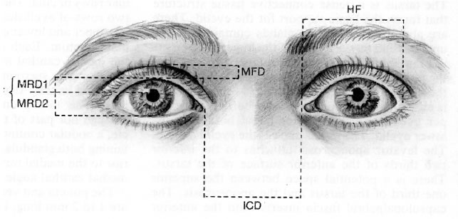
NORMAL MEASUREMENTS

The normal palpebral fissure is 26 to 30 mm in horizontal dimension and 8 to 10 mm in height (Fig. 2).7,8,15 The distance from the pupillary light reflex to the upper eyelid (MRD-1) ranges between 2.5 and 5.0 mm and the distance from pupillary light reflex and the lower eyelid (MRD-2) is between 4.5 and 5.5 mm.7,8 The normal upper eyelid rests 1 to 2 mm below the 12 o'clock border of the limbus, and the lower eyelid rests just at the 6 o'clock border of the limbus, although it may also be slightly above or below this level.16,17 The distance between the two medial canthi is normally 30 to 34 mm.15 In young individuals, 6 to 8 mm of full-thickness eyelid and margin may be removed and the defect repaired primarily. In older individuals it may be possible to remove as much as 12 to 14 mm of eyelid and still achieve primary closure. The puncta are approximately 6 to 7 mm from the medial canthal angle. The medial canthal angle is 0 to 2 mm below the level of the lateral canthal angle in most individuals.15 The tarsus is approximately 1 mm in thickness and 24 to 28 mm in length.1 The height of the upper eyelid tarsus is usually 10 mm, and the average height of the lower eyelid tarsus is 3.7 mm.1,12

Fig. 2. Eyelid measurements: margin fold distance (MFD); margin reflex distance--one (MRD-1); margin reflex distance--two (MRD-2); palpebral fissure (PF [MRD-1 + MRD-2]); horizontal fissure distance (HF); intercanthal distance (ICD).

Back to Top
PATIENT ASSESSMENT

HISTORY

Obtaining a detailed history concerning the nature of a patient's injury forms the basis for determining various management options. The time, cause, and circumstances of the injury must be determined. Since many periorbital and eyelid injuries can also involve the globe, a history of any change in visual acuity or severe ocular pain after the injury should be obtained. For children, consideration must be given to the possibility of child abuse as the cause of ocular and periocular injury. In criminal cases, bullets and other projectiles must be retained and marked by the physician so that there is no break in the chain of evidence.

Treatment provided at the time of injury or in the emergency department must be determined, especially regarding wound debridement, irrigation, manipulation of tissues, the removal of foreign bodies, and the administration of antibiotics and tetanus toxoid. If an animal or human bite is suspected, all information regarding the place of injury, the ownership of the animal, and abnormal animal behavior must be obtained and the local animal control department or state health department contacted. For human bites the possibility of human immunodeficiency virus infection or hepatitis must be considered.

A history of anticoagulant use, including aspirin and the use of any other medicines, and allergies is important. An immunization history, history of sickle cell disease or other serious illness, history of drug or alcohol use, and the name of the patient's family physician or internist should be obtained. Use of epinephrine is contraindicated in sickle cell disease. If surgical intervention is to be considered, the patient must be questioned regarding the time of last fluid or solid food ingestion.

CLINICAL EVALUATION

The goal of the examination of the patient with periorbital and eyelid injuries is to determine the exact extent of these injuries. Injury to the globe and intraocular contents as well as to other facial and intracranial structures can accompany seemingly trivial as well as obviously severe periorbital and eyelid injury. It is important to carefully document all injuries and if possible to photograph them. When the patient is severely injured, unstable, or unconscious, only a partial examination may be possible and a more thorough examination performed at a later time. In such instances and particularly if there is globe injury, it is important to place a protective shield over the injured side so that further damage does not take place during resuscitation efforts or other more urgent surgical procedures.

All patients with periocular or eyelid lacerations must have a complete ocular examination with particular attention paid to best corrected visual acuity, pupillary function and the presence of an afferent pupillary defect, intraocular pressure, and globe integrity. In the obtunded patient, the reaction of the pupil to light may be the only method for assessing the presence of optic nerve injury.

All periorbital and orbital injuries must be carefully examined for depth, extent, underlying bony fractures, and the presence of foreign bodies. Examination gloves should be worn during the evaluation of lacerations and fractures as part of universal precautions. Although some foreign bodies can be removed without anesthesia at the time of initial evaluation and wound irrigation, many foreign bodies will need to be removed intraoperatively at the time of injury repair. The eyelids should be everted to determine if foreign bodies are embedded into the palpebral conjunctiva or are present in the conjunctival cul-de-sacs. Wound depth can be estimated by gently separating the wound edges with a cotton-tipped applicator. The facial bones should be palpated to determine the presence of crepitus or orbital rim fractures.

The eyelids must be carefully examined to determine the integrity of the margin, skin, and tarsus. Lacerations of the medial eyelid may also involve the lacrimal system. It is important to probe and irrigate the lacrimal drainage system to determine if it is injured. Evaluation of levator function can be performed by measuring the excursion of the upper eyelid from far downgaze to far upgaze and comparing this with the uninjured side. Normally, the amount of excursion is 14 to 18 mm.7 The presence of swelling or impaired motility will interfere with determining if levator function is normal. The presence of orbital fat protruding from a laceration indicates extension of a wound into the orbit. In such cases consideration must be given to occult injury to the globe, extraocular muscle, and optic nerve as well as the possibility of retained orbital foreign bodies.18–21 Abnormalities of orbicularis function or the presence of lagophthalmos must also be assessed since this can result in corneal exposure. If there is no associated corneal laceration, ocular lubricating ointment can be placed over the cornea until surgical repair of the eyelid injuries.

Saline moistened dressings should be placed over facial and eyelid lacerations if there will be a delay between evaluation and surgical intervention. This prevents drying of the wound edges and desiccation of avulsed tissues. In addition, a protective shield should be placed over the injured tissue to prevent further injury before surgical intervention. It is important to monitor a patient's vision before and after surgery. Occult persistent post-traumatic hemorrhage can result in damage to the optic nerve.

LABORATORY AND RADIOLOGIC EVALUATION

Only a few laboratory tests are needed to complete the evaluation of the patient with ocular injury. A complete blood cell count and serum chemistries are often required for anesthesia purposes. A sickle cell test should be obtained in those patients at risk for this disease. Coagulation studies are helpful if a patient is using anticoagulant medication or vitamin K to counteract a prolonged prothrombin time. State laws may prevent testing for human immunodeficiency virus infection or hepatitis without patient permission. However, if possible these tests should be done. In select cases it may be necessary to obtain blood alcohol levels or to determine the presence of various chemical substances.

Ultrasonographic examination of the contents of the globe, extraocular muscles, orbit, and optic nerve is often helpful, especially in the obtunded patient. The presence of foreign bodies can be evaluated with this technique. Computed tomography is more helpful than plain radiologic films of the orbits and sinuses to determine the presence and extent of orbital fractures, foreign bodies, and extraocular injury to intracranial, nasal, intraoral, and sinus structures.18,20,22

DOCUMENTATION OF INJURIES

All injuries must be carefully documented in the patient's chart. This should include drawings and measurements of all lacerations and injuries. Photodocumentation is an essential aspect of all traumatic injuries. Polaroid and 35-mm photographs should be obtained at the time of initial evaluation. The medical record is a legal document, and comprehensiveness of the patient's evaluation may prove essential in future legal actions.

Back to Top
GENERAL SURGICAL CONSIDERATIONS23–32

TIMING OF REPAIR

Although it is optimum to repair eyelid injuries within 24 hours after their occurrence, these injuries can easily be repaired up to several days later. It is occasionally possible, although difficult, to primarily repair eyelid and canalicular injuries as long as 2 or conceivably more weeks after injury by carefully separating the edges of the wound, excising granulation tissue, and anatomically repositioning the tissues in their normal position. It is essential that the patient and globe be in a stable condition before acutely repairing non-life-threatening or non-vision-threatening injuries. This is particularly true for those cases in which globe integrity has been compromised and manipulation of the periorbital soft tissues or the bony orbit may cause increased pressure on the globe, resulting in the loss of the intraocular contents or elevated intraocular pressure. Postoperative soft tissue swelling can also cause significant pressure increases within the globe owing to decreased venous outflow. In the multiply injured patient, particularly when there are severe intracranial, cardiac, and pulmonary injuries as well as ocular and facial injuries, it is important to consult with the other physicians managing the patient to determine the optimum timing for repair of each of these injuries. Intraocular and optic nerve injuries take precedence over periorbital injuries, but it must be stressed that adequate eyelid coverage is essential for ensuring that the cornea does not dry and ultimately ulcerate.

In general, it is important to expertly repair all lacerations of the eyelids and periorbital areas acutely. However, if it appears that definitive repair is extremely difficult at the time of initial surgical management, as is the case in significant tissue loss or in severely macerated tissue, no attempt should be made to provide definitive repair at the time of initial surgical intervention. Skin grafts and complicated tissue flaps and tissue transfer techniques in these cases should be reserved for later use. A delay of 3 to 9 months, allowing swelling to resolve and scars to mature, is important.31,33,34 At this point it will be considerably easier to determine the exact approach needed to achieve the best result. It is often the case with post-traumatic ptosis or postsurgical ectropion and eyelid retraction that with resolution of edema and hematoma and with scar maturation there will be a considerable improvement in eyelid position.

In those instances when it is essential to provide an immediate covering for the globe, as in cases of severe burns or partial avulsions of the eyelids, a temporary suture tarsorrhaphy and aggressive ocular lubrication can be utilized. When there is total loss of the upper and lower eyelids, any remaining conjunctiva can be extensively undermined and sutured to itself over the cornea. A full-thickness skin graft from an area that would not ordinarily be used for eyelid reconstruction (e.g., the inner thigh or the inside of the arm) or banked donor skin can then be applied over this marsupialized conjunctiva. Suture tarsorrhaphies should be used in those cases when the patient is obtunded, when there is facial nerve injury, or when severe lagophthalmos is present. The use of a releasable suture will facilitate observation of the globe in those cases in which it is injured. Finally, although it is not common to permit the healing of eyelid lacerations by granulation, it is well known that after excision of medial canthal tumors and in instances of eyelid margin dehiscence, wound healing by granulation and scar formation can yield excellent results.35

PATIENT PREPARATION AND THE INFORMED CONSENT

In addition to careful and thorough preoperative evaluation and surgical planning, patient preparation and meticulous surgical technique are essential for successfully managing eyelid trauma. The patient needs to be informed about the likely postoperative results of surgery and possible complications, including the chance that additional surgical intervention may be necessary for correcting postoperative problems or intraoperative conditions that could not be adequately addressed. A seemingly successful result can be completely undermined if a patient does not understand the purpose of the surgery and the results that can be expected, the extent of postoperative swelling and ecchymosis, the possible need for additional scar management or eyelid surgery, the length of time it may take for incisions to heal, and the possibility of postoperative ocular irritation. Also, patients need to be informed that certain pre-existing conditions such as hyperthyroidism, congestive heart failure, diabetes, hypertension, renal failure, liver disease, and immune deficiency states can interfere with wound healing, resulting in a less than optimum outcome.34 In addition, a variety of other problems can interfere with the results of surgery. These are mostly related to nutritional abnormalities, irradiated and inflamed tissues, vascular compromise as is seen in patients who smoke and those with chronic lung disease, and patients taking corticosteroids, anticoagulants, and chemotherapeutic agents.34 In the young child the occurrence of amblyopia is often unavoidable after repair of severe facial injuries. It is important to inform the child's parents of this possibility and to manage it appropriately as soon as it is feasible.

PROPHYLAXIS FOR INFECTION36–39

It is important to determine an injured patient's tetanus immunization history after injury.37,38 All patients who have not received tetanus prophylaxis for 10 years or more need to be reimmunized. For clean, nonpuncture wounds tetanus toxoid should be administered to all patients except those who have received a full course of three boosters within the previous 10 years. For unclean wounds or puncture wounds all patients should receive tetanus toxoid again except those patients with an up-to-date immunization history within the past 5 years. In addition, they should receive tetanus immune globulin. The usual dose of tetanus toxoid is 0.5 ml intramuscularly or subcutaneously, and the dose of tetanus immune globulin is 250 IU intramuscularly.

The other important prophylaxis is for rabies.39 Rabies is still common in wild animals in many parts of the United States, and a number of individuals die of this disease each year. If the animal involved in the injury is a pet, it should be observed for 10 days for signs of illness. If possible, wild animals should be caught and killed and their brains tested for the presence of the rabies virus. If rabies is present, if the wild animal cannot be found but is suspected to be rabid, or if a pet develops signs of rabies, both human rabies immune globulin (20–40 IU/kg) and human diploid cell rabies vaccine must be administered. One milliliter of vaccine is given initially and again 3, 7, 14, and 28 days later. Immune globulin is divided in two parts, one of which is infiltrated, if possible, into the wound and the other given intramuscularly.

Antibiotic prophylaxis is probably not necessary for most injuries of the eyelids and periorbital tissues owing to the extensive vascularity of these tissues.40 Antibiotic prophylaxis for bite injuries is described below. Vigorous irrigation and wound debridement and removal of all foreign material is usually sufficient to prevent wound infection without the use of antibiotics.

In “clean, contaminated” surgery of the head and neck, that is, those operations involving the sinuses, oral cavity, pharynx, or nasal passages, preoperative antibiotic prophylaxis can be provided by a single dose of clindamycin (600–900 mg IV) or cefazolin (1–2 g IV) without the need for additional postoperative antibiotic coverage.40 However, in traumatic injuries, antibiotic coverage should be continued for 5 to 10 days after surgery. It is best not to use third-generation cephalosporins for prophylaxis since they are expensive, they may not be as effective against staphylococci as cefazolin, and their use as prophylaxis promotes the emergence of resistant organisms. Amoxicillin plus clavulanic acid (Augmentin), 250 to 500 mg three times daily, or clindamycin, 300 mg every 6 hours given orally, can be used postoperatively instead of intravenous antibiotics since these will provide adequate prophylaxis for most infections. It must be remembered that clindamycin is associated with pseudomembranous colitis secondary to Clostridium difficile.40 The treatment for this is oral vancomycin, 500 mg to 2 g daily in three or four divided doses over 7 to 10 days.

COAGULATION STATUS

The soft tissues of the face, mucosal lining of the nasal passages and sinuses, and the eyelids and orbit are quite vascular. Persistent bleeding often occurs after injury as well as during surgery. Delaying repair of lacerations for several hours will allow normal coagulation to take place and make the initial intraoperative wound evaluation easier. Patients on chronic aspirin therapy or anticoagulants may have extensive bleeding associated with their injuries and the subsequent surgical repair. Preoperatively, intramuscular or subcutaneous injections of vitamin K (10 to 25 mg every 4 hours) in patients taking anticoagulants can reduce a prolonged prothrombin time within 6 to 8 hours.26 The intraoperative use of lidocaine with 1:100,000 epinephrine and the intranasal or intrasinus use of 1% epinephrine or 4% to 10% cocaine-soaked pledgets is also helpful to control persistent bleeding. Reducing the systolic pressure to 100 mm Hg or less intraoperatively can also be helpful in selected situations. Intraoperative use of unipolar or bipolar cautery is also essential for hemostasis.

WOUND HYGIENE

As part of the evaluation and repair of injuries to the eyelids and periorbital tissues, it is important to thoroughly cleanse all such wounds. Care must be taken to ensure globe integrity before either irrigating or investigating the extent of any soft tissue injury. Both superficial and deep wounds should be forcefully irrigated with sterile saline or water to remove dirt and other materials that are clinging to the injured tissues. A large syringe with an attached 16- or 18-gauge catheter is often helpful for this purpose. Firmly embedded foreign bodies may be removed with scraping or with forceps and scissors. It may be necessary to scrub wounds with a stiff sterile scrub brush and mild soap. Chlorhexidine gluconate should not be used around the eyes, ears, or mouth because it is known to cause severe ocular injury and damage to mucous membranes. The depths of each individual laceration should be examined for debris and large foreign material. Oil, tar, or grease around or in wounds may need to be removed with solvents such as acetone, xylol, or ether.31 Since these chemicals can cause significant irritative conjunctivitis, the eyes should be protected when they are used. Failure to remove oil, tar, or grease as well as superficially embedded particles of dirt will result in permanent tattooing of the skin.31,41

FOREIGN BODIES

The management of superficially embedded foreign bodies has been discussed earlier.31,41 Deeply embedded material can be more problematic to manage. All organic material such as wood, plant debris, hair, or fecal material must be removed. Retention of such material is associated with both acute infection and chronic inflammation and suppuration.31 Glass foreign bodies are simple to remove but may be difficult to locate. Although they do not cause a significant inflammatory reaction they may be found to protrude from the skin after wound healing has occurred. Metallic foreign bodies such as bullet fragments are usually sterile and can often be removed. However, deeply embedded glass, plastic, or metal can be difficult to find. Plain radiographic films of the orbit and face will demonstrate metal- or lead-containing glass but not most glass fragments. Computed tomography or magnetic resonance imaging may be more helpful for locating deeply embedded foreign objects and wood or other organic material.22 Inorganic foreign bodies when deeply embedded in the orbit do not need to be removed unless they are associated with some functional ocular deficit such as ptosis, ocular motility disturbance, or optic nerve injury. They often become encapsulated with time and usually do not migrate. However, late migration and extrusion of retained foreign bodies may occur.

ANIMAL AND HUMAN BITES42–48

Lacerations and tissue avulsions due to animal or human bites are frequently associated with infection from oral flora. Wound debridement and irrigation will help to reduce the number of organisms remaining in the wound site. Puncture wounds should be allowed to granulate. Other wounds should be closed in layers to eliminate dead space where fluid can accumulate and become a nidus for infection. Bite wounds over 24 hours old should be allowed to granulate, making sure that there is adequate corneal protection. Intravenous antibiotics should be given preoperatively for prophylaxis against infection. Postoperatively these antibiotics can be continued or changed to oral antibiotics. Antibiotic coverage can be maintained for 5 to 7 days.

The most common flora in bite wounds are Streptococcus species, Pasteurella species, anaerobic bacteria including Bacteroides species, Eikenella corrodens, and dysgonic fermenter type 2 (DF2). Intravenous penicillin G (2 to 4 million units every 4 hours in adults and 75,000 U/kg/day divided into doses given every 4 to 6 hours in children) should be given for dog bites where DF2 infection is possible. This is a life-threatening fulminant infection from oral flora commonly found in dogs. Penicillin G is also effective against anaerobes found in human oral flora and can be combined with intravenous nafcillin (500 mg to 1 g IV every 4 hours) for improved Staphylococcus coverage.

Amoxicillin with clavulanic acid (Augmentin), 40 mg/kg/day in three divided doses (maximum dosage 500 mg three times daily), will provide excellent coverage against all other organisms, including anaerobes and penicillinase-producing staphylococci associated with both human and animal bites and is probably the antibiotic of choice for these injuries.40 Clindamycin (600 to 900 mg IV every 6 hours) also provides excellent coverage against both staphylococci and anaerobes.40 Other antibiotic choices include oral cefuroxime (20 mg/kg/day in two divided doses not to exceed 500 mg/dose), intravenous cefuroxime (75 mg/kg/day in three divided doses up to 1.5 g every 6 hours in adults with life-threatening infections), or ceftriaxone (50 mg/kg/day as a single IM dose or IV in two divided doses not to exceed 2 g daily in children and 4 g daily in adults).40

ANESTHESIA: GENERAL VERSUS LOCAL WITH OR WITHOUT SEDATION34

General anesthesia must be used in children, in some adults, and in cases of complicated wounds or injuries involving multiple bony and soft tissues structure of the face. Local infiltrative anesthesia tends to distort the tissues. However, it can be used in small lacerations. Usually, 1% or 2% lidocaine with 1:100,000 epinephrine is utilized, although longer-acting anesthetics can be substituted. The addition of epinephrine is essential for hemostasis and can be used in conjunction with general anesthesia. Regional anesthesia is the most effective approach for laceration repair since it minimally distorts tissues and will provide profound anesthesia to large areas. Injection of the infraorbital, supraorbital, infratrochlear and supratrochlear nerves is adequate for the repair of most lacerations involving the eyelids, lacrimal drainage system, and periorbital soft tissue. Another useful regional block involves the pterygopalatine canal just medial to the second molar and the greater palatine nerve, which will successfully anesthetize the lower eyelid, zygomatic area, and lateral aspect of the nose. Maxillary nerve blocks using a 3-inch long 22-gauge needle inserted externally just anterior to the coronoid process, inferior to the zygomatic arch, and through the pterygopalatine gap into the pterygopalatine fossa can be used instead of a regional block involving an intraoral route and the pterygopalatine canal. When intubation of the lacrimal drainage system is required for its successful repair, the intranasal use of 4% or 10% cocaine or 4% topical lidocaine are important to successfully carry this out. Cottonoids soaked with one of these solutions should be placed in the inferior meatus and around the inferior turbinate, remembering that the opening of the nasolacrimal duct into the nose is about 4 cm posterior to the nares just underneath the inferior turbinate.

Back to Top
MANAGEMENT OF SPECIFIC INJURIES

GENERAL SURGICAL CONSIDERATIONS24,28,31

Basic good surgical technique is essential to achieve a successful outcome in the repair of soft tissue injuries. Rough handling of tissues can lead to crushed skin edges and necrosis, with subsequent inflammation and scarring. Extensive desiccation and crusting of the wound may require rehydration with moist dressings before the initiation of surgical repair. Reactive suture materials used for cutaneous repair must be removed early to prevent permanent scarring. Tightly tied sutures will result in cross-hatching of the wound, leaving an unsightly scar and a less than acceptable outcome. This can be a particular problem with nylon and polypropylene since these materials stretch to some extent. Meticulous hemostasis is important since hematoma formation can cause wound edges to separate and may prove to be the nidus of a postoperative infection. Removal of any retained reactive foreign bodies, careful wound debridement, excision of areas of tissue necrosis, and control of infectious processes are also necessary to prevent excessive postoperative inflammation and scarring.

The repair of complicated lacerations requires some artistic vision and experience. The use of interrupted sutures is essential for correctly reapproximating wound edges in the correct anatomic position and with the appropriate tension. Interrupted sutures can be removed one at a time to evacuate accumulated fluid or for drainage of purulent discharge or hematoma. It is important to reapproximate the underlying muscle area to eradicate dead space and to prevent pockets of blood and serum from collecting and ultimately becoming thickened scar tissue. Adhesive paper strips such as the Steri-Strip or Micropore tape (3M Co. Medical Products Division, St. Paul, MN) can be used for postoperative skin closure support or for wound support after suture removal.

CONSERVATION OF TISSUES

It is essential to conserve all injured tissues. It is not unusual for apparently necrotic eyelid tissue or thin pedicles of tissue to survive owing to the extensive blood supply to the eyelids. In addition, it is not unusual in complicated lacerations for it to appear that tissue loss has occurred. Frequently this will turn out to be an erroneous conclusion as seemingly unrelated pieces of tissue are meticulously sutured into place. In general, there is retraction of eyelid tissue when it is lacerated and this is further enhanced by tissue shrinkage due to deficient vascularity and drying. Intraoperatively as blood flow returns and as tissue turgor improves, areas that appeared to be missing tissue or that were difficult to reapproximate will be able to be repaired.

METICULOUS REPOSITIONING OF TISSUES

Careful reapproximation of all injured tissues is another essential aspect of laceration repair. In some cases of severe eyelid and periorbital injury it can be difficult to orient the tissues and to determine precisely the tissue that should be reapproximated. The eyebrow, eyelashes, and eyelid margin provide keys to correctly repairing these injuries. The eyebrow tissues should be approached first. Carefully suturing the upper and lower edges of this structure will help to orient the direction of surrounding periorbital lacerations. After reapproximation of the eyebrows, the eyelash line should be sutured. It is helpful to remember that there are generally two or three rows of eyelashes in the upper eyelid and one or two in the lower, with the eyelashes ending just lateral to the punctum. Repair of the eyelid margin can be performed in conjunction with reapproximation of the eyelashes. Finally, resuturing the medial and lateral canthal angles will help to realign the eyelids into their normal position. The remaining lacerated tissues can then be repaired with the eyelid on some stretch. This will help to further define the tissue edges needing reapproximation.

SUTURE MATERIAL23,26,27,31

In general, interrupted absorbable 5-0 or 6-0 polyglactin 910 sutures are used to repair the tarsus and the orbicularis muscle while nonabsorbable sutures of 6-0 or 7-0 nylon, silk, or polypropylene are used for the skin. In children or other individuals for whom suture removal may be difficult, 6-0 fast-absorbing gut or mild chromic sutures can be used for lacerations involving the skin. Dehiscence of the levator aponeurosis can be repaired with either absorbable or nonabsorbable 6-0 sutures, although horizontal lacerations may be best repaired with nonabsorbable material. Generally, skin sutures are removed 5 or 6 days after repair while margin sutures are removed 10 to 14 days after repair. If the wound is on tension, skin sutures should be left in place longer than 5 to 6 days to reduce the incidence of wound separation after suture removal. However, leaving sutures in place for long periods of time will increase wound inflammation and scarring. The lateral canthal tendon should be reattached with nonabsorbable 5-0 nylon or polypropylene sutures since this will provide a more permanent reattachment to the lateral orbital tubercle and periosteum. This same suture material should be used for reattachment of the medial canthal tendon as well, although in cases of traumatic telecanthus the suture may be attached to a wire or a small bone screw.49,50 In these cases, a 4-0 or, in rare instances, a 3-0 nonabsorbable suture is used to attach the tendon to the wire since the reattached tendon is on considerable tension. Lacerations involving the canaliculi are most easily repaired using 6-0 or 7-0 absorbable chromic or polyglactin 910 sutures. Lacrimal sac lacerations are usually repaired with 4-0 or 5-0 absorbable sutures of the same material. Mattress sutures of 4-0 silk placed across repaired medial eyelid avulsion flaps will reduce tension across these wounds and possible postoperative wound separation. This suture is particularly helpful after repair of significant canalicular lacerations. Steri-Strips can be effectively used to support wounds involving the cheek, forehead, and lateral canthus and can also be used after suture removal to prevent wound separation. Tincture of benzoin or gum mastic can be used in conjunction with Steri-Strips to provide more skin adherence.

DRAPING AND PATIENT PREPARATION

The patient's entire face and hair should be thoroughly scrubbed with mild soap, diluted hexachlorophene solution, or povidone-iodine scrub solution. It is important to remove all make-up and debris. While the eyebrows should never be shaved or cut, a man's face may be shaved if necessary. This should be followed by either the use of povidone-iodine or hexachlorophene solution. The nasal cannula used in local anesthesia with sedation can be scrubbed along with the face. If the patient is undergoing surgery with local anesthesia, topical anesthetic drops should be instilled in the conjunctival cul-de-sacs to reduce ocular irritation before cleaning the face. After the surgical preparation of the face, the cul-de-sacs should be irrigated with saline to remove any remaining cleansing solution.

To successfully repair many extensive lacerations it is important to have the entire face in view to better visualize the interconnection between the various soft tissue structures of the face. A turban using two towels and a folded half sheet should be placed around the head. Towels can then be placed over the lower face, leaving the nose and, if necessary, the mouth exposed. The endotracheal airway used in general anesthesia can be draped off with towels. Head drapes should be positioned snugly so that they do not become loosened during surgery. A split sheet can be used to complete facial draping. Plastic adhesive polyethylene film may be helpful for covering difficult-to-drape areas.

Back to Top
SURGICAL MANAGEMENT OF SPECIFIC EYELID TISSUES23,25–27,29,51–54

SKIN

In general the skin is sutured with interrupted nonabsorbable sutures such as 6-0 or 7-0 silk, nylon, or polypropylene. Running sutures can be used in long linear lacerations. However, interrupted sutures are better able to reapproximate edges that are ragged or irregular lacerations. It is important to suture significant underlying orbicularis muscle lacerations to prevent the appearance of depressed areas of skin due to lack of tissue bulk.

TARSUS

The tarsus is a dense connective tissue structure that provides the eyelid with stability. For this reason it is important that this structure be carefully sutured as a separate layer when it is lacerated. Generally, 5-0 or 6-0 polyglactin 910 interrupted sutures are most effective for this, although any absorbable suture can be used. It is important to make sure the suture knot faces anteriorly so it does not abrade the cornea. In those instances where the tarsus is fractured in small pieces or is partially missing, this structure can be minimally trimmed to allow its reapproximation. Generally, the lacerated tarsal edges should be relatively parallel to each other for effective closure. The palpebral conjunctiva does not need to be sutured if the tarsus and orbicularis muscle are successfully reapproximated. Since at least a 3 mm wide band of tarsus is required to achieve a stable eyelid position, it may be necessary to develop a sliding tarsal flap to bridge an area of eyelid missing tarsal tissue. This can then be covered with a sliding skin-muscle flap.

LEVATOR APONEUROSIS55

Deep lacerations and puncture wounds of the eyelid can involve the levator muscle or its aponeurosis. In complicated injuries it can be difficult to visualize this structure. A knowledge of eyelid anatomy is essential for successfully repairing levator injuries. In linear vertical lacerations involving the tarsus reapproximating the tarsal edges may be sufficient for realigning the edges of the aponeurosis so that they can be sutured. Absorbable sutures are sufficient for this purpose. Horizontal or irregular lacerations are more problematic. In cases of simple horizontal lacerations, it may be possible to separate the edges of the wound, visualize the aponeurosis, and suture it to its normal position with a long-lasting absorbable or nonabsorbable 5-0 or 6-0 suture. However, in complicated or ragged lacerations, this can be a difficult task owing to the marked disruption of the normal eyelid anatomy. If the orbital fat and septum can be identified, the aponeurosis and muscle will be found immediately posterior or beneath them. If the surgical repair is being performed with the patient under local anesthesia with minimal sedation, asking the patient to look up and down will cause the aponeurosis to move, thereby facilitating its identification. Under general anesthesia this is not possible and only anatomic landmarks can be relied on. It may be possible to identify the levator muscle by mobilizing the superior rectus muscle and examining its dorsal surface posterior to the level of Whitnall's ligament. If the eyelid is extensively mangled, it may be impossible to accurately identify either the aponeurosis or the levator muscle. In such cases it is important not to widely dissect and explore looking for the edges of these structures. This can often cause more damage to already injured tissues. In such cases, delayed repair of these structures several months after initial wound repair and healing has occurred will allow a successful correction of the eyelid position to be performed.

ORBICULARIS MUSCLE

To take tension off of the eyelid closure and to cover septal lacerations, it is helpful to suture the orbicularis muscle with interrupted buried 5-0 or 6-0 absorbable sutures. When the orbicularis layer is adequately sutured it is not necessary to suture the septum. In fact, suturing this latter structure may result in lagophthalmos since it is very inelastic.

ORBITAL SEPTUM

When the septum is lacerated, orbital fat will prolapse into the wound. These deep injuries are associated with occult globe perforation, optic nerve injury, orbital hemorrhage, and orbital infection. Generally the septum does not need to be repaired, but the orbital fat should be repositioned, if possible, and the orbicularis muscle closed over it to retain it in its normal position. If the fat has been exposed for 24 hours or appears necrotic or hemorrhagic, it should be excised to prevent it from becoming a nidus for infection. Orbital fat is not well vascularized and frequently undergoes necrosis after injury.

EYELID MARGIN INJURIES AND AVULSIONS

Before closure of eyelid margin and tarsal lacerations, the would edges can be trimmed to allow better approximation of the repaired tissues. Initially a 5-0 or 6-0 interrupted absorbable suture, preferable polyglactin 910, should be placed in the tarsus 1 to 2 mm from the eyelid margin (Fig. 3). In the upper eyelid this should be a partial-thickness bite, while in the lower eyelid this may be a full-thickness bite since corneal abrasions are less likely from lower eyelid sutures. All tarsal sutures must be placed so that when tied the knots face toward the skin surface and not toward the cornea. This initial suture is crossed to pull the edges of the wound together to determine if the suture is properly placed to result in the correct approximation of the margin edges. After placement of this initial suture, interrupted 6-0 or 7-0 silk sutures are placed in the eyelid margin. One suture is placed in the mucocutaneous margin, one in the gray line, and one in the posterior eyelash line. The suture bites should be approximately 2 mm from the edge of either side of the laceration and 2 mm deep. It is important to have eversion of the edge of the margin laceration after the silk sutures are tied. This will prevent notching or recurrence of notching after wound healing has occurred. It may be helpful to employ mattress sutures placed in the eyelid margin in a near-near-far-far configuration to achieve the best approximation margin lacerations. Before the margin sutures are tied, additional absorbable sutures of polyglactin 910 can be placed intratarsally. Three such sutures are usually sufficient in the upper eyelid tarsus, and two are placed in the lower eyelid tarsus. The silk sutures in the margin are usually tied before the intratarsal sutures, but this may vary from surgeon to surgeon. When the margin sutures are tied, the two posterior margin sutures are tied through the anterior margin suture to prevent the suture ends from abrading the cornea. Traction on the three margin sutures is helpful for reapproximating the other eyelid tissues. The intratarsal suture closest to the margin is tied next. In some cases it may be helpful to tie this suture before the silk sutures in the margin are tied to properly reapproximate the edges of the wound. The other tarsal sutures are tied after the margin is correctly aligned. Absorbable sutures should be used to reapproximate the orbicularis muscle after the tarsal sutures are tied. The skin is closed with 6-0 or 7-0 silk, nylon, or polypropylene sutures. In children, 6-0 mild chromic or fast-absorbing gut sutures can be used for skin closure.

Fig. 3. A. Eyelid margin repair. Method demonstrated shows placement of tarsal sutures (1 and 2) followed by placement of interrupted sutures in eyelid margin (3) and routine closure of skin (4). Instead of interrupted sutures, the margin can be closed with vertical mattress sutures (5). B. Eyelid margin repair demonstrating closure of the tarsus and orbicularis muscle.

Avulsion flaps involving the eyelids are not uncommon. Those that are associated with medial or lateral canthal injury are discussed later in the chapter. When the body of the eyelid is part of an avulsion flap, the eyelid margin should be repaired as already described. The affected eyelid should then be placed on stretch using a 4-0 silk or nylon traction suture through skin, orbicularis muscle, and superficial tarsus 2 mm from the eyelid margin. The remaining laceration should then be repaired in a layered fashion using interrupted 5-0 or 6-0 polyglactin 910 sutures through the orbicularis muscle and interrupted 6-0 silk, nylon, or polypropylene for skin closure. It may be helpful to leave the eyelid on traction for 1 or 2 weeks after the initial repair of such extensive lacerations to prevent retraction secondary to scar formation.

SUTURE TARSORRHAPHY

Postoperatively, a suture tarsorrhaphy of 4-0 silk, nylon, or polypropylene can be quite helpful in splinting the eyelid when complicated lacerations have been repaired. This suture should be tied in a slip knot to allow for visualization of the globe. In addition, tying the suture through a rubber or silicone pledget will prevent it from eroding through the eyelid skin and will facilitate loosening of the slip knot and separation of the eyelids.

Back to Top
MANAGEMENT OF SPECIFIC EYELID AND PERIORBITAL INJURIES

EYELID CONTUSION AND HEMATOMA26,31

Contusions are bruising injuries frequently associated with blunt trauma. The injured tissue becomes edematous, and there may be an underlying hematoma. There may also be associated subconjunctival or orbital hemorrhage. Usually such injuries will resolve without the need for therapy beyond the use of cold compresses for 24 to 48 hours. Warm compresses should be used after the initial 48 hours. Dark glasses will help to further mask the presence of periorbital ecchymoses. Occasionally, the swelling may be severe and cause a dehiscence of the levator aponeurosis. However, this often cannot be determined until there has been resolution of the acute injury.

Significant hematomas of the eyelid and periorbital structures may not spontaneously resorb. If left untreated, this can provide a nidus for infection and result in significant scarring. While still in the “currant jelly” stage, a persistent hematoma can be easily evacuated with a simple skin incision. This incision should be placed within the eyelid crease, a skin fold, or a relaxed skin tension line to reduce the chance of postoperative scarring. When liquefied, the hematoma can be aspirated using a large-bore (16 or 18 gauge) needle. Loculated hematomas may require the aspiration needle to be inserted several times in different places. Patients should be warned that while initially unilateral, swelling and ecchymoses may spread to the uninjured side or to other portions of the face. Edema and hematoma will persist longer on the side on which the patient sleeps. Blunt trauma to the eyelids is often associated with orbital or periorbital fractures, rupture of the globe, or intraocular injury. For this reason, a complete ocular examination is essential after such injuries. In addition, the presence of proptosis may indicate an associated orbital hemorrhage. Evaluation of the patient's vision, pupillary function, and intraocular pressure for the first 24 hours after injury is important for identifying the presence or occurrence of compressive optic neuropathy associated with orbital hemorrhage.

EYELID ABRASION31

This type of injury involves the epidermis and superficial dermis. Deeper injuries involving full-thickness dermis are considered lacerations. After cleansing with mild soap or antiseptic preparations, such as diluted povidone-iodine or hexachlorophene, and removal of superficial foreign bodies, these wounds are usually left uncovered except for antibiotic ointment. Large abrasions or those that may become exposed to dirt or debris and are therefore at a risk for secondary infection may also be covered with a nonadherent dressing. Unless abrasions are quite deep, they usually heal without significant scar formation.

THERMAL AND CHEMICAL INJURIES56–61

In general, unless the depth of a chemical or thermal burn extends through the full thickness of the dermis, the cutaneous aspect of such injuries can be managed as an abrasion. After copious irrigation and debridement, superficial cutaneous injuries through partial-thickness dermis are allowed to granulate during the healing process. Antibiotic ointments and nonadherent dressings are used for wound coverage and as a prophylaxis for infection. Artificial tear ointments may be needed to lubricate the cornea if lagophthalmos is present after early eyelid edema subsides or if chemosis and conjunctival prolapse occurs. When ectropion or severe eyelid retraction occurs due to post-burn scarring, scar excision and skin grafting may be required after the acute phase of wound healing has passed.

When deep dermal injuries occur in association with severe third-degree burns, the wound would be gently cleaned and carefully debrided. The early application of a meshed or split-thickness skin graft will reduce post-burn scarring and ectropion. The use of banked or artificial skin substitutes may be considered. A suture tarsorrhaphy should be used to provide corneal protection and to keep the eyelids on stretch. The vigorous use of antibiotic ointments is essential in the early phase of wound healing to prevent infection. Definitive full-thickness skin grafting is usually performed after the acute phase of wound healing, usually at about 8 weeks after the initial burn. If there is significant lagophthalmos or ectropion and the patient is unstable or not yet ready for skin grafting, it may be necessary to create intermarginal adhesions that can later be released. Vigorous ocular lubrication must be continued until acceptable eyelid closure and corneal protection is present. In some cases it may be possible to perform full-thickness skin grafting somewhat earlier than 8 weeks after injury. Severely burned patients are hospitalized and usually under the care of a burn team, which is responsible for their overall management. The ophthalmologist must act in concert with the team members and their overall management approach.

INTRADERMAL TATTOOS31,41

Accidental tattoos occur when small embedded foreign bodies, especially asphalt or tar, are not removed from abrasions or lacerations. When left in place they become fixed to the tissue within 12 to 24 hours after injury. Removal of these particles requires energetic scrubbing and debridement. The use of solvents (e.g., acetone, xylol) may also be helpful in removing oil-based debris. If tattooing occurs, dermabrasion or surgical excision is required for removal of the pigmented areas after wound healing has taken place.

EYELID GANGRENE62,63

Eyelid gangrene is an unusual occurrence after trauma. The blood supply to the eyelid is quite rich, and even thin flaps of eyelid tissue will usually survive intact when sutured into place. However, skin infections associated with Proteus, Pseudomonas, Staphylococcus, and Streptococcus species have been associated with this entity. Treatment of gangrene is based on controlling the infectious process with intravenous and topical antibiotics combined with debridement of necrotic tissues and maintenance of corneal protection. It is important to obtain wound cultures to determine proper antibiotic coverage. Definitive repair of the eyelid tissues should be deferred until the infectious process and all associated inflammation has resolved. Considerable scarring can be expected after severe eyelid infections, and skin grafts or flaps as well as other more complicated procedures will be needed to surgically correct the resulting eyelid abnormalities.

INJURIES NOT INVOLVING EYELID MARGIN: PARTIAL THICKNESS

Partial-thickness cutaneous injuries are common. Repair should be done in layers including the orbicularis muscle and skin. When these injuries involve the upper eyelid, consideration must be given to possible injury to the levator aponeurosis. If obviously injured, this muscle should be repaired. If orbital fat is protruding from the wound, it should be repositioned, if possible, or excised. The septum should not be sutured because this may result in postoperative eyelid retraction. Small partial-thickness injuries, especially those only involving the epidermal layer, occasionally can be allowed to heal by granulation without causing any permanent eyelid abnormality. In particular, this may be necessary in the patient who is not stable enough to undergo surgical repair.

INJURIES NOT INVOLVING EYELID MARGIN: FULL THICKNESS18,20,21

Full-thickness injuries of the eyelid without involvement of the eyelid margin are quite unusual. They are most often associated with knife injuries or puncture wounds as may occur with nails or spikes. Globe injury is commonly associated with these injuries, and this possibility should be evaluated before soft tissue repair. Meticulous layered closure of the levator aponeurosis, orbicularis muscle, and skin should be performed to repair most of these injuries. The conjunctiva does not need to be sutured unless it is extensively lacerated. In this case, absorbable 7-0 or 8-0 plain gut or polyglactin 910 can be used. Occasionally small full-thickness puncture injuries will heal acceptably through granulation without resulting in any permanent eyelid defect.

TRAUMATIC PTOSIS55,64–66

Upper eyelid ptosis commonly occurs after both blunt and penetrating eyelid trauma. Often there is no associated injury of the levator muscle or aponeurosis. Edema and hematoma are the most common causes for post-traumatic ptosis, and as they resolve the eyelid will usually return to its pretrauma level. Occasionally edema or hematoma will result in a localized or diffuse dehiscence of the levator aponeurosis. However, it can be difficult to determine if this is the case until after there has been resolution of eyelid swelling. Recession of the eyelid crease and fold, if present, will suggest the presence of an underlying injury to the aponeurosis. Levator function can be assessed after trauma by measuring the excursion of the eyelid from extreme downgaze to extreme upgaze. Significant edema will compromise this movement, making evaluation unreliable. In general, movement greater than 8 mm suggests a normally functioning levator muscle but does not eliminate the possibility of aponeurosis injury or intramuscular edema or hematoma. Very poor levator function suggests that there may be injury to the nerve supply to the muscle or significant damage to the muscle itself.

A number of other factors can be associated with traumatic ptosis. Corneal irritation can cause the patient to “guard” the eye by closing the eyelid. Retained foreign bodies, exposed suture knots or ends, misdirected eyelashes, or inadvertent iatrogenic or traumatic corneal abrasions are the usual causes of this phenomenon. Removal of these factors will usually result in an improvement in the level of the ptotic eyelid. Traumatic disruption of Whitnall's ligament (which occurs after enucleation), orbital roof fractures or other disruptions of the bony orbit, infection, scars, pre-existing ptosis, and injury to the third nerve or sympathetic supply to the levator muscle are other causes of ptosis after eyelid or facial trauma. Unless there is an obvious laceration of the levator aponeurosis that can be repaired at the time of the patient's acute trauma, repair of post-traumatic ptosis should be delayed for 3 to 9 months or more because in many cases the eyelid level will improve without the need for surgical intervention.

Surgical repair of the lacerated or dehisced levator aponeurosis has been described previously. When late repair of the levator is carried out, the procedure should be performed through an eyelid crease incision in a similar manner to the repair of involutional ptosis. In children it may be necessary to perform an early repair of the eyelid to prevent amblyopia if the traumatized eyelid does not elevate above the pupil in a relatively short period of time. In such cases, a temporary sling procedure using a 4-0 nylon or polyester suture or silicone rod can be used. This allows for the removal of the sling material when eyelid function returns or when more definitive surgery is required. A simple pentagonal sling pattern positioned deep to the orbicularis muscle is usually sufficient to elevate the eyelid on a temporary basis.

PUNCTAL AND CANALICULAR LACERATIONS67–71

Careful examination of the lacrimal drainage system is an essential part of the evaluation of lacerations involving the medial aspect of the eyelid. The medial 6 to 8 mm of eyelid contains the puncta and canaliculi. Examination under the slit lamp is usually sufficient to demonstrate if the eyelid injury is at or medial to the punctum. If this is the case, the lacrimal system should be probed and irrigated to determine if it is damaged. It is essential to examine both the upper and the lower lacrimal canaliculi since it is not unusual for injuries to be small and self-sealing. These injuries can be missed on a cursory examination.

Although some authors have suggested that upper canalicular lacerations do not need to be repaired and that marsupialization of a canaliculus into the conjunctival sac may be acceptable in canalicular lacerations proximal to the punctum followed by repair of the eyelid, it is best to carefully and accurately repair all eyelid lacerations to achieve a normally functioning eyelid and lacrimal drainage system. Any other approach is to be discouraged. If a canalicular laceration or other injury to the tear drainage system appears to be too difficult to repair, the patient should be acutely referred to and treated by a surgeon experienced in the repair of such injuries. The American Society of Ophthalmic Plastic and Reconstructive Surgerymaintains a list of such surgeons. In any but the remotest areas of the continent such skilled individuals are within no more than a several-hour drive and can effectively and successfully repair almost any lacrimal system laceration.

Canalicular lacerations are best repaired 12 to 24 hours after they occur. By this time bleeding is controlled and little edema is present. It is easier to see the distal end of the lacerated canaliculus by delaying repair for several hours. This structure appears as an irregular or flattened oval with pearly-gray shiny rolled edges. In the eyelid it is approximately 1 mm from the eyelid margin. It is several millimeters (as many as 4 or 5 mm) deep to the margin at the medial canthal angle and can often be confused with separations between the orbicularis muscle fibers. For this reason, it is essential to use either high-powered (at least 2.5× and preferably 3.5×) loupes or an operating microscope to facilitate the repair of this structure. Regional anesthesia is preferred to local infiltration since there is less distortion of the tissues and the lacerated end of the canaliculus is easier to visualize. Infraorbital and infratrochlear nerve blocks are excellent for this purpose.

A number of techniques have been suggested for finding the lacerated ends of the canaliculus. The end nearest the punctum is found by placing a probe through the punctum and observing the laceration for its exit position. If there remains a portion of the eyelid medial to this, the medial end of the canaliculus can usually be found quite easily by following the probe and examining the other side of the laceration looking for the characteristic shape and glistening epithelium of the canaliculus. Complete avulsions of the eyelid from the medial canthus or lacerations at the medial canthal angle make finding the medial end of the canaliculus considerably more difficult. An assistant is very helpful for retracting tissues and keeping the wound dry. Cotton-tipped applicators to blot and gently separate the tissues, forceps to hold tissues apart, and gentle suction are essential for exploring the tissues. Probes will create false passages. Irrigation of fluid such as milk, saline, sodium hyaluronate, or diluted fluorescein solution through an intact canaliculus looking for its appearance at the medial aspect of the laceration may be helpful. Methylene blue should not be used because it will permanently dye the tissues and obscure the canaliculus. Pigtail probes should be avoided except by the most experienced surgeon. They are difficult to use and can cause considerable damage by tearing tissues as an attempt is made to pass them around the common canaliculus.72–74 If a pigtail probe must be used, only the probe with the suture eyelet should be used since the one with the crochet hook end can cause significant tearing of the tissues as it is rotated and manipulated. In rare cases it may be necessary to open the lacrimal sac through a medial canthal incision used for a dacryocystorhinostomy and to try to find the canaliculus by probing the common canaliculus. Patience, careful observation, and meticulous exploration are important for the successful identification of the medial lacerated end of the canaliculus.

Except possibly those eyelid lacerations only involving the puncta, canalicular lacerations should be stented as part of their repair.75–77 Both monocanalicular and bicanalicular stents have been described. Monocanalicular stents have included various metal rods (such as those designed by Viers, Johnson, and Beyer), 25-gauge venous catheters, paper clips, and small polyethylene or silicone tubes.78–80 These stents are passed across the laceration so that when sutured the lacerated canaliculus completely encases the stent, which is sutured to the outside of the eyelid to prevent extrusion. Bicanalicular stents usually consist of silicone tubing attached to metallic probes, which are passed through the upper and lower canaliculi, across the lacerated portion of the canaliculus, and down the nasolacrimal duct into the nose.81–84 The probes are then removed and the ends of the silicone shortened and sutured or tied to themselves and to a larger bolster such as a piece of catheter to prevent the tube from being inadvertently pulled out of the canaliculi. The nose must be anesthetized with either topical cocaine (4% or 10%), 4% topical lidocaine, or benzocaine spray for successful passage of the tubes. Every surgeon has his or her own preferred technique for stenting a lacerated canaliculus. In general, the repaired canaliculus should remain stented for at least 3 months and if possible longer to prevent a stricture from forming within the repaired canaliculus.

In my experience bicanalicular stenting with Crawford tubes containing an indwelling suture (Jedmed Instrument Company, St. Louis, MO) is the simplest and most successful of all methods (Fig. 4).83 These tubes are very easy to pass through the lacrimal system and retrieve from the nose with a Crawford hook. They are very well tolerated and associated with few complications.85 Mild corneal abrasions, stretching or erosion of the punctum and canaliculus, tube displacement, and granuloma formation have been reported. To prevent tube extrusion, an indwelling suture is present in the lumen of the silicone tube. After the probes are removed from the tubing after its passage into the nose, the tubes are shortened by removing a portion of the silicone from around the suture, leaving a tail of suture exposed from both ends of the silicone tubing. The suture ends are then tied to each other, creating a loop of silicone. The ends of the silicone that are connected by the suture knot are then sutured to the lateral wall of the nares with a 5-0 or 6-0 polyglactin 910 suture. This prevents early extrusion of the silicone especially in children.

Fig. 4. Repair of canalicular laceration with Silastic intubation. Canalicular sutures are placed on either side of the canaliculus and not through the lumen (A and B). The eyelid margin and skin are closed in standard fashion using interrupted sutures. The Silastic tubing is the Crawford variety (Jedmed Instrument Company, St. Louis, MO) and contains an indwelling suture that is tied to itself within the nares after exiting from the opening of the nasolacrimal duct beneath the inferior turbinate (C).

After identifying the lacerated ends of the canaliculus and stenting the lacrimal system, repair of the laceration can be performed. Although some surgeons recommend directly suturing the edges of the canaliculus to each other using 8-0 absorbable sutures, placing mattress sutures anterior, posterior, and, if possible, inferior to the sides of the canaliculus without actually suturing the edges of the canaliculus together will be sufficient for canalicular reformation over a silicone stent.68 Double-armed 6-0, 7-0, or 8-0 polyglactin 910 sutures work quite well for this repair. Tightening the silicone tubing by pulling down on it in the nose will help in approximating the wound edges as they are tied. After the pericanalicular sutures are tied, 5-0 or 6-0 polyglactin 910 sutures can be used to close the deep tissue layers and orbicularis muscle. If possible, one of these sutures should be used to connect the medial end of the tarsus to the lateral end of the cut medial canthal tendon. This will aid in holding the eyelid in place against the globe. In deep lacerations, every attempt should be made to place this suture into the deep portion of the medial canthal tendon, which inserts onto the posterior lacrimal crest. The skin is closed with nonabsorbable 6-0 or 7-0 sutures. The eyelid margin skin over the canaliculus can also be sutured at the same time, making sure that the suture ends do not abrade the cornea when they eye is adducted. To further reinforce the closure of a canalicular laceration, especially if it is deep and involves the medial canthal tendon, a 4-0 silk mattress suture can be placed across the incision through the deep eyelid and medial canthal tissue. This will help to overcome some of the tension placed on the wound and keep the wound edges closely approximated. This suture should be left in place for 2 weeks.

Almost all canalicular lacerations should be repaired by stenting and reapproximating the edges of the canaliculus. The only exception may be those lacerations that directly cut through the punctum. While some surgeons would stent the wound and suture the punctum around the stent, others would merely repair the eyelid in the usual fashion and marsupialize the punctum in a manner similar to a three-snip procedure.76,86 Since there is no tarsus in this area, the eyelid can be repaired only with well-placed deep sutures of 5-0 or 6-0 polyglactin 910 and nonabsorbable interrupted sutures through the eyelid skin and the skin anterior to the punctum. One important benefit of intubating such injuries, and this is certainly a benefit afforded more medially placed injuries, is that the silicone tubing helps to hold the wound edges together.

MEDIAL CANTHAL LACERATIONS AND AVULSIONS25,87

In any injury of the medial canthus the canaliculi and lacrimal drainage system should be probed and irrigated to determine if the injury involves these structures. Superficial injuries to the medial canthal area not involving the canalicular system or medial canthal tendon can be repaired by reapproximating the edges of the wound. These injuries should be repaired so that the suture line is oriented in a vertical direction. This can be a simple skin closure using 6-0 or 7-0 nonabsorbable suture material or, if deeper, a layered closure with the deep layers closed using 5-0 or 6-0 absorbable suture material. In some cases it may be necessary to undermine surrounding tissues to close the wound. When there are small avulsions of skin and muscle in the medial canthal area and the patient's condition is not stable enough to allow surgical repair, these avulsions can be allowed to heal by granulation. Large avulsions of skin and muscle may require skin grafting with postauricular skin, although usually such injuries can be repaired with undermining and primary closure.

Although lacerations of the superficial head of the medial canthal tendon do not lead to traumatic telecanthus, nonetheless, these should be repaired. Usually such lacerations are associated with canalicular injury and these should be managed as previously described. The canthal tendon can then be repaired by suturing the lacerated ends together with 5-0 polyglactin 910 sutures; or if the tendon is avulsed from the anterior lacrimal crest, it can be directly resutured to the periosteum in this area with absorbable sutures. Care must be taken during this procedure not to damage the lacrimal system or lacrimal sac.

Lacerations of the deep head of the medial canthal tendon must be repaired to prevent traumatic telecanthus and to ensure the appearance of a normal canthal angle.11,25,87 Again, injury to the lacrimal system must be repaired first. Occasionally the deep head of the tendon can be reattached to the bone of the posterior lacrimal crest or medial orbit using a microscrew from an orbital fracture plating system.49,50 The tendon is sutured to the microscrew or to a microplate using 4-0 or 5-0 nonabsorbable suture material. Rarely, the tendon can be directly sutured to the fracture by passing the suture around remaining bone. Usually, the bone is too thin for this. If neither of these procedures can be utilized, it is necessary to reattach the tendon using transnasal wiring as described in the later section on telecanthus.88,89

LATERAL CANTHAL LACERATIONS AND AVULSIONS51,90–92

It is relatively unusual for the eyelid to be avulsed from its lateral canthal attachment or for a laceration to completely divide the lateral canthal tendon. Medial canthal injuries are considerably more common. If there has been no significant tissue loss and the lateral orbital periosteum is intact, the eyelids can be resutured into their normal position. Usually two 5-0 nylon or polypropylene sutures are placed in an interrupted fashion through the lateral tarsus and then through the lateral orbital periosteum in the area of the lateral orbital tubercle, approximately 5 mm posterior to the lateral orbital rim. Some surgeons prefer to use polyglactin sutures instead. It may be necessary to create a small lateral tarsal strip to reattach the eyelids. This is done by excising pretarsal skin and orbicularis muscle over 2 or 3 mm of the lateral eyelid. A thin strip of eyelid margin is then excised over the same area and the conjunctiva scraped from the strip. This is then used to fixate the eyelid in place. After the eyelids are sutured in place, an interrupted absorbable suture is placed intramuscularly and through the lateral upper and lower eyelid gray lines to recreate the lateral canthus in a fashion similar to the tarsal strip procedure described below.

If periosteum is absent from the lateral orbit, as often occurs with severe trauma, drill holes can be made in the lateral orbit and the eyelids reattached by suturing them to 30-gauge wires placed through these holes. Twisting the wires over a larger-gauge wire will tighten the eyelids. Microscrews can be used in a similar fashion for medial canthal tendon repair.50 An alternative procedure is to elevate a U-shaped periosteal strip based at or near the lateral orbital rim from the outside of the lateral orbital rim. The lateral aspect of this strip is then flipped on itself to face medially, and the remaining lateral eyelid tissue sutured to it.91,93 This type of tendon reconstruction tends to leave a lateral gap between the globe and the eyelids. The lacerated skin can usually be directly approximated over the reattached eyelids. It is important to minimize vertical tension that can result in bowstring folds during skin closure. Using a periosteal strip as an eyelid tendon is also acceptable for repairing large avulsive defects of the lateral portion of the upper eyelid. In this case the periosteum becomes the posterior eyelid lamella and the anterior lamella can be a advancement skin flap. For lower eyelid defects it is easier to use a laterally based tarsal flap from the upper eyelid that is sutured laterally and transposed inferiorly to act as the posterior lamella for the lower eyelid.

TISSUE LOSS: SKIN AND ORBICULARIS MUSCLE27,91,94,95

Complete avulsions of eyelid skin and orbicularis muscle are relatively uncommon eyelid injuries. Often, what initially appears to be an avulsion of tissue is actually an avulsion flap or the post-traumatic separation of wound edges. Careful reapproximation of the wound and the salvage of all tissues will usually successfully result in wound closure. However, when present, areas of avulsed tissue can usually be managed by wound debridement and direct layered closure. Small areas of skin loss, especially those in the medial canthal area, can be allowed to granulate with excellent results. It is important to remember that upper eyelid and lateral canthal defects should be closed to leave a horizontal suture line while lower eyelid and medial canthal defects should be closed to leave a vertical suture line. This will reduce the incidence of postoperative ectropion. In some cases the tissue loss may be extensive. It is helpful in such situations, especially if eyebrow tissue is lost, to try to find the avulsed tissue and suture it in its proper place as a free graft. These are usually successfully revascularized owing to the extensive blood supply to the face and eyelids. If such free grafting is not possible, the initial approach to the repair of large tissue avulsions is the wide undermining of the skin and muscle surrounding the injury and the creation of advancement flaps, which are then used to close the wound (Fig. 5). In rare occasions skin loss may be so extensive as to require skin grafting combined with creation of an orbicularis muscle flap to achieve adequate wound closure.

Fig. 5. Creation of skin muscle advancement flap to close area of avulsed eyelid skin (A and B). Burrow's triangles prevent the formation of “dog ears” and to facilitate flap advancement.

TISSUE LOSS: FULL-THICKNESS EYELID27,53,91,94,95

Full-thickness avulsive injuries of the eyelids can require the use of a number of surgical techniques, many of which have been adapted from the surgical management of eyelid defects after tumor excision (Figs. 6 THROUGH 8).One important difference in the repair of the traumatically injured eyelid is that it is usually necessary to trim the edges of the wound to allow for adequate apposition of the wound edges. This is especially true for the eyelid margin and tarsus. Before repairing such injuries an attempt should be made to locate the avulsed tissue, which can then be sutured back into place as a free graft, often with excellent results owing to the rich vascular supply of the eyelids and periorbital areas. In young individuals, direct closure of tissue loss involving one fourth to one third of the upper or lower eyelid is possible. In older individuals, a defect involving up to one half of the eyelid and sometimes even more can usually be repaired with a primary eyelid margin closure. The addition of a canthotomy and the cantholysis of the limb of the lateral canthal tendon of the involved eyelid will allow closure of an additional 10% to 15% of the eyelid. When this procedure is carried out to close an avulsion injury, the lateral eyelid conjunctiva is sutured to the lateral eyelid skin in the area of the cantholysis since there is no tarsus in this area. Absorbable 6-0 interrupted suture material is used.

Fig. 6. Canthotomy and cantholysis for repair of full-thickness eyelid avulsion defects. Lateral portion of skin incision has been carried out as a small myocutaneous sliding semicircular flap A. Canthotomy. B. Cantholysis. C. Closure of eyelid margin. D. Skin closure. Semicircular flap can also be used to close a partial-thickness defect due to avulsion of eyelid skin and muscle. The semicircular flap can also be reversed to close an upper eyelid defect.

Fig. 7. Repair of a full-thickness eyelid laceration with “freshening” of jagged wound edge followed by formation of a semicircular myocutaneous flap and cantholysis of the inferior limb of the lateral canthal tendon (A) A periosteal flap is used as a tarsal replacement to reattach the eyelid laterally (B). Conjunctiva can be mobilized and attached to either the superior or inferior edge of the periosteal flap (C). The myocutaneous flap is advanced to form the anterior lamella of the eyelid with the periosteal flap as the posterior lamella (C). Creation of a Burrow's triangle laterally prevents formation of a “dog ear” after flap advancement.

Fig. 8. Repair of a full-thickness eyelid defect with the use of a Hewes flap consisting of upper eyelid tarsus (A) and a Tripier flap consisting of upper eyelid skin and muscle (B). The Tripier flap can be based medially to close medial defects or laterally to close lateral defects.

Larger defects involving the middle of the eyelid can be repaired by aggressively undermining the surrounding soft tissue laterally, cutting the eyelid retractors and septal attachments, and creating a subciliary incision to dissect a myocutaneous flap.27,95 This allows all remaining normal lateral eyelid to be slid medially and sutured to any remaining medial eyelid. The lateral portion of the eyelid can then be reconstructed successfully with a laterally based tarsal flap (i.e., Hewes flap) transposed into the lateral defect with a myocutaneous pennant flap (i.e., Tripier flap), semicircular flap or cheek advancement flap to cover the tarsus.95–99 This procedure alone can be successfully used to reconstruct as much as three fourths or more of the lateral eyelid, especially the lower eyelid. It is important to reinforce the lateral aspect of the tarsal flap by suturing it to the lateral orbital rim periosteum to create the semblance of a lateral canthal tendon. Instead of a tarsal flap, a free graft of nasal septal cartilage with attached mucosa, ear cartilage, hard palate mucosa, or tarsus from the opposite eyelid can be used to reconstruct the posterior lamella of the eyelid.100–102 As an alternative to these types of free grafts, a composite graft from the opposite normal eyelid can be used.103 This graft includes eyelid margin, cilia, and tarsus and is usually 10 to 15 mm and occasionally more in width. The pretarsal skin is removed from the graft, and the graft is covered by any type of myocutaneous flap to provide a vascular supply. If a large enough transposition or advancement flap (e.g., a cheek flap) is used, it is possible to reconstruct almost an entire eyelid using this technique.95

Total loss of an upper eyelid can be repaired using a Cutler-Beard full-thickness bridge flap from the lower eyelid.104,105This flap is a full-thickness section of lower eyelid used as an inferiorly based advancement flap. A 4-mm bridge of lower eyelid margin is left intact across the entire lower eyelid, and the flap is advanced superiorly under this bridge to fill in the upper eyelid defect. This flap is divided 6 weeks after placement. Total lower eyelid defects are effectively repaired by advancing a superiorly based flap of upper eyelid tarsus with attached conjunctival pedicle into the lower eyelid defect.106 This acts as a new posterior eyelid lamella. A skin graft or myocutaneous advancement flap can then be used to cover this flap and acts as the new anterior eyelid lamella. This flap can be divided 4 to 6 weeks after placement.

In those rare cases when there is total loss of both the upper and lower eyelids, as sometimes occurs with bite injuries, gunshot wounds, or degloving injuries, the most important priority is to provide corneal protection. If it is not possible to easily reconstruct the eyelids with flaps, it may be necessary to defer definitive reconstruction until a later date. In the meanwhile, the conjunctiva still attached to the globe and any remaining palpebral conjunctiva or tissue are extensively mobilized and then sutured to themselves so that the cornea is completely covered by a conjunctival sac. A split-thickness skin graft can then be placed over this. It is important to save tissue such as postauricular skin and grafts that can be used for posterior lamellar replacement until definitive repair is performed and to use less acceptable tissue during primary wound closure in such severe injuries.

PERIORBITAL AND EYEBROW LACERATIONS

The fundamental aspect of eyebrow repair is careful and accurate repositioning of the soft tissues combined with preservation of the eyebrow contour and cilia. The eyebrows must never be shaved as part of laceration repair. They are needed as landmarks for accurate soft tissue realignment. In addition, once shaved, they may not grow back. The muscle layer needs to be sutured for all eyebrow, forehead, and cheek lacerations since these tissues are on considerable stress to pull apart. It may be necessary to use 4-0 or larger absorbable sutures for this, depending on the size of the injury. Subcuticular interrupted 4-0 or 5-0 absorbable sutures should also be placed to reduce tension on the wound closure. The upper and lower edges of the eyebrow must be carefully realigned so that no permanent deformity of the brow contour is present. The skin can be sutured with 5-0 or 6-0 interrupted nonabsorbable sutures. In windshield injuries it is not unusual for multiple small U-shaped avulsion flaps to be present involving the forehead or cheek. A less conspicuous scar results if these very small flaps are excised to create a spindle-shaped wound that will close horizontally to fall within the natural forehead creases or relaxed skin tension lines. Excision of these small flaps should not be done too aggressively since closure of other lacerated areas may be made more difficult due to wound tension or there may be a change in the height of the eyebrow. In the cheek area, excision of significant tissue may lead to lower eyelid retraction and, therefore, this should be avoided anywhere below the eyelid but may be judiciously done in the temporal or lateral cheek areas.

AVULSION FLAPS31

Avulsion flaps are often quite disfiguring even after meticulous repair. They are frequently associated with injuries to the forehead and cheek. Such injuries result in large tangentially placed scar bands involving both the cutaneous tissues and subdermal and dermal areas. Often associated with these lacerations is significant injury to venous and lymphatic flow. Large avulsion flaps often remain edematous for long periods and with healing can have a heaped-up or “pin-cushion” appearance owing to the long linear scars that occur beneath the flap. To minimize disfigurement it is helpful to excise the beveled edge of the flap to allow for a closer approximation of both deep and superficial tissues and more perpendicular wound edges. If beveled edges cannot be excised or if the dermal portions of the flap are beveled, the deep tissues should not be closed and an attempt should be made to slide the beveled edges together with deep suture bites. Creating a staggered or jagged incision from curved laceration segments can help to camouflage scar lines. It is not unusual for large avulsion flaps to require later revision to correct some of the defects associated with their repair. This should be delayed for 6 more months since it takes at least this long for scar maturation to take place. Pressure dressings over the flap or face masks can help to reduce the edema associated with these injuries; however, they may need to be kept in place for weeks or months.

When possible, small avulsion flaps should be excised. This will result in small linear scars that heal in a much more acceptable fashion. Small areas of total tissue avulsion should also be excised to create an elliptical defect that can also be repaired to create a linear scar. If this is not done, the healing of such injuries will result in a pebbly appearance to the skin. This can be almost impossible to repair at a later time.

AVULSION DEFECTS

An avulsion defect represents a total loss of tissue in a particular area. As already noted, small avulsion defects can be excised, creating an elliptical wound that can then be sutured to create a simple linear scar. Large avulsion defects require a more complicated repair, which can include skin grafts, transposition or rotation-advancement flaps, pedicle flaps, or free flaps combined with microvascular anastomosis. A discussion of most of these procedures is beyond the scope of this chapter. Skin grafts and myocutaneous transposition or rotation-advancement flaps are discussed below.

Back to Top
POSTOPERATIVE CARE

GENERAL CARE107

Small lacerations can be managed on an outpatient basis. Ice compresses should be used for 24 to 48 hours to reduce postoperative edema. Warm compresses can be used after that time period. Patients should be informed about the signs of wound infection, including increasing pain, erythema, edema, and purulent discharge. This will facilitate early antibiotic intervention. Other serious postoperative complications include corneal ulcers secondary to exposed suture ends and knots or intraoperative abrasions and visual loss from a retrobulbar hemorrhage. It is essential that patients be informed concerning the signs and symptoms of these problems, such as foreign body sensation, conjunctival injection, and decreasing vision, so that these complications may be treated promptly. Patients must also understand that the postoperative presence of a subconjunctival hemorrhage, periorbital edema and ecchymoses, mucous discharge and associated decreased vision that improves with blinking, and moderate pain are to be expected and will improve with time. Patients may also experience some temporary change in vision owing to soft tissue swelling and pressure from the eyelids on the cornea.

Postoperative pain management is very important for patient comfort. Generally, codeine or a synthetic codeine-type medication or a nonsteroidal anti-inflammatory agent such as ibuprofen will be required for the first several days until there has been some resolution of postoperative edema. It is very unusual for such medications to be required for more than this amount of time except in severe facial trauma. If pain medications are required for longer periods of time some other source of postoperative pain such as a retained foreign body, indolent infection, or psychological or physical dependency should be suspected.

To reduce the incidence and severity of postoperative edema and hematoma it is helpful to have patients sleep with their head elevated. If possible, patients should also try to sleep on the side opposite to the one that underwent surgery. The use of an antinausea medication such as prochlorperazine (10 mg IM, IV, or orally or 25 mg rectally) or trimethobenzamide (200 mg IM or rectally or 250 mg orally) may also be necessary after anesthesia to reduce emesis, which can result in significant postoperative hemorrhaging.

Patients with severe trauma should be hospitalized for at least 24 hours and possibly longer, depending on the extent of the trauma and the stability of the patient's condition. These patients must be evaluated immediately postoperatively and daily or more frequently, depending on the extent of injury and repair, for any change in vision, increased intraocular pressure, retrobulbar hemorrhage, or incipient infection.

An often overlooked aspect of postoperative patient management is related to nutritional status. Although severely injured hospitalized patients closely observed by the surgical team will usually receive appropriate nutritional supplementation, on discharge home, wound breakdown can occur if this is not continued. A nutritionist may need to be consulted to outline an acceptable home diet, especially if the patient has oral injuries and is only able to eat by means of a straw.

One of the most difficult problems to manage postoperatively is the patient's expectations regarding his or her postoperative appearance and the process of wound healing. Patients must be told that adequate wound healing, scar maturation, reduction in edema, and disappearance of ecchymoses may take weeks or months. It is not unusual for scars to continue to change for 1 to 3 years after trauma. In addition, patients need to understand that all such postoperative problems may not completely resolve and that further surgical intervention may be required. Related to this is that in complicated lacerations with loss of tissue or large flaps, it is to be expected that additional surgery or multiple operations may be needed and that despite the best efforts, even in the hands of the most skilled and experienced surgeon, it may not be possible to achieve results that resemble the patient's pretraumatic appearance. The surgeon should not feel forced by the patient to secondarily intervene and revise a wound or scar before the best result can be achieved. Scar revision, no matter how unsightly, should not be carried out until adequate maturation has taken place. This is usually about 6 months after the initial laceration repair in severe lacerations. Patients will complain about their appearance but must also understand that early intervention will not achieve an acceptable result. The use of dark glasses and cover-up make-up may be required to mask unsightly scars until revision can be performed.

TISSUE NECROSIS AND HYPERBARIC OXYGEN108,109

In cases of possible postoperative wound necrosis due to an inadequate vascular supply especially if extensive tissue loss is anticipated, hyperbaric oxygen may be useful. This therapy is also quite useful as an adjunct to antibiotic therapy in cases of severe anaerobic infection. This mode of treatment is not readily available except in large medical centers. When small areas of necrosis are present, debridement associated with semi-occlusive or occlusive dressings and antibiotic ointment can be used to allow wound granulation and healing to take place. Scar revision may be required when tissue necrosis is extensive but should not be performed until the acute healing phase has taken place and there has been scar maturation. This may take as long as 6 months after initial surgical repair.

MEDICATIONS: ANTIBIOTICS40

Postoperative ocular antibiotic ointments containing tobramycin or gentamicin applied two to four times daily to repaired lacerations may be helpful in wound prophylaxis. Similar dermatologic ointment containing gentamicin or bacitracin can be used on facial lacerations. Ointments will also help to reduce the extent of wound crusting and will facilitate suture removal. As already noted (see section on prophylaxis for infection) it is usually not necessary to use oral or intravenous antibiotics in the postoperative period except in cases of “dirty” wounds or bite injuries. In these cases intravenous clindamycin or cefazolin or oral amoxicillin (Augmentin) are the antibiotics of choice to provide coverage for both resistant staphylococci and anaerobes.

MEDICATIONS: CORTICOSTEROIDS

Topical corticosteroid preparations can be used to reduce scaring associated with laceration repair. The use of these medications should be deferred until after the acute healing phase has taken place, usually several weeks after wound repair. They can be continued for several months if necessary. Topical steroidal preparations such as triamcinolone acetonide 0.1% (Aristocort), fluocinolone acetonide 0.01% or 0.025% (Synalar), or fluocinonide 0.05% (Lidex) or the intralesional injection of small amounts (0.1 to 0.3 ml) of corticosteroids such as triamcinolone acetonide (10 or 40 mg/ml) is usually employed to control postoperative scarring.110

SUTURE REMOVAL

Cutaneous sutures should generally be removed 5 to 7 days after placement. Skin sutures in areas under extensive tension such as the cheek and forehead may be left in place for 7 days. Silk sutures are associated with an inflammatory reaction and if left in place for too long will be associated with unsightly scarring. Nylon and polypropylene sutures do not have this problem. Eyelid margin sutures should be left for 10 to 14 days to prevent wound separation. After suture removal if there appears to be some gaping of the wound or if the wound is still on some tension, Steri-Strips or Micropore tape can be used for support and to physically close the wound until adequate healing has taken place.

EARLY SCAR MANAGEMENT34,110

During the wound healing process multiple cellular and environmental events take place that influence wound healing and scar formation. Retained foreign materials, infection, and inflammation can prolong wound healing and promote excessive scar formation. Nutritional deficiencies are also important for successful wound healing. Protein deficiency states and vitamin C and K and zinc deficiencies will all adversely effect wound healing. Diabetes, smoking, vascular disorders, and immune deficiency states are just a few of the systemic conditions that can prolong wound healing. It is important to correct these conditions if minimal scarring and adequate wound healing is to be achieved.

Normally, a surgical wound develops tensile strength at about the seventh postoperative day.34 Collagen formation increases until it reaches a maximum after 3 to 5 weeks. Wound contraction is maximal between 5 and 15 days. Collagen deposition and scar formation continues for up to 6 or more months after wound formation. The maximum tensile strength of a wound is not achieved until after this time. Scar massage several times daily either alone or with vitamin E cream, cocoa butter, or topical steroid ointments can be initiated at about 3 weeks postoperatively and continued for several weeks. In many cases this will help in reducing scar bulk and improving the postoperative scar appearance.58,111

Back to Top
SCAR REVISION AND LATE POSTOPERATIVE INTERVENTIONS

COVER-UP COSMETICS AND THE COSMETOLOGIST112,113

A variety of post-traumatic problems can be very effectively managed by patient referral to a cosmetologist skilled in the art of concealing scars. A variety of waterproof camouflaging cosmetics are available that can be used to conceal scars or other skin color or texture changes. Not only do these products create consistent skin tones over the entire face, but they can be used to fill pits and depression to smooth out facial irregularities. In addition, artificial eyelashes and hair styling changes are other adjunctive methods for disguising unsightly facial abnormalities. Some of the companies producing these products are: Cover Mark, (Moonachie, NJ); Dermablend, (Farmingdale, NJ); Derma Color, (Cofasco Incorporated, Concord, CA); and Nature Cover Cosmetics, (Baltimore, MD)

THE Z-PLASTY AND TISSUE FLAPS27,95,114

Deficiencies in lower eyelid and upper eyelid skin as may occur after avulsive injuries or excision of scar tissue can frequently be rectified through the use of sliding or transpositional skin flaps. Similar techniques can be used to correct avulsive injuries of the forehead and cheek. Making use of relaxed skin tension lines, skin from the lateral lower eyelid and lateral cheek can be slid medially.115 Examples of this are the Mustardé cheek flap and the semicircular flap.27,96 Reversing the semicircular flap, that is, designing it so that it is facing downward, facilitates its use in repairing upper eyelid defects.98 Simple sliding finger flaps with Burrow's triangles and extensive undermining can also be used.95 Midline forehead flaps have been frequently used to reconstruct loss of medial canthal tissue and the medial upper and lower eyelid.27 Rhomboid flaps and glabellar flaps are also useful for medial canthal reconstruction.95 Transposition of a pennant flap from the lateral upper eyelid to the lateral lower eyelid is one of the most useful techniques for managing lateral lower eyelid skin loss.97,99 There is an extensive literature on flaps, with many surgeons having different favorite flaps for similar problems.95 Although it is important to have some knowledge of a variety of flap techniques, it is equally important for a surgeon to pick and feel comfortable with a few useful flaps that can be modified for many situations.

The Z-plasty is an effective method for correcting eyelid retraction secondary to linear scar contracture with or without a minimal loss of skin (Figs. 9 AND 10).114 Classically, it is a rotational skin flap technique consisting of two equal angles and three equal members. In this technique, the distance between the tips of the two parallel members of the Z-plasty flaps is substituted for the length of the central member of the Z. Incising, undermining, and exchanging the flaps is necessary to achieve this result. The two concepts contained in this technique are the change in direction of a scar to conform more closely to the relaxed skin tension lines and the movement of seemingly excess skin from one direction to another where the skin is deficient.33,115 An important principle of scar revision is that conformity to relaxed skin tension lines will result in the most cosmetically acceptable postoperative scar.

Fig. 9. Repair of lower eyelid retraction (A) with a multiple Z-plasty myocutaneous flap rotation technique (B). The area vertical axis represents the line to be lengthened. Scar tissue along this line should be excised as part of the procedure.

Fig. 10. The multiple Z-plasty technique: Formation of the transposition flaps (A); partial flap rotation (B); the closure after complete rotation of the flaps (C). Interrupted sutures are used to close the incision. Generally only one or two sutures are required per side to close the small flaps used to correct eyelid retraction or canthal webbing.

The eyelids are somewhat different from other areas of the face in that for the upper eyelid and lateral canthus, horizontal incisions are required to achieve optimal results while for the lower eyelid and medial canthus, vertical incisions are usually required.91,94 Nonetheless, the lower eyelid is a particularly difficult area as it has minimal vertical skin and horizontal curvilinear relaxed skin tension lines. The reasoning behind making a vertical incision in the lower eyelid when removing full- or partial-thickness lesions is that on closure tissue will be moved in from the horizontal direction to fill the defect. This will reduce the incidence of postoperative lower eyelid retraction. However, should retraction occur, it is easily managed with a Z-plasty. In lower eyelid retractions secondary to a vertical deficiency of skin and linear scarring, performance of a Z-plasty will take skin from the horizontal dimension of the eyelid and move it into the vertically deficient area. For both lower and upper eyelid retraction, this technique can easily be combined with a skin graft if there is a marked deficiency of skin after complete excision of scar tissue.114

Vertical incisions in the medial canthal area have a separate problem, the creation of a bowstring effect. Again, the Z-plasty is useful in breaking up the linearity and pull of such a scar. Extensive undermining of the flaps and surrounding areas is important to achieve adequate rotation and sliding of the skin. Careful manipulation of the tips of the flaps is essential to prevent necrosis. In general, angles of 60° result in 75% theoretical gain in skin length along the shortened scar line while angles of 75° result in a 100% theoretical gain.114 Multiple Z-plasties are useful for long linear scars or when more lengthening of a scar is required or less lateral distortion of the surrounding area is needed.114 Z-plasties with unequal angles or unequal members are at times necessary to close rhomboid or round defects or as part of rotating lateral upper eyelid skin into a lateral lower eyelid defect. This is more often thought of as a rotational or transpositional pennant or finger flap. At times, the flaps of the Z-plasty may need to be rounded to result in more cosmetically acceptable scarring. Flexibility of the surgeon is an essential element in creating flaps for each individual situation.

SKIN GRAFTING27,116–120

The management of acute avulsions of periorbital skin or large scars associated with eyelid or eyebrow retraction is best managed with skin grafting or any of a number of rotational or transpositional skin flaps. In general, larger defects are best managed with skin grafts while smaller ones can be corrected with skin flaps. To make sure that an adequately sized graft or flap is placed, wide undermining of the skin with excision of all underlying scar tissue is essential. It is not unusual for the amount of skin missing in a particular area to be considerably more extensive than is initially anticipated based on preoperative evaluation. For defects involving the lower eyelid or cheek after scar excision, having a patient look up while opening and closing the mouth will allow the surgeon to see if there is continued pull on the lower eyelid due to inadequate undermining of the skin. For the upper eyelid, having the patient look down while comparing the palpebral fissure to the opposite normal side or having the patient gently attempt to close the eyelids will help in determining if there has been adequate undermining of the tissues. Paralysis of the orbicularis muscle with local anesthesia will, to a certain extent, influence the apparent amount of retraction. The surgeon should err to the side of overaggressive undermining and thorough scar excision or lysis to ensure an adequate bed for the skin graft or flap. In acute injuries, it is still important to perform extensive undermining on the skin surrounding the avulsed area to slide as much surrounding tissue over the defect. Many times aggressive undermining of tissues will be enough to allow closure of many defects.

The next consideration in skin grafting relates to the site for harvesting the graft. Important in this regard is color match, appropriate thickness, and absence of hair from the graft site. Since the upper eyelid skin is the thinnest in the body, its best substitute is skin from the opposite upper eyelid. In lieu of this, postauricular skin is the next best choice for both the upper and lower eyelids. This is followed by the supraclavicular area and the inside of the arm. In those cases where foreskin is present, it can be used and provides excellent thin skin for grafts. The inside of the leg is usually too hairy for eyelid grafts, but it may be possible to use this in some individuals. Color match may be difficult in some cases because the eyelids are exposed to the sun while most skin utilized for grafting purposes is not exposed. Usually with time the color of the graft will blend with the surrounding tissues. Additionally, in darkly pigmented patients, there may be either a loss of pigmentation from the graft or hypopigmentation or hyperpigmentation of the surrounding tissues after surgery. Although these changes usually resolve with time, they may also persist in some instances. Cover-up cosmetics may be needed to mask any color differences when this occurs.

In general, full-thickness grafts obtained by freehand excision of skin are used in eyelid reconstruction. It is important to carefully trim all subcutaneous tissue and fat from the graft to make sure that it is not too thick. This will reduce the chance of necrosis secondary to inadequate vascularization. Split-thickness and meshed grafts are usually reserved for severe thermal injuries or when little tissue remains for grafting. Synthetic or banked skin may also be helpful in these cases. Thick split-thickness skin grafts are ideal for large areas of avulsed tissue. They readily vascularize, are easy to handle, and will give a more satisfactory result than thin split-thickness grafts. If necessary, several grafts can be sutured together to cover very large defects. In general, split-thickness grafts harvested with a drum-type dermatome are about 4 inches in width. Subcutaneous injection of saline may help to smooth and tighten neck skin when this is the source for the graft. Otherwise grafts can easily be obtained from the inside of the arm or leg by keeping the skin taut while the dermatome is run over the area. The donor site is left to granulate. Antibiotic ointment and petrolatum-impregnated gauze are used in the postoperative period until healing takes place. Usually, the final result in the donor area will be a flat, slightly whitened scar that is only minimally visible. However, this may take several years.

The two major problems associated with skin grafts are inadequate size and postoperative scarring. A 30% to 50% shrinkage of grafted tissue is to be expected. There are two ways to prevent this from compromising the postoperative results. First, placement of the eyelid, whether upper or lower, on traction is important to maximize the area to be grafted. The use of traction sutures intraoperatively and during the first 1 to 2 weeks postoperatively will help keep the eyelid and graft on stretch so that the maximum area will be grafted and the graft will be flat. Second, obtaining a graft one-third larger than is necessary will compensate for graft shrinkage. In addition, creating slits in the graft will allow for drainage of blood and serous fluids from beneath the graft, thereby enhancing graft adherence. Finally, placing a bolster over the graft for the first week will help ensure good graft adherence. Generally, this bolster consists of a nonadherent dressing material wrapped around cotton or some other firm dressing that is held in place over the graft by means of sutures placed in the tissue around the outside of the graft.

Scarring after skin grafting can be reduced by careful approximation of the edges of the graft to the surrounding tissue. The use of nylon sutures and the early removal of sutures, especially if silk is used, is also important. Careful tissue manipulation is essential for preventing tissue damage and necrosis. Should postoperative inflammation, scarring, and necrosis occur, tincture of time is often all that is required for resolution of these problems and may be sufficient to lead to an acceptable result. However, this can be enhanced by massage of the scar either alone or with topical steroidal preparations such as triamcinolone acetonide 0.1%, vitamin E cream, or cocoa butter.110 Intralesional corticosteroid injections are helpful for markedly hypertrophic scars.110 Avoidance of exposure to the sun for up to 6 months is essential to prevent permanent scar discoloration. The use of a sun block with a sun protective factor of 15 as well as a hat and dark glasses will help in this regard. Occasionally dermabrasion of some scars or excision of markedly hypertrophic scars may be required, but this should be reserved until medical treatment has been exhausted.41

DERMABRASION41

Small embedded foreign bodies, accidental tattooing with tar or asphalt, and small thickened scars can be managed with dermabrasion. Various types of dermabraders are available, including battery-powered, electric, and air-driven models. Hand-held wire brushes and tungsten carbide sandpaper can also be used. Small areas can be easily dermabraded using local anesthesia and sedation. Large areas require general anesthesia. When dermabrading it is important not to enter the deep dermal layers because this will result in permanent scarring. Deeply embedded foreign bodies should be surgically teased out before dermabrasion. At times it may be more effective to excise large scars and perform a layered closure of the area rather than dermabrading the area. Postoperatively, antibiotic ointment and nonadherent acetate gauze dressings are placed over the dermabraded area until healing takes place.

CANALICULAR AND LACRIMAL DRAINAGE SYSTEM OBSTRUCTION70,71,121,122

Persistent epiphora and periorbital skin excoriation and irritation are associated with inadequate lacrimal drainage due to canalicular or lacrimal duct or sac scarring. The management of lacrimal drainage abnormalities depends on the location of the blockage. In those cases where this is near or at the punctum it may be possible to incise the eyelid just proximal to the occlusion, find the canaliculus, and marsupialize it toward the conjunctival sac. In some cases the lacrimal sac may need to be opened and the canaliculus retrointubated and marsupialized. If the blockage is closer to the lacrimal sac or common canaliculus, it may be possible to excise the area of scarred canaliculus, intubate the lacrimal system, and resuture the eyelid with a silicone stent in place to restore adequate drainage. Approximately 8 mm of one canaliculus is needed to establish adequate tear drainage. In cases of damage to the lacrimal duct or the inferior portion of the lacrimal sac, a dacryocystorhinostomy is necessary for tear drainage. In those instances when the canaliculi or common canaliculus and lacrimal sac are extensively scarred or completely occluded, a glass by-pass tube (Jones tube) and conjunctivodacryocystorhinostomy is required to restore tear outflow.

TELECANTHUS AND CANTHAL DYSTOPIA

Abnormalities of canthal position are most often associated with orbital or periorbital fractures. However, failure to adequately repair a medial or lateral canthal tendon laceration can result in a misplaced eyelid or canthus or an unsightly scar or fold. These latter problems have already been addressed as part of scar excision and Z-plasty. It should be noted that it is possible to perform a large medial or lateral canthal Z-plasty as part of repositioning of the canthal tendon to move the canthal angle either upward or downward.54 In such a case a large myocutaneous pennant-type flap is transposed from either the upper to the lower eyelid or vice versa while at the same time the tendon and any scar tissue is incised, releasing the canthus, which is then resutured to the periosteum at the desired level. This procedure will not correct any telecanthus and may result in a gap forming between the globe and eyelid unless the canthal tendon is sutured far enough posteriorly to the periosteum.

For repositioning an improperly positioned lateral canthus two procedures have proven effective.50,90 In the first, a canthotomy is performed and both the upper and lower limbs of the lateral canthal tendon are incised and released. A tarsal strip is formed from the lateral lower eyelid by excising several millimeters of the lateral eyelid margin and pretarsal skin and orbicularis muscle. The retractors are cut from the lateral tarsal strip and the conjunctival epithelium scraped from the strip to prevent formation of an epithelial-lined cyst. The strip is then sutured to the periosteum on the inside of the lateral orbital rim at the desired level using two interrupted 5-0 nylon sutures. After this, a small tarsal strip is formed from the lateral upper eyelid in a similar fashion, and this is resutured to the periosteum just superior to the position of the lower eyelid strip. The canthal angle is then reformed using a buried 5-0 polyglactin 910 suture that runs from the orbicularis muscle to exit from the lateral gray line of one eyelid to enter the lateral gray line of the opposite eyelid before exiting the orbicularis muscle. The angle is reformed when this suture is tied. Skin closure is carried out in a normal fashion or combined with a Z-plasty as necessary.

The other option for correcting an abnormally positioned lateral canthus is to make a lateral canthal incision and to mobilize the entire canthus by excising all attached scar tissue and dissecting the canthal tendon from its attachment to the lateral orbital periosteum. The periosteum or scar tissue is extensively elevated from both the internal and external aspects of the lateral orbital rim, and a drill hole is made through the lateral wall of the orbit approximately 5 mm inside the rim. A 30-gauge wire is then sutured through the lateral canthal tendon, and both ends of the wire are placed through the drill hole. The wire is then twisted over a larger 26- or 28-gauge wire or a piece of a stylet from a catheter until the canthus is pulled into the desired position. This procedure is most valuable when there is no significant periosteum remaining laterally for suturing a tarsal strip in place.50

Traumatic telecanthus has traditionally been managed through the use of transnasal wiring.88,89 In a manner similar to that described for the lateral canthus a hole is made through the bone of the medial orbit preferably at the level of the posterior lacrimal crest and above or at the superior aspect of the lacrimal sac. Care must be taken not to damage the sac and lacrimal drainage system during the creation of this hole. Incisions must be made on both sides of the nose to allow for the placement of an ostium on each side of the nose. Excess bone is burred down as necessary to narrow the bony dimension between the eyes. Thickened soft tissue or scar should also be excised as necessary. It is important to make sure that the medial canthal tendon and tissue is released from all attachments so that it can be successfully moved. Thirty-gauge wire is placed through the tendon or the tendon is sutured to a loop of wire with a 4-0 nylon suture on a cutting needle. The ends of the wire are then passed through the drill hole and twisted around a larger gauge wire until the canthal tendon is pulled into position. It is helpful to pass a 16-gauge spinal needle through the drill holes and pass the wire through the cannula. If telecanthus is bilateral, wires can be passed through both tendons so that when the wire is tightened each tendon will be brought into position. The skin incision is then closed in layers, making sure that the cut ends of the twisted wire are turned into the drill hole and do not protrude. This procedure may also be done by inserting a small titanium screw or microplate into the desired position posteriorly and suturing the released medial canthal tendon to it with a 4-0 nylon suture. The skin is closed in standard fashion or combined with a Z-plasty to correct post-traumatic canthal webbing.

MARGINAL NOTCHING AND COLOBOMAS

Eyelid notching occurs when there is inadequate reapproximation of margin lacerations. Management of this problem includes excision of the notched area and reapproximation of the eyelid margin, as is done after a wedge resection of the eyelid or in a primary repair of a margin laceration. This is described previously under repair of margin lacerations (see Figs. 6 THROUGH 8).

Traumatic colobomas of the eyelids occur when there is a failure to achieve primary wound closure or there is dehiscence of a repaired laceration and healing takes place through granulation. To repair such a defect it is necessary to replace all layers of the eyelid. After the wound edges are excised and freshened, the extent of the defect is evaluated and repair is carried out in a manner similar to eyelid repair after full-thickness eyelid excision for removal of skin cancer or for full-thickness eyelid lacerations as already described. Occasionally such a defect can be repaired primarily or a canthotomy and cantholysis may be required to allow the edges of the wound to approximate each other.117 If the defect is extensive, various grafts and flaps may be needed to achieve adequate wound closure. For lateral full-thickness defects of the lower eyelid a 4-mm wide finger flap of tarsus attached laterally at the lateral canthal angle can be rotated down into the defect and covered with either a semicircular flap or a myocutaneous finger flap based laterally with the incision for the inferior portion of the flap made at the upper eyelid crease.117 A free graft of either auricular cartilage, tarsus, nasal septal cartilage with attached mucosa, or hard palate mucosa can be used as replacement tissue for the posterior eyelid lamella and the anterior lamella and blood supply for the graft provided by a cheek flap, rotational flap, or advancement flap.25 These techniques allow the eyelids to be left open so that vision is not compromised. Other alternatives for the management of large eyelid defects include a tarsal-conjunctival flap from the upper eyelid to the lower eyelid, which is then covered by a skin graft or rotational or advancement myocutaneous flap, a Cutler-Beard full-thickness bridge flap from the lower eyelid to the upper eyelid, or an Abbé-type full-thickness flap that can be used for either upper or lower eyelid defects.25 All of these techniques require a second procedure under local anesthesia for flap division. One other useful graft is the composite free full-thickness graft from an opposite normal eyelid.

PTOSIS REPAIR55,64

An extensive discussion of the many techniques for correcting upper eyelid ptosis is beyond the limits of this chapter. Ptosis repair should not be undertaken until there has been resolution of all postoperative or post-traumatic edema and hematoma, foreign bodies retained in the eyelid have been removed, scar maturation has taken place, and vertical muscle imbalance has been surgically corrected. Considerable elevation of a traumatically ptotic eyelid may occur over a 6- to 12-month period after upper eyelid trauma or surgery, which ultimately may render surgical correction of the eyelid level unnecessary.

It is useful to perform most ptosis repairs with the use of local anesthesia that provides minimal sedation. This will facilitate intraoperative evaluation of the eyelid height by having the patient sit up on the operating table so that the levels of the upper eyelids can be compared. As part of the preoperative evaluation the orbicularis function and level of tear secretion should be evaluated along with levator function to choose the appropriate surgical intervention and to reduce the incidence of overcorrection and lagophthalmos. The eyelid height can then be adjusted to achieve the optimum level. In general, when there is evidence of a levator aponeurosis dehiscence, this should be repaired in a manner similar to the repair of involutional ptosis. Severe neurogenic ptosis is best managed either with a sling-type procedure using either silicone rods or nonabsorbable suture material such as polyester or with a full-thickness or split-thickness eyelid resection procedure. A conjunctiva-Müller's muscle resection is useful in those cases in which the eyelid responds to phenylephrine (Neo-Synephrine) and there is good levator function. Finally, levator resection will elevate a ptotic eyelid when there is fair to good levator function and no evidence of a dehisced aponeurosis.

ABNORMALITIES OF THE EYELID CREASE AND FOLD123–125

The normal attachments of the levator aponeurosis to the orbicularis muscle and eyelid skin are frequently disrupted after laceration repair and eyelid surgery. This results in downturned eyelashes and asymmetry of the eyelid crease and fold when the eyelids on each side are compared. This can be made more obvious if there is an asymmetric loss of orbital fat. Correction of this problem may require a blepharoplasty on the unoperated side or on both sides. It is considerably easier to raise a crease that is too low than to lower a crease that is too high. Lowering a crease that is too high usually requires insertion of a fat graft to prevent adhesions from forming between the aponeurosis and orbicularis muscle at an unacceptable level. Fat necrosis and postoperative inflammation often prevent an adequate result. The skin incision should be placed at the level desired for the new crease. The pretarsal skin is then dissected down to just above the eyelash follicles across the entire tarsus. Superiorly the aponeurosis is exposed by excising orbicularis muscle and, if necessary, opening the septum. The upper edge of the pretarsal skin is then fixated with either 6-0 or 7-0 long-lasting absorbable or nonabsorbable sutures to the lower extent of the levator aponeurosis. These sutures can be either placed in a buried fashion through the orbicularis muscle above and below the area of aponeurosis that is being sutured or placed from skin edge to skin edge incorporating both the orbicularis muscle and the aponeurosis as part of the overall repair of the skin incision. At least three sutures placed nasally, centrally, and temporally are required to achieve a successful repair.

Occasionally, minimally downturned upper eyelid eyelashes or upturned lower eyelid eyelashes can be redirected with an eyelash curler without the need for surgical intervention. However, even in the lower eyelid it may be necessary to create a new crease by dissecting down to the capsulopalpebral fascia, excising a strip of orbicularis muscle and a minimal amount of skin, and suturing the wound with long-lasting absorbable sutures that incorporated the eyelid retractors and orbicularis muscle into the skin closure.

ECTROPION, EYELID RETRACTION, AND LAGOPHTHALMOS

Abnormalities of the lower eyelid position or inadequate upper eyelid closure are due to postoperative scarring of either the anterior or posterior lamella of the eyelids. Skin grafts and rotational skin flaps for the correction or anterior lamellar deficiencies have already been discussed. Posterior lamellar deficiencies and scars can be corrected with a variety of surgical techniques, including grafts, flaps, and correction of eyelid laxity. These techniques are noted here only to be comprehensive. However, they are not described fully and the appropriate references should be consulted for further elaboration.

Conjunctival scarring and symblepharon may require small Z-plasties or scar excision and placement of a conjunctival graft from the opposite eye or an oral mucous membrane graft taken from the buccal mucosa.27,100 When there is also scarring of the eyelid retractors, it is necessary to divide these from their attachment to the inferior tarsal border, recess them, and place an interpositional graft between them and the tarsus. Some of the more common graft materials include auricular cartilage, nasal septal cartilage with attached mucosa, and hard palate mucosa.101,102,126–130 Banked fresh or preserved sclera may also be used.131–133 Ear cartilage or hard palate mucosa are easy to obtain and associated with minimal complications and, therefore, are probably most effective for grafting. The grafts are sutured in place to both the inferior tarsal border and the recessed retractors with 5-0 or 6-0 polyglactin 910 sutures using taper needles for ear cartilage (this will prevent the cartilage from fracturing) or cutting needles for all other materials. Suture knots must be buried toward the skin. After the grafts are inserted the eyelid should be placed on upward traction for 1 to 2 weeks to prevent it from re-retracting.

A useful adjunctive procedure for correcting lower eyelid retraction and one that is commonly used to surgically correct involution ectropion is the horizontal shortening of the eyelid through the formation of a tarsal strip.90,92 This procedure tightens the lower eyelid and will correct without the need for grafting 1 or 2 mm of retraction when combined with aggressive recession of the eyelid retractors and excision of scar tissue. In this procedure, a canthotomy and cantholysis of the inferior limb of the lateral canthal tendon is performed. The retractor and scar tissue are then cut from their attachments across the entire eyelid until the lower eyelid is completely mobilized. The eyelid is stretched laterally and marked at the point where the lower eyelid margin crosses the still attached lateral aspect of the upper eyelid. Pretarsal skin and a thin strip of eyelid margin are then excised from the lateralmost aspect of the lower eyelid up to the point where it crossed the lateral portion of the upper eyelid. The conjunctival epithelium is then scraped from the lateral lower eyelid tarsal strip, and the strip is shortened so that it does not create a lump when sutured back into place. At this point any interpositional graft material is sutured into place and the tarsal strip reattached to the lateral orbital tubercle with two interrupted 5-0 nylon sutures. It is important to remember that the normal eyelid is attached several millimeters inside the lateral orbital rim. Failure to do this will cause a gap to be present postoperatively between the lower eyelid and the globe. A 5-0 or 6-0 absorbable buried suture is then used to attach the lateral upper and lower eyelids, effectively recreating the lateral canthal angle; and normal skin closure is performed. This same procedure can also be combined with lower eyelid skin grafting or the transposition of a lateral skin flap from the upper eyelid or temporal area to correct anterior lamellar deficiencies.

Upper eyelid retraction as sometimes occurs after ptosis repair is managed in a similar fashion to that which has just been described for lower eyelid retraction.134 The upper eyelid is everted over a Desmarres retractor and an incision made at the superior tarsal border in the area that is retracted. The incision is carried down through conjunctiva, Müller's muscle, the levator aponeurosis, and any scar tissue until an acceptable eyelid level is achieved. If necessary, interpositional grafts of sclera or hard palate or nasal septal cartilage with attached mucosa are sutured in place to maintain the eyelid at an acceptable height.117 It is important to remember to keep all suture knots buried away from the cornea. The eyelid is kept on downward traction with traction sutures for 1 to 2 weeks postoperatively to prevent re-retraction of the eyelid. This same procedure can be carried out through an anterior approach by means of an eyelid crease incision. When this approach is used it is important to reform the eyelid crease at the time of wound closure to prevent asymmetry between the two upper eyelids.

EYELASH AND EYEBROW LOSS

Eyebrow and eyelash loss can be disfiguring and psychologically disturbing. The use of artificial eyelashes is probably the most successful method for managing eyelash loss. Microtransplantation of hair follicles to the eyelash line is possible but has not been evaluated extensively.135–137 The most successful surgical method for replacing large areas of missing eyebrow tissue is the transplantation of temporal hair-bearing skin on a vascular pedicle flap.31,138 The desired amount of skin with the attached vascular supply is placed through a deep subcutaneous tunnel and sutured in place in the desired area after scar tissue is excised. Postoperative the new hair-bearing area will need to be shaped through epilation and cut on a regular basis since this hair, unlike normal brow hair, will continue to grow. For small areas of brow loss, scar excision with reapproximation of the edges of the brow or the use of punch grafts in a manner similar to hair transplantation may be quite effective. Camouflage cosmetics can be used in conjunction with this technique.

TRICHIASIS AND CICATRICIAL ENTROPION

Trichiasis is a frustrating problem that occurs when eyelash follicles are misdirected after repair of eyelid trauma or after eyelid inflammation. These eyelashes tend to rub on the cornea in the various positions of gaze, resulting in significant ocular irritation, erosions, and, occasionally, ulceration. The position of the eyelashes must be carefully evaluated in all gaze positions since it is not unusual for eyelashes that appear to be directed away from the cornea in one position to turn inward against the cornea when the patient is looking up or down or in some other direction. It is also important to examine the conjunctival surface of the upper and lower eyelids since occasionally eyelashes will grow out of this surface and abrade the cornea. This is due to the presence of scars involving the palpebral conjunctiva. Forceps epilation is the easiest approach to managing trichiasis when only a few eyelashes are involved. Cryoepilation or electroepilation are necessary for recurrent trichiasis or extensive areas of abnormally positioned eyelashes.117,139,140 When there is frank cicatricial entropion, a transmarginal blepharotomy and eyelid margin rotation (Weis or Ballen procedure) is necessary to reposition the eyelashes.117,141,142

Back to Top
REFERENCES

1. Doxanas MT, Anderson RL: Clinical Orbital Anatomy, pp 57–107. Baltimore, Williams & Wilkins, 1984

2. Zide BM, Jelks GW: Surgical Anatomy of the Orbit, pp 13–50. New York, Raven Press, 1985

3. Lemke BN: Anatomy of the ocular adnexa and orbit. In: Smith BC, Della Rocca RC, Nesi FA, Lisman RD (eds): Ophthalmic Plastic and Reconstructive Surgery, vol 12, pp 3–74. St. Louis, CV Mosby, 1987

4. Bergin DJ: Anatomy of the eyelids, lacrimal system, and orbit. In McCord CD Jr, Tanenbaum M (eds): Oculoplastic Surgery, 2nd ed, pp 41–71. New York, Raven Press, 1987

5. Smith BC, Bosniak S: Reconstructing the supratarsal crease. Adv Ophthalmic Plast Reconstr Surg 1:75–87, 1982

6. Gavaris P: Editors' note: The lid crease. Adv Ophthalmic Plast Reconstr Surg 1:89, 1982

7. Putterman AM: Basic oculoplastic surgery. In Peyman GA, Sanders DR, Goldberg MF (eds): Principles and Practice of Ophthalmology, pp 2248–2250. Philadelphia, WB Saunders, 1980

8. Putterman AM: Evaluation of the cosmetic oculoplastic surgery patient. In Putterman AM (ed): Cosmetic Oculoplastic Surgery, 2nd ed, pp 12–26. Philadelphia, WB Saunders, 1993

9. Van Gemert JV, Leone CR Jr: Correction of a deep superior sulcus with dermis-fat implantation. Arch Ophthalmol 104:604, 1986

10. Rose GE, Collin R: Dermofat grafts to the extraconal orbital space. Br J Ophthalmol 76:408, 1992

11. Anderson RL: Medial canthal tendon branches out. Arch Ophthalmol 95:2051, 1977

12. Wesley RE, McCord CD Jr, Jones NA: Height of the tarsus of the lower eyelid. Am J Ophthalmol 90:102, 1980

13. Wulc AE, Dryden RM, Khatchaturian T: Where is the gray line? Arch Ophthalmol 105:1092, 1987

14. Jones LT, Wobig JL: Surgery of the Eyelids and Lacrimal System, pp 57–70. Birmingham, AL, Aesculapius, 1976

15. Gorlin RJ, Cohen MM Jr, Levin LS: Syndromes of the Head and Neck, pp 924–926. New York, Oxford University Press, 1990

16. Mackinnon SE, Fielding JC, Dellon AL, Fisher DM: The incidence and degree of scleral show in the normal population. Plast Reconstr Surg 80:15, 1987

17. Frueh BR, Musch DC, Garber FW: Lid retraction and levator aponeurosis defects in Graves' eye disease. Ophthalmic Surg 17:216, 1986

18. Spoor TC: Penetrating orbital injuries. Adv Ophthalmic Plast Reconstr Surg 7:193, 1988

19. Mono J, Hollenberg RD, Harvey JT: Occult transorbital intracranial penetrating injuries. Ann Emerg Med 15:589, 1986

20. Mutlukan E, Fleck BW, Cullen JF, Whittle IR: Case of penetrating orbitocranial injury caused by wood. Br J Ophthalmol 75:374, 1991

21. Fanning WL, Willett LR, Phillips CF, Wallman LJ: Puncture wound of the eyelid causing brain abscess. J Trauma 16:919, 1976

22. Orcutt JC: Orbital foreign bodies. In Linberg JV (ed): Oculoplastic and Orbital Emergencies, pp 183–197. Norwalk, CT, Appleton & Lange, 1990

23. Deutsch TA, Feller DB: Management of Ocular Injuries. Philadelphia, WB Saunders, 1985

24. Wilkins RB, Kulwin DR, McCord CD Jr, Tanenbaum M: Skin and tissue techniques. In McCord CD Jr, Tanenbaum M (eds): Oculoplastic Surgery, 2nd ed, pp 1–39. New York, Raven Press, 1987

25. McCord CD Jr: System of repair of full-thickness eyelid defects. In McCord CD Jr, Tanenbaum M (eds): Oculoplastic Surgery, 2nd ed, pp 117–127. New York, Raven Press, 1987

26. Catalano RA: Eyelid injuries. In Catalano RA (ed): Ocular Emergencies, pp 109–130. Philadelphia, WB Saunders, 1992

27. Mustardé JC: Repair and Reconstruction in the Orbital Region, 3rd ed, pp 11–331. New York, Churchill Livingstone, 1991

28. Mathog RH: Basic management of soft tissue injury. In Spoor TC, Nesi FA (eds): Management of Ocular, Orbital, and Adnexal Trauma, pp 365–379. New York, Raven Press, 1988

29. Ramocki JM, Nesi FA, Spoor TC: Management of injuries to the ocular adnexa. In Spoor TC, Nesi FA (eds): Management of Ocular, Orbital, and Adnexal Trauma, pp 381–426. New York, Raven Press, 1988

30. Woog JJ: Penetrating eyelid trauma. In Shingleton BJ, Hersh PS, Kenyon KR (eds): Eye Trauma, pp 323–333. St. Louis, CV Mosby, 1991

31. Schultz RC: Facial Injuries, pp 1–227, 509–563. Chicago, Year Book Medical Publishers, 1988

32. Mindlin AM: Prioritizing the repair of adnexal trauma. Adv Ophthalmic Plast Reconstr Surg 6:91, 1987

33. Thomas JR: Scar analysis. In Thomas JR, Holt GR (eds): Facial Scars, pp 103–122. St. Louis, CV Mosby, 1989

34. Goslen JB: Physiology of wound healing and scar formation. In Thomas JR, Holt GR (eds): Facial Scars: Incision, Revision, and Camouflage, pp 10–26. St. Louis, CV Mosby, 1989

35. Fox SA, Beard C: Spontaneous lid repair. Am J Ophthalmol 58:947, 1964

36. Zloty P, Belin MW: Antibiotics, steroids, and tetanus immunization. In Catalano RA (ed): Ocular Emergencies, pp 497–512. Philadelphia, WB Saunders, 1992

37. Committee on Trauma of the American College of Surgeons: Bull Am Coll Surg 69:22, 1984

38. Immunization Practice Advisory Committee: General guidelines on immunization. MMWR 1983:1, 1983

39. Immunization Practice Advisory Committee: Rabies prevention--United States. MMWR 33:393, 1984

40. Antimicrobial prophylaxis in surgery. Med Lett 35:91, 1993

41. Koranda FC: Dermabrasion. In Thomas JR, Holt GR (eds): Facial Scars: Incision, Revision, and Camouflage, pp 123–136. St. Louis, CV Mosby, 1989

42. Gonnering RS: Orbital and periorbital dog bites. Adv Ophthalmic Plast Reconstr Surg 7:171, 1988

43. Gonnering RS: Periorbital animal bites. In Linberg JV (ed): Oculoplastic and Orbital Emergencies, pp 215–220. Norwalk, CT, Appleton & Lange, 1990

44. Gonnering RS: Dog bite injuries. In Shingleton BJ, Hersh PS, Kenyon KR (eds): Eye Trauma, pp 347–352. St. Louis, CV Mosby, 1991

45. Spinelli HM, Sherman JE, Lisman RD, Smith B: Human bites of the eyelid. Plast Reconstr Surg 78:610, 1986

46. Avram DR, Hurwitz JJ, Kratky V: Dog and human bites of the eyelid repaired with retrieved autogenous tissue. Can J Ophthalmol 26:334, 1991

47. Serrano F, Starck T, Esquenazi S: Surgical treatment of human bites of the upper eyelid. Ophthalmic Plast Reconstr Surg 5:127, 1989

48. Shannon GM: The treatment of dog bite injuries of the eyelids and adnexa. Ophthalmic Surg 6:41, 1975

49. Mindlin AM, Nesi FA, Smith B: A new suturing material for medial canthal tendon plication and repair. Ophthalmic Surg 15:210, 1984

50. Callahan MA: Canthal fixation with a stainless steel screw: A report of ten patients. Adv Ophthalmic Plast Resconstr Surg 7:33, 1988

51. McNab AA, Collin JRO: Eye and canthal lacerations. In: Linberg JV (ed): Oculoplastic and Orbital Emergencies, pp 1–14. Norwalk, CT, Appleton & Lange, 1990

52. Garber PF, Macdonald D, Beyer-Machule CK: Management of trauma to the eyelids. In Smith BC, Della Rocca RC, Nesi FA, Lisman RD (eds): Ophthalmic Plastic and Reconstructive Surgery, vol 1, pp 437–469. St. Louis, CV Mosby, 1987

53. Tenzel RR: Lid reconstruction. In Smith BC, Della Rocca RC, Nesi FA, Lisman RD (eds): Ophthalmic Plastic and Reconstructive Surgery, vol 2, pp 771–886. St. Louis, CV Mosby, 1987

54. Mindlin AM, Nesi FA, Silver B, Lisman RD: Basic concepts in eyelid repair. In Smith B, Della Rocca RC, Nesi FA, Lisman RD (eds): Ophthalmic Plastic and Reconstructive Surgery, vol 1, pp 417–436. St. Louis, CV Mosby, 1987

55. Silkiss RZ, Baylis HI: Management of traumatic ptosis. Adv Ophthalmic Plast Reconstr Surg 7:149, 1987

56. Kulwin DR, Kersten RC: Acute eyelid and periocular burns. In Linberg JV (ed): Oculoplastic and Orbital Emergencies, pp 77–85. Norwalk, CT, Appleton & Lange, 1990

57. Glover AT: Eyelid burns. In Shingleton BJ, Hersh PS, Kenyon KR (eds): Eye Trauma, pp 315–322. St. Louis, CV Mosby, 1991

58. Frank DH, Wachtel T, Frank HA: The early treatment and reconstruction of eyelid burns. J Trauma 23:874, 1983

59. Sloan DF, Huang TT, Larson DL, Lewis SR: Reconstruction of eyelids and eyebrows in burned patients. Plast Reconstr Surg 58:340, 1976

60. Kulwin DR: Treatment of periorbital burns. Adv Ophthalmic Plast Reconstr Surg 7:167, 1987

61. Neale HW, Billmire DA, Carey JP: Reconstruction following head and neck burns. Clin Plast Surg 13:119, 1986

62. Ross J, Kolhepp PA: Gangrene of the eyelids. Ann Ophthalmol 5:84, 1973

63. Lyon DB, Lemke BN: Eyelid gas gangrene. Ophthalmic Plast Reconstr Surg 5:212, 1989

64. Levine MR: Traumatic ptosis. Adv Ophthalmic Plast Reconstr Surg 1:195, 1982

65. Berke RN: Surgical treatment of traumatic blepharoptosis. Am J Ophthalmol 72:691, 1971

66. Arden RL, Moore GK: Complete post-traumatic ptosis: A mechanism for recovery? Laryngoscope 99:1175, 1989

67. Garber PF: Management of injuries to the lacrimal system. Adv Ophthalmic Plast Reconstr Surg 3:175, 1984

68. Zolli CL: Microsurgical technique of lacrimal canalicular repair in the management of traumatic medial canthal eyelid avulsions. Adv Ophthalmic Plast Reconstr Surg 3:197, 1984

69. Hawes MJ: Canalicular lacerations. In Linberg JV (ed): Oculoplastic and Orbital Emergencies, pp 15–27. Norwalk, CT, Appleton & Lange, 1990

70. Hawes MJ: Trauma of the lacrimal drainage system. In Linberg JV (ed): Lacrimal Surgery, pp 241–262. New York, Churchill Livingstone, 1988

71. Iliff WJ: Lacrimal system trauma. In Shingleton BJ, Hersh PS, Kenyon KR (eds): Eye Trauma, pp 334–340. St. Louis, CV Mosby, 1991

72. Worst JGF: Method of reconstructing torn lacrimal canaliculus. Am J Ophthalmol 53:520, 1962

73. Saunders DH, Shannon GM, Flanagan JC: The effectiveness of the pigtail probe method of repairing canalicular lacerations. Ophthalmic Surg 9:33, 1978

74. Walter WL: The use of the pigtail probe for silicone intubation of the injured canaliculus. Ophthalmic Surg 13:488, 1982

75. Anderson RL, Edwards JJ: Indications, complications and results with silicone stents. Ophthalmology 74:1474, 1979

76. Older JJ: Editorial comment: Silicone stents for lacrimal drainage surgery. Adv Ophthalmic Plast Reconstr Surg 3:273, 1984

77. Dortzbach RK, Angrist RA: Silicone intubation for lacerated lacrimal canaliculi. Ophthalmic Surg 16:639, 1985

78. Beyer-Machule CK: Lacrimal stents. Adv Ophthalmic Plast Reconstr Surg 3:171, 1984

79. Veirs ER: Malleable rods for immediate repair of the traumatically severed lacrimal canaliculus. Trans Am Acad Ophthalmol Otolaryngol 66:263, 1962

80. Johnson CC: A canaliculus wire. Am J Ophthalmol 78:854, 1974

81. Quickert MH, Dryden RM: Probes for intubation in lacrimal drainage. Trans Am Acad Ophthalmol Otolaryngol 74:431, 1970

82. Pashby RC, Rathbun JE: Silicone tube intubation of the lacrimal drainage system. Arch Ophthalmol 97:1318, 1979

83. Crawford JS: Intubation of obstructions in the lacrimal system. Can J Ophthalmol 12:289, 1977

84. Hawes MJ, Segrest DR: Effectiveness of bicanalicular silicone intubation in the repair of canalicular lacerations. Ophthalmic Plast Reconstr Surg 1:185, 1985

85. Snead JW, Rathbun JE, Crawford JB: Effects of the silicone tube on the canaliculus: An animal experiment. Ophthalmology 87:1031, 1980

86. Older JJ: Treatment of the lacrimal excretory system after resection of medial canthal and eyelid tumors. Ophthalmic Surg 10:29, 1979

87. Stasior OG, Callahan A, Korn EL et al: Medial canthal reconstruction. In Smith BC, Della Rocca RC, Nesi FA, Lisman RD (eds): Ophthalmic Plastic and Reconstructive Surgery, vol 2, pp 807–821. St. Louis, CV Mosby, 1987

88. Beyer CK, Fabian RL, Smith B: Naso-orbital fractures, complications and treatment. Ophthalmology 89:456, 1982

89. Petrelli RL: Naso-orbital fractures: Management and complications. Adv Ophthalmic Plast Reconstr Surg 7:27, 1988

90. Anderson RL, Gordy DD: The tarsal strip procedure. Arch Ophthalmol 97:2192, 1979

91. McCord CD Jr, Nunnery WR: Reconstruction of the lower eyelid and outer canthus. In McCord CD Jr, Tanenbaum M (eds): Oculoplastic Surgery, 2nd ed, pp 93–115. New York, Raven Press, 1987

92. Jordan DR, Anderson RL: The lateral tarsal strip revisited: The enhanced tarsal strip. Arch Ophthalmol 107:604, 1989

93. Tenzel RR: Treatment of lagophthalmos of the lower eyelid. Arch Ophthalmol 81:366, 1969

94. McCord CD Jr, Wesley R: Reconstruction of the upper eyelid and medial canthus. In McCord CD Jr, Tanenbaum M (eds): Oculoplastic Surgery, 2nd ed, pp 73–91. New York, Raven Press, 1987

95. Jackson IT: Local Flaps in Head and Neck Reconstruction, pp 273–326. St. Louis, CV Mosby, 1985

96. Tenzel RR, Stewart WB: Eyelid reconstruction by the semicircle flap technique. Trans Am Acad Ophthalmol Otolaryngol 85:1164, 1978

97. Frueh BR: A method for reconstruction of the lower eyelid. Am J Ophthalmol 75:710, 1973

98. Tenzel RR: Reconstruction of the central one half of an eyelid. Arch Ophthalmol 93:125, 1975

99. Hewes EH, Sullivan JH, Beard C: Lower eyelid reconstruction by tarsal transposition. Am J Ophthalmol 81:512, 1976

100. Siegel RJ: Palatal grafts for eyelid reconstruction. Plast Reconstr Surg 76:411, 1985

101. Bartley GB, Kay PP: Posterior lamellar eyelid reconstruction with a hard palate mucosal graft. Am J Ophthalmol 107:609, 1989

102. Baylis HI, Perman KI, Fett DR, Sutcliffe RT: Autogenous auricular cartilage grafting for lower eyelid retraction. Ophthalmic Plast Reconstr Surg 1:23, 1985

103. Putterman AM: Viable composite grafting in eyelid reconstruction. Am J Ophthalmol 85:237, 1978

104. Cutler NL, Beard C: A method for partial and total upper lid reconstruction. Am J Ophthalmol 39:1, 1955

105. Smith B, Obear MF: Bridge flap technique for reconstruction of large upper lid defects. Plast Reconstr Surg 38:45, 1966

106. Cies WA, Bartlett RE: Modification of the Mustarde and Hughes methods of reconstruction of the lower lid. Ann Ophthalmol 7:1497, 1967

107. Holt GR: Dressings, medications, and postoperative care. In Thomas JR, Holt GR (eds): Facial Scars: Incision, Revision, and Camouflage, pp 75–82. St. Louis, CV Mosby, 1989

108. Gonnering RS, Kindwall EP, Goldmann RW: Adjunct hyperbaric oxygen therapy in periorbital reconstruction. Arch Ophthalmol 104:439, 1986

109. Heng MC: Topical hyperbaric therapy for problem skin wounds. J Dermatol Surg Oncol 19:784, 1993

110. Goslen JB: The role of steroids in preventing scar formation. In Thomas JR, Holt GR (eds): Facial Scars: Incision, Revision, and Camouflage, pp 83–97. St. Louis, CV Mosby, 1989

111. Bodian M: Use of massage following lid surgery. Eye Ear Nose Throat Mon 48:542, 1969

112. Hurwitz JJ, Eplett CJ, Sharpe J: Nonsurgical aesthetic alteration of the eyelids in pathological conditions. Can J Ophthalmol 25:197, 1990

113. Thomas JR, Bluestein JH: Cosmetics and hairstyling as adjuvants to scar camouflage. In Thomas JR, Holt GR (eds): Facial Scars: Incision, Revision, and Camouflage, pp 349–351. St. Louis, CV Mosby, 1989

114. Davis WE, Renner GJ: Z-plasty and scar revision. In Thomas JR, Holt GR (eds): Facial Scars: Incision, Revision, and Camouflage, pp 137–149. St. Louis, CV Mosby, 1989

115. Borges AF: Preoperative planning for better incisional scars. In Thomas JR, Holt GR (eds): Facial Scars: Incision, Revision, and Camouflage, pp 41–56. St. Louis, CV Mosby, 1989

116. Grabosch A, Weyer F, Gruhl L, Bruck JC: Repair of the upper eyelid by means of the prepuce after severe burns. Ann Plast Surg 26:427, 1991

117. Shore JW, McCord CD Jr, Popham JK: Surgery of the eyelids. In Tasman W, Jaeger EA (eds): Clinical Ophthalmology, vol 6, chap 123. New York, JB Lippincott, 1992

118. Field LM: The preauricular site for donor grafts of skin: Advantages, disadvantages, and caveats. J Dermatol Surg Oncol 6:40, 1980

119. Parkash S: The use of preputial skin to replace conjunctiva and to correct ectropion. Br J Plast Surg 35:206, 1982

120. Sawada Y: Silicone gel sheet tie-over for skin graft on the eyelid following release of scar contracture. Br J Plast Surg 41:325, 1988

121. Jones LT: The cure of epiphora due to canalicular disorders, trauma and surgical failure of the lacrimal passages. Trans Am Acad Ophthalmol Otolaryngol 66:506, 1962

122. Callahan MA: Microsurgical late repair of canalicular lacerations. Adv Ophthalmic Plast Reconstr Surg 3:207, 1984

123. Putterman AM, Urist MJ: Reconstruction of the upper eyelid crease and fold. Arch Ophthalmol 94:1941, 1976

124. Small RG: Supratarsal fixation in ophthalmic plastic surgery. Ophthalmic Surg 9:73, 1978

125. Putterman AM: Surgical treatment of upper eyelid dermatochalasis with reconstruction of the upper eyelid crease: Skin-muscle flap approach. In Putterman AM (ed): Cosmetic Oculoplastic Surgery, 2nd ed, pp 110–121. Philadelphia, WB Saunders, 1993

126. Callahan A: Reconstruction of the eyelids with cartilage and mucosa from the nasal septum. Trans Ophthalmol Soc UK 96:39, 1976

127. Lipshutz H: Experiences with upper lateral nasal cartilage in reconstruction of the lower eyelid. Am J Ophthalmol 73:592, 1972

128. Kersten RC, Kulwin DR, Levartovsky S et al: Management of lower-lid retraction with hard-palate mucosa grafting. Arch Ophthalmol 108:1339, 1990

129. Marks MW, Argenta LC, Friedman RJ, Hall JD: Conchal cartilage and composite grafts for correction of lower lid retraction. Plast Reconstr Surg 83:629, 1989

130. Silver B: The use of mucous membrane from the hard palate in the treatment of trichiasis and cicatricial entropion. Ophthalmic Plast Reconstr Surg 2:129, 1986

131. Crawford JS, Easterbrook M: The use of bank sclera to correct lid retraction. Can J Ophthalmol 11:309, 1976

132. Feldman K, Putterman AM: Combined scleral graft, tarsal strip, and tarsorrhaphy to treat thyroid lower eyelid retraction. Ophthalmic Plast Reconstr Surg 8:278, 1992

133. Hurwitz JJ, Archer KF, Gruss JS: Treatment of severe lower eyelid retraction with scleral and free skin grafts and bipedicle orbicularis flap. Ophthalmic Surg 21:167, 1990

134. Karesh JW, Putterman AM: Correction of postoperative eyelid retraction with a simplified levator palpebrae superioris (LPS) muscle recession: Analysis of a 10-year experience. Ophthalmic Surg 18:815, 1987

135. English FP, McDermott ND, Siu SK: Cilia graft. Br J Ophthalmol 57:763, 1973

136. Marritt E: Transplantation of single hairs from the scalp as eyelashes: Review of the literature and a case report. J Dermatol Surg Oncol 6:271, 1980

137. Beyer CK, Smith B: Cilia transplant. Arch Ophthalmol 85:445, 1971

138. Gonnering RS: Eyebrow reconstruction. In Hornblass A (ed): Oculoplastic, Orbital and Reconstructive Surgery, vol 1, pp 2, 91–297. Baltimore, Williams & Wilkins, 1988

139. Sullivan J, Beard C, Bullock J: Cryosurgery for treatment of trichiasis. Trans Am Ophthalmol Soc 64:189, 1976

140. Harrington JN: Trichiasis, distichiasis, madarosis, and poliosis. In Hornblass A (ed): Oculoplastic, Orbital and Reconstructive Surgery, vol 1, pp 298–306. Baltimore, Williams & Wilkins, 1988

141. Wies FA: Surgical treatment of entropion. J Int Coll Surg 21:758, 1956

142. Ballen PH: A simple procedure for the relief of trichiasis and entropion of the upper lid. Arch Ophthalmol 72:239, 1964

Back to Top