|
|
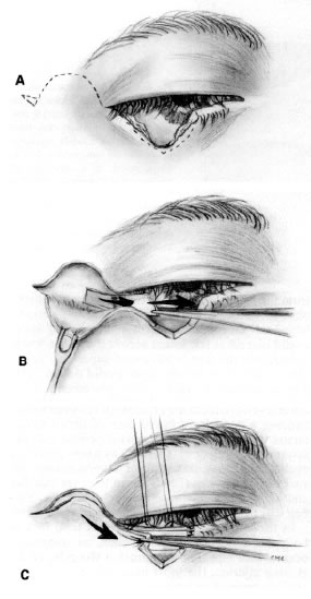
| Fig. 7. Repair of a full-thickness eyelid laceration with “freshening” of jagged wound edge followed by formation of a semicircular myocutaneous flap and cantholysis of the inferior limb of the lateral canthal tendon (A) A periosteal flap is used as a tarsal replacement to reattach the eyelid laterally (B). Conjunctiva can be mobilized and attached to either the superior or inferior edge of the periosteal flap (C). The myocutaneous flap is advanced to form the anterior lamella of the eyelid with the periosteal flap as the posterior lamella (C). Creation of a Burrow's triangle laterally prevents formation of a “dog ear” after flap advancement. |