Chapter 27
The Eye in Skin and Mucous Membrane Disorders
C. STEPHEN FOSTER
Main Menu   Table Of Contents

Search

BULLOUS-VESICULAR DISORDERS
PIGMENTARY DISORDERS AND DISEASES PRECIPITATED BY LIGHT
HYPERKERATOTIC DISORDERS
DISORDERS OF SKIN ELASTICITY
DISORDERS INVOLVING NEOPLASIA
MISCELLANEOUS SKIN DISORDERS
CONCLUSIONS
REFERENCES

Skin and eye are closely connected through at least three avenues:
  1. Skin and mucous membranes and corneal epithelium are derived from common embryologic ancestry, with the surface epithelium deriving from surface ectoderm and the subepithelial connective tissues and vascular components deriving from mesoderm.
  2. Juxtaposition between surface of the eye and edge of the eyelids can result in “innocent bystander” ocular damage when a particular disorder is affecting only the skin but is affecting the skin of the eyelids up to the lid margin.
  3. Certain immune disorders, such as cicatricial pemphigoid, eczema, xeroderma pigmentosum, and Vogt-Koyanagi-Harada syndrome, to name a few, may result in an autoimmune attack of both ocular structures, such as conjunctiva, and skin.

The importance of these shared properties between the eye and skin is relatively underappreciated by ophthalmologists, and it is the purpose of this chapter to emphasize this connection and its importance to the ophthalmologist, particularly the ability to diagnose systemic disease on the basis of its ocular manifestations. In addition, it is important for the ophthalmologist to recognize that proper care of the eye will categorically require concomitant care of the skin and treatment of the underlying systemic disease.

The diseases chosen for discussion here are primarily the more serious, potentially blinding skin disorders that can affect the eye, and some of the more common disorders with which the ophthalmologist will have contact on multiple occasions (e.g., rosacea). The reader is referred to other excellent resources for additional reading.1,2

Ocular abnormalities associated with disease of the skin and mucous membranes may involve any part of the eye or its adnexae. In this discussion, the dermatologic conditions will be grouped in broad categories, and the eye manifestations will be discussed with each disease as follows:

  Bullous-Vesicular Disorders
  Pemphigus
  Cicatricial pemphigoid
  Bullous pemphigoid
  Dermatitis herpetiformis
  Erythema multiforme
  Toxic epidermal necrolysis
  Epidermolysis bullosa
  Hydroa vacciniforme
  Acrodermatitis enteropathica
  Pigmentary Disorders and Diseases Precipitated by Light
  Incontinentia pigmenti
  Xeroderma pigmentosum
   Chédiak-Higashi syndrome
  Nevus of Ota
  Hyperkeratotic Disorders
  Ichthyosis
  Psoriasis
  Atopic dermatitis
  Disorders of Skin Elasticity
  Ehlers-Danlos syndrome
  Pseudoxanthoma elasticum
  Disorders Involving Neoplasia
  Basal cell nevus syndrome
  Juvenile xanthogranuloma
  Hemangioma
  Miscellaneous Skin Disorders
  Acne rosacea
  Anhidrotic ectodermal dysplasia
  Malignant atrophic papulosis
  Focal dermal hypoplasia syndrome

Back to Top
BULLOUS-VESICULAR DISORDERS

PEMPHIGUS

Description

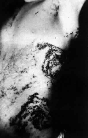
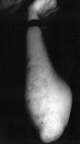
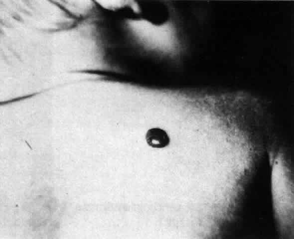
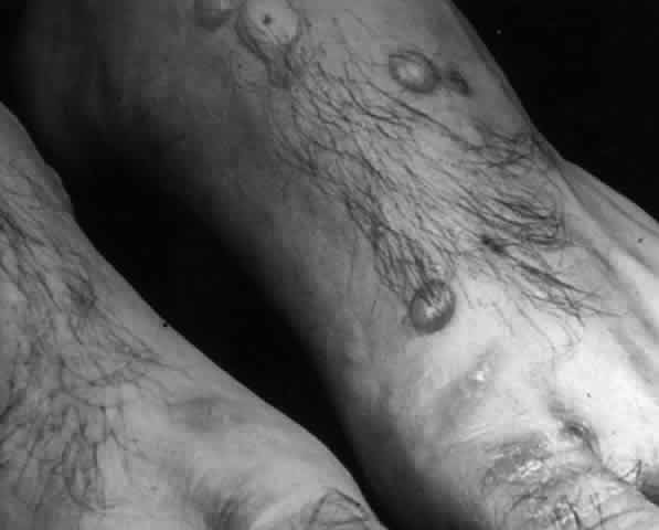
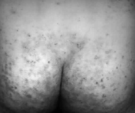
Pemphigus (from the Greek meaning puff or blister) comprises a group of autoimmune vesiculobullous eruptions of the skin and mucous membranes that are seen primarily in middle-aged people, with both sexes being affected equally.3 Involvement of mucosae may occur as the initial manifestation in as many as 25% of cases, and it occurs at some point during the course of disease in 95% of cases.4 Pemphigus vulgaris and its variant, pemphigus vegetans, constitute a more acute form of the disease in which the earliest lesions appear in the oral mucosa and are then followed by a bullous eruption of the skin over the trunk and extremities within 4 to 12 months (Fig. 1). This form of pemphigus was almost uniformly fatal prior to the use of systemic corticosteroid therapy. The mortality rate was reduced to approximately 40% by the use of steroids and has been further reduced to 5% by the addition of immunosuppressive chemotherapeutic regimens for the most difficult cases. Pemphigus foliaceus and its variant, pemphigus erythematosus, follow a more chronic but benign course. Mucosal lesions are unusual in this form, which is primarily an exfoliative dermatitis.3

Fig. 1. Pemphigus vulgaris. Bullae are flaccid, particularly when they enlarge. (Courtesy of Dr. Howard Bader, Massachusetts General Hospital)

The characteristic histologic feature of pemphigus is acantholysis or loss of interepidermal cell bridges. This leads to bulla formation within the epidermis. The epidermis may be readily dislodged by lateral sliding finger pressure, the Nikolsky sign, a clinical feature shared with toxic epidermal necrolysis and epidermolysis bullosa.4

In pemphigus vulgaris, the bullae form above the basal cell layer; in the foliaceus and erythematosus forms of pemphigus, such changes occur just below the stratum corneum. Marked acanthosis, with formation of intraepidermal eosinophilic abscesses, is present in pemphigus vegetans. These changes are found in biopsy specimens of both skin and mucous membranes.5

Epidemiology

Pemphigus vulgaris is by far the most common form of pemphigus, occurring predominantly in people of Jewish or Mediterranean origin, but it is reported in all races and ethnic groups, with no sex predilection.

Pathogenesis

Antibodies to intercellular components of epidermis have been demonstrated in the sera of patients with pemphigus.6 Tissue-fixed immunoglobulins are present in the interepidermal cell spaces, and a large percentage of these patients have circulating antibodies that react with an intercellular antigen present in many stratified squamous epithelia.5 These intercellular substance-reactive antibodies are typically IgG, are complement fixing, are pathogenic to tissue culture preparations of epidermal cells,7 and produce blistering skin lesions, identical to pemphigus vulgaris lesions, in mice injected intraperitoneally with the antibodies.8 The relevant antigen in the intercellular substance is a cadherin.9 The disease develops in the genetically susceptible individual, probably after he or she is exposed at some point in life to an environmental trigger. The disease is especially prevalent in Ashkenazi Jews, and 92% of these patients express the major histocompatibility complex class II alleles HLA-DR4, DQw8.10 It is presumed that because the disease is attributable to the presence of an antibody to an intraepidermal intercellular cement substance, the class II susceptibility gene controls the production of the antibody as a dominantly expressed immune response gene.

The intercellular cement substance against which the autoantibody is directed, pemphigus vulgaris antigen (PVA), is a novel cadherin, limited in tissue distribution to stratified squamous epithelia, with significant homology with the other known cell adhesion molecules within the cadherin family (E-cadherin, N-cadherin, P-cadherin, L-CAM, and DGI). PVA like DGI binds to plakoglobin, and with DGI forms a subfamily of cadherins; the other cadherins bind to catenions and to CAP102. DGI is the target antigen in pemphigus foliaceus and is present in all desmosome-containing tissues.9

Diagnosis

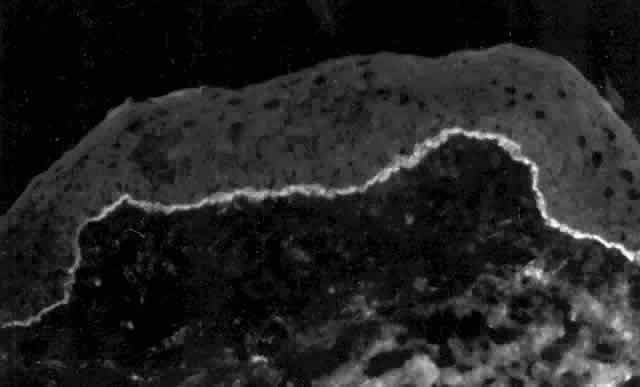
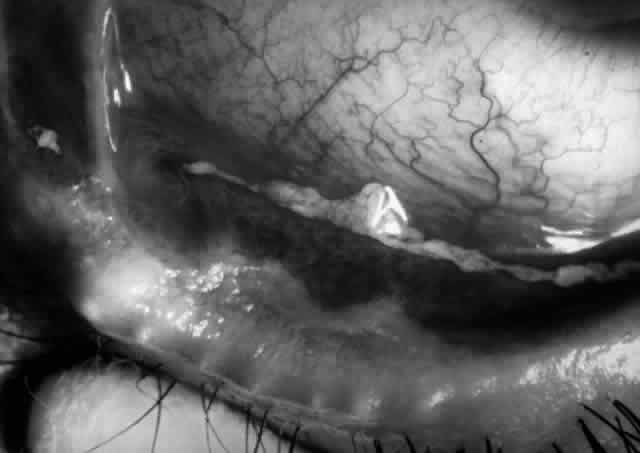
Pemphigus can be differentiated from bullous pemphigoid and from cicatricial pemphigoid on histologic and immunologic bases. Whereas the bullae in pemphigus are intraepidermal, those occurring in bullous pemphigoid and in cicatricial pemphigoid are subepidermal, between epidermis and dermis, or between mucosa and submucosa. Acantholysis is not present in the latter two conditions.5 In bullous pemphigoid and in cicatricial pemphigoid there are tissue-fixed immunoglobulins in the subepidermal basement membrane zone (Fig. 2).11 This is in contrast to the interepidermal fixation of antibodies (Fig. 3) seen in pemphigus (Table 1).

Fig. 2. Fluorescence microscopy of conjunctiva from a patient with cicatricial pemphigoid affecting the eye. The antibody used was a fluorescein-conjugated antibody directed against human IgG. Note the bright, continuous, linear deposition of IgG at the epithelial basement membrane zone, a finding virtually diagnostic of cicatricial pemphigoid.

Fig. 3. Immunofluorescence microscopy of conjunctiva from a patient with pemphigus vulgaris. The antibody is a fluorescein-conjugated antibody directed against human IgG. Note the bright staining of the intercellular cement, brightly outlining the borders of all of the epithelial cells.


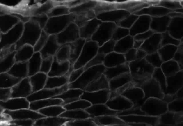
TABLE 1. Histologic and Immunologic Features of the Blistering Dermatoses


DermatosisTarget AntigenLocation of BullaeAcantholysisFixed ImmunoreactantsCirculating Antibodies to
Pemphigus vulgarisPVA cadherinIntraepidermalPresentIgG; C'PVA cadherin
Bullous pemphigoidBPA G1SubepidermalAbsentIgG, A, M; C'180 and 230
 BPA G2   kd/Hemi-desmosomes and BMZ lamina lucida
Cicatricial pemphigoid205 kd/BMZSubepidermalAbsentIgG, A, M; C'BMZ
Dermatitis herpetiformisAnchoring fibrilsSubepidermalAbsentIgAAnchoring fibrils
Erythema multiforme SubepidermalAbsentIgA-vessels 
Toxic epidermalnecrolysis IntraepidermalPresent  
Epidermolysis bullosa?SubepidermalAbsentIgG, A, M; C'Anchoring fibrils

PVA, pemphigus vulgaris antigen; C', complement components; BPA, bullous pemphigoid antigen; kd, kilodalton; BMZ, basement membrane zone.


Ocular Manifestations

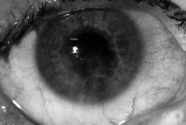
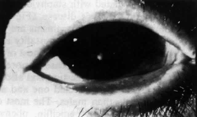
Ocular complications in pemphigus are rare,5 but ocular manifestations of pemphigus on lids, conjunctiva, cornea, lens, and iris have been reported. Conjunctival bullae are infrequently seen in pemphigus vulgaris. More typically, a catarrhal or purulent conjunctivitis develops.3 The ocular features are similar in pemphigus foliaceus and are usually confined to the palpebral conjunctiva, with erythema, edema, and purulent discharge.5,12 Pemphigus foliaceus may also produce entropion and trichiasis of both lids with resultant corneal damage. Lens opacities have been described in 5% of patients with a form of pemphigus foliaceus that is endemic to Brazil. Some of these patients also exhibit red, nodular iris lesions, the exact nature of which is not known.12

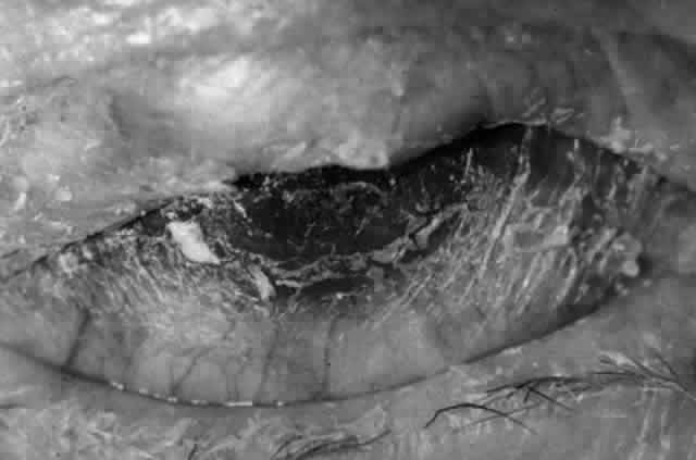
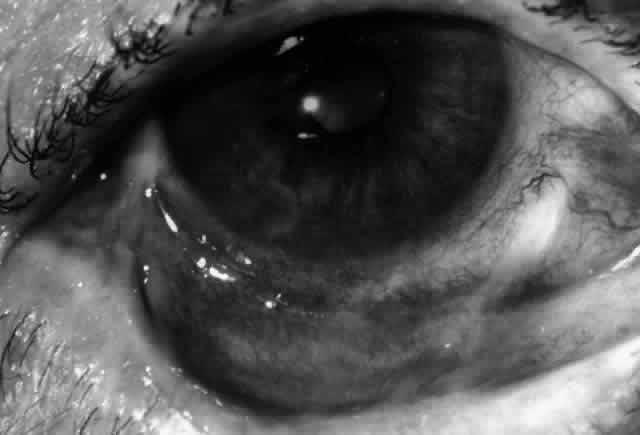
The conjunctival involvement in pemphigus may lead to symblepharon as a result of the formation and rupture of small vesicles. This does not, however, lead to progressive scarring and blindness, as is so often the case in cicatricial pemphigoid (Fig. 4).13

Fig. 4. Eye of a patient with ocular involvement in pemphigus vulgaris. Note the subepithelial fibrosis with fornix foreshortening and symblepharon formation. The degree of scarring stabilized since the patient's pemphigus vulgaris was brought under control 5 years before photograph was taken.

Therapy

Acute pemphigus vulgaris requires high-dose systemic corticosteroid therapy (100–200 mg prednisone/day), with gradual reduction of steroid dose with cessation of new blister formation.14 Infection is the leading cause of death in patients with pemphigus vulgaris; great vigilance for and vigorous treatment of any infection is obviously indicated. Immunosuppressive chemotherapy with methotrexate (15–25 mg once a week), azathioprine (2–3 mg/kg/day), or cyclophosphamide (2–3 mg/kg/day), used for long-term control of the disease, should be started once the risk of infection has diminished, with the usual caveats vis-a-vis monitoring for and managing drug toxicity.15,16

CICATRICIAL PEMPHIGOID

Definition

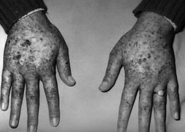
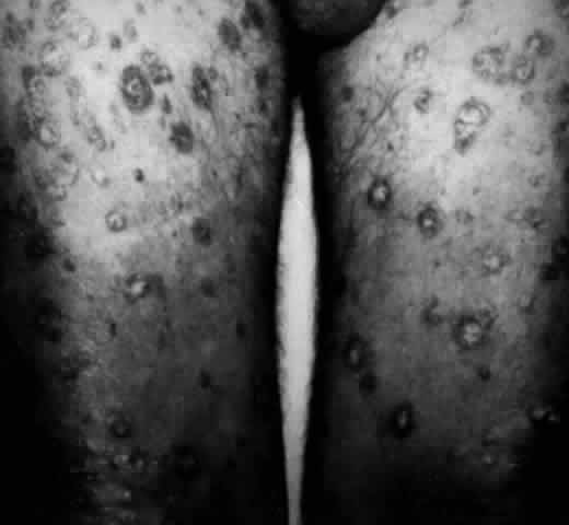
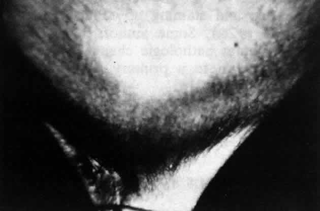
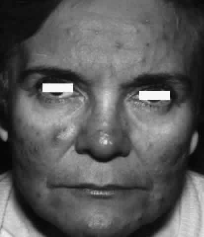
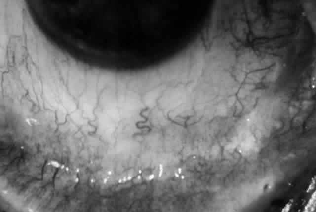
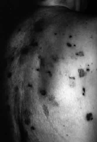
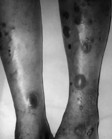
Cicatricial pemphigoid (CP) is a chronic cicatrizing autoimmune disease of the mucous membranes and skin. It is said to have an average age of onset of 65 years, but this figure under-reports the true epidemiologic features of this disease, because the cases are usually not in their earliest stages.17 Females are affected two to three times as frequently as males. Conjunctival involvement may occur as early as 10 years before other mucosal or skin lesions develop, or it may occur as late as 20 years after the onset of other lesions; the disease may be limited to the conjunctiva (Fig. 5). The scarring Brusting-Perry dermatitis occurs in approximately 25% of cases (Fig. 6), and cicatrizing conjunctivitis develops in 70% to 75%.17,18 Involvement of other mucosa may lead to scarring of the soft palate and oral and nasal mucosa, and esophageal, urethral, vaginal, and anal strictures may develop. Laryngeal involvement may cause pain and hoarseness,19 and the esophageal scarring can be lethal, because asphyxiation can result from a food bolus that is lodged during attempted swallowing by a patient with dysphagia from esophageal strictures that have been neglected by patient and physician alike.

Fig. 5. Eye of a patient with cicatricial pemphigoid, biopsy-proven, without extraocular manifestations. The disease has progressed to end stage and blindness. Note the total “leatherization” of the eye, with complete adherence of the eyelids to the globe.

Fig. 6. Brusting-Perry dermatitis of cicatricial pemphigoid affecting skin. Note the scarring characteristic of this dermatitis of cicatricial pemphigoid, a characteristic quite different from that of bullous pemphigoid.

Histologically, the conjunctival lesions show submucosal scarring,20 chronic inflammation, perivasculitis, and squamous metaplasia of the epithelium, with loss of goblet cells; mast cell participation in the inflammation is surprisingly great.

Tissue-fixed immunoglobulins and complement components are present in the epithelial basement membrane zone (BMZ) in patients with cicatricial pemphigoid20,21; indeed, such immunoreactant deposition is the sine qua non for definitive establishment of the diagnosis.20 Circulating antibodies to the basement membrane of conjunctiva are found in all of these patients if ultrasensitive radioimmunoassay techniques are employed,22 and circulating antibodies to conjunctival epithelium have been found (see Table 1).21,23,24 Circulating antinuclear antibodies (ANA) have also been demonstrated in patients with cicatricial pemphigoid.20,25

Epidemiology

Cicatricial pemphigoid is probably not quite as rare as published incidence figures indicate. Bittelheim26 estimated 1 in 15,000 ophthalmic patients; Hardy and Lamb27 and others28,29 estimated an incidence of 1 in 20,000 patients. Smith and colleagues30 estimated 1 in 46,000. Bedell31 estimated 1 in 8000, and Lever and Talbott32 stated frankly that it is impossible to make an accurate estimate from available data; they guess that the incidence of CP is somewhere between 1 in 12,000 and 1 in 60,000 ophthalmic patients. But these figures estimate the incidence of relatively advanced CP. Diagnosis of this disorder in its early stages is difficult,28 and most cases are not recognized as CP until they reach what we could categorize as stage III disease. There are, therefore, patients with stage I and stage II CP uncounted in the epidemiologic estimates.

Data on the average age of CP patients are also somewhat distorted by subtleties of the diagnostic signs. CP is said in many publications to be a disease of older people, with the average age given as 60)33 or 70)28 years. Although reports of CP in children may in fact represent cases of localized erythema multiforme, we certainly now recognize that CP can begin at least as early as the third decade of life.

Most reports describe a slight but clear female predilection. Thus, Klauder and Cowan34 reported 8 females and 3 males, Lever35 reported 3 females for every male, and Hardy and colleagues36 reported 52 females and 29 males with CP; 62 of these patients had ocular involvement, but Hardy and associates36 did not report the sex distribution for this subset of patients. No racial or geographic predilection is reported.36

Pathogenesis

Ocular cicatricial pemphigoid (OCP) is clearly an autoimmune disease with a genetic predisposition requirement and probably a “second-hit” environmental requirement to trigger the onset of the disease. We first reported an increase in the frequency of the HLA-DR4 and HLA-DQw3 alleles in OCP patients.37 Subsequently, in patient and family studies employing restriction fragment length polymorphism analysis of the DQw3 haplotypes, it was determined that the gene that provides enhanced susceptibility to OCP development is the HLA-DQw7 gene.38

The second-hit environmental trigger that stimulates the genetically susceptible individual to develop OCP may be microbial (we suspect that this is so for idiopathic OCP) or may be chemical, as in the case of so-called drug-induced or pseudo-OCP, which develops in some individuals exposed to practolol39 or to a limited variety of ocular medications.40–43 We know that patients with pseudo-OCP carry the same susceptibility gene (HLA-DQw7),44 but we further know that the autoantigens in these two forms of OCP are different. We have identified a 205-kd protein molecule in the basement membrane zone of conjunctiva and epidermis as the relevant target antigen in idiopathic OCP,45 and we have found that sera from patients with pseudo-OCP bind to 97- and 290-kd proteins in conjunctiva and epidermis lysates, and to 45-, 150-, 290-, and 400-kd proteins in dermal lysates.46 None of these autoantigen protein targets is absorbed from the tissue lysates by sera from patients with bullous pemphigoid, pemphigus vulgaris, or OCP, indicating that the autoantigens in OCP, pseudo-OCP, and bullous pemphigoid are distinct from each other.

The autoantibodies that develop in patients with OCP22 can be detected in all OCP patients when the disease is active, and the autoantibodies are probably pathogenic, just as the autoantibodies in patients with pemphigus are.7,8 The binding of the autoantibody to the autoantigen at the epithelial basement membrane then sets in motion a complex drama in which an impressive cast of characters participates.20 CD4 (helper) T lymphocytes far outnumber CD8 (suppressor) T cells in the inflammatory cell population that develops in the substantia propria of the conjunctiva; plasma cells, histiocytes, and mast cells are present in very large numbers, and a panoply of cytokines is elaborated from these cells, the effects of which are only now being dissected by molecular biologic techniques in our laboratory. However, the result of this complex drama is corneal epithelial damage from inflammatory cytokines and from the xerosis, meibomian gland dysfunction, and trichitic trauma developing as a consequence of cytokine-induced conjunctival fibroblast proliferation and activation, with resultant subepithelial fibrosis.

Diagnosis

The diagnosis of OCP is extremely important, given the natural history of the disease, the effectiveness but potential toxicity of therapy, and the potential confusion in differentiation from other causes of chronic cicatrizing conjunctivitis (Table 2). The clinical diagnosis requires immunohistochemical confirmation prior to institution of therapy20; the diagnosis is confirmed by the demonstration of one or more immunoreactants at the epithelial basement membrane zone (see Fig. 2).


TABLE 2. Causes of Cicatrizing Conjunctivitis


Cicatricial pemphigoidProgressive systemicd clerosis
Stevens-Johnson syndrome Sjögren's syndrome
Atopic conjunctivitisSarcoidosis
Rosacea conjunctivitisChemical burn
Adenoviral conjunctivitisTrauma
Diphtheria conjunctivitisToxic epidermal necrolysis
TrachomaEpidermolysis bullosa acquisita
Pemphigus vulgarisDermatitis herpetiformis
Porphyria cutanea tardaErythoderma ichthyosiform congenita


Ocular Manifestations

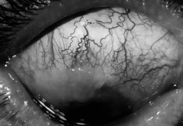
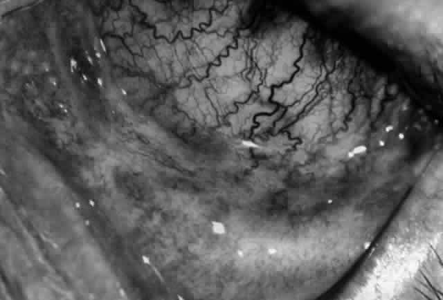
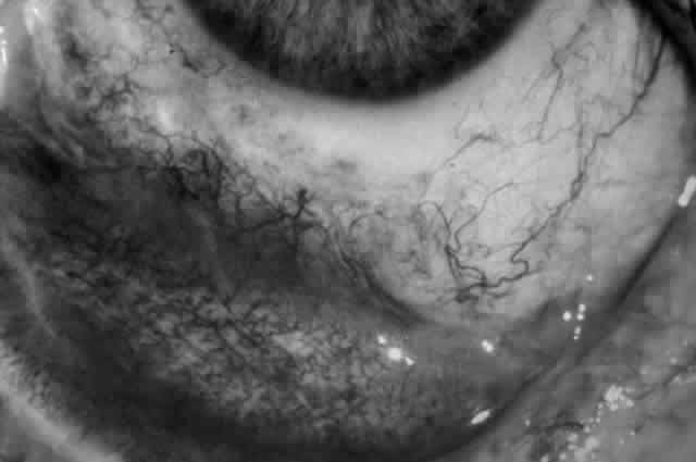
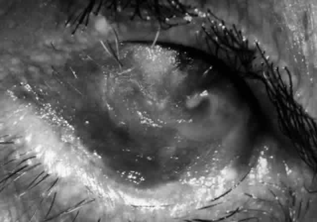
Clinically, the ocular disease in cicatricial pemphigoid (OCP) may present unilaterally in the form of a chronic, recurrent catarrhal conjunctivitis, but it eventually becomes bilateral. Subepithelial fibrosis is characteristic of stage 1 of OCP (Fig. 7). Stage 2 shows fornix foreshortening (Fig. 8), and symblepharon formation is the hallmark of stage 3 (Fig. 9). Stage 4, end-stage disease, is characterized by ankyloblepharon and surface keratinization (Fig. 10). Obstruction of the lacrimal ductules and meibomian gland ducts eventually produces an unstable tear film and progressive sicca syndrome, but it is to be emphasized that OCP is not a dry-eye syndrome until late in the disease course.20 Trichiasis and entropion occur because of the subepithelial fibrosis, with eventual keratopathy, corneal neovascularization, and corneal ulceration and scarring.20

Fig. 7. Stage 1 cicatricial pemphigoid, with cicatrizing conjunctivitis, and fine striae-type areas of subepithelial fibrosis, but without evidence of shrinkage of the conjunctiva.

Fig. 8. Stage 2 cicatricial pemphigoid, with fornix foreshortening and subepithelial fibrosis without frank symblepharon formation.

Fig. 9. Stage 3 of an eye affected by cicatricial pemphigoid. The conjunctival “shrinkage” continued and a frank symblepharon developed.

Fig. 10. Stage 4 cicatricial pemphigoid. Progressive shrinkage of the conjunctiva resulted in extreme trichiasis and distichiasis and keratopathy, with compromise of meibomian ductules and lacrimal ductules and the production of a totally dry eye.

In a series reported from the Mayo Clinic, 21 of 81 patients with cicatricial pemphigoid were blind at the time of the report; 17 of these patients were blind in both eyes. Sixty-two of the 81 patients had ocular involvement; approximately 88% of the 62 patients had the ocular disease bilaterally.18

Therapy

Medical treatment of the ocular complications of cicatricial pemphigoid was once difficult, protracted, and not very effective. Herron reported on the management of a case with ocular, skin, and mucosal involvement of the pharynx and larynx. Oral triamcinolone was used, along with injections of the drug into the areas of symblepharon. The skin and mucosal lesions responded in 10 weeks, but the ocular disease did not.47

CURRENT STATUS OF MEDICAL THERAPY PROGRAM FOR CP.

Our current approach to medical treatment of patients with active CP20 may be summarized as follows:

  1. Sicca syndrome is treated aggressively. We favor ointment lubricants without preservatives (e.g., Akwa Tears, Optilube, or Duolube ointments) applied at a frequency sufficient to maintain adequate surface lubrication and protection (e.g., every 2>4 hours and at bedtime). Artificial tears without preservatives (e.g., Refresh or Unisol drops) are employed as needed between ointment applications.d—Topical retinoid (0.01% tretinoin) ointment is used once or twice daily in one eye; we compare objective and subjective responses to the vitamin A ointment and continue treatment beyond 4 weeks of therapy only if we and/or the patient concludes that there is a distinct therapeutic benefit to this agent. Fewer than 30% of our CP patients have shown an apparent benefit from topical retinoids. We strongly caution against inappropriate enthusiasm for the efficacy of this component of the therapeutic plan. We observed the unfortunate results of such inappropriate preoccupation with topical therapy for this systemic disease in three patients who are now functionally blind because the disease progressed while they were on topical retinoids without concomitant appropriate systemic therapy.
  2. Chronic blepharitis and meibomianitis are treated with vigorous lid hygiene and oral doxycycline, 100 mg once or twice daily. Manual expression of plugged meibomian ducts is performed at the time of every evaluation. Microbial colonization of the lids and meibomian glands by any organism, including Staphylococcus, is treated with appropriate topical and/or systemic antimicrobial agents. We have discovered two cases of chronic Candida infestation and one of Nocardia.
  3. Chemotherapy for cases that are extremely active (4+ ) and rapidly progressive is begun with prednisone 1 mg/kg and cyclophosphamide 2 mg/kg/day. The cyclophosphamide dose is adjusted according to therapeutic response, bone marrow response, and drug tolerance. Prednisone is gradually tapered, with a switch to alternate-day dosing by 8 to 12 weeks and subsequent tapering and discontinuation by 4 to 6 months. We presently maintain patients on chemotherapy for a minimum of 1 year.

Dapsone and prednisone are used for cases that are less active and cases that are not rapidly progressive. The initial daily dose of dapsone we employ is 1 mg/kg; the maximum dose ever employed is 200 mg/day. We emphasize that dapsone is not a benign drug. We follow our patients who are on dapsone as often and as carefully as we do those who are on cyclophosphamide. Patients who are not glucose-6-phosphate dehydrogenase deficient, as determined by the screening test, may still hemolyze when treated with dapsone; therefore, the hemoglobin, hematocrit, and reticulocyte count, as well as the white blood count, are important monitoring parameters for these patients. We usually accept a low degree of hemolysis if dapsone is achieving the desired therapeutic response. However, reticulocytosis must adequately compensate for such hemolysis; a steadily falling hematocrit is unacceptable.

Approximately 70% of patients will respond well to dapsone. A large proportion of these (41% in our experience) will relapse within 6 months of cessation of dapsone therapy.48 We treat these patients with dapsone, methotrexate or azathioprine. Azathioprine, 2 mg/kg initial dose, is also our second-choice agent for the 30% of patients who do not respond adequately to dapsone. Adjustment of azathioprine dose is accomplished, as in the case of the other agents, through the judgment of the skilled chemotherapist, who, by virtue of training and experience, is an expert in the use of these agents.

Some authors have advocated surgical treatment in the early or “moist” phase of the disease: tarsectomy for correction of entropion, strip peritomy to provide an avascular barrier against corneal vascularization, superficial keratectomy for removal of corneal vascular and scar tissue, and fornix incision for release of symblepharon. All of these approaches not only will fail but in fact will make the disease worse if the patient is not adequately immunosuppressed prior to surgery. Mucous membrane grafting may be of benefit in cases of cicatricial entropion and lid marginal keratinization associated with this disease.49 Contact lenses and shells with concurrent use of lubricants may aid in the management of the chronic, dry phase of the disease with corneal involvement.50

BULLOUS PEMPHIGOID

Definition

Bullous pemphigoid (BP) is an autoimmune blistering dermatosis first recognized by Lever as a distinct entity in 1953.51 It clinically resembles, but is not identical to, pemphigus vulgaris (hence the name pemphigoid), and is one of four diseases on the spectrum of bullous pemphigoid, cicatricial pemphigoid, linear IgA disease, and herpes gestationis. The latter typically develops in pregnant and postpartum women, resolving within months and recurring with each pregnancy. Like pemphigus vulgaris, BP is characterized by skin bullae that may rupture; the disease can be fatal, usually as a result of sepsis.

Epidemiology

BP typically occurs in individuals of either sex between the ages of 50 and 80, without racial or geographic predilection. Its true prevalence and incidence are unknown.

Pathogenesis

A genetic predisposition to this disease, HLA-DQw7 accompanied by some (as yet unknown) environmental trigger, results in IgG autoantibodies that recognize and attach to 180-kd and 230- to 240-kd proteins associated with the hemidesmosomes and the lamina lucida of the epidermal basement membrane zone.52,53 The 230-kd autoantigen, or BPAG1, is a glycoprotein encoded on chromosome 6p,52 whereas the 180-kd autoantigen, or BPAG2, is a novel collagen encoded by a gene on the long arm of chromosome 10, at locus 10q24.3.53 These proteins or autoantigens are distinct from those recognized by the autoantibodies of cicatricial pemphigoid, pemphigus vulgaris, dermatitis herpetiformis, and epidermolysis bullosa.

The BP autoantibodies are complement fixing, and they are pathogenic. Fixation of the autoantibodies to the BP antigens, followed by complement fixation, results in complement split-product-mediated attraction of neutrophils and eosinophils, with release of their cytokines and production of tissue damage and blister formation.54 Mast cells also play a role in lesion development,55 and itch is a common symptom during acute phases of the disease. The disease is chronic, remitting, and recurrent when untreated, lasting from months to years, with little or no tendency for the healing skin lesions to scar. Death, however, may occur as a result of sepsis.

Diagnosis

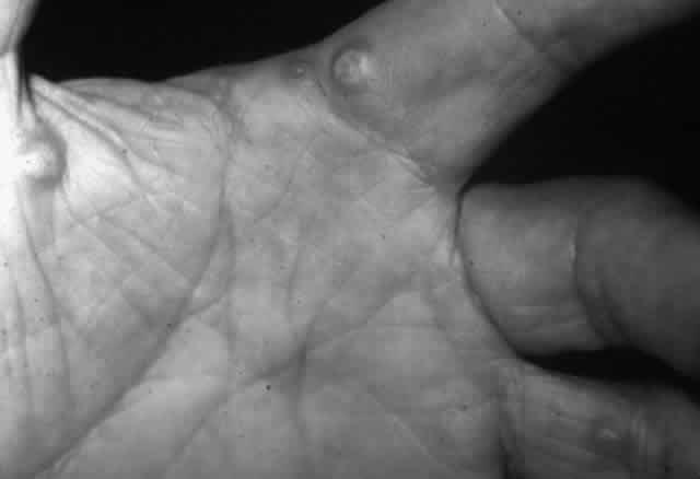
The diagnosis of BP is suspected in the patient (usually 50–80 years old) with a blistering dermatosis with large tense (rather than flaccid) bullae (on a normal or an erythematous base) that has a predilection for intertriginous areas, palms, soles, and abdomen (Fig. 11). Nikolsky's sign is negative. Mucosal lesions (blisters) may be present in approximately 30% of patients. The histopathologic characteristics of a biopsied skin lesion include the presence of subepidermal blister formation without acantholysis, with neutrophils, eosinophils, and lymphocytes in the blister cavity and a lymphocytic perivasculitis in the dermis. The hallmark immunopathologic feature of BP is the presence of IgG and the third component of complement (C3) at the dermal-epidermal (DE) junction and the presence of anti-BMZ antibodies, usually in high titer, in the serum of the patient. These can be detected by indirect immunofluorescence technique using guinea pig lip or monkey esophagus as substrate.

Fig. 11. Bullous pemphigoid. Note the tense bullae in this patient whose dermatitis is associated with bullous pemphigoid. This patient is in the erect position. If these were bullae of pemphigus vulgaris, they would “droop” because of their flaccidity.

Ocular Manifestations

Ocular manifestations of BP are not as impressive (Fig. 12) as those of cicatricial pemphigoid affecting the conjunctiva; therefore, they may go undetected. A study of 36 patients with BP in England disclosed that some had fine striae of tarsal subepithelial fibrosis, and one had frank symblepharon formation.56 Seventy-three percent of those who had conjunctival biopsy performed showed immunoreactant deposition at the BMZ.57

Fig. 12. Conjunctivitis and eyelid margin blepharitis in a patient with bullous pemphigoid. Note the small vesicle at the lid margin.

Therapy

The treatment of BP is essentially the same as that for pemphigus vulgaris, with systemic prednisone (50–100 mg/day) acutely and immunosuppressants, such as azathioprine, combined with alternate-day prednisone for long-term care.58 Sulfones may also be of some value in selected patients.59

DERMATITIS HERPETIFORMIS

Definition

Dermatitis herpetiformis (DH), first described by Duhring in 1884,60 is an autoimmune blistering dermatosis characterized by an intensely itching papulovesicular rash, especially on extensor surfaces, and associated (usually) with asymptomatic gluten-sensitive enteropathy.

Epidemiology

DH affects both sexes of any age, with peak incidence at ages 10 to 40 years. It is unclear how common this disease is.

Pathogenesis

Just as in cases of systemic lupus erythematosus, myasthenia gravis, celiac disease, and juvenile onset diabetes mellitus, dermatitis herpetiformis is associated with the HLA-B8, HLA-DR3, DQw2 phenotype. Such genetically predisposed individuals tend to make IgA autoantibodies directed against proteins in the dermal papillary tips, specifically in anchoring fibrils below the basal lamina.61 They may be provoked to make such antibodies through gastrointestinal contact with gluten, a protein in rye, barley, oats, and wheat.62 IgA and/or IgA-containing immune complexes deposit in the apices of the dermal papillae and activate complement via the alternative pathway, thereby attracting neutrophils, with resultant microabscess formation and the clinical appearance of the papulovesicular rash characteristic of DH.63

Diagnosis

Herpetiform (herpeslike) groupings of itchy papulovesicular eruptions on elbows, knees, shoulders, buttocks, and hands, preceded by an 8-hour itchy, burning prodrome, make the diagnosis nearly unmistakable (Fig. 13). The diagnosis is confirmed by the histopathologic findings, on lesion biopsy, of microabscesses in the apices of the dermal papillae, by the immunopathologic finding of granular IgA deposits in the same locus, and, most important, by the finding of granular IgA deposits in normal-appearing skin. The third component of complement (C3) is usually found along with IgA. Finally, the diagnosis is confirmed by the extremely rapid (within days) resolution of the lesions after starting dapsone therapy (see Treatment section).

Fig. 13. Dermatitis herpetiformis. Note the grouped vesicular and small bullous arrangement of this dermatosis, which is intensely pruritic.

Ocular Manifestations

Ocular manifestations of dermatitis herpetiformis are rare. Keratoconjunctivitis sicca, iritis, choroiditis, and papillitis have occurred in individual patients, but the causal relationship between dermatitis herpetiformis and the ocular problem is debatable.

Treatment

Dapsone revolutionized the care of patients with DH, with resolution of symptoms and new lesion development within 48 hours of initiation of therapy.64 The usual caveats regarding dapsone therapy vis-à-vis sulfa allergy and glucose-6-phosphate dehydrogenase deficiency must obviously be kept in mind. The dose of dapsone sufficient to induce remission, 100 to 300 mg per day, can usually be tapered to very low doses for maintenance of remission. Equally important, however, is the strict adherence to a gluten-free diet if one is to eliminate the dependence on medical therapy.65

ERYTHEMA MULTIFORME

Definition

Erythema multiforme (EM) is an acute inflammatory disease affecting primarily skin and mucous membranes.66 Major and minor variants exist, and it is the former, also known as Stevens-Johnson syndrome (SJS), that is of most concern to ophthalmologists.67 Prodromal symptoms of SJS include general malaise, myalgias, and fever.68 Skin lesions develop by 10 days to 4 weeks after the onset of the prodrome; they tend to be annular in shape (“iris lesions”) but may be maculopapular or bullous. Most commonly, involvement is limited to the skin of the dorsum of the hands and feet and the palms and soles. Later, the limbs, trunk, face, and neck may become involved (Fig. 14). EM major or SJS may be fatal, either from the systemic, visceral involvement of the disease or from complications arising from the extensive exfoliation of skin, producing consequences similar to those seen in patients with third-degree burns. The mortality rate is reported to be between 5% and 15% of cases, and 20% of cases recur.66

Fig. 14. Erythema multiforme major. This patient has oral mucous membrane involvement in addition to the classic target dermatitis lesions on his arms.

Epidemiology

SJS, although it has no racial or geographic predilection, occurs in young males more commonly than in other groups of individuals, with winter and spring seasonal peaks.67 The disease is relatively common, accounting for up to 1% of dermatologic outpatient visits.68,69

Pathogenesis

Histologically, the skin lesions exhibit diffuse inflammatory infiltration and edema of the dermis. When bullae are present, they are subepidermal in location and without acantholysis.66

The exact pathogenetic mechanism of this disease is unknown; autoimmune, infective, and allergic factors have all been considered possible causes. Most evidence now favors a hypersensitivity to microbes and drugs.70 Erythema multiforme has been associated with various bacterial (especially Mycoplasma pneumoniae), viral (especially herpes simplex virus), mycotic, and protozoan infections, with drugs and vaccines, with allergic contactants, with collagen disorders, and with internal malignancies. Some of the incriminated precipitating drugs include sulfonamides, tetracyclines, penicillin, bromides, iodides, salicylates, phenytoin sodium, barbiturates, phenylbutazone and other nonsteroidal antiinflammatory drugs, cortisone, and vaccines against polio, smallpox, influenza, diphtheria, and tetanus. The development of or exacerbation of Stevens-Johnson syndrome has been reported after ophthalmic application of proparacaine,71 sulfonamide,72 tropicamide,73 and scopolamine.73

Herpes simplex virus (HSV) is a vastly underappreciated cause of EM,74 with 15% to 63% of cases associated with this virus.75,76 Recurrent EM is especially associated with recurrent HSV infection,77 and herpes simplex virus has been isolated from the skin lesions of erythema multiforme.78

The results of our research,79 as well as that of others,80–87 suggest that episodic showers of circulating immune complexes may result in immune complex deposition in previously damaged, predisposed vessels, with resultant immune complex vasculitis/perivasculitis with lymphocyte and neutrophil participation in the vessel damage. Leakage of plasma and cells may progress to subepidermal blister formation and to necrosis of the epidermis over the blister.66

Diagnosis

The diagnosis of SJS is not difficult, given the dramatic characteristic skin and mucous membrane lesions.

Ocular Manifestations

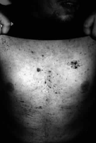
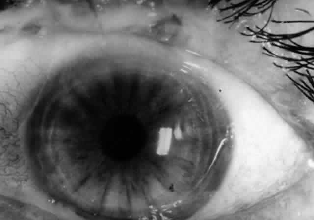
Ocular involvement is common in Stevens-Johnson syndrome, occurring in more than half of patients with this disease.70 The acute ocular manifestations vary from mild mucopurulent conjunctivitis to severe, pseudomembranous conjunctivitis leading to symblepharon, lid scarring, keratoconjunctivitis sicca, corneal ulceration, corneal scarring, and neovascularization. The chronic consequences of SJS depend on the degree of subepithelial fibrosis and tarsal conjunctival keratinization that occurs as a result of the acute exanthem. The former (fibrosis) results in variable degrees of meibomian duct and lacrimal ductule obstruction (with resultant deficient or unstable tear film problems), trichiasis, distichiasis (with resultant chronic abrasive trauma to the cornea), and cicatricial entropion; tarsal conjunctival keratinization results in a chronic sandpapering trauma effect on the cornea with each blink. The combined effects of these external aggravants produce chronic keratopathy, with eventual corneal scarring, neovascularization, or ulceration (Fig. 15).

Fig. 15. Eye of a patient with Stevens-Johnson syndrome. Note the conjunctivitis, the subepithelial fibrosis, the symblepharon formation, and the zone of variable width, posterior to the gray line of the eyelid that is obviously keratinized.

One other chronic, rare ocular manifestation of SJS may develop: recurrent, immunologically mediated conjunctivitis. Just as such recurrent episodes may occur in skin87 and in oral mucosa,88 so too may they occur in conjunctiva.79 The diagnosis of this phenomenon is not easy and depends on eradication of all external aggravants, eliminating these as confounding variables, and then demonstration, by immunohistochemical techniques employed on a biopsy specimen from the inflamed conjunctiva, of a lymphocytic microangiopathy with IgA deposition in the vessels. Herpes simplex virus is one inciting agent for recurrent SJS, and this possibility should be carefully investigated.

Therapy

Therapy of the acute phase of the ocular disease includes use of topical steroids as well as topical antibiotics applied frequently to combat superinfection, especially that from Staphylococcus aureus and (if the patient is hospitalized in a burn unit) Pseudomonas species. Lubricants should be generously employed, especially if lid involvement has resulted in even mild lagophthalmos. If anterior uveitis is present, mydriatic therapy is appropriate. Howard has pointed out that the pseudomembranes should not be stripped, because doing so leads to bleeding and increased scarring.89 Lid-globe adhesions should be lysed twice daily to maintain the fornices. I use my scleral depressor for this purpose after cleaning it with 70% isopropyl alcohol, having found no advantage to use of a glass rod. In an effort to prevent corneal scarring, Arstikaitis has advocated early tarsorrhaphy before deep stromal vessels invade the cornea.90 This is appropriate in instances of lagophthalmos and persistence of corneal epithelial defects.

The chronic phase of ocular involvement in Stevens-Johnson syndrome necessitates caring for the consequences of the acute exanthem: punctal occlusion for sicca syndrome, electro- or cryoepilation for trichiasis, soft or scleral contact lens therapy for chronic keratopathy arising from residual trichiasis and from tarsal conjunctival keratinization, mucous membrane grafting, and corneal surgery for preservation of globe integrity and, in rare instances, for attempted visual rehabilitation.91

Recurrent SJS may respond to chronic systemic acyclovir therapy if herpes simplex virus is the agent provoking the attacks. Some cases of recurrent SJS not associated with herpes have responded to long-term systemic immunosuppressive chemotherapy.79

TOXIC EPIDERMAL NECROLYSIS

Definition

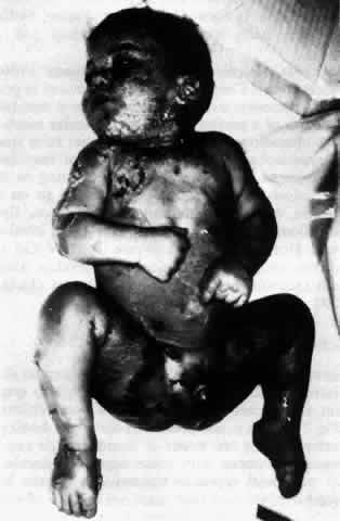
Toxic epidermal necrolysis (TEN) (Lyell's disease, Ritter's disease, scalded skin syndrome) is an acute systemic illness associated with a bullous eruption of the skin and mucous membranes that is, in many respects, similar to Stevens-Johnson syndrome.92–94 Lyell's disease presents with a prodromal fever accompanied by malaise and tender or painful skin. The skin of the face, trunk, and extremities becomes inflamed; within 6 to 24 hours, the epidermis separates in large sheets (Fig. 16). Large, flaccid bullae may develop within the epidermis; these are mobile, and Nikolsky's sign is positive. In contrast, the bullae that are present in Stevens-Johnson syndrome are subepidermal in location and are tense and nonmobile.94 The bullae of Lyell's disease usually remain sterile, even in children in whom staphylococcal infection is associated with the disease. Therefore, it is not a pyoderma. The mortality rate is approximately 10%, and most of these fatalities are in infants.

Fig. 16. Toxic epidermal necrolysis. (Courtesy of Dermatology Service, WRAMC)

Epidemiology

TEN has no ethnic or geographic predilections, and it can affect patients of any age. The adult type of disease is more commonly associated with drug ingestion and affects all ages, although females are affected one and a half times more frequently than males.

Pathogenesis

The disease may affect all age groups, but when seen in infants and children under 10 years of age, it is usually associated with staphylococcal infection and is called Ritter's disease (see Fig. 16). The most commonly involved organisms are phage group 2 staphylococci.92 The drugs often associated with this disease are penicillin, phenytoin, sulfonamides, phenolphthalein, and phenylbutazone. A case has been described in a patient on allopurinol therapy.95 The mortality rate in this group may be as high as 40%.93

Diagnosis

TEN secondary to drug or viral exposure is diagnosed by the history of such exposure or prodrome and the clinical picture of generalized bullae formation and exfoliation. Nikolsky's sign is positive, and lesion biopsy discloses blister formation with a cleavage plane high in the epidermis, in the granular cell layer, with little associated inflammatory reaction.96

Ocular Manifestations

Ocular involvement in TEN is limited to the conjunctiva; it is usually less severe than is the ocular involvement in Stevens-Johnson syndrome. A mucopurulent conjunctivitis is the most common early manifestation, and, unlike the involvement in erythema multiforme, this does not usually result in ulceration, scarring, symblepharon, or corneal pannus formation.70 It may do so, however, with corneal erosions and all the other consequences described for SJS.93,94

Therapy

The acute and chronic therapy for the ocular involvement in TEN is identical to that described above for SJS.

EPIDERMOLYSIS BULLOSA

Definition

The term epidermolysis bullosa (EB) refers to a group of hereditary, and one acquired, bullous eruptions of the skin and mucous membranes that are characterized by the formation of blisters and erosions at sites of injury.97 There are several recognized forms of hereditary epidermolysis bullosa (Table 3). Ocular involvement may occur in many of these disorders, including the acquired variety, epidermolysis bullosa acquisita.98–104


TABLE 3. Forms of Epidermolysis Bullosa


VariantInheritanceSite of LesionClinical Features
Simplex   
LocalizedAutosomal dominantIntraepidermal-suprabasalHands and feet, heals without scard
GeneralizedAutosomal dominantIntraepidermal-basal cellsHeals without scar
OgxaAutosomal dominantIntraepidermalLinkage to erythrocyte GPT
HerpetiformisAutosomal dominantIntraepidermalSevere, life-threatening at birth
Junctional (Atrophic)   
LetalisAutosomal recessiveLamina lucidaHeals without scar, death by age 2
BenignAutosomal recessiveLamina lucidaHeals with atrophy
LocalizedAutosomal recessiveLamina lucidaLegs, feet and nails
Dystrophic   
PasiniAutosomal dominantBasal laminaScarring, loss of nails
CockayneAutosomal dominantBasal laminaScarring, loss of nails
Touraine   
HallopeauAutosomal recessiveBasal laminaLoss of digits, eye involvement
Siemens  Mouth involvement
Acquired-Sub-lamina densaBlisters at sites of trauma, mouth and eye involvement

GPT, glutamic-pyruvic transaminase.


Symptoms in the hereditary form of EB tend to appear shortly after birth or in early childhood, and they reappear periodically during the patient's life. Epidermolysis bullosa simplex is the mildest form, and only 2% of these patients have any mucosal involvement. The mucosae are not involved in the hands-feet form. The recessive forms, epidermolysis bullosa letalis and dysplastic epidermolysis bullosa dystrophica, have mucosal involvement uniformly; conjunctival disease, although less common than oral disease, may be severe.105

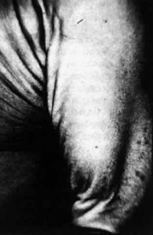
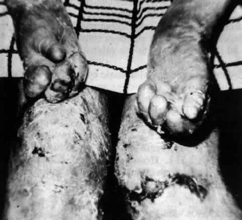
Epidermolysis bullosa dystrophica is characterized by scarring of the skin over limbs, hands, and feet. The scars become keloidal, resulting in deformities of the limbs, hands, and feet; these are most severe in the dysplastic form (Fig. 17). The most severe involvement of mucous membranes also occurs in the dysplastic, recessive form of epidermolysis bullosa dystrophica.106

Fig. 17. Epidermolysis bullosa dystrophica. (Courtesy of Dermatology Service, WRAMC)

Epidemiology

Males and females are affected equally by the disease,97 and there are no apparent racial, ethnic, or geographic predilections.

Pathogenesis

Epidermolysis bullosa is an autoimmune disease spectrum in which the patient produces an autoantibody reactive against protein(s) located in the epidermal sub-basal lamina anchoring fibril zone. Direct immunofluorescence studies of biopsied affected skin typically show IgG, IgA, IgM, and complement components deposited in a prominent linear distribution along the basement membrane zone (BMZ) of the epidermis. Subepithelial blister formation occurs as the anchoring fibrils are damaged and the basal epidermal cell attachments to the BMZ are lost.

Diagnosis

The diagnosis of EB is based on the clinical picture of blister formation occurring predominantly over joint surfaces, hands, feet, and other sites of minor trauma, with negative Nikolsky's sign and immunopathologic features on lesion biopsy as described above. The lesions occur predominantly on noninflamed skin and heal with scar and milia formation. Immunoelectron microscopy demonstration of immunoreactant deposition below the basal lamina distinguishes the lesion definitively from those of bullous pemphigoid.

Ocular Manifestations

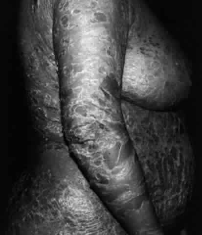
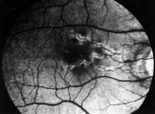
Conjunctival involvement in epidermolysis bullosa begins with vesicle formation, leading to ulceration and symblepharon formation.107 In the cornea, recurrent erosions occur that lead to vascularized scars (Fig. 18). Histologically, the epithelium exhibits vacuolation, particularly of the basal cell layer. The epithelium is poorly adherent to Bowman's membrane, whereas the remainder of the corneal structure is essentially normal.108 McDonnell reported that 8 of 11 patients with the dystrophic form of epidermolysis bullosa had ocular abnormalities, including conjunctival shrinkage, with symblepharon formation and corneal opacities.109 The authors remarked that these abnormalities were slowly progressive, and that except for recurrent corneal erosion, the patients were ocularly asymptomatic. Other ocular histopathologic changes have been noted in epidermolysis bullosa letalis by Aurora and co-workers.110 They have described a patient exhibiting edema of the trabeculum, iris, ciliary body, lens, and optic nerve. Focal edema and necrosis of the choroid and retina as well as total retinal detachment were also present in this patient. Citing research by other workers, these authors suggest that some of the ocular changes observed in epidermolysis bullosa may be due to focal accumulation of a proteolytic enzyme, possibly collagenase. Zierhut and associates, using modern criteria for the definitive diagnosis of epidermolysis bullosa acquisita (EBA), reported the case history of a 23-year-old man with EBA who developed bilateral tiny corneal subepithelial vesicles.111

Fig. 18. Ectropion and corneal ulcer in epidermolysis bullosa. (Courtesy of Dr. GR Beauchamp)

Therapy

All of the epidermolysis bullosa diseases are generally resistant to therapy. Supportive care, including vigorous therapy of infection, is recommended.112 The disease is chronic, but its protracted course may eventually end in disease remission. Avoidance of trauma, expert skin care, and proper nutrition are critical. Scattered reports of therapeutic benefits of supplemental vitamin E,113 phenytoin,114–117 retinoids,118 and corticosteroids have surfaced in the past two decades.

HYDROA VACCINIFORME

Definition

Hydroa vacciniforme (HV) is a particularly severe recurrent vesicular skin eruption that is related to polymorphic light eruption. It occurs in childhood, beginning in the first 4 years of life and disappearing soon after puberty. The disease is more common in boys and is manifested as large, recurrent, umbilicated bullae on skin areas that are exposed to the sun. These lesions affect mainly the face, nose, cheeks, ears, and the extensor surfaces of the arms and hands. As they heal, the lesions undergo an encrusted stage that makes them similar in appearance to the lesions of vaccinia. The lesions of hydroa vacciniforme usually heal with residual scarring.119 Histologically, the blisters exhibit necrosis of the epidermis as well as of the underlying dermis. Focal areas of necrosis are surrounded by inflammatory cells.120

Epidemiology

Hydroa vacciniforme is a very rare disorder affecting boys more commonly than girls. It is generally worse in summer because of the relationship to light exposure.

Pathogenesis

The cause of this disease, other than exposure to the sun, is unknown. An essentially identical photosensitivity eruption is seen in patients with porphyria, but porphyrin metabolism in patients with hydroa vacciniforme is normal. A similar disease, hydroa estivale, heals without scar formation.120

Diagnosis

Hydroa vacciniforme is diagnosed by the typical clinical picture.

Ocular Manifestations

Ocular complications may accompany hydroa vacciniforme. Perilimbal chemosis or severe vesicular reaction with conjunctival ulceration and necrosis may occur with resultant formation of symblephara. Corneal involvement may occur, with vesicular keratitis developing in the interpalpebral zone. These lesions may progress to epithelial erosion, vascularization, and scar formation. Bennion and associates reported inflammatory keratitis and uveitis (with stellate keratic precipitates) in a 6-year-old boy with hydroa vacciniforme, and they observed that development of new eye lesions was prevented by the use of sunglasses containing UVB filter.121

Eyelid lesions are usually a part of the facial involvement, and if severe and recurrent, cicatricial ectropion may develop, along with secondary conjunctival and corneal changes.122

Therapy

Therapy for hydroa vacciniforme is the same as for polymorphic light eruption, with sunscreen protection of exposed skin, antimalarial and topical steroid therapy, and oral psoralen photochemotherapy (PUVA).123

ACRODERMATITIS ENTEROPATHICA

Definition

Acrodermatitis enteropathica is a rare familial skin disorder that presents as a vesiculobullous eruption around body orifices in infants and children (Fig. 19). It is not present at birth but develops during the first few weeks or months of life and is usually associated with some degree of diarrhea. An auto-somal recessive transmission pattern has been identified, and both sexes are equally affected by the disease. These patients have been found to have a zinc deficiency, and prior to the introduction of therapy with diiodohydroxyquin, the disease was uniformly fatal. Oral zinc therapy is being successfully used, and virtually all patients now survive.124,125

Fig. 19. Acrodermatitis enteropathica. (Courtesy of Dermatology Service, WRAMC)

In addition to involvement around the mouth, ears, anus, and genitalia, the disease may affect the skin of the knees, elbows, and paronychial areas. The vesicular lesions are eventually replaced by erythematous psoriasiform plaques in the involved areas, and complete alopecia is a common feature of the disease.124

Epidemiology

Acrodermatitis enteropathica is a rare inherited, autosomal recessively transmitted disease affecting males and females equally. It has a worldwide distribution without racial, ethnic, or sexual predilection.

Pathogenesis

The disease develops as a result of an impairment of zinc presentation and/or absorption by the intestinal mucosal villus brush border.

Diagnosis

The triad of acral dermatitis, alopecia, and diarrhea, coupled with discovery of depressed serum zinc levels (below 80 μg/dL), establishes the diagnosis. Zinc absorption studies, employing radioactive zinc isotypes, may be performed in equivocal cases.

Ocular Manifestations

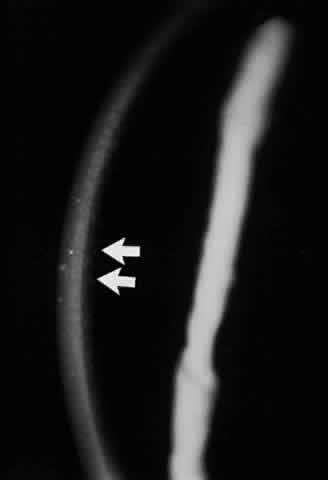
Ocular findings in acrodermatitis enteropathica include photophobia, blepharitis, punctal stenosis, conjunctivitis, and corneal opacification.126 Warshawsky and co-workers have described bilateral radiating linear superficial opacities in the periphery of the corneas in a 7-year-old boy with the disease. Histologically, the corneal epithelium was thickened and edematous in areas, with fragmentation of the underlying basement membrane and focal loss of Bowman's membrane. This child had been treated with diiodohydroxyquin and later with iodochlorhydroxyquin intermittently for 6 years.127

Therapy

Oral zinc therapy (50 mg Zn+ + /day) is associated with survival of all treated patients.

Back to Top
PIGMENTARY DISORDERS AND DISEASES PRECIPITATED BY LIGHT

INCONTINENTIA PIGMENTI

Definition

Incontinentia pigmenti (IP) is an inherited disorder of skin pigmentation that is associated with skeletal, dental, ocular, and central nervous system abnormalities. The disease is transmitted as an auto-somal dominant or sex-linked trait. It is usually seen in females, but a few cases have been reported in males who have survived to adulthood.128

Skin lesions are usually congenital or appear by the end of the first week of life, and they rarely appear after the second month. The lesions evolve, beginning first as tense bullae, frequently appearing on the limbs in linear groups. Smooth red nodules then appear on the trunk and limbs, followed by linear, warty lesions on the backs of the hands, feet, fingers, and toes. Pigmentation develops in the form of wavy, slate-gray or brown macules arranged in whorls or linear patches (Fig. 20).129

Fig. 20. Incontinentia pigmenti. (Courtesy of Dermatology Service, WRAMC)

Two forms of incontinentia pigmenti are recognized. The Naegeli type affects both sexes and is not congenital; its onset is usually around 2 years of age. This form manifests a reticulated cutaneous pigmentation without other abnormalities.129 The Bloch-Sulzberger type occurs almost exclusively in females, is congenital, and is associated with systemic disorders such as epilepsy, spastic paralysis, mental deficiency, congenital heart disease, chondrodysplasia, alopecia, and ocular and dental defects. A case has also been reported in association with pheochromocytoma.130

Histologically, the skin in the final stage exhibits increased amounts of melanin pigment within macrophages located in the upper dermis. The basal cell layer shows less melanin pigment than normal. These cells show degeneration and vacuolation and are incontinent of pigment.

Epidemiology

The disease is rare but ophthalmologically important. It occurs soon after birth, more commonly in girls (97% of cases). The disease is usually reported in whites, but individual cases in Asian, Indian, and African people have also been reported.130

Pathogenesis

The cause of this hereditary disease is unknown.

Diagnosis

The diagnosis of incontinentia pigmenti is based on the typical clinical picture.

Ocular Manifestations

Ocular involvement occurs in approximately 35% of patients with Bloch-Sulzberger syndrome.131,132 Cataracts, optic atrophy, strabismus, and nystagmus have been reported; in one third of those patients with ocular involvement, a unilateral retrolenticular mass is present that is clinically difficult to differentiate from retrolental fibroplasia.133 Zweifach has described retinal dysplasia with pigment proliferation and free pigment epithelial cells within the dysplastic retina, noting it to be a common finding in incontinentia pigmenti.134 Leib and Guerry described an exudative chorioretinitis and exudative retinal detachment in association with the disease.135

Pigmentary retinopathy is a frequent finding in incontinentia pigmenti, and this has been described as small, patchy, mottled areas of depigmentation presenting diffusely throughout the fundus.131,136 Menshena-Manhart and colleagues have described retinal pigment epithelial changes in the disease and have suggested that the underlying defect may exert a primary effect on the retinal pigment epithelial cells.137 Retinal vascular anomalies were present in 37% of a group of 19 patients reviewed by Watzke and associates.132 These consisted of arteriovenous anastomoses, associated with preretinal fibrous changes at the temporal equator. Peripheral to these lesions, the retina was not perfused. Jain and Willetts have described a case demonstrating frank proliferative neovascularization.138 These lesions may represent the early formation of the pseudoglioma that has been reported in Bloch-Sulz-berger syndrome. Spallone reported on the clear X-linked dominant genetic transmission of this disease and emphasized that his study of seven members from one family highlights the fact that vascular abnormalities of the retina and disorders of the retinal pigment epithelium are the most important ocular lesions in this disease.139

Conjunctival pigmentary changes have also been described in incontinentia pigmenti. McCrary and Smith examined a patient who had pigmentation that was confined to the interpalpebral zone of the bulbar conjunctiva. The pigmented lesions were near the limbus, were flat and dark brown in color, and resembled a “stellate cluster of nevi.”136 Histologically, these authors noted migration of pigment granules from the basal layer of the epithelium into the underlying subepithelial tissue, a finding similar to the changes in skin.

Therapy

There is no specific treatment for this disease.

XERODERMA PIGMENTOSUM

Definition

Xeroderma pigmentosum (XP) is a rare autosomal (or rarely sex-linked) recessive premalignant condition of the skin first described by Hebra and Kaposi in 1874.140 The skin is normal at birth, with the first signs of the disease presenting as increased dryness and freckling on skin areas with the greatest light exposure, particularly the face. Seventy-five percent of patients show the first skin changes between 6 months and 3 years of age. Skin tumors present in the affected areas, usually before 20 years of age (Fig. 21). Basal cell and squamous cell epitheliomas are the most frequently encountered lesions, but melanoma, angiosarcoma, and fibrosarcoma may occur. The disease may take an acute form in which malignant transformation occurs early, leading to death as early as 3 years of age. The most common form is subacute in which malignant transformation occurs after 8 to 10 years. In the chronic form, the life span may be 20 years or more.141 Patients with XP have an estimated 2000-fold increase in cutaneous neoplasm frequency when compared with the general population,142 and neurologic involvement from primary neuronal degeneration occurs in 18%.143

Fig. 21. Xeroderma pigmentosum. Notice the typical “freckles” and other pigmented lesions on the skin of the patient's hands.

Epidemiology

XP is a rare autosomal recessive dermatosis affecting 1 in 250,000 individuals in the general population. It affects males and females equally, has a worldwide distribution, and has no racial or ethnic predilections.

Pathogenesis

It is now believed that the pathophysiology in xeroderma pigmentosum is related to defective repair of actinic damage to DNA in skin cells. Cleaver has shown that fibroblasts from the skin of patients with the disease show either absent or markedly reduced DNA repair replication when compared with normal fibroblasts.144 He has suggested that alterations in the DNA occurring as a result of this defective repair may render the affected cells malignant. This damage to the cells is brought about by sunlight or ultraviolet light.

Histologically, in xeroderma pigmentosum, the epidermis is atrophic and there is basophilic degeneration of the dermal collagen, with irregular proliferation of dermal papillae and increased pigmentation.144

Diagnosis

The diagnosis of XP is not difficult, given the typical clinical picture and the hereditary characteristics of the disease. There is no diagnostic test for the condition.

Ocular Manifestations

More than 70% of patients with XP exhibit moderate to severe eye complications.145 Ocular changes may present as the earliest manifestation of the disease and may range from photophobia and blepharospasm with conjunctival hyperemia to severe corneal disease with ulceration or perforation.146 The most common ocular manifestation of the disease is a progressive atrophy of the lower eyelid. This begins, typically, at the lid margin and progresses to loss of the entire eyelid. The resulting exposure leads to inflammation, symblepharon, corneal ulceration, and corneal scarring.147 Histologically, the corneal changes are those of degenerative pannus.148 Conjunctival involvement begins as hyperemia with serous or mucopurulent discharge and may progress to xerosis, keratinization, and shrinkage that leads to ankyloblepharon.147 Corneal malignancy is seen in this disease, and the limbal zone appears to be a common site for involvement, usually by squamous cell carcinoma.149,150 Xeroderma pigmentosum has also been associated with band-shaped nodular corneal dystrophy in black patients.151

El-Hefnawi and Mortada have reviewed the ocular complications in a series of 46 patients with xeroderma pigmentosum.150 In this series, 91% presented with photophobia and differing degrees of conjunctivitis; 61% exhibited keratitis of varying degrees during their illness. Corneal opacification resulted in 22 of the 46 patients. Prior to our report,152 eight patients with XP in whom corneal transplantation had been performed for various problems had been described. The surgery is usually challenging, not only because of the corneal pathology but also because of the lid abnormalities. Ten benign conjunctival tumors occurred and two squamous cell carcinomas were seen. Five cases of squamous cell carcinoma of the cornea were recorded in this series.

Therapy

Therapy for patients with XP includes scrupulous protection from sun exposure, vigilance for and excision of cutaneous tumors, vigilance for internal malignancy (XP patients have a 20-fold increase in the incidence of internal neoplasms compared with the general population), and care of the lid, conjunctival, and corneal abnormalities developing in 70% to 90% of the patients.

CHÉDIAK-HIGASHI SYNDROME

Definition

Chédiak-Higashi syndrome is a rare form of oculocutaneous albinism consisting of pigmentary dilution, photophobia, neurologic abnormalities, and a lysosomal defect of the leukocytes that leads to recurrent infection. Patients with Chédiak-Higashi syndrome may also have hepatosplenomegaly and lymphadenopathy. The disease is transmitted as an autosomal recessive trait and is usually fatal by 10 years of age.153 Patients frequently develop an accelerated lymphomalike phase that results in infections and hemorrhage. They develop hepatosplenomegaly, lymphadenopathy, pancytopenia, and peripheral neuropathies and die at an early age.154 Usually, these patients have fair skin and dry, somewhat sparse hair. Their most common ocular complaint is photophobia, and examination reveals partial ocular albinism. In the end stage of the disease, they may have disc edema that is secondary to lymphocytoid infiltration of the optic nerve.155

Histologically, the cutaneous melanocytes in Chédiak-Higashi syndrome exhibit large, abnormal melanosomes that eventually degenerate. Similarly, circulating leukocytes contain enlarged, abnormal granules. These cells do not function normally, and the patient becomes susceptible to infections.

Epidemiology

Chédiak-Higashi syndrome is a rare disorder never reported in blacks. It affects both sexes and has been reported in America (North and South), Europe, and Asia.

Pathogenesis

The cellular and molecular details of this inherited disorder are not known.

Diagnosis

The diagnosis of Chédiak-Higashi syndrome is based on the typical clinical picture and the finding, on histopathologic studies, of giant azurophilic granules in the patient's leukocytes.

Ocular Manifestations

Spencer and Hogan have described the ocular histopathology in a 5-year-old boy who died of respiratory complications.155 Notable findings included absent pigmentation of the irides. The ciliary epithelium and the choroid had no pigment, and the retinal pigment epithelial cells were nearly devoid of pigment. The optic nerve and uveal tract were infiltrated by cells that resembled immature lymphocytes. No granulations were present in these cells, but the circulating leukocytes showed inclusions. Johnson and co-workers have described a case in which characteristic leukocytic inclusion bodies were demonstrated in the limbal zone, the iris, and the choroid.156 Reduced ocular pigmentation is believed to be due to abnormal aggregation of melanosomes into giant lysosomelike organelles. Thus, the pigmentary defect is not due to reduction or absence of melanin but to its abnormal distribution within the cells.153 The visual acuity of the patient is generally normal or only minimally decreased.

Therapy

High-dose vitamin C therapy may partially compensate for the leukocyte chemotactic and phagocytic defects characteristic of this disease.

NEVUS OF OTA

Definition

Nevus of Ota (oculodermal melanocytosis) is a blue-gray pigmentation of the skin of the cheek, temple, eyelids, brow, forehead, and nose that is associated with similar pigmentation of the sclera in the ipsilateral eye. The condition may be congenital or may appear in childhood or adolescence. Pigmentation typically follows the distribution of the ophthalmic and maxillary divisions of the fifth cranial nerve but may also occur in the distribution of the mandibular division. Other sites of involvement include oral mucosa; nasal mucosa; external auditory canal; tympanic membrane; within the orbital fat, vessels, and periosteum; and within the extraocular muscles. The dura may also be involved, and a case of associated melanoma of the frontal cortex has been reported.157,158 Associated ocular findings include unilateral glaucoma, Duane's syndrome, retinitis pigmentosa, and congenital cataract.157,159–163

Epidemiology

This disease is rare in whites and is more commonly found in Asians and blacks. It occurs five times more frequently in women than in men and is bilateral in approximately 5% of patients.157,164 It is present in 0.2% to 0.8% of dermatologic outpatients in Japan.

Histologically, the lesion consists of groups of dendritic-shaped cells that contain melanin pigment and that are scattered within the dermal collagen fibers.

Pathogenesis

The disease is an acquired pigmentary disorder of unknown etiology.

Diagnosis

The diagnosis is straightforward and is based on the clinical picture.

Ocular Manifestations

The ocular component of oculodermal melanocytosis cannot be differentiated from melanosis oculi on clinical appearance alone. The latter condition may undergo malignant change in as many as 25% of patients.165 Scleral pigmentation is the most common ocular finding in the nevus of Ota, but the change is not necessarily limited to the sclera. Increased pigmentation of the conjunctiva, cornea, iris, chamber angle, choroid, optic disc, and optic nerve sheath has been reported.157

Malignant change does occur in the nevus of Ota, although such change has been reported only in white patients.166 Melanomas developing in patients with the nevus of Ota have been reported in the choroid, iris, ciliary body, orbit, brain, and facial skin.165–168 Because the incidence of the nevus in whites is low, the incidence of malignant changes in this disease was considered, in the past, to be rare. In view of the known cases of melanoma in the nevus of Ota, these patients should be observed for this complication, particularly if they are white.

Therapy

There is no specific treatment for this disease.

Back to Top
HYPERKERATOTIC DISORDERS

ICHTHYOSIS

Definition

The term ichthyosis refers to a group of inherited disorders of the skin that are characterized by thickening and scale formation. They have traditionally been divided into four major types, although newer classifications have been proposed.169 Ichthyosis vulgaris and epidermolytic hyperkeratosis are both transmitted as autosomal dominant traits. Lamellar ichthyosis is an autosomal recessive defect (Fig. 22). Sex-linked ichthyosis is the fourth type.

Fig. 22. Lamellar ichthyosis. Note the severe, generalized lichenification and lamellar scaling of the skin.

A rare, severe form of congenital autosomal recessive ichthyosis occurs in which infants are born with grotesque, deeply fissured skin and deformities of the hands and feet. This is the harlequin fetus, a condition that proves fatal, usually within 6 weeks.170

On light microscopy, ichthyosis skin lesions demonstrate thickening of the epidermis with hyperkeratosis. There is usually some degree of thickening of the granular layer, with inflammatory infiltration of the dermis. However, in ichthyosis vulgaris, the granular layer is thinned or absent and the dermis is normal. The cells of the granular layer exhibit marked vacuolation in epidermolytic hyperkeratosis.

Epidemiology

The incidence of ichthyosis vulgaris is approximately 1 per 300 people, and that of epidermolysis hyperkeratosis and lamellar ichthyosis is 1 per 300,000 people. The incidence of X-linked ichthyosis is 1 per 6000 males. All these varieties of ichthyosis become clinically apparent soon after birth, all are worse in winter and in dry climates, and patients have the disease for life.

Pathogenesis

Hyperkeratosis is the most typical histopathologic feature. The granular layer of the epidermis is absent in ichthyosis but present in X-linked and lamellar forms, and giant keratohyaline granules are present in the granular layer of the skin of patients with epidermolytic hyperkeratosis. The genetic defects at the base of the pathogenesis of the various forms of ichthyosis are undoubtedly varied, but the end result is the same, with an epidermal defect that results in increased germinative cell hyperplasia, decreased desquamation (perhaps secondary to absent detachment process), and increased keratin production. In X-linked ichthyosis, a specific enzyme deficiency (steroid sulfatase and acylsulfate C) exists, and cholesterol sulfate builds up in the epidermis.

Diagnosis

The diagnosis of one of the ichthyoses is based on history, clinical appearance, and the typical histopathologic features on skin biopsy. The unique histopathologic features of epidermolytic hyperkeratosis are diagnostic, and X-linked ichthyosis is diagnosed by the finding of steroid sulfatase deficiency.

Ocular Manifestations

Eyelid involvement in ichthyosis varies with the form of the disease. Ectropion is seen only in the lamellar pattern of ichthyosis and is the most common ocular complication in these patients, having been found in 13 of 16 patients in one series.171 This ectropion is cicatricial in nature, and it occurs predominantly in the lower eyelids, although upper eyelid involvement has been reported.172–174 Histologically, the eyelid skin exhibits typical thickening of the epidermis, with hyperkeratosis and acanthosis.175

Eyelid involvement in ichthyosis may become severe and may lead to secondary changes in the conjunctiva and cornea as a result of exposure.172 Surgery may be required for this ectropion; however, spontaneous resolution of the condition has been noted in patients with a form of congenital ichthyosis known as collodion baby. In these patients, spontaneous resolution of the ectropion may occur as the surrounding skin desquamates.174,176

There is considerable evidence to support the concept that primary corneal and conjunctival changes occur in ichthyosis. In a series of 62 patients reported by Jay and co-workers, 16 patients had characteristic corneal changes.172 Of these, 13 had sex-linked ichthyosis and 3 had ichthyosis vulgaris. None of these patients had lamellar ichthyosis, the form most frequently complicated by ectropion. Sever and his colleagues also noted corneal changes only in male patients with X-linked ichthyosis.171 These opacities were diffuse and confined to one deep layer of the cornea, either Descemet's membrane or deep stroma. The opacities were dot or filament shaped (Fig. 23). The opacities became more apparent with increasing age and were not associated with any visual loss. None of the patients with lamellar ichthyosis showed this change, but it was present in most of the female carriers of X-linked ichthyosis. Katowitz and his co-workers have reported the primary involvement of conjunctiva in ichthyosis, citing keratinization, hyperkeratosis, parakeratosis, and papilla formation as characteristic histologic features.175 Because their biopsy material was taken from the fornix region, these authors believed that the observed changes were primary in nature and not due to exposure, as others have suggested.

Fig. 23. Corneal opacities in a patient with X-linked icthyosis.

Lamellar ichthyosis in combination with mental retardation, short stature, and pyramidal tract spasticity describes Sjögren-Larsson syndrome. These patients may have hypertelorism, dental osseous dysplasias, and epiphyseal-metaphyseal dysplasia. They may also suffer spastic diplegia or quadriplegia and may be subject to focal motor seizures. Their life expectancy is less than half that of the normal population. In 20% to 30% of these patients, chorioretinitis with macular and perimacular pigmentary degeneration develops.177 These changes were described by Gilbert and associates in a 2<fr1/2>-year-old black male.178 The lesions were sharply circumscribed, involved the macula, were tan-white, were approximately <fr1/2> disc diameter, and were surrounded by small, glistening intraretinal dots. Fluorescein angiography demonstrated retinal pigment epithelial loss. The electroretinogram was normal. These authors noted that such lesions appeared similar to the late stage of vitelliform macular degeneration.

Romanes has reported a case of severe keratitis with stromal scarring, vascularization, and descemetocele formation in a 24-year-old man with Sjögren-Larsson syndrome.179 However, this patient had autosomal recessive ichthyosis, and there is evidence to suggest that the corneal changes were due in part to the patient's rubbing of his eyes over a long period of time.

Treatment

Skin hydration and protection against skin moisture evaporation, keratolysis with 3% to 6% salicylic acid ointments or 60% propylene glycol, and, for short-term therapy, systemic methotrexate and/or etretinate are the therapeutic strategies most commonly employed. Epidermolytic hyperkeratosis is the most resistant to effective treatment. Topical sodium bicarbonate and topical antibacterial agents may help with the surface bacterial overgrowth and unpleasant odor so typically present.

PSORIASIS

Definition

Psoriasis is a chronic papulosquamous disorder of the skin characterized by sharply demarcated, round, erythematous patches that eventually are covered by gray or silver-white scales (Fig. 24). Scalp, extensor surfaces of the limbs, knees, and elbows, and the sacral region are typically affected, producing symptoms of itching and burning. Changes may also occur in the nails.

Fig. 24. Psoriasis. Note the erythematous patchy dermatitis with silvery scales.

Epidemiology

Psoriasis is more common in whites than in blacks and in women than in men. It is worldwide in distribution and affects approximately 1% of the population in the United States. It is rare in African-Americans and West Africans (from whom most African-Americans are descended), Asians, and American Eskimos and Indians. Age at onset usually is between 10 and 35 years, but it may begin as early as infancy or as late as the ninth decade. Involvement may vary from a relatively mild asymptomatic skin lesion to severe skin disease accompanied by psoriatic arthritis, an often mutilating disease (Fig. 25).180

Fig. 25. Generalized psoriasis (severe). Almost the entire body surface area of this patient has been affected by psoriasis. Note the multiple discrete patches of the dermatitis with its characteristic silvery scales.

Pathogenesis

The cause of psoriasis is not known, but it seems to be a genetic susceptibility disorder, inherited in one or more dominant patterns. Trigger factors (age, trauma, infection, cold, lack of sunlight, stress, drugs such as lithium and antimalarials) provoke the disease in the genetically susceptible individual.

Hyperproliferation of the epidermis is the central feature of psoriatic skin, with resultant hyperkeratinization and thickening of the epidermis. Inflammation, however, is a critical ingredient in the production of psoriatic skin lesions. Cyclic GMP (guanosine monophosphate), polyamines, and arachidonic acid are elevated in skin affected by psoriasis; therapy with corticosteroids and methotrexate dramatically alters these inflammatory features and produces clinically obvious improvement.

Diagnosis

The diagnosis is based on the pathognomonic clinical features and the histopathologic characteristics of biopsied skin.

Ocular Manifestations

Ocular signs occur in approximately 10% of cases of psoriasis. Such complications are seen twice as frequently in males as in females and may be the only manifestation of the disease.181 The eyelids are frequently involved, and if the scaling process affects the bases of the lashes, an inflammatory ectropion with trichiasis or madarosis may develop.182 The eyelid margins may become swollen, erythematous, and scaly while granulating lesions of the palpebral and bulbar conjunctiva and vascular infiltration of the cornea appear.181

Patients with psoriasis are usually symptomatic, with marked photophobia. Plaquelike lesions may appear on the conjunctiva or on the cornea alone, or in association with lesions of the eyelids or eyelid margins. Vrabec has described these changes in a 36-year-old male. The lesions have a xerotic appearance, and in the conjunctiva the surrounding vessels are dilated. The peripheral portions of the corneal lesions may be superficially vascularized, and the surface of the lesion may be irregular. Histologically, these areas exhibit changes similar to those seen in the skin. There is less abundant parakeratosis and acanthosis in the conjunctiva than in the skin, and because of its structure, there is no acanthosis in the corneal lesions.183

A significant association exists between psoriatic arthritis and the ocular involvement in psoriasis. Wright and Reed have reported a series of 11 patients with psoriatic arthritis. Six of these patients had ocular inflammation. Three patients had iritis alone, one had iritis and conjunctivitis, and two had conjunctivitis alone.184 Reed and Becker have also described the acute occurrence of conjunctivitis, urethritis, and arthritis in a patient with pustular psoriasis.185

Therapy

Therapy of psoriasis may be complex, and combination strategies are now common in the care of patients with severe psoriasis. Anthralin in zinc paste with 0.5% to 1% salicylic acid, 2% to 5% coal tar, topical and systemic corticosteroids, systemic methotrexate, systemic etretinate (0.5–1 mg/kg/day), oral nonsteroidal antiinflammatory drugs, and photo ultraviolet A wave light (PUVA: exposure to UVA light 2 hours after ingestion of a photosensitizer such as the psoralen 8-methoxypsoralen) are the main-line therapies in use today.

ATOPIC DERMATITIS

Definition

Atopic dermatitis (AD) or eczema, first described by Willen in 1808,186 is a chronic, inflammatory dermatosis that probably has both a genetic and an environmental basis. It is characterized by an itchy rash that remits and exacerbates, from childhood to adulthood, with evolution of the initial papular and follicular dermatitis to lichenification and sometimes exfoliative erythroderma. Patients with AD frequently have elevated levels of IgE in their serum, and they often suffer from allergic rhinitis or asthma. A family history of allergy or of AD is present in approximately 60% of patients with AD. Myriad environmental provocateurs of AD exacerbations have been identified in some individuals with AD, including mites, pollens, foods, danders, and other ubiquitous environmental allergens.

In 1892, Besnier cataloged the diseases in which itch was a prominent symptom (prurigo) and described the relationship between asthma, hay fever, and AD.187 Coca and Cooke coined the term atopy (no place ness, out of place, or inappropriate) to describe the hypersensitivity typical of asthma and hay fever, later including pruritic rash with these disorders.188 Sulzberger suggested the term atopic dermatitis in 1935.189

Epidemiology

AD affects approximately 7 out of every 1000 individuals in the United States.190 Slightly more males than females (1.2:1) develop AD.191 Almost everyone who develops AD does so before the age of 5 years.

Pathogenesis

The cause of AD is not known, although in some patients dysregulation of IgE responses to environmental materials to which the rest of the population does not react apparently occurs through complex genetically based defects, with the result that large amounts of IgE are made and mast cell and other inflammatory cytokine products interact with intrinsically hyperactive skin and trauma to that skin to produce the lesions of AD.

Diagnosis

The diagnosis of AD is generally straightforward, with the typical clinical picture evolving in a child (Fig. 26) with a positive family history of atopy. An elevated serum IgE level, blood and tissue eosinophilia, and skin test hyper-reactivity can be confirmatory.

Fig. 26. Eczema. This patient had atopic dermatitis on his face, arms, legs, chest, and back. Episodic intense pruritus was associated with intense scratching, resulting in excoriations.

Ocular Manifestations

The ocular manifestations of AD are protean and potentially serious. Hogan emphasized, in particular, the potentially blinding corneal complications of the disease.192 In the undertreated or inappropriately treated patient the following can occur:

  Keratoconus
  Increased prevalence of recurrent herpes simplex keratitis
  Secondary infection with staphylococcus organisms
  Superficial punctate keratopathy
  Nonhealing epithelial defects that are secondary to the inflammatory mediators found in the preocular tear film originating from the atopic disease itself
  Corneal stromal ulceration, scarring, and neovascularization

Our report of 45 patients with atopic keratoconjunctivitis (AKC) emphasized not only these and other corneal complications of the disease (Fig. 27), which were present in more than 50% of our patients, but also the extraordinary conjunctival pathology, including subepithelial fibrosis in 58% of the patients, with fornix foreshortening in 29% and frank symblepharon formation in 27%.193 Recurrent herpes simplex keratitis was present in 18% of our patient population; glaucoma, usually steroid-induced, was present in 15%; retinal detachment developed in 13%; and recurrent herpes keratouveitis developed in 4%. The disease is an under-recognized cause of serious, vision-limiting pathology.

Fig. 27. Atopic keratoconjunctivitis. Note the intense conjunctivitis and the associated gelatinous mound formation on the bulbar conjunctiva spilling over onto the peripheral cornea. This lesion was incorporated into the epithelium; it could not be scraped off the surface of the eye, but it vanished with topical steroid therapy.

Therapy

Environmental, systemic, and topical treatment should be designed to control the ocular inflammation, prevent repeated flare-ups, and restore, when possible, visual acuity. We limit our chronic topical medication to either 4% disodium cromoglycate four times daily or 0.1% lodoxamide four times daily. Some patients will require unpreserved preparations of these medicines if one discovers that they have developed sensitivities to preservatives typically present in the ocular medications. All patients will probably require systemic antihistamines, and when itch is quite pronounced and uncontrollable, scratching is a prominent feature in the patient's constellation of signs and symptoms, so hydroxyzine hydrochloride is used in a slowly escalating program. The dose begins with 50 mg at bedtime, with gradual introduction of daytime administration and dosage escalation to levels compatible with continued functioning but also with pronounced reduction in the neurodermatitis component of the patient's disease.

Patients and family are strenuously educated about the extreme importance of environmental controls; they must rigorously prevent contact with any putative allergen responsible for inflammatory recurrence. Measures such as removal of carpeting and pets, installation of air cleaners and air conditioning units, avoidance of wool blanketing and feather pillows, encasement of mattresses and box springs in plastic, and special attention to mite control and fungus control are routine in our management program and are to be strongly recommended.

Episodic breakthrough attacks of ocular inflammation are treated topically with short-term 1% methylprednisolone eyedrops. Systemic prednisone is given under only the most unusual circumstances, and only then under the direction of the dermatologist or allergist, when atopic dermatitis is not adequately controlled by antihistamines and topical dermatologic products. Hospitalization and/or systemic prednisone is rarely required to control the dermatologic or respiratory aspects of the disease but may be indicated under special circumstances.

Patients and family clearly suffering from emotional dysfunctions are educated about the long-term benefits of counseling, and appropriate referral for this service is performed.

Hyposensitization may or may not be indicated; if the patient can be appropriately skin tested and there is clear-cut evidence of major allergens identified as relevant to a particular patient, desensitization immunotherapy may be appropriate, but one must remember to begin with at least one log unit lower concentration of the desensitizing material than the allergist would ordinarily employ.

Oral acyclovir is to be strongly recommended for those patients who are suffering from herpes simplex keratitis with keratouveitis; we typically prescribe 800 mg with breakfast each day when we treat patients with this problem.

Immunomodulatory therapy may be necessary. It is to be strongly cautioned, however, that traditional immunomodulators may in fact have deleterious effects on a patient with atopy. Intravenous gamma globulin therapy is currently being experimentally tested, and systemic cyclosporine has enjoyed some limited success in our hands in the care of patients with extremely severe atopic dermatitis and associated atopic keratoconjunctivitis.

Back to Top
DISORDERS OF SKIN ELASTICITY

EHLERS-DANLOS SYNDROME

Definition

Ehlers-Danlos syndromes (cutis hyperelastica) are genetically transmitted disorders of skin elasticity in which the skin is characteristically fragile and subject to easy scar formation and calcification. The skin is excessively elastic, and there is an associated hyperextensibility of the joints, particularly those of the fingers and toes, and often the knees (Fig. 28). Gaping wounds may follow seemingly minor trauma to the skin, and these result in irregular, atrophic scars. Hemangiomatous-appearing pseudotumors frequently develop around joints and pressure points as a result of subcutaneous hemorrhages following trauma.194 This condition may be transmitted as an autosomal dominant trait but also may be recessive and even sex-linked in some families.195 At least 11 varieties of Ehlers-Danlos syndrome have been described.

Fig. 28. Hyperextensibility of skin in a patient with Ehlers-Danlos syndrome. (Courtesy of Dermatology Service, WRAMC)

In addition to this classic form of the syndrome, type I, ten other clinical types have been described, types II through XI. In the varicose form, the skin and joint manifestations are mild, and varicose veins are a prominent feature. The arterial form (type IV) demonstrates mild skin symptoms and joint hyperextensibility, usually limited to the hands. Aneurysms and ruptures of major arteries have been reported in this form.196 Type V is X-linked, type VI is autosomal recessive with scoliosis and ocular fragility as major features, and type X is fibronectin deficient (Table 4).


TABLE 4. Varieties of Ehlers-Danlos Syndrome


TypeInheritanceDefectClinical Features<qc
IAutosomal dominant?Skin fragility, joint hyperextensibility
IIAutosomal dominant?Finger hyperextensibility
IIIAutosomal dominant?Large joint hyperextensibility
IVAutosomal dominantdAutosomal recessiveDecreased type III collagenSkin fragility, arterial rupture, intestinal perforation
VX-linked recessiveLysyl oxidase deficiencyHeart valve prolapse, joint hypermobility
VIAutosomal recessiveLysyl hydroxylase deficiencyOcular fragility, joint and skin hyperextensibility
VIIAutosomal recessiveProcollagen aminoprotease deficiencyHip dislocation, joint hypermobility
VIIIAutosomal dominant?Periodontitis
IXX-linked recessiveCopper metabolism, lysyl oxidase deficiencyElastic skin, occipital horns
XAutosomal recessiveFibronectin deficiencyHyperextensible skin and joints
XIAutosomal dominant?Hyperextensible joints


Epidemiology

The Ehlers-Danlos syndromes are genetic, inherited disorders, with X-linked recessive, autosomal dominant, and autosomal recessive modes of inheritance.

Pathogenesis

Ehlers-Danlos syndrome was previously associated with a metabolic defect in collagen structure wherein most patients were found to be deficient in hydroxylysine because of a primary deficiency of the enzyme lysyl hydroxylase. Two cases having normal enzyme levels were described, however, suggesting that some degree of genetic heterogeneity exists in this condition.197 Additional studies then yielded the findings that established the currently recognized 11 types of the Ehlers-Danlos syndromes (see Table 4). For example, diminished type III collagen synthesis is seen in Ehlers-Danlos syndrome type IV, whereas lysyl oxidase deficiency is present in types V and IX, lysyl hydroxylase deficiency is present in type VI, procollagen aminoprotease deficiency exists in type VII, and abnormal fibronectin is characteristic of type X.

Light microscopy of the skin of patients with Ehlers-Danlos syndrome reveals a relative increase in the amount of dermal elastic tissue, although this is not a constant finding. The dermal collagen is decreased in amount and is irregularly arranged.

Diagnosis

Accurate diagnosis of one of the Ehlers-Danlos syndromes is based on the clinical, genetic, and biochemical features of the patient.

Ocular Manifestations

Multiple ocular abnormalities have been described in association with Ehlers-Danlos syndrome, the most common of which is the presence of marked epicanthal folds. Angioid streaks and associated macular pigmentary degeneration have been reported in patients with this disease alone or in combination with pseudoxanthoma elasticum.198 Other eye findings include strabismus, blue sclerae, microcornea, keratoconus, ectopia lentis, severe myopia, and familial retinal detachment.195 Pemberton and his co-workers have described a family in which six of seven members were afflicted with a severe myopic staphyloma and retinal detachment. Four of these six patients had systemic manifestations of the Ehlers-Danlos syndrome.199

Therapy

Truly effective treatment of these disorders will require gene therapy. Bracing and bone fusion, skin care, lens extraction, strabismus surgery, scleral grafting, and vascular surgery are the current approaches to the problems that emerge in these patients.

PSEUDOXANTHOMA ELASTICU

Definition

Pseudoxanthoma elasticum (PXE) is an inherited disorder of skin elasticity characterized by redundant folds of soft, wrinkled, and lax skin typically located on the neck, abdomen, perineum, and thighs, and in the axilla and groin areas (Fig. 29). Transmitted most often as an autosomal recessive trait, the disease usually appears by age 30, although it may appear in childhood or in old age. Cardiovascular complications accompany the disease, with patients frequently exhibiting hypertension, coronary insufficiency, arterial insufficiency in the extremities, dilatation of the aorta, and gastrointestinal hemorrhage.200

Fig. 29. Redundant folds of lax skin in a patient with pseudoxanthoma elasticum. (Courtesy of Dermatology Service, WRAMC)

Epidemiology

PXE affects women twice as often as men. Pope's type I dominantly inherited form is characterized by choroiditis, peau d'orange skin, hypertension, and angina, whereas Pope's type II dominant form is characterized by myopia, blue sclera, joint hypermobility, and hyperelastic skin; this latter form is four times more common than type I. Classic PXE (Pope's recessive type I) is the more common form.

Pathogenesis

Histologically, there is calcium deposition in the dermal elastic fibers in this condition. This change is characteristically seen with degeneration of the elastic fibers, and the relative amount of dermal hyaluronic acid is increased. Similar changes occur in the heart, the blood vessels, and the elastic lamina of Bruch's membrane.

The pathophysiology in this disorder is not clearly understood. Increased immunoglobulin levels have been reported in the sera of patients with PXE.201 Huang and colleagues have concluded, on the basis of their electron microscopic studies, that the disease is due to abnormal formation of the elastic fibers rather than to degeneration.202

Diagnosis

The diagnosis of PXE is based on the clinical picture and the characteristic features on histopathology of the skin.

Ocular Manifestations

Pseudoxanthoma elasticum is complicated by the formation of angioid streaks in the fundi of approximately 85% of patients, and this form of the disease is known as the Grönblad-Strandberg syndrome. It is complicated by disciform macular degeneration and subsequent loss of central vision in 70% of patients so affected.203,204 Angioid streaks usually occur bilaterally and develop in the second or third decades of life, gradually becoming wider over several years (Fig. 30).204,205 They are rarely seen in children. Diffuse mottling, often associated with widespread drusen-like spots, may be the earliest fundus change in this disease. Frequently, these drusen may have a rocklike atypical appearance, and 6% of PXE patients may have associated optic nerve drusen.206 These disturbances may be present without any evidence of angioid streaks. Gills and Paton have described a family in which two brothers were affected. The mother, a carrier, had the mottled fundus but no skin manifestations. One brother had mottling and angioid streaks, whereas the second brother had mottling only. Streaks later developed in the second brother after trauma to one of his eyes.207

Fig. 30. Angioid streaks in a patient with pseudoxanthoma elasticum.

Angioid streaks are clinically manifest cracks in an abnormal Bruch's membrane. Histologically, they exhibit basophilic staining of Bruch's membrane, breaks in the membrane, fibrovascular proliferation through these breaks into the subretinal space, and proliferative changes in the retinal pigment epithelium in and near the macula.204

Angioid streaks may not be visible on fundus examination but at the same time may be clearly seen on fluorescein angiography. The initial fluorescence observed is due to choroidal intravascular fluorescence seen through defects in the retinal pigment epithelium. These defects may be induced by abnormalities in the underlying Bruch's membrane. Late staining of the streaks results from scleral fluorescence and staining of collagen in Bruch's membrane.208 Some authors have attributed the basic ocular pathologic change in pseudoxanthoma elasticum to a primary defect in Bruch's membrane.205,209

Therapy

There is no specific therapy for patients with PXE.

Back to Top
DISORDERS INVOLVING NEOPLASIA

BASAL CELL NEVUS SYNDROME

Definition

Basal cell nevus syndrome is an inherited disorder in which multiple basal cell epitheliomas develop on the skin of the eyelids, nose, cheeks, and forehead, and, less frequently, on the trunk, on the neck, and in the axilla. These may be flesh-colored or pigmented papules or nodules, and they may be associated with fine telangiectasias (Fig. 31). These tumors may not become invasive for many years. Other cutaneous lesions, including lipomas, fibromas, and epithelial cysts, may be seen. The tumors may be congenital, although they usually appear in childhood or adolescence. An autosomal dominant pattern of inheritance has been demonstrated in families with this disease.210

Fig. 31. Basal cell nevus syndrome. (Courtesy of Dermatology Service, WRAMC)

Associated anomalies include pitted areas in the skin of the palms and soles, odontogenic cysts of the jaw, spina bifida occulta, rib anomalies, skull anomalies, cleft lip and palate, and hypogonadism in males. Agenesis of the corpus callosum has been documented in one case.211 Mental retardation is frequently present with this syndrome, and characteristic lamellar calcification of the falx cerebri and tentorium has been described radiographically.212 It is clearly not simply a skin disorder but rather a progressive degenerative multisystem disorder.

Epidemiology

The incidence and the prevalence of this disorder are not known.

Pathogenesis

Basal cell nevus syndrome is a genetic, inheritable disorder, autosomal dominant, with variable penetrance.

Diagnosis

The diagnosis of this syndrome is based on the clinical features.

Ocular Manifestations

Ophthalmic abnormalities have been present in over 30% of the reported patients with basal cell nevus syndrome. The most common ocular complication is orbital involvement by the tumors, and the second most frequent complication is strabismus.213 Other ocular findings include hypertelorism, coloboma of the choroid and optic nerve, congenital cataract, glaucoma, retinal atrophy, and epicanthal fold.211,214 The first neoplastic signs of the syndrome frequently appear on the eyelids, and multiple tumors of the eyelid skin may develop with direct extension to the orbit.213,215 These tumors may exhibit direct extension to the brain or may metastasize to the brain.216

Therapy

Treatment consists of careful excision of the skin tumors. Although they often follow a benign course, these tumors cannot be clinically or histologically differentiated from ordinary basal cell carcinoma.217 The clinical criteria suggested for surgical removal of the lesions are ulceration, elevation, or pedunculation.215

JUVENILE XANTHOGRANULOMA

Definition

Juvenile xanthogranuloma (JXG) (nevoxanthoendothelioma) is a disease of infants and small children (usually under 3 years of age) that presents as single or multiple yellow, red, orange, or brown papules 4 to 20 mm in diameter. These appear on the face, scalp, trunk, and extensor surfaces of the limbs (Fig. 32).

Fig. 32. Juvenile xanthogranuloma. (Courtesy of Dermatology Service, WRAMC)

Epidemiology

JXG affects males and females with equal frequency218; the disorder is uncommon.

Pathogenesis

The cause of JXG is unknown, although a genetic basis is likely.

Diagnosis

The lesions of JXG are pathognomonic, but biopsy of a lesion can confirm the clinical diagnosis, with findings of subepidermal nodular histiocytic proliferation and foam cell formation. Touton giant cells are also typically present.

Ocular Manifestations

Ocular involvement in juvenile xanthogranuloma often occurs in the absence of skin lesions, and although usually unilateral, the ocular disease may be bilateral.219–221 Ocular complications result from involvement of the iris and ciliary body with the tumor. The posterior uveal tract and retina are rarely involved.219 Eighty-five percent of the patients in Zimmerman's series who had iris involvement were younger than 1 year of age, and 64% were younger than 7 months of age.219

Iris involvement may present as a solitary nodule, or the iris may be diffusely thickened and have a muddy coloration. Histologically, the iris is infiltrated by large histiocytes with clear eosinophilic cytoplasm containing fat. These cells may be polygonal or spindle shaped, and they are usually found in association with inflammatory cells. Characteristically, as in the skin lesions, Touton giant cells are present.218,219 The histiocytes may collect in the anterior border layer of the iris and then be shed into the aqueous layer.220 Anterior chamber paracentesis may serve as an aid in the diagnosis of the disease when these cells can be obtained for examination.222

The iris lesions in juvenile xanthogranuloma may be highly vascular, with thin-walled vessels in the stroma, causing them to be confused histologically with hemangiomas and leading to the significant complication of hyphema. In Sanders' series of 20 patients, 17 presented with hyphema.218 Patients with anterior uveal involvement typically present in one of five ways: spontaneous hyphema, glaucoma, uveitis, heterochromia iritis, or an asymptomatic iris tumor.219 An unusual case of ciliary body involvement in an adult was reported by Smith and associates.223

Lesions may occur on the eyelids, in the orbit, in the epibulbar tissues, and on the cornea. Patients with eyelid lesions do not seem to have concurrent uveal involvement.219 Orbital lesions in this disease frequently present as congenital unilateral proptosis. All patients in Zimmerman's series were younger than 3 months of age.219 Approximately half of the patients with intraorbital disease show some involvement of the extraocular muscles, either primarily or after orbital surgery, and such involvement may also lead to destruction of orbital bones.224,225

Therapy

Iris lesions of juvenile xanthogranuloma may respond to topical treatment with steroids and mydriatics.220 However, spontaneous regression of these lesions, unlike the skin lesions, has not been reported; if complicated by hyphema or glaucoma, the lesion may require surgical removal or radiation therapy.226,227 Gaynes and Cotten have reported a case of orbital involvement that was successfully treated with oral steroids after failure to respond to radiation therapy.228

HEMANGIOMA

Definition

The ocular complications of hemangiomas occurring in the eyelids and orbit will be discussed here, but the reader is referred elsewhere in these volumes for a discussion of hemangiomatosis syndromes among the phakomatoses (please consult index).

Hemangiomas are commonly found on the eyelids and the facial skin near the eyes. Less commonly, they may involve the orbit, eyelid, or extraocular muscles, or the eye itself. These lesions are congenital hamartomas, or they appear shortly after birth, and they consist of capillary, cavernous, or mixed capillary-cavernous components.229 These tumors normally grow rapidly during the first 6 months of life and then follow a slowing growth pattern for several months until approximately age 2 years, when spontaneous regression begins in most cases. By age 7 years, 76% of cases show complete resolution of the tumor with minimal residual deformity.230

Epidemiology

Incidence and prevalence figures for hemangiomas are not available. The disorder affects all races and both sexes, without geographic predilection.

Pathogenesis

Proliferation of angioblastic mesenchyme occurs, resulting in a tumorous growth of vascular tissue, the hemangioma.

Diagnosis

Diagnosis is not difficult because the clinical features are so characteristic.

Ocular Manifestations

Stigmar and co-workers have reported the visual outcome and ocular complication rate among a series of 51 patients followed for these lesions.230 Twenty-seven patients, or 52%, had ocular complications. Forty-four percent were amblyopic, and 34% had strabismus. These authors related the observed amblyopia to strabismus, stimulus deprivation, and anisometropia, all three factors being caused by the hemangioma. As one would expect, the severity of amblyopia in this series correlated with the length of time the eyes were totally occluded by eyelids involved with tumor, but it is important to note that these authors found severe amblyopia in patients having complete eyelid closure for as little as 1 month. Anisometropia was present in 70% of the affected patients, with the involved eye being more hyperopic. With regard to delayed complications, 20% of these patients manifested late effects of orbital hemangiomas, including proptosis, optic atrophy, ptosis, and paralytic strabismus, suggesting that the mass may directly affect the muscles and optic nerve.

Therapy

Treatment of hemangiomas is difficult, and the literature reflects its controversial nature. Many forms of therapy have been advocated: x-ray, superficial radiation therapy, cryotherapy, sclerosing agents, surgical excision, and steroid therapy.231 None of these modes is without significant hazard, especially when one considers the natural history of regression in most cases. If the eye is totally occluded or if amblyopia is developing, however, therapy must be instituted. It is especially important to realize that patients with partial occlusion remain at risk because amblyopia may develop secondarily as a result of anisometropia or some other effects of the tumor. These patients can be effectively treated by careful patching of the uninvolved eye.231

Back to Top
MISCELLANEOUS SKIN DISORDERS

ACNE ROSACEA

DEFINITION

Acne rosacea is a chronic, acneiform, papular and pustular eruption that usually involves the skin of the midface, nose, cheeks, and, sometimes, forehead and chin (Fig. 33). There is cutaneous erythema, and hypertrophy of the sebaceous glands may occur, especially over the nose. This latter condition is known as rhinophyma. Multiple telangiectasias are present in the affected areas.

Fig. 33. Acne rosacea. Note the skin pustules, telangiectasias, and earliest stages of rhinophyma.

Epidemiology

Acne rosacea is seen more often in women than in men. It usually occurs between 30 and 50 years of age.83 Africans and Asians are rarely affected by rosacea. The disorder is especially common in individuals of Northern European ancestry.

Pathogenesis

Histologically, the collagen fibers of the upper dermis are disrupted and there is marked elastotic change. Inflammatory infiltrates of lymphocytes, histiocytes, plasma cells, foreign-body giant cells, and neutrophils are present.232 In the glandular hypertrophic form of the disease, the number and size of sebaceous glands are increased, and keratinous material obstructs the sebaceous ducts. Vasodilatation is present, and in 19% of biopsy specimens, Demodex folliculorum may be found.83,84

Diagnosis

The telangiectatic vascular lesions are the earliest tell-tale sign of rosacea, with acneiform skin lesions and constipated sebaceous and meibomian ducts following.

Ocular Manifestations

Ocular manifestations of acne rosacea include blepharitis, meibomian gland dysfunction, recurrent chalazion, hyperemic conjunctivitis associated with small marginal catarrhal ulcers of the cornea, nodular conjunctivitis, scleritis, and keratitis. The most severe complication is rosacea keratitis, which has an incidence of approximately 5%.233 Rosacea keratitis begins as an interstitial keratitis, followed by increasing subepithelial infiltration of white blood cells into the lower two thirds of the cornea (Fig. 34). Eyelid trauma produces erosion and scarring, with progressive infiltration so that the central cornea is eventually involved.233 Corneal surface epithelial breakdown and subsequent stromal ulceration may occur.

Fig. 34. Rosacea keratitis. Note the pericentral nebula and the frond of peripheral neovascularization inferiorly. The intense phase of the keratitis has been suppressed with judicious topical steroid use.

Therapy

Treatment of rosacea may be difficult and protracted. Brown and Shahinian have described successful treatment of a series of 18 patients with long-term tetracycline therapy,234 and our experience confirms the efficacy of the various tetracycline derivatives in treating not only the skin but also the meibomian gland dysfunction typical of the disease. Topical metronidazole solution may be quite beneficial for the dermatologic manifestations of the disease.

ANHIDROTIC ECTODERMAL DYSPLASIA

Definition

Anhidrotic ectodermal dysplasia is an inherited skin disease in which there is some degree of failure in development of the skin appendages, particularly the sweat glands. Associated defects include anodontia or hypodontia, hypotrichosis, mental retardation, and various ocular defects. The patients characteristically have soft, dry, and finely wrinkled skin, which gives the appearance of premature aging. Lack of sweating is a hallmark of the disorder, and dystrophic nail changes are common.

Epidemiology

This is a rare genetic disorder affecting only males.

Pathogenesis

Anhidrotic ectodermal hypoplasia is a sex-linked recessive genetic disorder typically seen in males.

Diagnosis

The altered cranium, absence of teeth, dry, thin, shining skin that doesn't sweat, and hypotrichosis generally establish the diagnosis in infancy.

Ocular Manifestations

Various forms of ocular involvement have been reported in anhidrotic ectodermal dysplasia. These include telecanthus, entropion, partial or complete absence of brows and eyelashes, absence of the meibomian gland openings, microphthalmos, congenital cataract, chorioretinal atrophy, absence of the lacrimal puncta, nasolacrimal atresia, deficient lacrimation, keratoconjunctivitis sicca, corneal pannus (Fig. 35), conjunctivitis, and blepharitis.235–238 Granular dystrophy of the cornea has been reported in association with ectodermal dysplasia in one family.239

Fig. 35. Cornea of a patient with ectodermal dysplasia. Note the diffuse corneal haze with extensive neovascularization.

Ectodermal dysplasia, in conjunction with ectrodactyly and cleft lip and palate, has been reported as a syndrome.235,240 These patients exhibit photophobia, chronic blepharitis, entropion, lacrimal system anomalies, and corneal scarring. Reported systemic anomalies have included congenital absence of one kidney, deformities of the external ear, hypoplastic nipples, and multiple small cutaneous melanomas.240

Corneal changes associated with ectodermal dysplasia have been attributed, in some cases, to deficient lacrimation.238 However, Baum and Bull believe that the corneal disease is due to an inherent epithelial defect. Their patient exhibited diffuse stromal opacification in the upper third of both corneas, beginning as superficial stromal infiltration and later becoming more diffuse, opaque, and vascularized. A keratoplasty was performed, but the graft became opacified when it failed to re-epithelialize. The authors suggest that this failure to re-epithelialize was due to the basic ectodermal defect in the host epithelium.235

Treatment

There is no known effective treatment for this disorder.

MALIGNANT ATROPHIC PAPULOSIS

Definition

Malignant atrophic papulosis, or Degos' syndrome, is a rare endovasculitis that affects the skin, intestine, eye, and other organs. It is usually a rapidly fatal disease affecting primarily young and middle-aged men.241,242

Clinically, these patients develop disseminated papular lesions of the skin over the front of the trunk and proximal parts and flexor surfaces of the extremities. Eyelids, hands, and oral mucosa may also be involved.241 Presenting as pink-to-red round or oval papules 2 to 10 mm in diameter, these lesions progress to form porcelain-white atrophic central depressions that are often surrounded by telangiectasia (Fig. 36).241

Fig. 36. Degos' syndrome. Note the three lesions at the base of the thumb, index finger, and ring finger.

Epidemiology

Degos' syndrome affects men three times more often than women, and because it is typically fatal, older people with the disease are generally not seen.

Pathogenesis

The lesions are characterized histologically by vascular endothelial proliferation with resultant thrombosis of capillaries and small arteries and infarction of the surrounding tissues.241,243 This leads to the porcelain-like atrophic lesion that is so distinctive as to be almost pathognomonic of the disease.

Gastrointestinal symptoms in Degos' syndrome are nonspecific and usually appear several months after the skin lesions, but they may be delayed by several years. In most instances, the gastrointestinal complications are silent until they present with a fulminant peritonitis.241 The interval between appearance of skin lesions and death has varied from days to more than 9 years.242

Diagnosis

The diagnosis is based on the characteristic skin lesions and the histopathologic features of the skin biopsy specimen.

Ocular Manifestations

Reported ocular involvement in Degos' syndrome has included atrophic skin lesions of the eyelids, conjunctival microaneurysms and telangiectasias, and atrophic, plaque-like lesions of the conjunctiva.241,242,244 Sharply demarcated atrophic chorioretinal scars have been noted in some patients.242,243 Henkind and Clark have described focal vascular changes in the eye in Degos' syndrome.243 Episcleral, disc, retinal, and choroidal vessels exhibited thickening of the walls, subendothelial fibrosis, and fibrinoid necrosis. These changes reflect the same process of endovasculitis seen in the skin. Winkelmann and his co-workers reported two cases of cerebral involvement in Degos' syndrome.244 Biopsy in one patient revealed focal infarction of the cerebral cortex due to thrombosis of small cortical arteries and veins. Cerebral edema was present as a result of this process, and the patient was noted clinically to have bilateral papilledema.242

Therapy

There is no effective treatment for Degos' syndrome. Steroids are of no benefit, and bowel resection is only a temporizing measure.

FOCAL DERMAL HYPOPLASIA SYNDROME

Definition

Focal dermal hypoplasia syndrome (Goltz syndrome, congenital cutis hypoplasia) is a rare inherited skin disorder that presents with multiple yellow papillomatous lesions of the skin that represent focal areas of thinning of the dermis with resultant herniation of the underlying fat through the dermis into the epidermis. The skin is thinned in these areas and is subject to ulceration after trauma. These patients also exhibit bony defects with particular involvement of the hands and feet; dental, cardiac, and renal anomalies have been described, as has mental retardation.

Epidemiology

This disease is rare, with 53 reported cases. All were white, and there were 8 females and 45 males.

Pathogenesis

Focal dermal hypoplasia may be an inherited genetic disorder, but in truth the etiology is not known.

Diagnosis

The diagnosis is based on the clinical picture and the characteristic presence, on histopathology of one of the skin lesions, of superficial fatty collections.

Ocular Manifestations

Forty percent of these patients have associated ocular abnormalities, with photophobia being the most common complaint.245

Willetts has surveyed the ocular findings in focal dermal hypoplasia.246 The major abnormalities reported have been anophthalmos; enophthalmos; microphthalmos; ectropion; strabismus; nystagmus; coloboma of the iris, choroid, and optic disc; aniridia; subluxation of the lens; and retinal pigment epithelial changes in the form of patches of hypopigmentation as well as hyperpigmentation. The inheritance pattern of focal dermal hypoplasia is uncertain but is thought to be X-linked or autosomal dominant with variable expression.246

Therapy

There is no known specific treatment for this disease.

Back to Top
CONCLUSIONS
Ocular manifestations of dermatologic disorders are epidemiologically important and are especially important to ophthalmologists because the ocular manifestations of these dermatologic disorders may, if not properly diagnosed and treated, result in profound vision loss. The collaboration of the ophthalmologist with a dermatologist and with an immunologist/allergist usually provides the best team approach to the care of a patient with these disorders. We have the distinct impression, in our clinic over the past two decades, that ophthalmologists in general are considerably more attuned to the importance of these matters than they were just 20 years ago, and hence the patients that we are seeing today are considerably better cared for than those we were seeing just that short time ago. This chapter is written in the hopes that it too can provide ophthalmologists with the information they need to continue this excellent care.

The shared properties between eye and skin with the resultant ocular manifestations of dermatologic disorders are incredibly important to ophthalmologists not only because of the inescapable fact that rarely will the ocular problem be adequately treated without treating the underlying dermatologic disorder, but also because of the ophthalmologist's opportunity to participate in the establishment of the diagnosis of a systemic dermatologic disorder through recognition of its ocular manifestations. Although this presentation was not meant to be encyclopedic, its intention was to address the more common dermatologic disorders with ocular manifestations, with special emphasis on those diseases with especially blinding potential. Enormous progress has been made in our understanding of many of these dermatologic disorders and the development of effective treatments for them; it follows that ophthalmologists and their patients with these diseases are likely to benefit from these advances.

Back to Top
REFERENCES

1. Korting GW: The Skin and Eye. Curth W, Curth HO, Urbach FF, Albert DM (trans). Philadelphia, WB Saunders, 1973

2. Albert DM, Jakobiec FA (eds): Principles and Practices of Ophthalmology. Philadelphia, WB Saunders, 1993

3. Korman N: Pemphigus. J Am Acad Dermatol 18:1219, 1988

4. Lever WF: Pemphigus and pemphigoid. A review of the advances made since 1964. J Am Acad Dermatol 1:2, 1979

5. Michel B, Thomas C, Levine M et al: Cicatricial pemphigoid and its relationship to ocular pemphigus and essential shrinkage of the conjunctiva. Ann Ophthalmol 7:11, 1975

6. Beutner E, Jordan R: Demonstration of skin antibodies in sera of pemphigus vulgaris patients by indirect immunofluorescent staining. Proc Soc Exp Biol Med 117:505, 1964

7. Farb RM, Dykesa R, Lazarus GS: Anti-epidermal-cell-surface pemphigus antibody detaches viable epidermal cells from plates by activation of proteinase. Proc Natl Acad Sci USA 75:459, 1978

8. Anhalt GJ, Labib RS, Voorhees JJ et al: Induction of pemphigus in neonatal mice by passive transfer of IgG from patients with the disease. N Engl J Med 306:1189, 1982

9. Amagni M, Klaus-Kortun V, Stanley JR: Autoantibodies against a novel epithelial cadherin in pemphigus vulgaris, a disease of cell adhesion. Cell 67:869, 1991

10. Ahmed AR, Yunis EJ, Khatu K et al: Major histocompatibility complex haplotype studies in Ashkenazi Jewish patients with pemphigus vulgaris. Proc Natl Acad Sci USA 87:7658, 1990

11. Bean S, Waisman M, Michael B et al: Cicatricial pemphigoid. Arch Dermatol 106:195, 1972

12. Amendola F: Ocular manifestations of pemphigus foliaceus. Am J Ophthalmol 32:35, 1949

13. Bean S, Holubar K, Gillett R: Pemphigus involving the eyes. Arch Dermatol 111:1484, 1975

14. Jordan RE: Bullous pemphigoid, cicatricial pemphigoid and chronic bullous dermatosis of childhood. In Fitzpatrick TB, Eisen AZ, Wolff K et al (eds): Dermatology in General Medicine, pp 580–586. New York, McGraw-Hill, 1987

15. Hemady R, Tauber J, Foster CS: Immunosuppressive drugs in immune and inflammatory ocular disease. Surv Ophthalmol 35:369, 1991

16. Foster CS: Immunomodulation in ophthalmology. Int Ophthalmol Clin 23:183, 1993

17. Hood C: Essential shrinkage of the conjunctiva, chronic cicatrizing conjunctivitis, and benign mucous membrane pemphigoid. Invest Ophthalmol 12:308, 1973

18. Hardy K, Perry H, Pingree G, Kirby T: Benign mucous membrane pemphigoid. Arch Dermatol 104:467, 1971

19. Perry H: Cicatricial pemphigoid. In Demis D (ed): Clinical Dermatology, Vol 2. Philadelphia, JB Lippincott, 1993

20. Foster CS: Cicatricial pemphigoid. Trans Am Ophthalmol Soc 84:527, 1986

21. Furey N, West C, Andrews T et al: Immunofluorescent studies of ocular cicatricial pemphigoid. Am J Ophthalmol 80:825, 1975

22. Ahmed AR, Khan KN, Wells PA et al: Preliminary serological studies comparing immunofluorescence assay with radioimmunoassay. Curr Eye Res 8:1011, 1989

23. Mondino B, Brown S, Rabin B: Autoimmune phenomena of the external eye. Ophthalmology 85:801, 1978

24. Bean S, Furey N, West C et al: Ocular cicatricial pemphigoid. Trans Am Acad Ophthalmol Otolaryngol 81:806, 1976

25. Waltman S, Yarian D: Circulating autoantibodies in ocular pemphigoid. Am J Ophthalmol 77:891, 1974

26. Bittelheim H, Kraft D, Zehetbauer G: Uber den sogenanneten Augenpemphigus (pemphigus ocularis; pemphigus conjunctivae). Klin Augenheilkd 160:65, 1972

27. Hardy WF, Lamb HD: Essential shrinking of the conjunctiva with report of two cases. Am J Ophthalmol 34:289, 1917

28. Duke Elder S: System of Ophthalmology, Vol 8, part 1, p 502. St. Louis, CV Mosby, 1965

29. Franke E: Pemphigus und de essential le Schrumpfung der Bindehart per des Auges, p 111. Wiesbaden JF, Bergmann, 1900

30. Smith RC, Myers EA, Lamb HD: Ocular and oral pemphigus: Report of case with anatomic findings in eyeball. Arch Ophthalmol 11:635, 1934

31. Bedell AJ: Ocular pemphigus: A clinical presentation. Trans Am Ophthalmol Soc 62:109, 1964

32. Lever WF, Talbott JH: Pemphigus: A historical study. Arch Dermatol Syphilis 46:800, 1942

33. Chaklkely TH: Chronic cicatricial conjunctivitis. Am J Ophthalmol 67:526, 1969

34. Klauder JV, Cowan A: Ocular pemphigus and its relation to pemphigus of the skin and mucus membranes. Am J Ophthalmol 25:643, 1942

35. Lever WF: Pemphigus. Medicine 32:1, 1953

36. Hardy KM, Perry OH, Pingree GC et al: Benign mucous membrane pemphigoid. Arch Dermatol 104:467, 1971

37. Zaltas PJM, Ahmed AR, Foster CS: Association of HLA DR4 with ocular cicatricial pemphigoid. Curr Eye Res 8:159, 1989

38. Ahmed AR, Foster CS, Zaltas PJM et al: Association of DQw7 (DQB1* 0301) with ocular cicatricial pemphigoid. Proc Natl Acad Sci USA 88:11579, 1992

39. Rahi AH, Chapman CM, Garner A et al: Pathology of practolol induced ocular toxicity. Br J Ophthalmol 60:312, 1976

40. Lass JH, Thoft RA, Dohlman CH: Doxyuridine-induced conjunctival cicatrization. Arch Ophthalmol 101:747, 1983

41. Norn MS: Pemphigoid related to epinephrine treatment. Am J Ophthalmol 83:138, 1977

42. Hurst LW, Worblin T, Novak M: Drug induced cicatrizing conjunctivitis simulating ocular pemphigoid. Cornea 1:121, 1983

43. Patten JT, Cavenaugh HD, Allensmith MR: Induced ocular pseudopemphigoid. Am J Ophthalmol 82:272, 1976

44. Yunis JJ, Mohimen A, Foster CS et al: A preliminary report on the immunogenetics of drug related or pseudo ocular cicatricial pemphigoid. Hum Immunol (in press) 1994

45. Mohimen A, Foster CS, Neumann R, Ahmed AR: Characterization of the autoantigen in ocular cicatricial pemphigoid. Curr Eye Res (in press) 1994

46. Ahmed AR, Mohimen A, Neumann R et al: Study of the autoantigens in pseudo ocular or drug induced cicatricial pemphigoid. N Engl J Med (in press) 1994

47. Herron B: Immunologic aspects of cicatricial pemphigoid. Am J Ophthalmol 79:271, 1975

48. Neumann R, Tauber J, Foster CS: Remission and recurrence after withdrawal of therapy for ocular cicatricial pemphigoid. Ophthalmology 98:858, 1991

49. Shore JW, Foster CS, Westfall CT, Rubin PA: Results of buccal mucosal grafting for patients with medically controlled ocular cicatricial pemphigoid. Ophthalmology 99:383, 1992

50. Ridley F: The role of contact lenses and shells. Proc R Soc Med 54:110, 1961

51. Lever WF: Pemphigus. Medicine (Baltimore) 72:1, 1953

52. Stanley JR, Hawley-Nelson P, Yuspa SH et al: Characterization of bullous pemphigoid antigen: A unique basement membrane protein of stratified squamous epithelia. Cell 24:897, 1981

53. Li KH, Sawamura D, Giudice GJ et al: Genomic organization of collagenous domains and chromosomal assignment of human 180-kDa bullous pemphigoid antigen-2, a novel collagen of stratified squamous epithelium. J Biol Chem 266:24064, 1991

54. Diaz-Perez JL, Jordan RE: The complement system in bullous pemphigoid: IV. Chemotactic activity on blister fluid. Clin Immunol Immunopathol 5:360, 1976

55. Wintroub BU, Mihm MC Jr, Goetzl EJ et al: Morphologic and functional evidence for release of mast cell products in bullous pemphigoid. N Engl J Med 298:417, 1978

56. Venning VA, Frith PA, Bron AJ et al: Mucosal involvement in bullous and cicatricial pemphigoid. A clinical and immunopathological study. Br J Dermatol 118:7, 1988

57. Frith PA, Venning VA, Wojnarowska F et al: Conjunctival involvement in cicatricial and bullous pemphigoid: A clinical and immunopathological study. Br J Ophthalmol 73:52, 1989

58. Ahmed AR, Maize JC, Provost TT: Bullous pemphigoid: Clinical and immunologic follow-up after successful therapy. Arch Dermatol 113(8):1043, 1977

59. Person JR, Rogers R III: Bullous pemphigoid responding to sulfapyridine and the sulphones. Arch Dermatol 113:610, 1977

60. Duhring L: Dermatitis herpetiformis. JAMA 3:225, 1884

61. Yaoita H, Katz SI: Immunoelectronmicroscopic localization of IgA in skin of patients with dermatitis herpetiformis. J Invest Dermatol 67:502, 1976

62. Fry L, Keir P, McMinn RM et al: Small intestine structure and function and hematological changes in dermatitis herpetiformis. Lancet 2:729, 1967

63. MacVicar DN, Graham JH, Burgoon CF Jr: Dermatitis herpetiformis, erythema multiforme and bullous pemphigoid: A comparative and histopathological and histochemical study. J Invest Dermatol 41:289, 1963

64. Katz SI: Dermatitis herpetiformis. In Fitzpatrick TB, Eisen AZ, Wolff K et al (eds): Dermatology in General Medicine, pp 593–597. New York, McGraw-Hill, 1989

65. Fry L, Seah PP, Riches DJ et al: Clearance of skin lesions in dermatitis herpetiformis after gluten withdrawal. Lancet 1:288, 1973

66. Elias PM, Fritsch PO: Erythema multiforme. In Fitz-pat-rick TB, Eisen AZ, Wolff K et al (eds): Dermatology in General Medicine, pp 555–562. New York, McGraw-Hill, 1989

67. Stevens AM, Johnson FC: A new eruptive fever associated with stomatitis and ophthalmia: Report of two cases in children. Am J Dis Child 24:526, 1922

68. Huff JC: Erythema multiforme. In Demis D (ed): Clinical Dermatology, Vol 2. Philadelphia, JB Lippincott, 1993

69. Hellgren L, Hersle K: Erythema multiforme: Statistical evaluation of clinical and laboratory data in 224 patients and matched healthy controls. Acta Allergel (Kbh) 21:45, 1966

70. Mondino BJ: Cicatricial pemphigoid and erythema multiforme. Ophthalmology 97(7):939, 1990

71. Ward B, McCulley J, Segal R: Dermatologic reaction in Stevens-Johnson syndrome after ophthalmic anesthesia with proparacaine hydrochloride. Am J Ophthalmol 86:133, 1978

72. Gottschalk H, Stone O: Stevens-Johnson syndrome from ophthalmic sulfonamide. Arch Dermatol 112:513, 1976

73. Guill MA, Goette DK, Knight CG et al: Erythema multiforme and urticaria: Eruptions induced by chemically related ophthalmic anticholinergic agents. Arch Dermatol 115:742, 1979

74. Tennesson MG, Soter NA: Erythema multiforme. J Am Acad Dermatol 1:357, 1979

75. Haff JC, Weston WL, Tennesson MG: Erythema multiforme: A critical review of characteristics, diagnostic criteria and causes. J Am Acad Dermatol 8:763, 1983

76. Yetiv JZ, Bianchine JR, Owen JA Jr: Etiologic factors of the Stevens Johnson syndrome. South Med J 73:599, 1980

77. Shelley WB: Herpes simplex virus as a cause of erythema multiforme. JAMA 201:153, 1967

78. Major PP, Morrisset R, Kurstak C, Kurstak E: Isolation of herpes simplex virus type 1 from lesions of erythema multiforme. Can Med Assoc J 118:821, 1978

79. Foster CS, Fong LP, Azar D, Kenyon KR: Episodic conjunctival inflammation after Stevens-Johnson syndrome. Ophthalmology 95:453, 1988

80. Haff JC, Swinehart JM, Weston WL et al: Immune complexes involving herpes antigen in erythema multiforme. Clin Res 27:242A, 1979

81. Wuepper KD, Watson PA, Kazmierowski JA: Immune complexes in erythema multiforme and the Stevens-Johnson syndrome. J Invest Dermatol 74:368, 1980

82. Kazmierowski JA, Wuepper KD: Erythema multiforme: Immune complex vasculitis of the superficial cutaneous microvasculature. J Invest Dermatol 71:366, 1978

83. Bushkell LL, Mackel SE, Jordon RE: Erythema multiforme: Direct immunofluorescence studies and detection of circulating and immune complexes. J Invest Dermatol 74:264,372, 1980

84. Howland WW, Golitz LE, Weston WL, Huff JC: Erythema multiforme: Clinical, histopathologic and immunologic study. J Am Acad Dermatol 10:181,438, 1984

85. Finan MC, Schroeter AL: Cutaneous immunofluorescence study of erythema multiforme: Correlation with light microscopic patterns and etiologic agents. J Am Acad Dermatol 10:497, 1984

86. Imamura S, Yanase K, Taniguchi S et al: Erythema multiforme: Demonstration of immune complexes in the sera and skin lesions. Br J Dermatol 102:161, 1980

87. Fitzpatrick JE, Thompson PB, Aeling JL, Huff C: Photosensitive recurrent erythema multiforme. J Am Acad Dermatol 9:419, 1983

88. Lozada F, Silverman S Jr: Erythema multiforme: Clinical characteristics and natural history in fifty patients. Oral Surg Oral Med Oral Pathol 46:628, 1978

89. Howard G: The Stevens-Johnson syndrome: Ocular prognosis and treatment. Am J Ophthalmol 55:893, 1963

90. Arstikaitis M: Ocular aftermath of Stevens-Johnson syndrome. Arch Ophthalmol 90:376, 1973

91. Tugal-Tutkun I, Akova YA, Foster CS: Penetrating keratoplasty in cicatrizing conjunctival diseases. Opthalmology 102:576, 1995

92. Elias P, Fritsch P, Epstein E: Staphylococcal scalded skin syndrome. Arch Dermatol 113:207, 1977

93. Esterly NB: Toxic epidermal necrolysis. In Demis D (ed): Clinical Dermatology, Vol 1. Philadelphia, JB Lippincott, 1993

94. Ostler H, Conant M, Groundwater J: Lyell's disease, the Stevens-Johnson syndrome and exfoliative dermatitis. Trans Am Acad Ophthalmol Otolaryngol 74:1254, 1970

95. Bennett T, Sugar J, Sudarshan S: Ocular manifestations of toxic epidermal necrolysis associated with allopurinol use. Arch Ophthalmol 95:1362, 1977

96. Swartz M, Weinberg A: Infection is due to gram positive bacteria. In Fitzpatrick TB, Eisen AZ, Wolff K et al (eds): Dermatology in General Medicine, p 1439. New York, McGraw-Hill, 1979

97. Baxter D: Epidermolysis bullosa. In Demis D (ed): Clinical Dermatology, Vol 2. Hagerstown, MD, Harper & Row, 1979

98. Adamis AP, Schein OD, Kenyon KR: Anterior corneal disease of epidermolysis bullosa. Arch Ophthalmol 111:499, 1993

99. Renie WA (ed): Genetic and Metabolic Eye Disease, 2nd ed, pp 527–528. Boston, Little, Brown & Co, 1986

100. McDonnell PJ, Schofield OMV, Spalton DJ, Eady RAJ: Eye involvement in junctional epidermolysis bullosa. Arch Ophthalmol 107:1635, 1989

101. Gans LA: Eye lesions in epidermolysis bullosa. Arch Dermatol 124:762, 1988

102. Lang PG, Tapert MJ: Severe ocular involvement in a patient with epidermolysis bullosa acquisita. J Am Acad Dermatol 16:439, 1987

103. Arurora AL, Madhavan M, Sudha R: Ocular changes in epidermolysis bullosa letalis. Am J Ophthalmol 79:464, 1975

104. Dahl MGC: Epidermolysis bullosa acquisita--A sign of cicatricial pemphigoid? Br J Dermatol 101:475, 1979

105. Gedde-Dahl T: Epidermolysis bullosa: A clinical, genetic and epidemiological study. Baltimore, Johns Hopkins University Press, 1971

106. Orlando RC, Bozymski EM, Briggaman RA, Bream CA: Epidermolysis bullosa: Gastrointestinal manifestations. Ann Intern Med 81:203, 1974

107. Jay B, Saunders M: The hand as a sign-post to ocular disease. Br J Ophthalmol 51:210, 1967

108. Forgacs J, Franceschetti A: Histologic aspect of corneal changes due to hereditary, metabolic, and cutaneous affections. Am J Ophthalmol 47(pt 2):191, 1959

109. McDonnell PJ: The ocular signs and complications of epidermolysis bullosa. J R Soc Med 81:567, 1988

110. Aurora A, Madhavan M, Rao S: Ocular changes in epidermolysis bullosa letalis. Am J Ophthalmol 79:464, 1975

111. Zierhut M, Thiel HJ, Weidle EJ et al: Ocular involvement in epidermolysis bullosa acquisita. Arch Ophthalmol 107:398, 1989

112. Cooper TW, Bauer EA, Briggaman RA: The mechanobullous diseases (epidermolysis bullosa). In Fitzpatrick TB, Eisen AZ, Wolff K et al (eds): Dermatology in General Medicine, 3rd ed, Vol 1, pp 610–626. New York, McGraw-Hill, 1989

113. Unger WP, Nethercott JR: Epidermolysis bullosa dystrophica treated with vitamin E and oral corticosteroids. Can Med Assoc J 108:1136, 1973

114. Eisenberg M, Stevens LH, Schofield PJ: Epidermolysis bullosa--New therapeutic approaches. Australas J Dermatol 19:1, 1978

115. Bauer EA, Cooper TW, Tucker DR, Esterly NB: Phenytoin therapy of recessive dystrophic epidermolysis bullosa: Clinical trial and proposed mechanism of action on collagenase. N Engl J Med 303:776, 1980

116. Cooper TW, Bauer EA: Therapeutic efficacy of phenytoin in recessive dystrophic epidermolysis bullosa: A comparison of short- and long-term treatment. Arch Dermatol 120:490, 1984

117. Wirth H, Nesch A, Ostapowicz B, Anton-Lamprecht I: Phenytoin therapy of recessive dystrophic epidermolysis bullosa (epidermolysis bullosa dystrophica type Hallopeau-Siemens and epidermolysis dystrophica in inversa). Z Hautkr 58:555, 1983

118. Bauer EA, Seltzer JL, Eisen AZ: Retinoic acid inhibition of collagenase and gelatinase expression in human skin fibroblast cultures: Evidence for a dual mechanism. J Invest Dermatol 81:162, 1983

119. Domonkos A: Andrews' Diseases of the Skin, 6th ed, p 51. Philadelphia, WB Saunders, 1971

120. Lever W, Schaumburg-Lever G: Histopathology of the Skin, 5th ed, p 195. Philadelphia, JB Lippincott, 1975

121. Bennion SD, Johnson C, Weston WL: Hydroa vacciniforme with inflammatory keratitis and secondary anterior uveitis. Pediatr Dermatol 4:320, 1987

122. Crews S: Hydroa vacciniforme affecting the eye. Br J Ophthalmol 43:629, 1959

123. Parish J, White MB, Pathac MA: Photomedicine. In Fitzpatrick TB, Eisen AZ, Wolff K et al (eds): Dermatology in General Medicine, p 963. New York, McGraw-Hill, 1979

124. Metzler C: Acrodermatitis enteropathica and aerodermatitis enteropathica-like zinc deficiency states. In Demis D (ed): Clinical Dermatology, Vol 1. Philadelphia, JB Lippincott, 1993

125. Leopold I: Zinc deficiency and visual impairment? Am J Ophthalmol 85:871, 1978

126. Mattia C, Felker G, Ide C: Eye manifestations in acrodermatitis enteropathica. Arch Ophthalmol 93:194, 1975

127. Warshawsky R, Hill C, Doughman D, Harris J: Acrodermatitis enteropathica. Arch Ophthalmol 93:194, 1975

128. Bargman H, Wyse C: Incontinentia pigmenti in a 21-year-old man. Arch Dermatol 111:1606, 1975

129. Stein DH: Incontinentia pigmenti. In Demis D (ed): Clinical Dermatology, Vol 2. Philadelphia, JB Lippincott, 1993

130. Fischbein F, Schub M, Lesko W: Incontinentia pigmenti, pheochromocytoma, and ocular abnormalities. Am J Ophthalmol 73:961, 1972

131. Carney R: Incontinentia pigmenti: A world statistical analysis. Arch Dermatol 112:535, 1976

132. Watzke R, Stevens T, Carney R: Retinal vascular changes of incontinentia pigmenti. Arch Ophthalmol 94:743, 1976

133. Scott J, Friedman A, Chitters M, Pepler W: Ocular change in the Bloch-Sulzberger syndrome. Br J Ophthalmol 39:276, 1955

134. Zweifach P: Incontinentia pigmenti: Its association with retinal dysplasia. Am J Ophthalmol 62:716, 1966

135. Leib W, Guerry D: Fundus changes in incontinentia pigmenti. Am J Ophthalmol 45:265, 1958

136. McCrary I, Smith JL: Conjunctival and retinal incontinentia pigmenti. Arch Ophthalmol 79:417, 1968

137. Menshena-Manhart O, Rodrigues M, Shields J et al: Retinal pigment epithelium in incontinentia pigmenti. Am J Ophthalmol 79:571, 1975

138. Jain R, Willetts G: Fundus changes in incontinentia pigmenti (Bloch-Sulzberger syndrome): A case report. Br J Ophthalmol 62:622, 1978

139. Spallone A: Incontinentia pigmenti (Bloch-Sulzberger syndrome): 7 case reports from one family. Br J Ophthalmol 71:629, 1987

140. Hebra F, Kaposi M: On disease of the skin including exanthemata. New Sydenham Soc 61:252, 1874

141. Kraemer KH: Xeroderma pigmentosum. In Demis D (ed): Clinical Dermatology, Vol 4. Philadelphia, JB Lippincott, 1993

142. Kraemer KH, Lee MM, Scotto J: DNA repair protects against cutaneous and internal neoplasia: Evidence from xeroderma pigmentosum. Carcinogenesis 5:511, 1984

143. Kraemer KH, Lee MM, Scotto J: Xeroderma pigmentosum. Cutaneous, ocular, and neurologic abnormalities in 830 published cases. Arch Dermatol 123:241, 1987

144. Cleaver J: Defective repair replication of DNA in xeroderma pigmentosum. Nature 2118:652, 1968

145. Newsome D, Kraemer K, Robbins J: Repair of DNA in xeroderma pigmentosum conjunctiva. Arch Ophthalmol 93:660, 1975

146. Bellows R, Lahav M, Lepreau F et al: Ocular manifestations of xeroderma pigmentosum in a black family. Arch Ophthalmol 92:113, 1974

147. Reese A, Wilber J: The eye manifestations of xeroderma pigmentosum. Am J Ophthalmol 26:901, 1943

148. Freedman J: Corneal transplantation with associated histopathologic description in xeroderma pigmentosum occurring in a black family. Ann Ophthalmol 11:445, 1979

149. Giller H, Kaufman W: Ocular lesions in xeroderma pigmentosum. Arch Ophthalmol 62:130, 1959

150. El-Hefnawi H, Mortada A: Ocular manifestations of xeroderma pigmentosum. Br J Dermatol 77:261, 1965

151. Freedman J: Xeroderma pigmentosum and bandshaped nodular corneal dystrophy. Br J Ophthalmol 61:96, 1977

152. Calonge M, Foster CS, Rice BA, Baer JC: Management of corneal complications in xeroderma pigmentosum. Cornea 11:173, 1992

153. Zelickson A, Windhorst D, White J, Good R: The Chédiak-Higashi syndrome: Formation of giant melanosomes and the basis of hypopigmentation. J Invest Dermatol 49:575, 1967

154. Blume RS, Wolff SM: The Chédiak-Higashi syndrome: Studies in four patients and a review of the literature. Medicine 51:247, 1972

155. Spencer WH, Hogan MJ: Ocular manifestations of Chédiak-Higashi syndrome: Report of a case with histopathologic examination of ocular tissues. Am J Ophthalmol 50:1197, 1960

156. Johnson D, Jacobsen L, Toyama R, Monahan R: Histopathology of eyes in Chédiak-Higashi syndrome. Arch Ophthalmol 75:84, 1966

157. Gold D, Henkind P, Sturner W, Baden M: Oculodermal melanocytosis and retinitis pigmentosa. Am J Ophthalmol 63:271, 1967

158. Sang D, Albert D, Sober A, McMeekin T: Nevus of Ota with contralateral cerebral melanoma. Arch Ophthalmol 95:1820, 1977

159. Fishman G, Anderson R: Nevus of Ota. Am J Ophthalmol 54:453, 1962

160. Weiss D, Krohn D: Benign melanocytic glaucoma complicating oculodermal melanocytosis. Ann Ophthalmol 3:958, 1971

161. Holtz S: Congenital ocular anomalies associated with Duane's syndrome, the nevus of Ota, and axial anisometropia. Am J Ophthalmol 77:729, 1974

162. Gerwirtzman G, Rasmussen J: Nevus of Ota with ipsilateral congenital cataract. Arch Dermatol 12:1284, 1976

163. Foulks G, Shields M: Glaucoma in oculodermal melanocytosis. Ann Ophthalmol 9:1299, 1977

164. Skalka H: Bilateral oculodermal melanocytosis. Ann Ophthalmol 8:565, 1976

165. Halaska A: Malignant melanoma in a case of bilateral nevus of Ota. Arch Ophthalmol 84:176, 1970

166. Font R, Reynolds A, Zimmerman L: Diffuse malignant melanoma of the iris in the nevus of Ota. Arch Ophthalmol 77:513, 1967

167. Frezotti R, Guerra R, Dragoni G, Bonanni P: Malignant melanoma of the choroid in a case of nevus of Ota. Br J Ophthalmol 52:922, 1968

168. Mohandessan M, Fetkenhour C, O'Grady R: Malignant melanoma of the choroid in a case of nevus of Ota. Ann Ophthalmol 11:189, 1979

169. Levisohn D, Prendiville JS: The ichthyoses: Classification. In Demis D (ed): Clinical Dermatology, Vol 1. Philadelphia, JB Lippincott, 1993

170. Esterly N: Harlequin fetus. In Demis D (ed): Clinical Dermatology, Vol 1. Philadelphia, JB Lippincott, 1993

171. Sever R, Frost P, Weinstein G: Eye changes in ichthyosis. JAMA 206:2283, 1968

172. Jay B, Blach R, Wells R: Ocular manifestations of ichthyosis. Br J Ophthalmol 52:217, 1968

173. Rose H: Lid changes in non-bullous ichthyosiform erythroderma. Br J Ophthalmol S5:750, 1971

174. Orth D, Fretzin D, Abramson V: Collodion baby with transient bilateral upper lid ectropion. Arch Ophthalmol 91:206, 1974

175. Katowitz J, Yolles E, Yanoff M: Ichthyosis congenita. Arch Ophthalmol 91:208, 1974

176. Shindle R, Leone C: Cicatricial ectropion associated with lamellar ichthyosis. Arch Ophthalmol 89:62,1973

177. Guilleminault C, Harpey J, Lafourcade J: Sjögren-Larsson syndrome. Neurology 23:367, 1973

178. Gilbert W, Smith JL, Nyhan W: Sjögren-Larsson syndrome. Arch Ophthalmol 80:308, 1968

179. Romanes G: Sjögren-Larsson syndrome. Br J Ophthalmol 52:174, 1968

180. Abel EA, Farber EM: Psoriasis. In Demis D (ed): Clinical Dermatology, Vol 1. Philadelphia, JB Lippincott, 1993

181. Kaldeck R: Ocular psoriasis. Arch Dermatol 68:44, 1953

182. Korting G: The Skin and Eye, p 4. Philadelphia, WB Saunders, 1973

183. Vrabec F: Description histologique d'un cas du psoriasis a localisation conjunctivale, cornee, et cutanee. Ophthalmologica 124:105, 1952

184. Wright V, Reed W: The link between Reiter's syndrome and psoriatic arthritis. Ann Rheum Dis 23:12, 1964

185. Reed W, Becker S: Psoriasis and arthritis. Arch Dermatol 81:S77, 1960

186. Willen R: On Cutaneous Diseases. London, Johnson, 1808

187. Besnier E: Première Note et Observations Pr éliminaires Pour Servir D'Introduction é L' étude Diathesique. Ann Dermatol Syphil (Paris) 4:634, 1892

188. Coca AF, Cooke RA: On the classification of the phenomena of hypersensitiveness. J Immunol 8:163, 1923

189. Hill LW, Sulzberger MB: Evolution of atopic dermatitis. Arch Dermatol 32:451, 1935

190. Johnson ML: Prevalence of dermatologic disease among persons 1–74 years of age: United States. In Advance Data from Vital and Health Statistics of the National Center for Health Statistics, no. 4, Jan 26, 1977

191. Rajka G: Atopic dermatitis. In Rook A (ed): Major Problems in Dermatology, p 2. London, WB Saunders 1975

192. Hogan MJ: Atopic keratoconjunctivitis. Trans Am Ophthalmol Soc 50:265, 1952

193. Foster CS, Colonge M: Atopic keratoconjunctivitis. Ophthalmology 97:992, 1990

194. Olansky AJ, Holzberg M: Ehlers-Danlos syndrome. In Demis D (ed): Clinical Dermatology, Vol 1. Philadelphia, JB Lippincott, 1993

195. Beighton P: Serious ophthalmological complications in the Ehlers-Danlos syndrome. Br J Ophthalmol 54:263, 1970

196. Barabas A: Heterogeneity of the Ehlers-Danlos syndrome: Description of three clinical types and a hypothesis to explain the basic defects. Br Med J 2:612, 1967

197. Judisch G, Waziri M, Krachmer J: Ocular Ehlers-Danlos syndrome with normal lysyl hydroxylase activity. Arch Ophthalmol 94:1489, 1976

198. Green W, Friedman-Kien A, Banfield W: Angioid streaks in Ehlers-Danlos syndrome. Arch Ophthalmol 76:197, 1966

199. Pemberton J, Freeman HM, Schepens C: Familial retinal detachment and the Ehlers-Danlos syndrome. Arch Ophthalmol 76:817, 1966

200. Neldner KH: Pseudoxanthoma elasticum. In Demis D (ed): Clinical Dermatology, Vol 1. Philadelphia, JB Lippincott, 1993

201. Johnson G, Bloch K: Immunoglobulin levels in retinal vascular abnormalities and pseudoxanthoma elasticum. Arch Ophthalmol 81:322, 1969

202. Huang S, Steele H, Kumar G, Parker J: Ultrastructural changes of elastic fibers in pseudoxanthoma elasticum: A study of histogenesis. Arch Pathol 83:108, 1967

203. Connor J, Juergens J, Perry H et al: Pseudoxanthoma elasticum and angioid streaks: A review of 106 cases. Am J Med 30:537, 1961

204. Goodman R, Smith E, Paton D et al: Pseudoxanthoma elasticum: A clinical and histopathological study. Medicine 42:297, 1963

205. Paton D: The Relation of Angioid Streaks to Systemic Disease, pp 13, 31. Springfield, IL, Charles C Thomas, 1972

206. Meislik I, Neldner K, Reeve E, Ellis P: Atypical drusen in pseudoxanthoma elasticum. Ann Ophthalmol 11:653, 1979

207. Gills J, Paton D: Mottled fundus oculi in pseudoxanthoma elasticum. Arch Ophthalmol 73:792, 1965

208. Kadri W, Rosen E, Harcourt B: Intraretinal changes in the Grönblad-Strandberg syndrome. Br J Ophthalmol 57:588, 1973

209. Hull D, Aaberg T: Fluorescein study of a family with angioid streaks and pseudoxanthoma elasticum. Br J Ophthalmol 58:738, 1974

210. Jackson R, Finkelstein H: Basal cell carcinoma syndrome. In Demis D (ed): Clinical Dermatology, Vol 1. Philadelphia, JB Lippincott, 1993

211. Binkley G, Johnson H: Epithelioma adenoides cysticum: Basal cell nevi, agenesis of corpus callosum and dental cysts. Arch Dermatol 63:73, 1951

212. Pollard J, New P: Hereditary cutaneomandibular polyoncosis: A syndrome of myriad basal-cell nevi of the skin, mandibular cysts, and inconstant skeletal anomalies. Radiology 82:840, 1964

213. Fehman S, Apt L, Roth A: The basal cell nevus syndrome. Am J Ophthalmol 78:222, 1974

214. Gorlin R, Vickers R, Kellen E, Williamson J: The multiple basal-cell nevi syndrome. Cancer 18:89, 1965

215. Markovits A, Quickert M: Basal cell nevus. Arch Ophthalmol 88:397, 1972

216. Taylor W, Anderson D, Howell J, Thurston C: The nevoid basal-cell carcinoma syndrome: Autopsy findings. Arch Dermatol 98:612, 1968

217. Gorlin R, Goltz R: Multiple nevoid basal cell epithelioma, jaw cysts, and bifid rib. N Engl J Med 262:908, 1960

218. Sanders T: Intraocular juvenile xanthogranuloma. Am J Ophthalmol 53:445, 1962

219. Zimmerman L: Ocular lesions of juvenile xanthogranuloma. Trans Am Acad Ophthalmol Otolaryngol 69:412, 1965

220. Clements D: Juvenile xanthogranuloma treated with local steroids. Br J Ophthalmol 50:663, 1966

221. Smith J, Ingram R: Juvenile oculodermal xanthogranuloma. Br J Ophthalmol 52:696, 1968

222. Schwartz L, Rodrigues M, Hallet J: Juvenile xanthogranuloma diagnosed by paracentesis. Am J Ophthalmol 77:243, 1974

223. Smith M, Sanders T, Bresnick G: Juvenile xanthogranuloma of the ciliary body in an adult. Arch Ophthalmol 81:813, 1969

224. Sanders T: Infantile xanthogranuloma of the orbit: A report of three cases. Am J Ophthalmol 61:1299, 1966

225. Staple T, McAlister W, Sanders T, Miller J: Juvenile xanthogranuloma of the orbit: Report of a case with bone destruction. Am J Roentgenol Radium Ther Nucl Med 91:629, 1964

226. Gass JDM: Management of juvenile xanthogranuloma of the iris. Arch Ophthalmol 71:344, 1964

227. Maumenee A, Longfellow D: Treatment of intraocular nevoxanthoendothelioma. Am J Ophthalmol 49:1, 1960

228. Gaynes P, Cotten G: Juvenile xanthogranuloma of the orbit. Am J Ophthalmol 63:755, 1967

229. Leider M (ed): Ophthalmologic considerations of hemangiomas in and about the eyes. J Dermatol Surg Oncol 4:862, 1978

230. Stigmar G, Crawford J, Ward C, Thomson H: Ophthalmic sequelae of infantile hemangiomas of the eyelids and orbit. Am J Ophthalmol 85:806, 1978

231. Hiles D, Pilchard W: Corticosteroid control of neonatal hemangiomas of the orbit and ocular adnexa. Am J Ophthalmol 71:1003, 1971

232. Hoang-Xuan T, Rodriguez A, Zaltas M et al: Ocular rosacea, a histologic and immunopathologic study. Ophthalmology 97:1468, 1990

233. Goldsmith A: The ocular manifestations of rosacea. Br J Dermatol 65:448, 1953

234. Brown S, Shahinian L: Diagnosis and treatment of ocular rosacea. Ophthalmology 85:779, 1978

235. Baum J, Bull M: Ocular manifestations of the ectrodactyly, ectodermal dysplasia, cleft lip-palate syndrome. Am J Ophthalmol 78:211, 1974

236. Gregory I: Congenital ectodermal dysplasia. Br J Ophthalmol 39:44, 1955

237. Beckerman B: Lacrimal anomalies in anhidrotic ectodermal dysplasia. Am J Ophthalmol 75:728, 1973

238. Wilson F, Grayson M, Pieroni D: Corneal changes in ectodermal dysplasia. Am J Ophthalmol 75:17, 1973

239. Kline A: Occurrence of ectodermal dysplasia and corneal dysplasia in one family. J Pediatr 55:355, 1959

240. Kaiser-Kupfer M: Ectrodactyly, ectodermal dysplasia, and clefting syndrome. Am J Ophthalmol 76:992, 1973

241. Feuerman E: Papulosis atrophicans maligni Degos'. Arch Dermatol 94:440, 1966

242. Howard R, Klaus S, Savin R, Fenton R: Malignant atrophic papulosis. Arch Ophthalmol 79:262, 1968

243. Henkind P, Clark W: Ocular pathology in malignant atrophic papulosis. Am J Ophthalmol 65:164, 1968

244. Winkelmann R, Howard F, Perry H, Miller R: Malignant papulosis of skin and cerebrum. Arch Dermatol 87:54, 1963

245. Thomas J, Yoshizumi M, Beyer C et al: Ocular manifestations of focal dermal hypoplasia syndrome. Arch Ophthalmol 95:1997, 1977

246. Willetts G: Focal dermal hypoplasia. Br J Ophthalmol 58:620, 1974

Back to Top