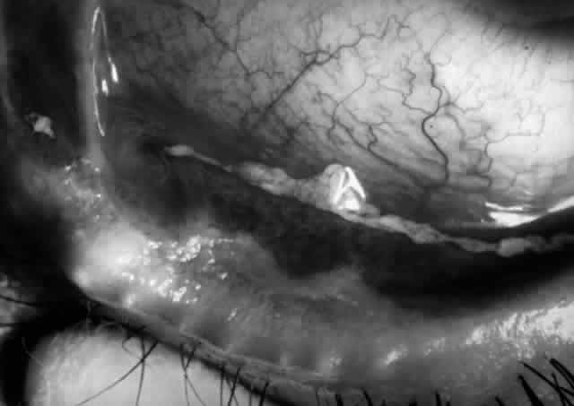
Fig. 15.
Eye of a patient with Stevens-Johnson syndrome. Note the conjunctivitis, the subepithelial fibrosis, the symblepharon formation, and the zone of variable width, posterior to the gray line of the eyelid that is obviously keratinized.