Chapter 26
Ocular Manifestations of the Rheumatic Diseases
JENNIFER E. THORNE and DOUGLAS A. JABS
Main Menu   Table Of Contents

Search

RHEUMATOID ARTHRITIS
SERONEGATIVE SPONDYLOARTHROPATHIES
ANKYLOSING SPONDYLITIS
REITER'S SYNDROME
ARTHRITIS WITH INFLAMMATORY BOWEL DISEASE
PSORIATIC ARTHRITIS
JUVENILE RHEUMATOID ARTHRITIS
FAMILIAL JUVENILE SYSTEMIC GRANULOMATOSIS
SYSTEMIC LUPUS ERYTHEMATOSUS
SCLERODERMA
POLYMYOSITIS AND DERMATOMYOSITIS
SJÖGREN'S SYNDROME
RELAPSING POLYCHONDRITIS
SYSTEMIC VASCULITIDES
LARGE VESSEL VASCULITIDES
MEDIUM VESSEL VASCULITIDES
SMALL VESSEL VASCULITIDES
REFERENCES

The rheumatic diseases are a heterogeneous collection of diseases that are multisystem in nature and immunologically mediated. They are loosely grouped into three general categories: the arthritides, the connective tissue diseases, and the vasculitides (Table 1). Behçet's disease has been classified variously but in this chapter is discussed with the vasculitides. The diagnosis of each of the rheumatic disorders is a clinical diagnosis. To ensure homogeneity in clinical research and assist in reporting in the literature, the American College of Rheumatology (ACR) has developed criteria for most of the rheumatic diseases. These criteria are for research purposes and cannot always be used in the care of every patient. However, they often provide a useful description of the clinical features of the disease.


Table 1. The Rheumatic Diseases

Arthritis
Rheumatoid arthritis
Seronegative (HLA-B27 associated) spondyloarthropathies
  Ankylosing spondylitis
  Reiter's syndrome
  Inflammatory bowel disease
  Psoriatic arthritis
Juvenile rheumatoid arthritis

Connective Tissue Diseases
  Systemic lupus erythematosus
  Scleroderma
  Polymyositis and dermatomyositis
  Sjögren's syndrome
  Relapsing polychondritis

Vasculitides
  Polyarteritis nodosa
  Churg-Strauss syndrome
  Hypersensitivity vasculitis
  Henoch-Schönlein purpura
  Wegener's granulomatosis
  Lymphomatoid granulomatosis
  Giant cell arteritis (temporal arteritis)
  Takayasu's arteritis
  Behçet's disease


The clinical features of the rheumatic disorders are based on the anatomic pattern of disease. This situation is analogous to the definitions of ocular uveitis syndromes, in which the anatomic pattern of disease is used as characteristic criteria. Thus, rheumatoid arthritis generally is described as an additive, symmetric, deforming polyarthritis, while the seronegative spondyloarthropathies often are associated with a migratory, asymmetric oligoarthritis. Similarly, the different vasculitides are defined by the pattern of vascular involvement and the size of blood vessels commonly involved. As such, individual laboratory tests (e.g., rheumatoid factor [RF] or antinuclear antibody [ANA]) are supporting evidence in the diagnosis of the disease but do not define the disease.

The major ophthalmic manifestations of the rheumatic diseases include scleritis, Sjögren's syndrome, uveitis, retinal vascular disease, and neuroophthalmic lesions.1,2 Each of these is most characteristically associated with a few but not all of the rheumatic disorders. Hence, scleritis most often is seen with rheumatoid arthritis or with vasculitis. Acute anterior uveitis most often is seen with the seronegative spondyloarthropathies. Retinal vascular and neuroophthalmic lesions are seen with disorders that have either a vasoocclusive component, such as systemic lupus erythematosus or one of the vasculitides.

Back to Top
RHEUMATOID ARTHRITIS
Rheumatoid arthritis (RA) is the most common rheumatic disorder affecting an estimated 1% of the general population.3–7 The overall incidence of RA has remained fairly stable across geographic locations.6 Classically RA is described as an additive, symmetric, deforming polyarthritis. The diagnosis of RA is a clinical one, based on the features outlined below. The ACR criteria for the diagnosis of RA are outlined in Table 2.8


Table 2. American College of Rheumatology (ACR) Revised Criteria for the Diagnosis of Rheumatoid Arthritis


CriterionDefinition
1. Morning stiffnessMorning stiffness in and around the joints lasting at least 1 hour before maximal improvement
2. Arthritis of three or more joint areasAt least three joint areas have simultaneously had soft tissue swelling or fluid (not bony overgrowth alone) observed by a physician. The 14 possible joint areas are right or left PIP, MCP, wrist, elbow, knee, ankle, and MTP joints.*
3. Arthritis of hand jointsAt least one joint area swollen as above in wrist, MCP, or PIP.
4. Symmetric arthritisSimultaneous involvement of the same joint areas (as in 2) on both sides of the body (hilateral involvement of PIPs, MCPs, or MTPs is acceptable without absolute symmetry).
5. Rheumatoid nodulesSubcutaneous nodules, over bony prominences, or exterior surfaces, or in juxtaarticular regions, observed by a physician.
6. Serum rheumatoid factorDemonstration of abnormal amounts of serum “rheumatoid factor” by any method that has been positive in less than 5% of normal control subjects.
7. Radiologic changes 

For classification purposes, a patient shall be said to have rheumatoid arthritis if he/she has satisfied at least 4 of the above 7 criteria. Criteria 1–4 must have been present for at least 6 weeks. Patients with 2 clinical diagnoses are excluded. Designation as “classic,” “definite,” or “probable” rheumatoid arthritis is not to be made.
*PIP, proximal interphalangeal joint; MCP, metacarpophalangeal joint; MTP, metatarsophalangeal joint; PA, posteroanterior (Arnett FC, Edworthy SM, Block DA, et al: The American Rheumanism Association 1987 revised criteria for the classification of rheumatoid arthritis. Arthritis Rheum 31:314, 1988. Copyright © 1988 Wiley-Liss. Reprined by permission of Wiley-Liss. Inc. a subsidiary of John Wiley & Sons lnc.)


ETIOLOGY AND PATHOGENESIS

The etiology of RA remains unknown, although both genetic and environmental factors likely are involved. Theories on the pathogenesis of RA postulate an inciting immunologic event in a genetically susceptible individual with subsequent inflammation in the joint resulting in synovial proliferation and joint destruction. The initial inciting immunologic event is unknown, but both immune complex disease and cellular immune events have been hypothesized.9,10 Immunohistologic studies of the joint space have shown an early accumulation of T lymphocytes, which localize to the synovial tissue and initiate the inflammation that causes the clinical manifestations of RA.11 Proliferation of synovial fibroblasts, synovial hypertrophy, and pannus formation occur.11,12 In established RA there is an accumulation of polymorphonuclear leucocytes (PMNs) in the synovial fluid, suggesting recruitment of PMNs in later stages of the disease. These changes lead to erosion of bone at the joint margin, osteoporosis at the joint margin, and in long-standing disease, joint destruction.

These immunologic processes cause the release of cytokines and metalloproteinases that perpetuate the joint destruction and leads to the production of serum RF.11,12 RF is a polyclonal autoantibody directed against immunoglobulin G (IgG), which results in circulating immune complexes. It can be detected by several tests, including sheep red blood cell agglutination and enzyme-linked immunosorbent assay (ELISA). RF is a useful marker for the presence of RA and is present early in the disease course in 85-90% of patients with RA.7,11 However, its contribution to the pathogenesis of the disease remains unclear, and the presence of RF is neither necessary nor sufficient for the diagnosis of RA.7,11 High levels of RF have been associated with more severe clinical disease and poorer prognosis.11,13

There is a genetic predisposition to RA, with an association of the disease with the human leukocyte antigen (HLA) types, HLA-DR4 and HLA-DR1.14 Both HLA-DR4 and HLA-DR1 are thought to be involved in both the susceptibility to and the severity of RA.11 In population studies, more than 90% of patients with RA have HLA-DR4, or HLA-DR1, or both.14 Studies of white Americans have shown a consistent association with HLA-DR4, with 70% of white patients with RA expressing the DR4 haplotype compared to 28% of patients with non-RA arthritis.15 Polymerase chain reaction techniques have identified a specific sequence in the DRβ1 gene that encodes an amino acid motif or shared epitope that has been found to occur in both HLA-DR4 and HLA-DR1 haplotypes.11 This shared epitope has been hypothesized to serve as either a binding site for an antigenic peptide or as an autoantigen that activates T-cell inflammation.11

CLINICAL FEATURES

In the majority of patients with RA, the onset is insidious. However, in 10% to 20% of patients there may be an abrupt or explosive onset. The arthritis classically is described as an additive, symmetric, deforming polyarthritis. Typically, the small joints of the hands and feet are involved, although all joints may be affected. As with all inflammatory arthropathies, the disease is characterized by a gel phenomenon, that is, stiffness at rest improving with movement. Most patients complain of morning stiffness. X-ray studies of the affected joints show erosions of bone at the joint margin, which evolve into joint destruction with long-standing disease. Because of the damage to surrounding structures, joint laxity may develop; this process results in the characteristic ulnar deviation of the fingers.7

Extraarticular features are common in patients with RA.7,16 The most common extraarticular lesion is the presence of rheumatoid nodules. These nodules occur in approximately 25% of patients with RA and are located primarily on extensor surfaces. On biopsy, they have a characteristic histologic picture with central fibrinoid necrosis surrounded by a palisade of elongated histiocyte-like cells. This core is enveloped by an outer zone of mononuclear inflammatory cells, primarily lymphocytes and plasma cells. Immunohistochemical analysis has demonstrated that the cells within the palisade layer stain for monocyte markers and that peripheral lymphocytes are predominantly T cells.17

Pulmonary lesions that may occur include pleural disease, pulmonary effusions, pleural or pulmonary nodules, arteritis with resultant pulmonary hypertension, and interstitial fibrosis. Pleuritis develops in 20% of patients with RA and may occur concurrently with the initial arthritic symptoms. Pulmonary effusions may be large enough to cause dyspnea. Rheumatoid nodules may develop in the pleural or within the lung. In the lung, the nodules may develop in clusters and cavitate, leading to bronchopleural fistula.7

Cardiac involvement includes pericarditis, which may be detected in as many as 50% of patients with RA at autopsy. Pericardial effusion secondary to pericarditis may be seen on echocardiogram in up to 31% of patients with RA, although the majority of patients are asymptomatic and left ventricular dysfunction because of the effusions is rare.7,18 Cardiac conduction defects as a consequence of rheumatoid nodules present in the conducting system can occur. Rheumatoid nodules also have been detected on heart valves.7,16

Rheumatoid vasculitis occurs in less than 1% of patients with RA and typically is seen in patients with severe arthritis and high RF titers. It presents as a polyneuropathy, particularly with lower extremity anesthesia or hypesthesia, poorly healing leg ulcers, or as palpable purpura. Digital gangrene and occasionally visceral ischemia also may be manifestations of rheumatoid vasculitis.7,16,19,20

Felty's syndrome consists of RA, splenomegaly, and leukopenia, and may represent up to 3% of patients with RA. Rheumatoid factor present in 98% of patients with Felty's syndrome, and titers typically are high. The pathogenesis of the leukopenia is unknown, but possible mechanisms include accelerated removal of leukocytes and suppression of granulopoiesis. Chronic leg ulcers with recurrent infections and hyperpigmentation of the skin also are characteristic features.7,21

TREATMENT

The goals of treatment for RA include the prevention and control of joint damage, prevention of loss of function, and pain reduction.22 Prior to the 1990s, the treatment of RA typically was approached in a stepwise fashion with nonsteroidal antiinflammatory drugs (NSAIDs) being the first line of therapy followed by institution of disease modifying antirheumatic drugs (DMARDs) in more severe or refractory cases.22 However, recent studies suggest that the destruction of the joints and loss of function occurs soon after the onset of synovitis.23,24 It is estimated that one half of the joint destruction that is seen over an 8-year period occurs in the first 2 years.25 Therefore, now a DMARD typically is instituted within the first 3 months of diagnosis of RA in addition to an NSAID.23 NSAID therapy includes salicylates, older nonselective cyclooxygenase (COX) inhibitors, and the newer COX-2 inhibitors. These NSAIDs are used to control pain and swelling and to improve the joint function but typically do not alter disease course or prevent joint destruction long term.22 Because joint destruction can occur within the first 2 years of the disease, early treatment with a DMARD or remittive agent is recommended.23 Remittive agents include hydroxychloroquine, sulfasalazine, methotrexate, etanercept, infliximab, leflunomide, and anakinra. Hydroxychloroquine and sulfasalazine are effective for milder disease.26–31 Hydroxychloroquine often is used first because of its relative lack of toxicity. Chloroquine is slightly more efficacious than hydroxychloroquine in the treatment of RA but also is more toxic; therefore, it is used infrequently.32 Methotrexate, an antimetabolite, typically is the first choice of remittive agents for more severe or advanced disease and has been found to be efficacious in the treatment of RA in multiple randomized clinical trials.33–36 Methotrexate was found to improve clinical signs and symptoms of RA, improve functional status, and slow radiographic disease progression. Leflunomide also has been found to be effective in controlling RA progression and in improving clinical and functional status both as monotherapy and in combination with methotrexate.37–39 Triple therapy with methotrexate, hydroxychloroquine, and sulfasalazine also has been shown to be effective in the treatment of RA, although simpler treatment regimens typically are used.40,41

Biologics are engineered agents designed to block cytokines or cytokine receptors. Biologic agents, including etanercept, infliximab, and anakinra have been found to be potent remittive agents in several randomized clinical trials.23,42–46 Etanercept is a recombinant tumor necrosis factor α (TNF-α) receptor Fc segment fusion protein and is effective in controlling progression of RA as initial monotherapy in early RA and as therapy for patients who have failed previous DMARD therapy.23,42,43 The typical dose of etanercept is 25 mg administered subcutaneously twice per week. Many patients taking etanercept improve more rapidly than those taking methotrexate, sometimes within 2 weeks after instituting etanercept.23,43 Etanercept also is effective in combination with methotrexate for controlling ongoing active RA in patients already taking methotrexate alone.43 Infliximab is a chimeric (mouse-human) anti-TNF-α monoclonal antibody and only is available as an intravenous preparation given either 3 to 5 mg every 4 weeks or 3 to 10 mg every eight weeks. Infliximab in combination with methotrexate has been used successfully to improve clinical and functional status in patients with RA who have failed methotrexate monotherapy.44,45 Anakinra, a recombinant human interleukin-1 receptor antagonist (IL-1Ra), also is approved for use in RA in a 100-mg daily dose given by subcutaneous injection.23 Anakinra inhibits the binding of the cytokines IL-1α and IL-1β to the IL-1 receptor, which prevents the activation of its target cells and subsequent synovial inflammation and joint destruction.23 Randomized clinical trials found anakinra to be superior to placebo in controlling clinical symptoms of RA.23 Another randomized clinical trial found that anakinra in combination with methotrexate was more effective in controlling RA progression than methotrexate alone.46

Immunosuppressive drugs, such as azathioprine,47,48 cyclosporine,49 or cyclophosphamide,48,50 also are efficacious in the treatment of RA but are used less frequently because of their toxicity. Occasionally they may be used in the treatment of rheumatoid vasculitis.22 Intramuscular and oral gold and d-penicillamine also are effective remittive agents but are used rarely because of more efficacious and better-tolerated newer drugs.22 Minocycline has been shown to be modestly effective in a randomized trial and is used for mild disease, although infrequently.51,52 Traditionally low-dose prednisone (5 mg every other day to 10 mg/d) was used to decrease stiffness and increase mobility. Although the newer remittive agents have decreased this practice, prednisone still is used.

OCULAR MANIFESTATIONS

The most common ocular manifestations of RA are keratoconjunctivitis sicca (secondary Sjögren's syndrome), scleritis, and rheumatoid corneal melts.2,53 Sjögren's syndrome originally was defined as dry eyes, dry mouth, and RA. It subsequently has become evident that Sjögren's syndrome can exist in association with a connective tissue disease, in which case it is termed secondary Sjögren's syndrome, or by itself with no definable associated connective tissue, in which case it is termed primary Sjögren's syndrome. In Sjögren's syndrome, there is a lymphocytic infiltration of the lacrimal and salivary glands resulting in glandular destruction and dysfunction. This process ultimately results in a characteristic loss of tearing and saliva. Approximately 11% to 13% of patients with RA have secondary Sjögren's syndrome.2

Scleritis (Fig. 1) is the second most common ocular manifestation of RA, affecting an estimated 1% to 6% of patients with RA and 14% of patients with rheumatoid vasculitis.2,54,55 Scleral inflammation can be classified as episcleritis or scleritis. Episcleritis presents with discomfort rather than pain, more superficial ocular inflammation, less frequent and severe ocular complications, and typically a less frequent association with systemic disease.56 Conversely, scleritis often is characterized by pain, presents with deeper inflammation and edema in the sclera, often has ocular complications, which may be severe, and in approximately one half of the cases, is associated with systemic disease.56,57 Episcleritis may be self-limited and spontaneously remitting, whereas scleritis typically requires therapy. Scleritis may be classified as anterior, posterior, or necrotizing. Anterior scleritis is subdivided into diffuse and nodular types. Scleromalacia perforans is a separate category, in which there is an insidious but destructive scleral process and is seen in patients with long-standing RA. Any of the above types of scleritis may be seen in association with RA, although anterior scleritis is most common.2,56

Fig. 1. Scleritis in a patient with rheumatoid arthritis (Courtesy of David Knox, MD)

Although estimates of the frequency of scleritis in patients with RA have been as high as 6%, large series have shown that approximately 1% of patients with RA have scleral disease.2,55,58 Of patients with scleritis, 10% to 33% have RA, and RA represents the most commonly seen rheumatic disease in patients with scleritis.53,56 In patients with RA, scleritis is associated with more severe systemic disease and with a greater frequency of extraarticular manifestations.53 Patients with RA are more likely to develop necrotizing scleritis than those patients with idiopathic scleritis.59 Necrotizing scleritis is more likely to be associated with systemic vasculitis and a poor systemic prognosis. In a retrospective study by Foster and colleagues, 29% of patients with RA and necrotizing scleritis or necrotizing keratitis died, typically because of complications of vascular disease.54

Rheumatoid corneal melts, also known as marginal corneal ulcers, may be either bland (not inflammatory) or necrotizing (peripheral ulcerative keratitis [PUK]) in nature. Noninflammatory marginal keratitis often can be treated with local measures. Necrotizing keratitis requires aggressive medical therapy with systemic corticosteroids and often immunosuppressive drugs. Patients with PUK and RA have high mortality rate, possibly because of an underlying rheumatoid vasculitis.2,54,55,59

Other uncommon features include Brown's syndrome,60–62 orbital myositis,63 and hydroxychloroquine retinal toxicity.64–71 Brown's syndrome has been described in a few patients with RA and is caused by a stenosing tenosynovitis of the superior oblique tendon.60–62 Rare cases of a retinal microangiopathy with cotton-wool spots have been reported,2 but these probably are the result of an associated rheumatoid vasculitis.

Patients with RA on hydroxychloroquine or chloroquine often are referred for ophthalmic evaluation and follow-up in order to monitor the patient for drug toxicity. Hydroxychloroquine is used much more frequently than chloroquine because it has less toxicity than chloroquine.66 Both drugs have ocular toxicity as their dose-limiting side effect.64–71 They are accumulated in pigmented tissues, such as the retinal pigment epithelium, and may produce a bull's eye pigmentary retinopathy. This retinopathy is thought to be reversible if discovered early,69 but irreversible, despite discontinuation of the drug, when allowed to progress. Because of this toxicity, patients taking hydroxycloroquine are referred for screening ophthalmic examinations in order to detect the early signs of toxicity. It is recommended that all patients in whom hydroxychloroquine is to be instituted receive a baseline ophthalmic examination including visual acuity, dilated fundus examination, Amsler grid testing, and visual field testing with the Humphrey automated perimetry 10-2 test. Color testing and fundus photography also are suggested at the baseline examination. The timing of follow-up examinations to screen for signs of toxicity is controversial. The frequency of retinopathy is less than 5% when doses less than 6.5 mg/kg per day (less than 400 mg/d) of hydroxychloroquine are used.67 It appears that patients can be treated with large cumulative doses of hydroxychloroquine without evidence of toxicity when daily doses at this level are used.68 However, the risk for patients taking less than 6.5 mg/kg per day of hydroxychloroquine remains greater than zero over long-term follow-up.69 Cost-effectiveness analyses have suggested that a baseline ophthalmic examination followed by annual follow-up ophthalmic examination may be the most cost-effective approach.70 However, one study in which rheumatologists and ophthalmologists were surveyed revealed that the majority recommended monitoring every 6 months for hydroxychloroquine toxicity.71 Therefore, our practice has been to follow patients who are taking hydroxychloroquine at a dose of 400 mg daily every 6 months and to follow those patients taking 200 mg daily annually.

Back to Top
SERONEGATIVE SPONDYLOARTHROPATHIES
The seronegative spondyloarthropathies include ankylosing spondylitis (AS), Reiter's syndrome, arthritis with inflammatory bowel disease, and psoriatic arthritis. Historically, these disorders were called seronegative because they lacked rheumatoid factor, which distinguished them from RA, in which patients were said to be seropositive. The seronegative spondyloarthropathies are better termed the HLA-B27–associated spondyloarthropathies, because of their statistical association with the gene HLA-B27. In addition, these disorders are linked by several clinical features and sometimes are difficult to differentiate in the earlier stages of the disease. Clinical findings include a predilection for axial skeletal involvement, extraarticular features, enthesopathy (inflammation of the insertion of tendons), and an asymmetric, migratory oligoarthritis.72 The most common ocular manifestation of the seronegative spondyloarthropathies is a nongranulomatous, recurrent, acute anterior uveitis. Acute anterior uveitis shares with the spondyloarthropathies an association with the gene HLA-B27; approximately 50% of patients with acute anterior uveitis possess HLA-B27,73–75 and if the uveitis is unilateral and recurrent, up to 71% of patients will be HLA-B27 positive.75 Approximately 50% of patients with HLA-B27–associated acute anterior uveitis have a seronegative spondyloarthropathy.75,76

ETIOLOGY AND PATHOGENESIS

The seronegative spondyloarthropathies (Table 3) are thought to be multifactorial in nature and triggered by an environmental insult in a genetically predisposed person. This genetic predisposition is evidenced by the strong association with HLA-B27.77–83 AS has the strongest association in that more than 90% of white patients with AS are HLA-B27–positive, whereas the general population frequency of HLA-B27 is approximately 8%,2,77,78


Table 3. The Seronegative (HLA-B27 Associated) Spondyloarthropathies


DisorderFrequency HLA-B27 (%)Frequency Uveitis (%)
Ankylosing spondylitis9025
Reiter's syndrome70–8020–40
Inflammatory bowel disease62–12
 With sacroiliitis50–7050
Psoriatic arthritis18–227
 With sacroilitis50–60 

(Adapted from Jabs DA: The rheumatic diseases. In Ryan S (ed): The Retina. Vol II. pp. 457–480. Copyright 1989, with permission from Elsevier Science.)


The best insight into a possible environmental trigger for these disorders is provided by reactive arthritis and epidemic Reiter's syndrome. Reactive arthritis and epidemic Reiter's syndrome occur after an infectious gastroenteritis with a limited number of organisms, including Shigella, Salmonella, and Yersinia. The subsequent arthritis occurs well after the infectious gastroenteritis has resolved and is a sterile arthritis. The arthritis appears to represent an immunologic response to the infectious organism, which triggers the arthritis in a susceptible person. Reactive arthritis is distinguished from epidemic Reiter's syndrome by the other extraarticular features present in Reiter's syndrome. In reactive arthritis and epidemic Reiter's syndrome, the presence of HLA-B27 again is associated strongly with the development of disease.72,84

The exact molecular mechanisms by which HLA-B27 predisposes to disease are unknown. Theories include molecular mimicry,72,85 and the possibility that HLA-B27 is associated with a defective class I antigen-mediated cellular response.83 The molecular mimicry hypothesis suggests that there is a similarity at the molecular level between the HLA-B27 molecule and the inciting organisms allowing for the triggering of an immune response and the subsequent development of clinical disease.83,85 The process is felt to be analogous to rheumatic fever or acute poststreptococcal glomerulonephritis, where the immune response to a cross-reactive antigen results in a subsequent immune-mediated disease. Alternatively it has been suggested that the HLA-B27 molecule may be a defective molecule associated with an aberrant cytotoxic T-cell response.83,86

Back to Top
ANKYLOSING SPONDYLITIS

CLINICAL FEATURES

AS is characterized by involvement of the axial skeleton and bony fusion (ankylosis).87 The estimated prevalence of AS is 0.1% to 0.2% of the general population, and is 2% in HLA-B27–positive patients.83 Young adult males are affected most frequently, with a male to female ratio of approximately 3:1. Earlier studies suggested a much higher male to female ratio, but it subsequently has become evident that women may have mild or atypical forms of the disease.88,89 Between 10% and 30% of HLA-B27–positive first-degree relatives of AS patients will develop the disease.90

The characteristic symptom of AS is chronic low back pain, which improves with exercise. The symptoms may be mistaken by patients as a strain-related injury, and the diagnosis may be delayed by several years.83,93 As with other inflammatory arthropathies, the disease is associated with morning stiffness. The inflammation results in fusion of the axial skeleton (spinal ankylosis) and sacroiliitis. Physical examination reveals restricted motion of the spine and may reveal tenderness over the sacroiliac joints. Radiographic examination of the spine and sacroiliac joints are important in the diagnosis of the disease. The demonstration of sacroiliitis on radiographic examination of the sacroiliac joints is almost a sine qua non for the diagnosis of spondylitis. The end stage of this process is a completely fused and immobilized spine, also known as a bamboo or poker spine. In addition to the spinal arthritis, patients may also develop an arthritis of the shoulders and hips, limited chest expansion, restrictive lung disease, apical pulmonary fibrosis, aortic insufficiency because of arteritis, and heart block.72,87,92

TREATMENT

The treatment of AS relies on the judicious use of NSAIDs and a program of physical therapy and exercise. The more potent NSAIDs, indomethacin and phenylbutazone, appear to be more effective in the treatment of spondylitis than many of the others. Although phenylbutazone is effective for the treatments of spondylitis, it has been associated with the occurrence of aplastic ankylosis, which has limited the use of this drug. Antiinflammatory medications do not reverse and generally do not prevent the ultimate spinal ankylosis. However, they relieve symptoms and allow the patient to participate in an exercise program. The program of physical therapy and exercise allows the patient to maximize the mobility of the spine and to allow spinal fusion to occur in a posture most advantageous to the patient.72,87 Methotrexate and infliximab have been reported to be effective in the management of more severe disease.72,93–95

OCULAR MANIFESTATIONS

The primary ocular manifestation of AS is a nongranulomatous, recurrent, acute, anterior uveitis.76,87,90 Generally, one eye is affected at a time, although both eyes may suffer attacks.76,90 Occasionally anterior chamber inflammation may be severe enough to produce hypopyon, fibrin deposition, and posterior synechiae (Fig. 2).87,92 An estimated 25% to 40% of patients with AS suffer an attack of uveitis at some time during the course of their disease.83,87,90 Studies of patients with acute anterior uveitis have reported that AS is present as the underlying systemic disease in 18% to 34% of these patients.90,92,96,97 Rarely, a spillover vitritis is seen in patients with acute anterior uveitis and AS, but it is substantially less frequent than the typical iridocyclitis.99

Fig. 2. Fibrin clot and posterior synechiae in a patient with acute, anterior uveitis and ankylosing spondylitis.

Back to Top
REITER'S SYNDROME

CLINICAL FEATURES

Reiter's syndrome originally was characterized by the classic triad of arthritis, urethritis, and conjunctivitis. In 1981, the American Rheumatism Association expanded this original set of criteria to: (1) the presence of a peripheral arthritis with a duration of greater than or equal to 1 month and (2) an associated urethritis, cervicitis, or diarrhea.100 The increased scope of the disease encompasses reactive arthritis in which a sterile arthritis ensues after the infection has resolved.100 Typically it is rare for a patient to present with the classic triad originally described. Historically, Reiter's syndrome existed in two forms: epidemic and endemic. Epidemic Reiter's syndrome occurs after an infectious gastroenteritis; the ensuing arthritis occurs after the gastroenteritis has resolved, and it is a sterile arthritis. Although a triggering agent can be identified for epidemic Reiter's syndrome, none as yet has been identified for endemic Reiter's syndrome. Because patients with Reiter's syndrome often developed urethritis and presented to venereal disease clinics, it initially was believed that endemic Reiter's syndrome was because of Chlamydia, but studies have not demonstrated a higher prevalence of chlamydial infection in patients with Reiter's syndrome than in controls, and the role of Chlamydia remains debated.72,100

The arthritis of Reiter's syndrome is migratory, asymmetric, episodic oligoarthritis affecting primarily the large joints at the lower extremities, such as the knees or ankles. Other articular features include heel pain, sausage digits caused by interphalangeal arthritis of the toes and/or fingers, and sacroiliitis. Mucocutaneous lesions include urethritis in men and cervicitis in women, circinate balanitis, painless oral ulcers, keratoderma blennorrhagica, and dystrophic nail lesions. Systemic symptoms including fever and weight loss also may occur. The disease tends to follow an episodic and relapsing course.100–102 The arthritis may be recurrent in 15% to 50% of patients.100 Another 15% to 30% of patients will develop chronic arthritis over 10 to 20 years of follow-up.100,101

OCULAR MANIFESTATIONS

Conjunctivitis was one of the original triad described by Reiter and is one of the hallmarks of the disease. It tends to be a feature of early disease, particularly of the initial attack, and may be missed if patients are seen only during subsequent attacks. Depending on the source of the reported series, and on the diagnostic criteria used, the frequency of conjunctivitis in patients with Reiter's syndrome has varied from 33% to 100%. The more serious ocular manifestation is recurrent, acute, nongranulomatous anterior uveitis. It occurs in approximately 5% to 20% of patients with the initial attack but may occur in as many as 50% of patients with Reiter's syndrome over long-term follow-up.83,90,103 Some studies of patients with acute anterior uveitis suggest that Reiter's syndrome is more frequently seen in patients with acute anterior uveitis than is AS,75,98 although this finding may be the result of a referral bias or of geographic variations in the prevalence of these disorders. As with AS, occasionally a spillover vitritis has been described in Reiter's syndrome and is found more commonly with Reiter's syndrome than with AS. Rarely other forms of posterior ocular involvement such as multifocal choroiditis have been reported in Reiter's syndrome.104,105 Rarely, patients may develop a keratitis with punctate epithelial lesions, progressing to a central loss of the corneal epithelium and subepithelial infiltrates.106–108

Back to Top
ARTHRITIS WITH INFLAMMATORY BOWEL DISEASE
Inflammatory bowel disease (IBD) consists of two distinct diseases: ulcerative colitis and Crohn's disease. Ulcerative colitis is an inflammatory disorder of the colonic mucosa. Crohn's disease, also known as regional enteritis, granulomatous ileocolitis, or granulomatous colitis, is a focal granulomatous disease involving any area of the gastrointestinal tract.

CLINICAL FEATURES

The clinical features of IBD are diarrhea and abdominal pain. Diarrhea may or may not be bloody. Extraintestinal manifestations of IBD include dermatitis, mucous membrane disease, ocular inflammation, and arthritis. Skin disorders occur in 6.5% to 15% of patients with IBD and include erythema nodosum and pyoderma gangrenosum.109,110 Arthritis occurs in approximately 22% of patients with IBD. Two distinct variants of enteropathic arthritis are seen. One form is a large-joint, lower-extremity, nondeforming oligoarthritis. The activity of this enteropathic arthritis parallels that of the bowel disease. The frequency of this type of enteropathic arthritis is approximately 15% in Crohn's disease and 10% in ulcerative colitis.111 The second form of arthritis associated with IBD is an HLA-B27–associated axial-sacroiliac spondylitis, which is seen in approximately 20% of patients with Crohn's disease and in 10% to 15% of patients with ulcerative colitis.111 In one series of 103 patients with IBD, arthritis occurred in 39% of patients, and of the patients with arthritis, 90% met the criteria for spondyloarthropathy.112 Between 50% and 70% of patients with spondylitis and IBD are HLA-B27 positive.111 Of the HLA-B27–positive patients with IBD, 50% will develop acute anterior uveitis.109 The activity of this spondylitis is unrelated to that of the bowel disease, and in 18% to 20% of patients, the spondylitis will be asymptomatic.83,111,113

TREATMENT

Treatment of the IBD is individualized and dependent on the extent and severity of the disease. Typically treating the underlying bowel disease improves the joint disease. Localized proctitis often may be treated with corticosteroid enemas. More extensive bowel disease may require systemic corticosteroid therapy. Generally, nonsteroidal therapy with sulfasalazine is used first in an effort to minimize steroid complications and often is used to decrease the does of corticosteroids needed. Severe corticosteroid-dependent IBD also can be treated with corticosteroid-sparing agents, such as azathioprine or methotrexate.111,113 Infliximab has been shown to induce remissions in Crohn's disease and is effective in the treatment of Crohn's-related fistulas.114 However, infliximab appears to be ineffective in treatment of ulcerative colitis.111 Because of the risk of colonic malignancy with long-standing ulcerative colitis, a colectomy sometimes is performed. After colectomy the mucocutaneous manifestations and the enteropathic arthritis may resolve.111

OCULAR MANIFESTATIONS

Ocular inflammation has been reported to occur in up to 13% of patients with IBD, but typically occurs in approximately 2% to 6% of patients.90,110,113,115 The ocular manifestations include anterior uveitis, scleritis, and keratitis. Anterior uveitis is the most common ocular manifestation.115–118 More often it presents as a nongranulomatous, recurrent, acute anterior uveitis. This type of uveitis is seen in association with spondylitis and HLA-B27. Chronic and bilateral uveitis also occurs occasionally and may be seen more frequently in women with IBD.90,119,120 There have been occasional case reports of retinal vascular disease, including retinal artery occlusion and retinal vasculitis, and ischemic optic neuropathy in patients with IBD.121–123 It has been suggested that these retinal vascular lesions are caused by a thrombic diathesis seen in some patients with IBD.121 All types of scleritis have been associated with IBD including anterior scleritis, necrotizing scleritis, and posterior scleritis. The scleral inflammation may parallel the activity of the underlying bowel disease (Fig. 3).117,118 Rarely, keratitis118,124 and Brown's syndrome125 may be seen in association with IBD.

Fig. 3. Scleritis in a patient with inflammatory bowel disease. A. Active scleritis. B. Healed scleritis after treatment of the inflammatory bowel disease.

Back to Top
PSORIATIC ARTHRITIS

CLINICAL FEATURES

Psoriatic arthritis is a syndrome defined by the presence of psoriasis and an associated seronegative inflammatory arthritis.126 Psoriasis is a common cutaneous disorder, affecting 1% to 3% of the population and is characterized by erythematosus, well-demarcated macular lesions with silvery scales. It characteristically occurs on extensor surfaces, particularly the elbows and scalp, but also the chest or back. Approximately 10% to 20% of patients with psoriasis have arthritis.126,127 The skin disease generally precedes the onset of the arthritis by several years, but the arthritis precedes the onset of skin disease in 15% of patients with psoriatic arthritis.126 Nail changes are present in 90% of patients with psoriatic arthritis as compared to 41% of patients with psoriasis but without arthritis.128 The nail changes include pitting, transverse ridges, crumbling, and onycholysis.

Psoriatic arthritis has been classified into five groups based on the clinical presentation of the arthritis. Classic psoriatic arthritis is manifested by involvement of the distal interphalangeal joints. Arthritis multilans is a severely deforming, destructive, and usually widespread arthritis with ankylosis of joints and characteristic erosive joint changes seen radiographically. A symmetric, additive, deforming polyarthritis similar to RA can be seen. However, psoriatic arthritis usually is negative for RF. The most common presentations of psoriatic arthritis are monoarthritis or an asymmetric oligoarthritis, usually affecting the distal interphalangeal, proximal interphalangeal, or metatarsal interphalangeal joints. The fifth type of psoriatic arthritis is psoriatic spondylitis. Psoriatic spondylitis is an HLA-B27–associated disorder, and approximately 50% of patients with psoriatic spondylitis are HLA-B27–positive.72,126

OCULAR MANIFESTATIONS

Conjunctivitis occurs in approximately 20% to 33% of patients with psoriatic arthritis, iritis in 4% to 7%, and scleral disease in 2%.90,126,127,129 Patients with psoriatic spondylitis typically develop a nongranulomatous, recurrent, acute anterior uveitis, as is the case with other HLA-B27–associated arthropathies, but chronic uveitis also may occur. Brown's syndrome also has been reported in patients with psoriatic arthropathy.130

Back to Top
JUVENILE RHEUMATOID ARTHRITIS
Juvenile rheumatoid arthritis (JRA), also known as juvenile chronic arthritis (JCA) or juvenile idiopathic arthritis (JIA), is defined as an arthritis of greater than 3 months' duration with onset at less that 16 years of age.131 Other causes of arthritis in children, such as leukemia, must be excluded. The traditional classification of JRA depends on the pattern of presentation of the arthritis and has been classified as polyarticular, oligoarticular, and systemic disease. Polyarticular JRA is defined as presentation with five or more joints involved, and is seen in 30% of children presenting with JRA. Oligoarticular JRA involves four or less joints, and comprises 60% of children with JRA. Systemic disease is the disorder originally described by Still, affects 10% of children with JRA, and has prominent systemic features with a variable arthritis.132

CLINICAL FEATURES

The incidence and prevalence of JRA have been estimated as 10 to 20 cases per 100,000 population per year and 65 to 148 cases per 100,000 population, respectively.133–135 Five distinct subgroups of JRA have been described. The systemic variant of JRA also is known as Still's disease. The male to female ratio is approximately 1:1 and the onset of disease is generally at less than 5 years of age. The arthritis has a variable relation to the onset of the systemic disease but is usually a polyarthritis. The systemic features include fever, a salmon-colored evanescent maculopapular rash, lymphadenopathy, hepatitis, splenomegaly, and serositis. An elevated erythrocyte sedimentation rate and leukocytosis are seen, but ANA and RF tests are negative in at least 90% of cases.132 Ocular disease generally is not associated with this variant.131

Two types of oligoarticular JRA have been described. ANA-positive oligoarticular disease typically is seen in children younger than 5 years of age. The mean age of onset is 3 years. Girls are affected more often than boys with a male to female ratio of 1:5. The arthritis is an oligoarticular, large-joint, lower-extremity arthritis that tends to spontaneously remit and not leave significant articular deformities. More than 80% of these patients have a positive ANA test. This subgroup of patients is associated with HLA-DR5, DR6, DR8, DQ1, and DQ2.136–138 A chronic anterior uveitis is the characteristic ocular feature of this group.131,139–141

A second type of oligoarticular disease is seen in older children, with a mean age of onset at 12 years. Boys are affected more often than girls and the male to female ratio is approximately 5:1. The arthritis is a large-joint, lower-extremity oligoarthritis. Sacroiliitis is common, and these children tend to progress to AS over time. Ninety percent of these children are HLA-B27 positive, and approximately 25% develop a nongranulomatous, recurrent, acute anterior uveitis. This variant shares the clinical and immunogenetic features of the seronegative spondyloarthropathies in adults, including the same type of uveitis.131,142,143

Two types of polyarticular disease have been described. In older children, older than 10 years of age, an RF-positive (seropositive), rheumatoid-like polyarthritis may develop. Girls are affected more commonly than boys. The arthritis is an additive, symmetric, deforming polyarthritis similar to that of adult RA. The arthritis is persistent and results in long-term articular morbidity with joint erosions and deformity seen. This type of arthritis is associated with HLA-DR4, the HLA antigen seen in adult RA. Ocular disease in uncommon in this subgroup.131,132,142

A second type of polyarticular disease has been defined.132,144 The children are older and tend to present with an additive, symmetric, deforming polyarthritis. However, patients are RF-negative but are HLA-B27–positive. Sacroiliitis and spondylitis are common, and a characteristic fusion of the posterior spines of the C2 and C3 vertebrae in the cervical spine can be seen. Micrognathia occurs in approximately 25%. The ocular lesion seen tends to be a recurrent acute iridocyclitis and occurs in 25% of patients.144

TREATMENT

The goal of treatment for JRA is to control the clinical symptoms of the disease and to limit joint deformity using the most conservative and safest therapy available.132 Initial therapy typically begins with either aspirin or NSAIDs, such as ibuprofen or naproxen. The NSAID dose is dependent on the child's age and weight. Because of the potential association between aspirin use and Reye's syndrome, any aspirin or NSAID should be discontinued in a sick child who potentially has influenza or varicella. Systemic corticosteroids generally are avoided, except in severe systemic disease, because of the potential for growth retardation with long-term use. If the patient fails to respond to NSAIDs alone, methotrexate typically is used because it is well-tolerated, has been shown to be effective in the treatment of JRA in a randomized clinical trial, and there is extensive experience with the drug over long-term follow-up.145–147 Methotrexate also has been found to be effective in the treatment of JRA-associated uveitis.148–150 Other immunosuppressive drug therapies, including cyclosporine and azathioprine, have been used to treat JRA that is refractory to treatment with methotrexate.132 Etanercept also has been shown to be effective in the treatment of polyarticular JRA in a multicenter randomized clinical trial.151

OCULAR MANIFESTATIONS

Approximately 12% to 17% of children with any type of JRA have uveitis.152–154 A population-based study performed in Finland reported the mean annual incidence and prevalence rates for JRA-associated uveitis as 0.2 and 2.4 cases per 100,000 population, respectively.155 The ocular manifestations of JRA are two different types of anterior uveitis. Acute anterior uveitis is seen in the HLA-B27–associated subgroups and is similar to that seen in adult HLA-B27–associated disease. Chronic anterior uveitis is seen primarily in the ANA-positive oligoarticular subgroup where frequencies have been reported to range from 20% to 56%.132,139,156–159 Although traditionally it has been thought that these cases occur primarily in girls, a recent retrospective study of 90 children with JRA found no gender difference in the risk of developing uveitis.152 The chronic anterior uveitis tends to be asymptomatic or minimally symptomatic; therefore, it is recommended that these patients be evaluated every 3 months for screening in an effort to detect the uveitis early.160 Two thirds of patients have bilateral disease, and in patients with unilateral disease, the majority will develop uveitis in the fellow eye within 1 year of presentation.132 The chronic uveitis typically requires chronic therapy. Severe visual disability may develop. Band keratopathy and cataracts may be seen in up to one third of patients. Secondary glaucoma occurs in 10% to 20% of patients with JRA-associated uveitis and is a poor prognostic finding.154,156,157,161 Posterior synechiae are common. Other complications include macular edema in 8% to 10% of cases and phthisis in 4% to 10% of patients. Although blindness developed in 40% of patients reported in early series, more recent series have reported a better prognosis.154,156–158,162–164 One study from Finland reported on 426 incident cases of JRA diagnosed between 1989 and 1996, of whom, 24% developed uveitis. In this series only 3% of those with uveitis were reported to have a best-corrected visual acuity of 20/60 or worse. The improved prognosis appears to be because of earlier detection, better treatment, and better surgical management of the complications. Although pars plana lensectomy with vitrectomy and aphakic correction traditionally had been considered the best method of cataract surgery in these patients,157,165 more recent case series have suggested that intraocular lenses can be placed in these patients with good visual outcome.166,167

Treatment for the uveitis associated with JRA depends on the type of uveitis. Acute anterior uveitis typically can be controlled with topical corticosteroid eye drops such as prednisolone acetate 1%. The medication is instituted at one drop every one to two hours while awake until the uveitis is controlled and then tapered slowly and discontinued. Milder forms of chronic uveitis also may be controlled with topical corticosteroids, although the medication may be required indefinitely to keep the uveitis quiet. The dose of topical corticosteroid is tapered to the lowest dose in which the uveitis remains quiet. In cases of more severe uveitis, methotrexate or cyclosporine may be used.148–150 Systemic corticosteroids are given in the cases of severe, sight-threatening uveitis and are used in conjunction with a steroid-sparing agent such as methotrexate. The systemic corticosteroids are tapered and discontinued as quickly as possible in order to avoid side effects such as growth retardation.

Back to Top
FAMILIAL JUVENILE SYSTEMIC GRANULOMATOSIS
Familial juvenile systemic granulomatosis, also known as Jabs syndrome, Blau syndrome, and autosomal dominant granulomatous disease of childhood, is an uncommon genetic disease with polyarthritis and uveitis that often is misdiagnosed as JRA or sarcoidosis.168–172 The disease is inherited in an autosomal dominant fashion, and the joint disease is a granulomatous, deforming, nonerosive polysynovitis. Other more variable features of the syndrome include rash, cranial nerve palsies, and vasculopathy.168–172 The syndrome is caused by mutations in the CARD15 gene, which is involved in apoptosis and is preferentially expressed on monocytes.173 The CARD15 gene also has been linked to Crohn's disease, but the mutations for the two disorders are in different segments of the gene.173

Children with this syndrome often are misdiagnosed as having either JRA or sarcoidosis. Familial juvenile systemic granulomatosis may be distinguished from JRA by the presence of polyarthritis on presentation, the absence of ANA, and the pattern of inheritance. Although patients with familial juvenile systemic granulomatosis may have a chronic anterior uveitis similar to JRA, these patients also may have multifocal choroiditis, which is not seen in patients with JRA.172 Patients with JRA who have chronic uveitis tend to have oligoarticular arthritis and positive ANA antibodies. Familial juvenile systemic granulomatosis differs from sarcoidosis by the pattern of arthritis, the absence of pulmonary involvement and adenopathy, and the inheritance pattern.172 The uveitis associated with familial juvenile systemic granulomatosis may be a chronic anterior uveitis or a chronic panuveitis with multifocal choroiditis. Patients may require aggressive medical therapy to control the uveitis, sometimes including immunosuppressive drugs. Ocular complications are common.

Back to Top
SYSTEMIC LUPUS ERYTHEMATOSUS

ETIOLOGY AND PATHOGENESIS

Systemic lupus erythematosus (SLE) is a multisystem autoimmune disease. Although the etiology of SLE is unknown, the association of SLE with HLA-DR2, HLA-DR3, and the association of lupus nephritis with Fcγ receptors IIA and IIIA located on chromosome 1 all suggest a genetic predisposition.174,175 Pathogenetically SLE is characterized by B-cell hyperactivity, polyclonal B-cell activation, hypergammaglobulinemia, autoantibody formation, and abnormal T-cells.174 Autoantibodies seen in patients with SLE include ANA, antibodies to DNA, both single-stranded DNA (anti-ssDNA) and double-stranded or native DNA (anti-dsDNA), and antibodies to cytoplasmic components, such as anti-Sm, anti-Ro (SSA), and anti-La (SSB).174 Studies have demonstrated multiple defects in T-cell signaling pathways in patients with SLE that are thought to lead to autoreactivity and hyperactivated T-cell lymphocytes.176–178 SLE classically has been regarded as an immune complex disease, in which circulating immune complexes are deposited in tissue and incite an inflammatory response, which may lead to end-organ damage. Immunoregulatory abnormalities in SLE lead to an inadequate clearing of immune complexes and further hyperactivation of both B- and T-cells causing further inflammation and tissue damage.174

CLINICAL FEATURES

The incidence of SLE has been estimated as 2 to 9 cases per 100,000 population per year.179 SLE is a multisystem disease that may affect almost any organ system.179–184 Because of the multisystem nature of the disease, criteria for the diagnosis have been established and are outlined in Table 4.185 To establish a diagnosis of SLE, four or more criteria must be met. SLE is a clinical diagnosis, and no single laboratory test defines the disease. These criteria are for use in clinical investigations and may not always be applicable in patient management.


Table 4. The 1982 Revised Criteria for the Diagnosis of Systemic Lupus Erythematosus

   Criteria*

  1. Malar rash
  2. Discoid lupus
  3. Photosensitivity
  4. Oral ulcers
  5. Arthritis
  6. Serositis (pleuritis, pericarditis)
  7. Renal disorder (proteinuria, nephritis)
  8. Neurologic disorder (seizures, psychosis)
  9. Hematologic disorder (hemolytic anemia, leukopenia, lymphopenia or thrombocytopenial)
  10. Immunologic disorder (positive LE cell preparation, anti-DNA test, anti-Sm or false-positive test for syphilis)
  11. Antinuclear antibody

*Persons shall be said to have SLE if he or she has 4 or more of the 11 criteria. (Reprinted from Jabs DA: The rheumatic diseases. In Ryan S (ed): The Retina, Vol II. pp. 457–480. Copyright 1989, with permission from Elsevier Science.)


Cutaneous disease occurs in approximately 85% of patients with SLE. The most classic manifestation is the characteristic butterfly rash across the nose and cheeks, also known as a malar flush. Other cutaneous lesions include discoid lupus erythematosus, vasculitic skin lesions such as cutaneous ulcers or splinter hemorrhages, purpuric skin lesions, alopecia, and livedo reticularis, a reticular purplish rash seen most commonly on the legs. Less common skin lesions include a maculopapular eruption, lupus profundus, bullous skin lesions, and urticarial skin lesions. Mucosal lesions occur in 30% to 40% of patients with SLE and characteristically are painless oral ulcers. Photosensitivity is a common feature of the skin lesions in SLE affecting up to 70% of patients, and patients may be exquisitely sun sensitive.179–183

Raynaud's phenomenon occurs in approximately 20% of patients with SLE. Cardiac disease includes pericarditis, occasionally myocarditis, and Libman-Sacks endocarditis. Libman-Sacks endocarditis was common in old autopsy series but now is unusual. Pleuropulmonary lesions include pleurisy and, less commonly, pneumonitis. Adenopathy and hepatosplenomegaly can be seen in 50% of patients with SLE.179–183 Involvement of the central nervous system (CNS) by lupus occurs in 35% of patients with SLE.183,184 Peripheral nervous system involvement with a peripheral neuropathy and cranial nerve palsy is less common. CNS lupus may present as seizures, an organic brain syndrome, or psychosis.184 Transverse myelitis is an uncommon manifestation, occurring in only 4% of patients with SLE but often is seen in association with optic neuritis.

Eighty to 85% of patients with SLE have articular disease at some point during the course of their SLE. Articular involvement includes polyarthralgias and a nondeforming migratory polyarthritis. Cutaneous nodules, myalgias, and myositis are less common. Systemic features occur in more than 80% of patients with lupus and include fatigue, fever, and weight loss. Renal disease is present in approximately 50% of these patients. Lupus nephritis clinically presents as either proteinuria with a nephritic picture or as glomerulonephritis with active urinary sediment. Lupus nephritis is a major cause of morbidity and mortality in SLE.179–183

Patients with SLE often have a mild chronic anemia, known as anemia of chronic disease. However, they may also develop an autoimmune hemolytic anemia. Leukopenia, in particular lymphopenia, is a characteristic feature. Thrombocytopenia occurs in approximately one third of patients.179–183

The lupus anticoagulant may be found in 28% to 34% of patients with SLE.186–191 The lupus anticoagulant is a member of a family of antiphospholipid antibodies, which also includes the anticardiolipin antibodies. These antibodies are found in combination with clinical features including arterial or venous thrombosis and pregnancy morbidity in patients with antiphospholipid antibody syndrome (APS), which may be a primary disease unassociated with other autoimmune diseases or may be secondary to SLE. Approximately 15% of patients with SLE have APS.192–195 The APS is associated with occlusive disease, which can lead to tissue ischemia and end-organ damage, and with thrombotic disorders, such as deep vein thrombophlebitis and strokes.186–195 The mechanism by which antiphospholipid antibodies cause thrombosis is not known, but two possible explanations are induction of platelet aggregation by the antibody and inhibition of prostacyclin production by the vascular endothelium. Both of these mechanisms could cause vessel occlusion.195

TREATMENT

Because SLE is a multisystem disease with highly variable features in different patients, treatment is directed at the specific symptoms in any given patient. Nonsteroidal antiinflammatory agents or low-dose prednisone are used for treatment of the arthritis and serositis.196 Hydroxychloroquine often is used for the treatment of the skin disease or as a corticosteroid-sparing agent for mild skin disease, although it may require 3 to 6 months of therapy before a complete response is noted.196–198 High-dose corticosteroids, including pulse intravenous (IV) corticosteroids, often are necessary for end-organ or life-threatening disease, such as severe hematologic abnormalities (i.e., hemolytic anemia, severe thrombocytopenia), lupus nephritis, or CNS lupus.196 In patients with rapidly progressive renal disease, pneumonitis, systemic vasculitis, or CNS lupus, IV pulse corticosteroids have been used successfully with clinical improvement within days.199–201 Long-term therapy after pulse corticosteroids is required.

Cytotoxic drugs have proven to be effective in the treatment of patients with severe lupus nephritis. Monthly IV cyclophosphamide at a dose of 0.5 to 1.0 g/m2 was found to cause remission of renal disease over a 5-year period in 85% of patients if given in combination with IV methylprednisolone, compared to 62% of patients treated with IV cyclophosphamide only, and 29% of patients treated with intravenous methylprednisolone only.202 Daily oral cyclophosphamide 1 to 2 mg/kg per day or azathioprine 1 to 2 mg/kg per day have been used alone or in combination with oral corticosteroids to control life-threatening active lupus nephritis.196,203 However, because pulse intravenous cyclophosphamide causes less side effects than oral cyclophosphamide, pulse intravenous cyclophosphamide typically is the preferred treatment for lupus nephritis. For severe, refractory disease, bone marrow immunoablation with or without autologous stem cell transplantation has been successful in inducing remission in a small number of patients over 1 to 2 years of follow-up.204 Anticoagulation therapy is instituted in patients with SLE and APS. Although aspirin or low-dose heparin are used commonly, it has been suggested that warfarin (Coumadin, Bristol-Myers Squibb) is the only intervention that significantly lowers clotting risk in these patients.205,206

OCULAR MANIFESTATIONS

The major ocular manifestations of SLE can be classified into five general categories: (1) involvement of the skin of the eyelids with cutaneous disease; (2) secondary Sjögren's syndrome; (3) retinal vascular lesions; (4) neuroophthalmic lesions; and (5) scleritis.2 Eyelid disease is most often discoid lupus erythematosus involving the margins of the lids.207 Secondary Sjögren's syndrome occurs in approximately 20% of patients with SLE and is indistinguishable from the sicca complex seen in patients with other rheumatic disorders.183

Retinal vascular lesions are the most common form of intraocular involvement in patients with SLE. The retinopathy most frequently consists of cotton-wool spots (Fig. 4) with or without intraretinal hemorrhages.208–210 The frequency of this finding varies depending on the patient population studied and the activity of the disease. One study209 found that only 3% of ambulatory outpatients had cotton-wool spots, while two separate studies180,210 found that 28% to 29% of hospitalized patients with active SLE had retinal vascular findings. This retinopathy occurs independently of hypertension and is thought to be related to the underlying microangiopathy of SLE. The histopathology of CNS lupus is that of a microangiopathic disease with small vessel vasoocclusion, a process presumed to be similar to that in the retina.211,212 Studies of autopsy material of patients with SLE have demonstrated immune reactants, primarily immunoglobulin and complement in the ocular vessel walls, and it has been suggested that these are the cause of the microangiopathy.213 Fluorescein angiographic studies have suggested that mild background retinopathy with microaneurysms and telangiectatic vessels may be relatively common.210

Fig. 4. Cotton-wool spots along the superotemporal arcade in a patient with active systemic lupus erythematosus.

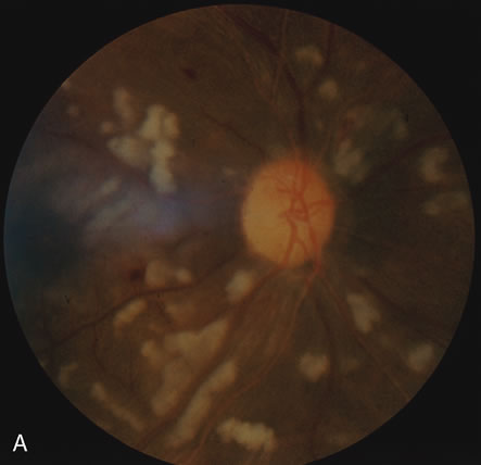
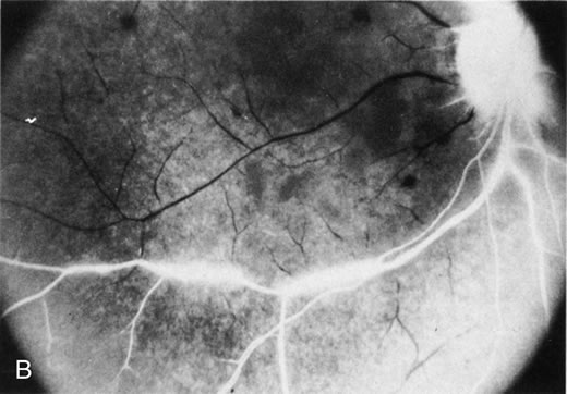
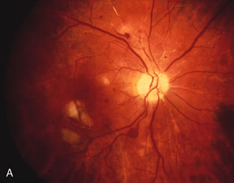
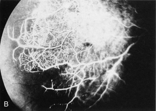
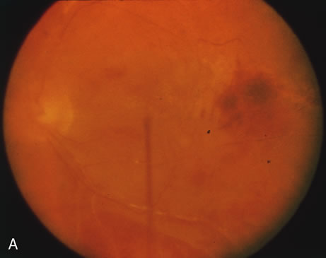
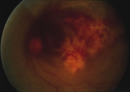
The finding of retinal vascular changes in patients with SLE correlates with the activity of the disease.214 The relationship of cotton-wool spots alone to CNS lupus has been debated, but to date no relationship has been established. Most experts feel that the finding of cotton-wool spots does not indicate the presence of CNS lupus. A much less frequent, severe retinal vasoocclusive disease has been described and does appear to be associated with CNS lupus, in particular, diffuse CNS dysfunction, such as an organic brain syndrome.214–223 This more severe retinal vasoocclusive disease may present as a central retinal artery occlusion, central retinal vein occlusion, branch artery occlusion, or most frequently, a diffuse retinal vasoocclusive process (Fig. 5), sometimes called retinal vasculitis. Although this last process sometimes has been called retinal vasculitis, the exact pathogenesis may not be true vasculitis. Cases of severe retinal vasoocclusive disease in SLE in association with the lupus anticoagulant also have been reported, and the retinal disease in these cases is presumed to be secondary to this autoantibody (Fig. 6).190,191 The prognosis for vision with this diffuse retinal vascular disease is poor and retinal neovascularization commonly develops. Panretinal photocoagulation may be of value in the treatment of the neovascularization of severe lupus retinopathy. Approximately 50% of eyes affected with this severe vaso-occlusive disease become blind. Although visual loss is common in those patients with severe retinal disease, in the more common mild retinopathy visual loss is unusual.223

Fig. 5. A. Fundus photograph and B. fluorescein angiogram, demonstrating diffuse vaso-occlusive disease in a patient with systemic lupus erythematosus. Fluorescein angiogram shows extensive nonperfusion of the retina. (Jabs DA, Fine SL, Hochberg MC, et al: Severe retinal vaso-occlusive disease in systemic lupus erythematosus. Arch Ophthalmol 1104:558, 1986. Copyrighted 1986, American Medical Association. Reprinted with permission.)

Fig. 6. A. Fundus photograph and B. fluorescein angiogram demonstrating diffuse vaso-occlusive disease in a patient with systemic lupus erythematosus. This is peripheral nonperfusion of the retina. The patient had anticardiolipin antibodies. (Jabs DA, Fine SL, Hochberg MC, et al: Severe retinal vaso-occlusive disease in systemic lupus erythematosus. Arch Ophthalmol 104:558, 1986. Copyrighted 1986, American Medical Association. Reprinted with permission.)

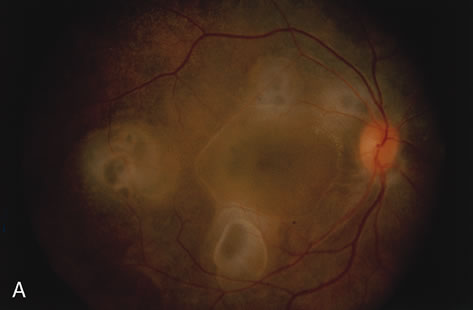
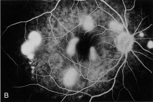
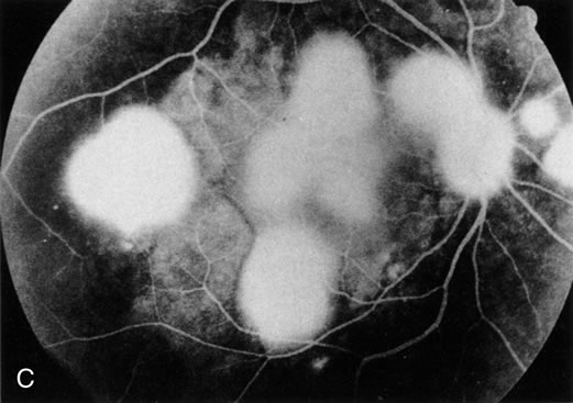
Even less common than retinopathy is lupus choroidopathy, of which only a few cases have been reported to date.214,224–226 Early autopsy studies demonstrated the frequent presence of mononuclear inflammatory cells in the choroid of patients with untreated SLE.208 The clinical changes seen in patients with lupus choroidopathy include serous elevation of the retina, most often of the neurosensory retina, serous elevations of the retinal pigment epithelium, and combined elevations of both (Fig. 7). These clinical findings are associated with systemic vascular disease, either hypertension because of lupus nephritis or systemic vasculitis.226 Treatment of the underlying disease, with systemic corticosteroids and immunosuppressive agents if needed, and control of any hypertension can resolve these serous retinal detachments.

Fig. 7. A. Fundus photograph and B. and C. fluorescein angiogram of a patient with choroidopathy and systemic lupus erythematosus. The fluorescein angiogram demonstrates multiple “smokestack” areas of fluorescein leakage. (Jabs DA, Hanneken A, Schachat AP, et al: Choroidopathy in systemic lupus erythematosus. Arch Ophthalmol 106:230, 1988. Copyright 1986, American Medical Association. Reprinted with permission.)

Neuroophthalmic involvement in SLE includes cranial nerve palsies, lupus optic neuropathy, and central retrochiasmal disorders of vision.227–237 The central disorders include hallucinations and visual field loss.232,237 The optic nerve lesion most often seen in SLE is reported as retrobulbar optic neuritis. However, other optic nerve lesions can be seen, including anterior optic neuritis with optic disc edema, ischemic optic neuropathy, and a slowly progressive visual loss from lupus optic neuropathy.227–236 Autopsy studies have suggested that a microangiopathic process may be the pathogenesis of lupus optic neuropathy. Focal demyelination can be seen, but more severe lesions with axonal damage and even optic nerve infarcts have been demonstrated.227–229,231 One case demonstrated both demyelination and loss of axons with foci of total destruction of the optic nerve.229 This finding suggests that the same process may cause milder, reversible lesions and more severe irreversible infarct-like lesions. Transverse myelitis is seen in over 50% of patients with lupus optic neuropathy as compared with its overall frequency of 4% in patients with SLE.236 The frequency of clinical optic neuropathy in SLE patients has been estimated at 1% to 2% of patients with SLE.182–184

Scleritis occurs less commonly in patients with SLE. In one retrospective study of 97 patients with scleritis, 4% had SLE.56 Any type of scleritis may be seen, but anterior scleritis appears most commonly. Therapy that controls the underlying SLE typically controls the scleritis.2,56

Back to Top
SCLERODERMA
Scleroderma is a systemic connective tissue disease of unknown etiology characterized by fibrous and degenerative changes in the skin and the viscera. The hallmark of scleroderma is the skin change, which consists of a thickening, tightening, and induration of the skin. It caused by fibrous replacement of the dermis of the skin, with subsequent loss of mobility and contracture. In addition, vascular insufficiency and vasospasm are present.238–240 Localized forms of scleroderma, such as linear scleroderma and morphea, also exist.238

ETIOLOGY AND PATHOGENESIS

The etiology of scleroderma is unknown. The most characteristic feature of scleroderma is the increased collagen content in the skin and visceral organs.241 Some investigators studying fibroblasts from patients with scleroderma in vitro have demonstrated increased collagen production.242 The second characteristic feature is the vascular disease, including Raynaud's phenomenon and diffuse microvasculopathy, which typically is present at the first recognizable stages of the disease.241 Although no hypotheses of pathogenesis have been proved, several have been proposed, including a primary vascular disorder, an abnormality of fibroblast function (i.e., a collagen production abnormality), and a primary immunologic disorder. However, it is thought that the abnormality of fibroblast function, which causes the accumulation of collagen and extracellular matrix, is secondary to either a primary vascular or immunologic process.241 The vascular theory suggests that the primary lesion is a vascular abnormality, which subsequently triggers a fibroblastic response and the abnormal collagen deposition. The fibroblastic response to this initial vascular insult further aggravates the problem by the intimal proliferation seen in blood vessels and the increase in dermal collagen in the skin.238,243 Several investigators have found that the sera from patients with scleroderma are cytotoxic for endothelial cells, and abnormalities of platelet aggregation and platelet activation have been detected in patients with scleroderma.244–246 However, it is unknown whether these changes are primary or are secondary to platelet activation because platelets pass through damaged blood vessels. The demonstration of a scleroderma-associated antigen that is released under the conditions of hypoxemia-reperfusion (i.e., Raynaud's phenomenon) suggests that the initial insult in scleroderma could be vascular.247

Because of the presence of a mononuclear inflammatory cell infiltrate in the dermis of patients with scleroderma,248 an immunologic process has been proposed. The infiltrating lymphocytes in the dermis of patients with scleroderma consist largely of CD4+ T cells.249In vitro studies have demonstrated that lymphocytes from these patients can produce cytokines, which will augment collagen production by fibroblasts.238 Furthermore, dermal antigens have been shown to stimulate lymphocytes from scleroderma patients to produce a variety of cytokines. Patients with scleroderma also have polyclonal hypergammaglobulinemia, and autoantibodies, such as ANA, are seen in greater than 90% of patients with scleroderma. These features suggest an immune-mediated or autoimmune process.

CLINICAL FEATURES

Scleroderma characteristically affects middle-aged women with a peak age of onset of 30 to 50 years of age. The incidence has been calculated at 18 to 20 cases per million population per year.250,251 It is estimated that there are 75,000 to 100,000 cases of scleroderma in the United States.238 Scleroderma is defined by the skin changes, which consist of thickening, tightening, induration, and hidebound changes, resulting in a loss of mobility and contracture. The disease characteristically begins peripherally and involves the fingers and hands, with a subsequent centripetal spread up the arms to involve the face and body. Early in the disease, an edematous phase of the skin changes may exist, but later-stage disease consists of contracture and shiny, taut, hidebound skin. Telangiectasia and calcinosis are common. Early in the disease process, skin biopsy specimens demonstrate a mononuclear inflammatory cell infiltrate. In later-stage disease there is a subcutaneous fibrosis with thinning of the skin, loss of the epidermis, and an absence of epidermal appendages.238–240

Approximately 90% of patients with scleroderma have Raynaud's phenomenon, which is the initial complaint in approximately 70% of patients.238 Raynaud's phenomenon consists of pallor, cyanosis, suffusion, and tingling that occur in an abrupt and phasic manner after a known exposure. Most often this exposure is cold but other precipitating factors, including emotion, have been described. Raynaud's phenomenon is the result of an exaggerated vasospasm and subsequent vasodilation in response to the cold challenge. Microvascular abnormalities may be detected in the digital arteries and in the nail bed capillaries of patients with scleroderma.252 Severe narrowing of this vasculature is seen in greater than 75% of patients.238 Raynaud's phenomenon may be more systemic in nature, and similar vasospastic events have been detected in the cardiac, pulmonary, and renal vasculature in scleroderma patients after cold exposure.253,254 Histologically, the blood vessels often show intimal proliferation (in patients with severe vascular disease), which may be associated with fibrinoid necrosis. As a consequence of Raynaud's phenomenon and vascular insufficiency, digital ulcers may develop.238

Gastrointestinal involvement may occur in more than 75% of patients with scleroderma. The esophagus is involved most frequently and shows dysmotility, gastroesophageal reflux and stricture formation. Small bowel involvement is less frequent and presents with decreased motility, bacterial overgrowth, and malabsorption. Large bowel involvement produces widemouth diverticula.238

Pulmonary disease is found in 75% to 90% of patients at autopsy but is less frequently detected clinically. As the management of scleroderma-associated renal crisis has improved, pulmonary disease has become the leading cause of mortality.238 Patients develop pulmonary fibrosis and may have exertional dyspnea, decreased lung capacity, and a restrictive lung disease. Pulmonary hypertension and right-sided heart failure may develop as a consequence of this process.238,243,255 Cardiac disease may be detected in approximately one third of patients because of fibrotic involvement of the cardiac conduction system, and resulting in conduction abnormalities and arrhythmias.256

Renal disease is a major cause of mortality and often is associated with the onset of malignant hypertension and a rapid progression to renal failure.257,258 Scleroderma renal crisis or scleroderma kidney was almost uniformly fatal until the late 1970s; however, aggressive antihypertensive therapy can successfully reverse scleroderma renal crisis.259–261

Musculoskeletal lesions include polyarthralgias, tendon friction rubs, infrequently polyarthritis, and occasionally myositis. Myositis may occur as an overlap syndrome, sometimes termed sclerodermatomyositis.238–240 A peripheral neuropathy because of entrapment caused by the fibrotic process impinging on the nerve has been seen. Trigeminal neuralgia is the most common example of this neuropathy.262

There is a spectrum of disease ranging from the more mild CREST variant to severe diffuse scleroderma. The CREST variant was named for its features of calcinosis, Raynaud's phenomenon, esophageal disease, sclerodactyly, and telangiectasia. This syndrome is more benign than diffuse scleroderma and more slowly progressive. In patients with the CREST syndrome, visceral involvement is less common than in diffuse scleroderma. The CREST syndrome is associated with anticentromere antibodies in 50% to 96% of patients, which are a marker for this variant.263–266 Patients with diffuse scleroderma, also known as progressive systematic sclerosis, have a more rapidly progressive skin disease with more severe and progressive visceral involvement. These patients have a poorer prognosis than those with the CREST variant.

Overlap syndromes occur between scleroderma and other connective tissue diseases. The best known overlap syndrome is mixed connective tissue disease, which has features of SLE, systemic sclerosis, and myositis.267 It was characterized by antibodies to ribonuclear protein (RNP). It originally was suggested that there was a corticosteroid-responsive nature to many of the clinical features. Long-term follow-up studies have suggested that mixed connective tissue disease generally evolves into systemic sclerosis.268

TREATMENT

No specific treatment has been determined definitely to be efficacious in altering the progression of scleroderma, and traditionally treatment has been directed toward management of the complications and symptomatic relief. Corticosteroids are ineffective in the treatment of scleroderma itself, but occasionally may be used for treatment of the inflammatory stage of interstitial lung disease in scleroderma or of the associated myositis in patients with an overlap syndrome.238,269 Immunosuppressive agents, including chlorambucil, azathioprine, and 5-fluorouracil, have not shown any consistent value in the treatment of scleroderma.238

Scleroderma renal crisis, which previously was a fatal complication, often can be controlled by aggressive treatment of the hypertension and dialysis for renal failure. Control of the hypertension involves the use of multiple antihypertensive agents including angiotensin converting enzyme blockade.259–261 Raynaud's phenomenon most often is treated with calcium channel antagonists, such as nifedipine. These agents are effective in symptomatic relief and reducing the number of attacks of the Raynaud's phenomenon. Calcium channel antagonists are more effective in patients with idiopathic Raynaud's phenomenon than in those with scleroderma, possibly reflecting the fixed vascular changes caused by luminal narrowing in patients with scleroderma.270,271

OCULAR MANIFESTATIONS

The eye is frequently involved in patients with scleroderma.272,273 Most often, this involvement consists of scleroderma of the eyelids, resulting in tightness of the lids and blepharophimosis. Lid involvement occurs in 30% to 65% of patients with scleroderma. Despite the frequent involvement of the eyelids, corneal exposure is fortunately uncommon. Conjunctival vascular abnormalities, including telangiectasia and vascular sludging, often occur. Estimates of this finding have run as high as 71%. These changes in the conjunctival vasculature may be analogous to those seen in the nailfold capillary bed.

Keratoconjunctivitis sicca with a Sjögren's-like picture has been described.274,275 Minor salivary gland biopsy studies have suggested that two different pathogenetic mechanisms may be present. Some patients appear to have inflammation of the glands, similar to Sjögren's syndrome, while others appear to have a glandular fibrosis. Early studies suggested a correlation between these two variants and the variants of scleroderma, with CREST patients having salivary gland inflammation and diffuse systemic sclerosis patients having minor salivary gland fibrosis.274 Subsequent studies have suggested that this association is not as clear-cut.275

Other reported lesions have included periorbital edema occurring as part of the early edematous phase of scleroderma,276 cranial nerve palsies,277 scleral pits,278 restrictive ophthalmopathy,279 and extraocular muscle myositis occurring in a patient with sclerodermatomyositis.280 All these lesions are uncommon and have been described only as case reports. There are occasional reports of retinal vascular lesions in patients with scleroderma but without scleroderma renal disease. They are uncommon and occur in less than 5% of patients. Such cases include retinal hemorrhage, branch retinal vein occlusion, and central retinal vein occlusion.272,273,281 More common has been the retinopathy of malignant hypertension seen in patients with scleroderma renal crisis. The clinical findings include cotton-wool spots, intraretinal hemorrhages, and optic disc edema. Both clinical and histologic studies have demonstrated that the process in patients with scleroderma is identical to that seen in patients with idiopathic malignant hypertension.2,282–285

In contrast to the infrequent findings of retinopathy in patients with scleroderma, small case series of fluorescein angiographic studies have suggested more frequent abnormalities of the choroidal vasculature in patients with systemic sclerosis, including patchy and irregular perfusion of the choroid and variable hyperfluorescence of the retinal pigment epithelium in one third to one half of the patients. The lesions were clinically silent, and ophthalmoscopy results were normal. These angiographic abnormalities were interpreted as damage to the retinal pigment epithelium caused by vascular lesions in the choroid.286–289

In summary, retinal complications are uncommon in patients with scleroderma unless hypertension is present. Occasionally, retinal vascular disease may herald the onset of malignant hypertension and a medical emergency for the scleroderma patient. Choroidal vascular changes may be more common but are clinically silent.

Back to Top
POLYMYOSITIS AND DERMATOMYOSITIS
Polymyositis and dermatomyositis are inflammatory diseases of skeletal muscles characterized by pain and weakness in the involved muscle groups. In the typical case, weakness begins insidiously and involves the proximal muscle groups, particularly those of the shoulders and hips. Dermatomyositis is distinguished from polymyositis by the presence of cutaneous lesions. The skin lesions of dermatomyositis include an erythematosus to violaceous rash, which may affect the eyelids (heliotrope rash), cheeks, nose, chest, and extensor surfaces, and a rash over the knuckles of the fingers, known as Gottron's papules.290–292

ETIOLOGY AND PATHOGENESIS

The etiology of the myositis is unknown. The pathogenesis appears to be an immune-mediated attack against the skeletal muscle fibers triggered by environmental factors in a susceptible individual.290 Muscle biopsy specimens may show vascular deposits of immunoglobulin and complement. These vascular deposits represent immune complexes that produce vascular damage.293,294 The presence of vasculopathy in patients without a defined inflammatory vasculitis may explain the occasional occurrence of retinopathy and small vessel vasoocclusion in patients with dermatomyositis. Other pathologic changes in muscle suggest cellular immune mechanisms, with lymphocyte proliferation in response to skeletal muscle antigens reported by some investigators.294,295

Immunogenetic studies have suggested a genetic predisposition in both childhood and adult polymyositis and dermatomyositis. The strongest associations exist for the HLA-B8, DR3, and DR52 antigens.296,297 The association with HLA-DR3 and B8 are especially high in white patients with an antibody to a saline extractable nuclear antigen, Jo-1.290 Anti-Jo-1 antibody is one of several myositis-specific autoantibodies identified in a small subset of patients with idiopathic myositis. The presence of this antibody has been associated with an increased prevalence of interstitial pulmonary fibrosis, arthritis, and Raynaud's phenomenon.290,298,299

CLINICAL FEATURES

The estimated incidence of myositis in the United States is 5 new cases per million population per year and reports have ranged from 0.5 to 8.4 new cases per million population per year.300 Women are affected more often than men in adult myositis. The age of onset is bimodal, affecting children ages 10 to 15 years and adults ages 45 to 60 years most frequently.290 In the typical case, symmetric, proximal muscle weakness begins insidiously either in the lower or the upper extremities. The patient slowly becomes aware of difficulty in running, climbing stairs, and ultimately in getting out of a chair. The gait then becomes clumsy, and the patient has difficulty in arising without assistance. Systemic symptoms including malaise and weight loss are frequently present. On examination, a symmetric, proximal upper limb and lower limb weakness is found in almost every patient. Cutaneous lesions are found in patients with dermatomyositis but not in those with primary idiopathic polymyositis.290

Laboratory abnormalities include an elevated erythrocyte sedimentation rate and elevated skeletal muscle enzymes. The muscle enzymes found to be abnormal include creatinine phosphokinase (CPK), aldolase, aspartate aminotransferase (AST), alanine aminotransferase (ALT), and lactic dehydrogenase (LDH). Although the pattern of abnormal enzymes varies from individual to individual, the CPK is most frequently abnormal. Electrmyographic abnormalities are present in more than 90% of patients with myositis and consist of an increase in insertional activity of muscle, fibrillation potentials, and positive sharp waves at rest. Motor unit action potentials show myopathic changes with decreased amplitude and duration and an increase in the proportion of polyphasic potentials. Bizarre, high-frequency, repetitive discharges also are seen.290

Muscle biopsy specimens reveal the degeneration of muscle fibers with occasional areas of regeneration. Interstitial muscle necrosis is present in approximately 60% of biopsy specimens, and interstitial fibrosis in another 20%. Inflammatory infiltrates of muscle are present in 75% of muscle biopsy specimens. The inflammatory infiltrate consists primarily of mononuclear inflammatory cells with lymphocytes and plasma cells. Vasculitis often is seen in childhood dermatomyositis.290

Polymyositis and dermatomyositis have been classified into five groups as outlined in Table 5.301,302 Malignancy occurs in 13% of adults with dermatomyositis. Patients with malignancy are likely to be over 45 years of age and have dermatomyositis. The tumors present occur at typical cancer sites and are generally diagnosed either concurrently or within 1 year of the diagnosis of myositis.303,304 Vasculitis is frequent in patients with childhood dermatomyositis. Inflammatory myositis associated with a connective tissue disease is most often seen with either SLE, primary Sjögren's syndrome, or scleroderma.


Table 5. Classification of Idiopathic Inflammatory Myopathies

Primary idiopathic polymyositis
Primary idiopathic dermatomyositis
Dermatomyositis (or polymyositis)
Childhood dermatomyositis (or polymyositis)
Polymyositis or dermatomyositis associated with collagen vascular disease (overlap group)


TREATMENT

Corticosteroids are the mainstay of treatment for myositis. Ninety percent of patients improve on corticosteroid therapy alone, and 50% to 75% have complete remission.305 Corticosteroid therapy typically is begun at a dosage of 1 mg/kg per day (60 to 80 mg daily) and continued until the muscle enzymes have normalized. Once the myositis is controlled, the corticosteroid doses are tapered, utilizing continued monitoring of the skeletal muscle enzymes. When corticosteroids fail or when corticosteroid-related complications become unacceptable, immunosuppressive agents such as methotrexate or azathioprine have been used as steroid-sparing agents in the treatment of myositis.290,306–309 Hydroxychloroquine has been used effectively for the skin lesions in patients with dermatomyositis but has no effect on the myositis.310 Case series have suggested that pulse intravenous cyclophosphamide and cyclosporine also may be useful in controlling the myositis, particularly in children.290

OCULAR MANIFESTATIONS

The most common ocular manifestation of patients with dermatomyositis is the heliotrope rash affecting the eyelids. Occasionally ophthalmoplegia caused by to involvement of the extraocular muscles by myositis may be seen.311 Cotton-wool spots occasionally have been seen in association with polymyositis and dermatomyositis.312–317 The majority of these reports are in children with dermatomyositis, and this association may be the result of the vasculitis commonly seen in these children.312–317 However, a microangiopathy also has been reported in an occasional dermatomyositis patient without demonstrable systemic vasculitis.317 The retinal vasculopathy may be caused by the presence of inflammatory mediators in the microvasculature.

Back to Top
SJÖGREN'S SYNDROME
Sjögren's syndrome originally was described as a triad of dry eyes, dry mouth, and RA. It subsequently has become apparent that Sjögren's syndrome may coexist with a variety of other connective tissue diseases (secondary Sjögren's syndrome), or without a defined connective tissue disease (primary Sjögren's syndrome). Secondary Sjögren's syndrome is seen most often in association with RA, SLE, or scleroderma.318–322 Primary Sjögren's syndrome is associated withHLA-DR3, while secondary Sjögren's with RA is associated with HLA-DR4, the HLA type associated with RA itself.322

ETIOLOGY AND PATHOGENESIS

The cause of the dry eyes and dry mouth in patients with Sjögren's syndrome is a mononuclear inflammatory infiltrate in the lacrimal and salivary gland resulting in glandular destruction and dysfunction. Multiple studies have demonstrated that the minor salivary gland biopsy is a useful tool for documenting the presence of this inflammatory infiltrate.322–324 Immunohistologic studies of minor salivary gland biopsy specimens have demonstrated that the majority of cells in this infiltrate are CD4+ T cells, with lesser numbers of CD8+ T cells and B cells.325,326 Lacrimal gland biopsy specimens have shown a higher percentage of B cells.327 This difference in the relative proportion of B cells in minor salivary gland biopsy and lacrimal gland biopsy specimens may reflect intrinsic differences in the glands or different sampling in an evolving process.328

CLINICAL FEATURES

Although several criteria for the diagnosis of Sjögren's syndrome have been proposed, none have been widely accepted as yet. Most investigators use some variation of a system in which two of the following three elements are present: (1) keratoconjunctivitis sicca; (2) evidence of salivary gland dysfunction, such as an abnormal minor salivary gland biopsy or abnormal salivary flow study; and (3) presence of a defined rheumatic disease, such as RA, SLE, or scleroderma.319 Secondary Sjögren's syndrome is diagnosed when a defined connective tissue disease is present, and primary Sjögren's syndrome when no defined connective tissue disease is present. There appears to be a subgroup of patients with primary Sjögren's syndrome who have an aggressive systemic illness associated with serologic abnormalities, cutaneous vasculitis, CNS disease, and hematologic abnormalities.329–331

Patients with Sjögren's syndrome frequently have multiple autoantibodies including ANA, RF, and antibodies to the two Sjögren's syndrome antigens, Ro (SSA) and La (SSB). Studies using immunodiffusion as the technique for detecting anti-Ro and anti-La have demonstrated antibodies to these antigens in approximately 50% and 30% of patients with Sjögren's syndrome, respectively.318–322 Antibody studies using ELISA techniques have shown antibodies to Ro in 96% of patients and to La in 87% of patients with Sjögren's syndrome.332

OCULAR MANIFESTATIONS

Keratoconjunctivitis sicca is the ocular hallmark of Sjögren's syndrome. This disorder is most often documented by an abnormal Schirmer's test demonstrating decreased tear production and damage to the ocular surface using rose bengal staining. Treatment of keratoconjunctivitis sicca consists of the frequent instillation of tear substitutes. These drops may need to be given hourly, and sometimes patients need them even more frequently. The use of bland ointments for nighttime lubrication may also be necessary. More severe cases require punctal occlusion in order to reduce the frequency of instillation of tears. Punctal plugs often are used as a temporary measure to see if permanent punctal occlusion will be of benefit. Topical cyclosporine also may benefit patients with severe dry eye because of Sjögren's syndrome. A small, randomized, double-masked, placebo-controlled clinical trial demonstrated that patients treated with topical cyclosporine 2% had improved tear film break-up times and decreased rose bengal staining of the corneal surface.333

Occasionally, patients with RA and secondary Sjögren's syndrome may develop a corneal melting disorder. Patients in that subgroup of primary Sjögren's syndrome with systemic autoimmune features may develop CNS lesions mimicking multiple sclerosis.331 Ischemic optic neuropathy has been described and is presumed to be a consequence of CNS vasculitis.329,330 A small case series of patients with anterior and intermediate uveitis and primary Sjögren's syndrome also has been reported.334

Back to Top
RELAPSING POLYCHONDRITIS

ETIOLOGY AND PATHOGENESIS

Relapsing polychondritis is a rare disease characterized by recurrent, widespread, and potentially destructive inflammation of cartilage, the cardiovascular system, and the special sense organs, particularly the eyes and ears.335 It appears to be the result of an immunologic attack on cartilage itself. The etiology is unknown, but the pathogenesis appears to be an immunologic reaction to type II collagen. Type II collagen is widespread in cartilage and is present in the eye. Autoantibodies, and cellular immune reactions to type II collagen have been demonstrated in patients with relapsing polychondritis.335,336 In one case series,336 the autoantibody titer correlated with the clinical course and severity of the disease.

CLINICAL FEATURES

The most common clinical features are outlined in Table 6. They include auricular inflammation, polyarthritis, and nasal cartilage inflammation. Laryngotracheal-bronchial disease occurs in approximately one-half of the patients with relapsing polychondritis and may lead to death from laryngeal collapse. Involvement of the internal ear, cardiovascular system, and skin are less common. Relapsing polychondritis may be seen as an overlap syndrome with other systemic connective tissue autoimmune diseases such as RA.335 Systemic vasculitis occurs in 17% of patients with relapsing polychondritis and may help to account for the occasional retinal vasculitis reported.335,337–341


Table 6. Clinical Features of Relapsing Polychondritis


FindingFrequency (%)
Auricular inflammation85–95
Polyarthritis67–76
Nasal cartilage inflammation64–66
Ocular inflammation54–59
Laryngotracheal-bronchial disease42–49
Cardiovascular disease24–28
Systemic vasculitis11
Cutaneous disease16

(Adapted from Jabs DA: The rheumatic diseases. In Ryan S (ed): The Retina. Vol II, pp. 457–480. Copyright 1989, with permission from Elsevier Science.)


Joint involvement in relapsing polychondritis is a migratory, asymmetric, episodic, nondeforming polyarthritis. Costochondritis is a classic feature. Cardiovascular lesions include aortic insufficiency caused by progressive dilation of the aortic ring and ascending aorta. Skin lesions most often are caused by a cutaneous vasculitis, and include palpable purpura and livedo reticularis. Auricular chondritis and nasal chondritis are the features that most often suggest the diagnosis. Most laboratory tests are nonspecific; however, antibodies to native and denatured type II collagen are found in approximately 50% of patients. Titers of this antibody may correlate with disease activity. On biopsy of cartilaginous structures such as the ear, there is a loss of the basophilic staining of the cartilage matrix and perichondral inflammation. Eventually there is destruction of the cartilage and replacement of it by fibrous tissue.335–340,342

TREATMENT

Dapsone and corticosteroids have been the mainstay of treatment for relapsing polychondritis.335,343–345 Dapsone at doses of 50 to 200 mg/d often is used first and seems to be particularly effective in mild disease without cardiac or respiratory involvement.335,343,344 For patients whose disease is refractory to dapsone, systemic corticosteroids (e.g., 0.5 to 1 mg/kg per day of prednisone) may be needed. Pulse intravenous methylprednisolone has been used successfully in patients with airway compromise.335 Immunosuppressive agents including azathioprine, cyclosporine, and methotrexate have been useful in corticosteroid-resistant cases.335,346–349 In patients with ocular disease, indomethacin may be used to try to treat anterior scleritis, but systemic corticosteroids often are needed. Necrotizing scleritis or posterior scleritis requires systemic corticosteroids.

OCULAR MANIFESTATIONS

Ocular manifestations are frequent in relapsing polychondritis and occur in 50% to 54% of reported patients.335,349–356 The most common ocular manifestations are conjunctivitis, episcleritis, scleritis, and uveitis. In patients with associated vasculitis, optic nerve involvement and cranial nerve palsies may be seen. Scleral involvement is the most common ocular manifestation of relapsing polychondritis and occurs in 35% to 41% of patients.335,349 Although diffuse anterior scleritis is seen most frequently, all types of scleral inflammation including recurrent episcleritis, necrotizing scleritis, and posterior scleritis have been reported. Uveitis occurs in approximately 17% to 25% of patients and most often is either an anterior uveitis (iridocyclitis) or a sclerouveitis. Keratitis, including marginal corneal ulceration, has been seen in 10% to 15% of patients with relapsing polychondritis. One series reported retinopathy in 10% of their patients with relapsing polychondritis, consisting of a few cotton-wool spots and intraretinal hemorrhages.351 Branch retinal vein occlusion and central retinal vein occlusion were also reported. Ischemic optic neuropathy has been reported and appears to be because of an associated systemic vasculitis.

Back to Top
SYSTEMIC VASCULITIDES
The systemic vasculitides are primary vasculitic disorders in which no underlying disease is found, and are listed in Table 7. Each of the different vasculitides is characterized by its clinical pattern of vessel involvement and the histopathologic features of the vasculitis.357–360 The classification system for the systemic vasculitides was modified in 1994 by the Chapel Hill Consensus Conference, and categorizes the vasculitides by the size of the vessels involved: large-, medium-, or small-sized blood vessels.361 The ocular manifestations of the systemic vasculitides generally fall into two general categories: scleritis, and vasculitis-mediated damage to the retina, optic nerve, or cranial nerves.2 Although any of these eye lesions may occur with any of the vasculitides, the frequency of ocular involvement and the pattern of ocular involvement in dependent on the size and pattern of blood vessels involved by a given type of vasculitis. Thus, ocular manifestations are relatively uncommon in polyarteritis nodosa (PAN), which generally involves vessels of the viscera, but are very common in Wegener's granulomatosis, which frequently involves the vessels of the head and neck.


Table 7. Classification of the Vasculitides

Large vessel
  Giant cell arteritis (temporal arteritis)
  Takayasu's arteritis
  Cogan's syndrome

Medium vessel
  Polyarteritis nodosa

Small vessel
  Churg-Strauss syndrome
  Wegener's granulomatosis
  Behçet's disease
  Henoch-Schönlein purpura
  Hypersensitivity vasculitis
  Lymphomatoid granulomatosis


ETIOLOGY AND PATHOGENESIS

The etiology of most vasculitides is unknown. The pathogenesis of vasculitis is complex with genetic, infectious, and environmental factors triggering autoantibody- and immune complex-mediated injury to endothelial cells of blood vessels, leading to inflammation of vessel walls, thrombosis, and tissue ischemia and necrosis. Studies of HLA antigens have found associations between HLA-DRβ1 and giant cell arteritis,362 HLA-DR2 and Takayasu's arteritis in Japanese populations,363 and HLA-B51 with Behçet's disease.364,365 Infectious agents are thought to induce vasculitis by molecular mimicry, in which either antibodies or T cells cross-react with self-antigens that are similar to the infectious antigen.366,367 Possible infectious agents have been considered as inciting agents in the etiology of Wegener's granulomatosis, PAN, and giant cell arteritis but none have been proven.367 Antineutrophil cytoplasmatic antibodies (ANCA) have been found to activate neutrophils, which damage vessel endothelial cells and enhance the release of cytokines that promote further inflammation in ANCA-associated vasculitides such as Wegener's granulomatosis.367 Examples of an immune complex-mediated vasculitis have been reported in hepatitis B-associated PAN,368–371 and in hepatitis C-associated essential mixed cryoglobulinemia.367 Approximately 10% of patients with PAN are hepatitis B-antigen–positive. Circulating immune complexes composed of hepatitis B antigen and antibodies to the hepatitis B antigen have been found in vessel walls early in the disease process.371 It is thought that the hepatitis B antigen containing circulating immune complexes deposited in the vessel wall provides the initial immunologic insult leading to the subsequent occurrence of vasculitis. In essential mixed cryoglobulinemia, an estimated 85% to 95% of patients have circulating antibodies to hepatitis C virus.372–374 The serum cryoglobulins consist of immune complexes containing polyclonal IgG antibodies to hepatitis C viral RNA. In one prospective study, 54% of patients with hepatitis C and chronic liver disease had serum cryoglobulins.374

Back to Top
LARGE VESSEL VASCULITIDES

GIANT CELL ARTERITIS

Giant cell arteritis (GCA), also known as temporal arteritis, is a disease of patients age 50 years or older.375 All size vessels may be affected, although the temporal arteries are involved most frequently. The vasculitis is granulomatous in nature with a variable number of giant cells present. Occasionally the infiltrate may show only lymphocytes and plasma cells without evident giant cells.360

Clinical Features

GCA has been described in all races, although whites most often are affected. It is particularly common in northern climates, such as Scandinavia, Great Britain, and the northern United States, and is the most common systemic vasculitis in Western countries.375–378 Autopsy studies have estimated the frequency of GCA at 1.1% of the population.379 Diagnostic criteria were established by the ACR in 1990 for GCA and have been found to be highly sensitive and specific for the diagnosis of GCA when used in research studies involving patients with vasculitis.380 The criteria require finding three of more of the following signs or symptoms: age greater than 50 years; new-onset headache; tenderness over the temporal artery or a pulseless temporal artery; erythrocyte sedimentation rate (ESR) greater than 50 mm/hr using the Westergren technique; arteritis with a mononuclear infiltrate or multinucleated giant cells on biopsy; and granuloma formation380–383 (Table 8). Polymyalgia rheumatica refers to a symptom complex of proximal muscle pain and stiffness, and may present as a disease entity itself, or in association with GCA.384 Approximately 40% to 50% of patients with polymyalgia rheumatica will have GCA.385 Jaw claudication, with pain on chewing, often is associated with the positive temporal artery biopsy.382


Table 8. Clinical Features of Giant Cell Arteritis


SymptomsFrequency (%) (range)SignsFrequency (%) (range)
Headache60 (36–100)Temporal artery tenderness55 (28–91)
Polymyalgia rheumatica47 (20–86)Nodular artery35 (8–48)  
Jaw claudication36 (4–67)Pulseless artery51 (23–72)
Anonexia36 (15–69) Scalp tenderness57 (28–97)
Malaise29 (12–97)Fever48 (71–98)
Extremity claudication 8 (3–43)Weight loss45 (16–76)
Depression25 (4–46)Blindness36 (7–66)
Facial neuralgia21 (2–41)  
Blurred vision16 (6–23)  
Vertigo12 (3–20)  
Amaurosis fugax12 (2–19)  
Diplopia12 (2–43)  

(Adapted from Goodman BW: Temporal arteritis, Am J Med 67: 839, 1979. Copyright 1979, with permission from Exerpta Medica Inc.)


More than 90% of patients with GCA have an elevated ESR. Keltner386 has stated that only 2% of patients have a normal ESR, but rates of normal ESR in patients with polymyalgia rheumatica and suspected GCA have been reported as high as 6% to 22%.387–388 Some care must be taken in interpreting the ESR, because the method used must be known. Markedly elevated ESRs are obtained with the Westergren method, and in patients with GCA, the Westergren method yields a median ESR of 96 mm/hr, with a range of 50 to 132 mm/hr. Similarly, studies stating that the normal ESR in elderly patients can be as high as 40 mm/hr have used the Westergren method. The Wintrobe method, which uses a close-ended tube, produces a mean ESR in patients with GCA of 51 mm/hr, with a range of 38 to 59 mm/hr.388–394 C-reactive protein (CRP) has been suggested as a good marker because it is a more sensitive marker of the acute phase response, and a normal CRP has been reported in only 1% of patients with suspected or confirmed GCA.387,388

Temporal artery biopsy is the gold standard for making the diagnosis of GCA. The pathologic features in a temporal artery biopsy include occlusion of the vessel lumen with either thrombus formation or subintimal edema and cellular proliferation. There is fragmentation of the internal elastic lamina and a patchy degeneration of smooth muscle cells. Granulomatous inflammation may affect all portions of the vessel wall. Temporal artery biopsies occasionally may be negative on one side and positive on the second side. Two studies have suggested that the frequency of false-negative unilateral biopsies is 4% to 5%, but one study reported the rate of false-negative biopsies as high as 15%. These false-negative unilateral biopsies are caused by the presence of “skip areas” in temporal artery biopsy specimens.387,395–401

Treatment

The treatment of GCA typically consists of systemic corticosteroids. The initial dose of prednisone for patients with GCA generally is 1 mg/kg per day (60 to 80 mg daily). Symptoms may respond promptly within several days. Treatment with daily oral corticosteroids is prophylactic for loss of vision.375,402,403 Treatment generally is instituted at the initial high dose and then slowly tapered, utilizing the clinical symptoms to monitor the disease. Alternate-day steroids are ineffective in the initial treatment of GCA.402 Some studies have suggested that GCA is a self-limited disease and will resolve itself after a period of 1 to 2 years. However, most patients require treatment with corticosteroids for 2 to 3 years. Other steroid-sparing agents, such as methotrexate, azathioprine, cyclosporine, and cyclophosphamide, as well as hydroxychloroquine and dapsone have been tried in the treatment of GCA with mixed results. Methotrexate has been the most extensively and rigorously studied. However, the data from randomized placebo-controlled clinical trials have not consistently supported its use.387,404,405

Ocular Manifestations

The most frequent ocular manifestation of GCA is ischemic optic neuropathy (ION). ION is caused by involvement of the posterior ciliary arteries supplying the optic nerve and clinically may be either anterior or retrobulbar. Overall, visual loss from ischemic optic neuropathy has been reported to occur in 36% of patients. Early series reported 50% to 70% of patients with visual loss, but in more recent series this has been reduced to 13%.406,407 This reduction in visual loss is thought to represent earlier recognition of the disease and institution of corticosteroid therapy.386,408–411 The characteristic clinical features of ION are a painless, sudden loss of vision, loss of color vision, and the characteristic altitudinal visual field defect. The visual field loss sometimes may be a Bjerrum-type scotoma rather than a complete altitudinal loss. Anterior ION is diagnosed when optic disc edema is present, whereas retrobulbar ION is diagnosed when the initial appearance of the disc is normal.

Amaurosis fugax has been reported in 2% to 19% of patients with giant cell arteritis. Amaurosis fugax is felt to be a preischemic optic neuropathy symptom and an indication for immediate corticosteroid therapy in these patients.382 In one series, approximately 40% of patients with amaurosis fugax and GCA developed ION and another 15% developed central retinal artery occlusions.406

Retinal findings are less frequent and include central and branch retinal artery occlusions, cilioretinal artery occlusion, and ischemic retinopathy with cotton-wool spots and/or intraretinal hemorrhages.412,413 Diplopia as a result of ophthalmoplegia has been reported in approximately 12% of patients with GCA. The pattern of extraocular muscle involvement may fluctuate with time and often is not indicative of a particular cranial nerve paresis. Instead, these lesions are thought to represent ischemia of the extraocular muscles. Such patients are at high risk for the development of ION and require prompt corticosteroid therapy.382,414–417 Other reported ocular complications include the ocular ischemic syndrome,418 hypotony,419 choroidal ischemia,411 Horner's syndrome and other pupillary abnormalities, visual field defects, and cortical blindness.386,420 The high frequency of ocular disease in patients with GCA is because of the pattern of vascular involvement, with almost universal involvement of vessels of the head and neck.421

Although corticosteroid use in a patient who has developed ION is primarily for prophylaxis of involvement of the fellow eye, there have been case reports of the reversal of ischemic optic neuropathy with the immediate institution of high-dose pulse IV corticosteroid therapy.422,423 This form of therapy is the use of 1 g of methylprednisolone daily as an intravenous infusion for 1 to 3 days. A response has been reported when patients were treated within the first 24 hours of presentation. Although this aggressive form of therapy would seem to be appropriate in a patient with newly diagnosed ION and GCA, it may not be of benefit in many, or perhaps most, patients.

TAKAYASU'S ARTERITIS

Takayasu's arteritis is a chronic, systemic, obliterative vasculitis that affects large arteries, particularly branches of the aorta. It occurs primarily in children and young women. The disease is rare in the Western world but common in Asia, where the estimated incidence rate is 2.6 cases per million people per year. Other names for Takayasu's arteritis include aortic arch arteritis, aortitis syndrome, and pulseless disease.424–427

The disease affects primarily the large elastic arteries and selected muscular arteries. The aorta, the aortic arch vessels, and other major branches of the aorta commonly are involved. The inflammatory lesion is a panarteritis in which active granulomatous inflammtion may coexist with sclerosing fibrosis, vessel narrowing, and thrombosis. Active inflammation has been documented in arterial vessels at autopsy in patients with clinically inactive disease. In 20% of patients, the lesions are aneurysmal. Weakened vascular walls may develop dissection, or an aneurysm may rupture.357,428

Clinical Features

Takayasu's arteritis is a disease of children and young adults. Almost all patients are younger than 40 years of age at onset of disease, and the peak onset of disease occurs between 10 and 24 years of age. Women are affected more often than men. Clinically, the disease course may be divided into pre-pulseless and late pulseless phases. Systemic features, such as fatigue, weight loss, or low-grade fever are seen in more than 50% of patients in the pre-pulseless phase. Alterations in circulation may be present in the acute pre-pulseless phase and include claudication of the upper and lower extremities, syncope, Raynaud's phenomenon, and angina. Progressive vascular insufficiency caused by large artery narrowing leads to the characteristic pulseless phase. Intermittant claudication of the extremities, hypertension, and bruits are common findings in the pulseless phase and are seen in 45%, 60%, and 80% of patients respectively in several large series. A blood pressure difference usually can be detected between the two arms. The disease is diagnosed based on characteristic clinical findings and confirmed using arteriography, which shows narrowing or occlusion of the aorta, its branches, or large arteries in the proximal upper or lower extremities. The vascular involvement is segmental, with smooth-walled, tapered, and focal narrowing.424–429

Treatment

Takayasu's arteritis may be rapidly progressive and fatal. Treatment in the acute phase is critical and typically consists of systemic corticosteroids, which may successfully suppress the disease, relieve symptoms, and reverse absent pulses. The initial dose of prednisone generally is 1 mg/kg per day. Cyclophosphamide, methotrexate, cycolsporine, and mycophenolate mofetil have been used with some benefit in corticosteroid-resistant cases or in cases in which the corticosteroid could not be tapered to appropriately low maintenance doses. Surgical arterial reconstruction occasionally may be necessary.424,429

Ocular Manifestations

The most characteristic ocular findings of Takayasu's arteritis are hypertensive retinopathy and Takayasu's retinopathy. Takayasu's retinopathy may be categorized into four stages: stage 1 is dilation of small vessels; stage 2 is capillary microaneurysm formation; stage 3 is retinal arteriovenous (AV) anastomoses; and stage 4 is AV anastomoses with other ocular complications. In a series of 78 patients with Takayasu's arteritis, 30.8% of patients had hypertensive retinopathy and 13.5% had Takayasu's retinopathy.430 The AV anastomoses generally are seen around the disc in a wreath-like pattern and in the midperiphery, and are thought to be caused by ocular ischemia from narrowing of the carotid and vertebral arteries. These retinal changes are demonstrated best by fluorescein angiography. Long-standing and more severe ischemia may result in peripheral retinal nonperfusion, neovascularization, and consequent vitreous hemorrhage.431

COGAN'S SYNDROME

Cogan's syndrome is defined as vestibuloauditory dysfunction accompanied by an interstitial keratitis and an arteritis similar to polyarteritis nodosa. The arteritis typically is a focal necrotizing inflammation that involves medium-sized muscular vessels and vessel bifurcations predominantly. Other systemic symptoms include arthralgias and myalgias, arthritis, and cardiovascular lesions such as aortic insufficiency.2,55

Ocular disease in Cogan's syndrome typically is that of a bilateral patchy interstitial keratitis, which later may develop into multiple, yellowish nodular lesions in the deep stroma of the cornea. Rarely scleritis or uveitis may complicate Cogan's syndrome. The vestibular and auditory disease in Cogan's syndrome typically responds to oral corticosteroids, which may prevent long-term hearing loss.55 The interstitial keratitis is treated with topical corticosteroids, such as 1% prednisolone acetate, at a dose of one drop every hour while awake until the disease is quiet. Once the keratitis is quiet, the corticosteroid drops may be tapered.

Back to Top
MEDIUM VESSEL VASCULITIDES

POLYARTERITIS NODOSA

Polyarteritis nodosa affects medium and small muscular arteries typically within the visceral and cutaneous blood vessels. The vasculitis is classified as necrotizing with a mixed inflammatory cell infiltrate. There tends to be a focal and segmental involvement of the vessels with the coexistence of both acute and healed vascular lesions. Characteristic histologic findings are microaneurysm formation and infarction.357–360,432

Clinical Features

The average age of onset of PAN is 40 to 50 years. Constitutional symptoms are seen in approximately 70% of patients with PAN. Renal involvement is common, affecting 40% of patients, and may be related either to the vasculitis or to glomerulonephritis. Hypertension may develop as a consequence of the renal disease and affects one third of patients. Gastrointestinal disease with infarction of the viscera also may occur, although infrequently. Neurologic disease is common in the peripheral nervous system, presenting with mononeuritis multiplex in 42% to 70% of patients with PAN. Involvement of the central nervous system is rare.357,359,427,433,434

Treatment

Survival in untreated patients with PAN was poor, with the majority of patients dying within 1 to 2 years.432 The most common causes of death were renal failure, gastrointestinal infarction or hemorrhage, and cerebral vasculitis. High-dose oral corticosteroid therapy may quiet the disease and prolong survival. However, the long-term outcome of patients treated with corticosteroid therapy alone has been poor.435 Most patients with PAN are now treated with high-dose oral corticosteroids and immunosuppressive drugs, such as cyclophosphamide. This treatment regimen improves disease control and the long-term outcome. The initial dose of cyclophosphamide generally is 2 mg/kg per day. The corticosteroids are continued until the disease is controlled and then tapered over a period of 2 to 3 months. The cyclophosphamide is continued for a period of one year and then tapered and discontinued. Long-term remissions may be induced by this type of treatment.436,437 As an alternative, intravenous cyclophosphamide has been used at 10 to 15 mg/kg monthly and may be less toxic.438 Azathioprine, methotrexate, and mycophenolate mofetil appear to be effective for milder disease and have less side effects.427

Ocular Manifestations

Ocular involvement occurs in 10% to 20% of patients with PAN.433,439–456 Older series reporting ocular disease and PAN did not distinguish between PAN and other forms of vasculitis. In the older literature, many of the ocular lesions attributed to PAN were caused by Wegener's granulomatosis. The most common ocular manifestations of PAN are related to the vascular disease and include hypertensive retinopathy in patients with renal disease, retinopathy from the vasculitis itself, and retinal arterial occlusive disease, including central retinal artery occlusion. Occasionally central lesions resulting in visual loss (e.g., visual field deficits), cranial nerve palsies, scleritis, and marginal corneal ulceration are seen. Any type of scleritis can occur, although the anterior and necrotizing types are more common than posterior scleritis. Choroidal ischemia with serous detachments of the retina has also been reported.

Back to Top
SMALL VESSEL VASCULITIDES

CHURG-STRAUSS SYNDROME

Churg-Strauss syndrome, also known as Churg-Strauss angiitis or allergic granulomatosis, was described by Churg and Strauss in 1951 and is characterized by necrotizing vasculitis, eosinophilic infiltrate, and extravascular granulomas. The vasculitis affects small arteries and veins, often arterioles and venules. Vessels involved include those of the upper and lower respiratory tract, the viscera, heart, and skin. The vasculitis may be necrotizing or granulomatous, and the inflammatory infiltrate is mixed in cell type. Eosinophils are prominent. Biopsy specimens often show extravascular necrotizing granulomas with prominent eosinophils.357,360,457–461

Clinical Features

In Churg-Strauss syndrome there is an allergic diathesis of unknown etiology. Nearly 70% of patients have allergic rhinitis, which may precede the vasculitis by several years. Lung disease, particularly asthma, is the sine qua non for the diagnosis of Churg-Strauss syndrome. Pulmonary infiltrates may be present and are migratory and transient. Pleural effusions also have been reported. Eosinophilia is present and generally is greater than 15,000 cells per microliter. A mononeuropathy or polyneuropathy attributed to the vasculitis is common. Cardiomyopathy, coronary arteritis, gastrointestinal, and renal disease also can occur, although infrequently.457–462

Treatment

Treatment for Churg-Strauss syndrome typically is with high-dose oral corticosteroids either alone or in combination with oral cyclophosphamide. The 5-year survival rate is 79%. The presence of renal, gastrointestinal, or cardiac disease predicts poorer outcome and typically mandates more aggressive treatment.462

Ocular Manifestations

Ocular disease in Churg-Strauss syndrome is uncommon. Case reports have described uveitis, marginal corneal ulcers, scleritis, and neuroophthalmic lesions including cranial nerve palsies, papilledema, and optic neuropathy in association with the disease. Conjunctival granulomas also have been reported.458,46–466

WEGENER'S GRANULOMATOSIS

Wegener's granulomatosis was originally described as a triad of necrotizing granulomatous lesions of the upper and lower respiratory tracts, focal necrotizing vasculitis, and glomerulitis.462,467–471 Subsequently, a limited form of Wegener's granulomatosis was described that consisted of the upper and lower airway granulomatous disease without involvement of the kidneys.472,473 Studies using renal biopsy have demonstrated that often there are subclinical renal lesions in patients with limited Wegener's granulomatosis.470

The vasculitis usually involves small arteries and veins, but occasionally larger blood vessels may be involved. It primarily affects the blood vessels of the upper and lower respiratory tract and the kidney, but infrequently the skin, heart, viscera, or brain also may be involved. The vasculitis may be necrotizing or granulomatous, and the inflammatory infiltrate mixed in nature. There is a geographic pattern of tissue necrosis.357

Clinical Features

The clinical features of Wegener's granulomatosis are outlined in Table 9. Men and women are affected equally by Wegener's granulomatosis, and the mean age of onset of the disease is 41 years. Sinus disease is the most characteristic feature clinically although biopsy specimens of sinus tissue may show only necrotic material without characteristic histologic features. Superinfection of the sinuses caused by the presence of necrotic material occurs commonly. Many patients have a history of improvement in their sinus disease with the use of antibiotics, which treat the superinfection and lessen the patients' symptoms. Pulmonary disease also is frequent and lung biopsy specimens often are diagnostic. Renal disease may be present on biopsy, even when it is not clinically evident. Early detection of renal disease is essential, as up to 85% of patients with Wegener's granulomatosis will develop renal disease during the course of the disease, and if left untreated, the mean survival time in a patient with Wegener's-associated renal disease is 5 months.364,467–470


Table 9. Clinical Features of Wegener's Granulomatosis


Organ SystemFrequency at Presentation (%)Frequency over Disease Course (%)
Upper Respiratory7090
   Paranasal sinuses6685
   Nose3364–80
   Ear2560
Lung4587
Kidney11–1877–85
Joints32–5367–76
Eye8–1628–58
Skin13–2540–50
Nervous system<122–50

(Adapted from Jabs DA: The rheumatic diseases. In Ryan S (ed): The Retina. Vol II. pp. 457–480. Copyright 1989, with permission from Elsevier Science.)


Treatment

Before the use of immunosuppressive drugs for treatment, Wegener's granulomatosis was a fatal disease. Mean survival of untreated patients was 5 months, and the 1-year mortality was 82%.474 Corticosteroid therapy alone modestly improved survival to 12.5 months, but disease progression and relapses were common.475 Combination therapy with prednisone 1 mg/kg per day and oral cyclophosphamide 2 mg/kg per day improved the survival rate to greater than 80% at 5 years.462 Typically the corticosteroids are used until the disease is controlled and then subsequently tapered and discontinued. Treatment with oral cyclophosphamide is continued for 1 year after complete remission has been achieved, and then tapered and discontinued. The best results with this treatment regiment have been reported by the National Institutes of Health, where up to 90% of patients successfully achieved remission.467,468,470 In one study in which 158 patients with Wegener's granulomatosis were treated with combination prednisone and cyclophosphamide, 91% of patients had improvement in their disease and 75% had complete remission.476 Although some patients have relapsed after the discontinuation of therapy, a second remission often can be achieved. The complications of cyclophosphamide therapy include a dose-dependent and reversible leukopenia, hemorrhagic cystitis, and an increased risk of cancer, including a 33-fold increased risk of bladder cancer.474 Gonadal dysfunction, alopecia, and infection also may occur.470 In a randomized controlled trial of patients taking oral cyclophosphamide, 30% developed Pneumocystis carinii pneumonia.477 For this reason, P. carinii prophylaxis is recommended in patients taking daily oral cyclophosphamide. Although pulse IV cyclophosphamide therapy has fewer side effects than oral cyclophosphamide and has equal initial control of the disease, IV cyclophosphamide is less effective in inducing a remission than is daily oral cyclophosphamide. Other therapies for Wegener's granulomatosis have included methotrexate, which has been used typically for milder disease, and mycophenolate mofetil, which has been used successfully in some patients for remission maintenance. However, the results of clinical trials using these therapies are limited and inconsistent; therefore these are not recommended as treatment at this time.

Ocular Manifestations

Ocular disease occurs in 28% to 58% of patients with Wegener's granulomatosis during the course of the disease.470,478–482 Orbital involvement is common, ranging from 15% to 50% of patients, and in some series, is the most common form of ophthalmic involvement.470,480 Orbital disease often presents as an extension of the granulomatous inflammation from the sinus into the orbit (Fig. 8). This inflammation can lead to a compartment syndrome within the orbit, proptosis, orbital apex syndrome, compressive optic neuropathy, and subsequent irreversible vision loss. Orbital pseudotumor, separate from the sinus inflammation, also may be seen.483 Orbital cellulitis may occur as sinus superinfection extends into the orbit. Nasolacrimal duct obstruction and subsequent dacryocystitis occur in approximately 10% of patients and caused by involvement of the nasal mucosa.

Fig. 8. Orbital computed tomography (CT) scan demonstrating contiguous spread of sinus inflammation into both orbits in a patient with Wegener's granulomatosis.

Scleritis also is common in Wegener's granulomatosis, occurring in 16% to 38% of patients, and like orbital involvement, is either the first or second most frequent ocular feature, depending on the series.466,470 Scleritis may be of any type, particularly diffuse anterior or necrotizing scleritis (Fig. 9). Marginal corneal ulcers often are seen in association with the scleritis (necrotizing sclerokeratitis), and occasionally without scleritis.478–482,484 Posterior scleritis also has been reported.481

Fig. 9. Necrotizing scleritis in a patient with Wegener's granulomatosis.

Retinal vascular and optic nerve lesions occur in 10% to 18% of patients with ocular involvement from Wegener's granulomatosis. A retinopathy consisting of cotton-wool spots with or without intraretinal hemorrhages is more common, but retinal arterial occlusion has been reported as well. Ischemic optic neuropathy, optic disc vasculitis, and even a retinal vasculitis, have been reported. In patients with retinal vasculitis, neovascularization, vitreous hemorrhage, and rubeotic glaucoma may develop.450,478–482 Vision loss in Wegener's granulomatosis has been reported in approximately 8% of patients.476 Occasionally, the ocular disease in Wegener's granulomatosis will be active despite apparent control of the systemic disease. Although the ocular disease typically responds to treating the underlying disease, some patients, particularly those with severe orbital involvement, may require more aggressive therapy than that required to control other involved organs.55

BEHÇET'S DISEASE

Behçet's disease was initially described by Behçet in 1937 as a triad of oral ulcers, genital ulcers, and hypopyon uveitis. The disease is most common in the Middle East and Far East, particularly in Japan.485 In these populations, the disease is associated with the HLA-B51 antigen.365 It is believed that the HLA-B51 antigen may be associated with more severe disease.486,487 The pathogenesis of Behçet's disease remains unknown.

Clinical Features

The clinical features of Behçet's disease are outlined in Table 10.485,488–491 Oral ulcers are the most common clinical feature and essentially are required to make the diagnosis of Behçet's disease. The oral ulcers are painful, which distinguishes them from the painless ones of Reiter's syndrome, and range in size from 2 to 10 mm. They often come in crops. The genital ulcers have similar features but are diagnosed less often. Ocular disease, described in more detail below, occurs in 68% to 85% of patients. Skin involvement includes erythema nodosum, superficial thrombophlebitis, or pyoderma. Pathergy is the phenomenon in which a pustule occurs after the breaking of the skin by a needle, such as in blood drawing.488–491 The arthritis is an asymmetric, nondeforming, large joint polyarthritis, which frequently is corticosteroid responsive.492 Vascular lesions include migratory superficial thrombophlebitis, major vessel thrombosis, arterial aneurysms, and peripheral gangrene. The CNS lesions include brain stem syndrome, meningoencephalitis, or confusional states. Patients most often present with combinations of these three symptoms. CNS involvement by Behcet's disease is associated with an increased mortality.493–495


Table 10. Clinical Features of Behçet's Disease


FeatureFrequency (%)
Oral ulcers98–99
Genital ulcers80–87
Ocular disease68–85
Skin disease68–90
Arthritis44–59
Thrombophlebitis24–30
Central nervous system disease14–18

(Adapted from Jabs DA: The rheumatic diseases. In Ryan S (ed): The Retina, Vol II. pp. 457–480. Copyright 1989, with permission from Elsevier Science.)


Several different criteria have been proposed for the diagnosis of Behçet's disease. These include the Japanese criteria,496 the criteria of Mason and Barnes,488 the O'Duffy criteria,485 and in 1990, the criteria proposed by the International Study Group for Behçet's Disease,497 which were found to be 91% sensitive and 96% specific for the diagnosis of Behçet's disease. The International Study Group criteria are outlined in Table 11.


Table 11. The International Study Group Criteria for the Diagnosis of Bechet's Disease


CriterionDefinition
Recurrent oral ulcerationMinor aphthous, major aphthous, or herpetiform ulceration observed by physician or patient, which recurred at least 3 times in one 12-month period
Plus 2 of: 
 Recurrent genital ulcerationAphthous ulceration or scanning, observed by physician or patient
 Eye lesionsAnterior uveitis, posterion uveitis, or cells in vitreous on slit lamp examination; or retinal vasculitis observed by an ophthalmologist
 Skin lesionsErythema nodosum observed by physician or patient, pseudofolliculitis or papulopustular lesions; or acneiform nodules observed by physician in postadolescent patients on corticosteroid treatment
 Positive pothergy testRead by physician at 24–48 hours

(Adapted from International Study Group for Behçet's Disease: Criteria for the diagnosis of Behçet's disease, Lancet 335 1078, 1990. Copyright 1990, with permission from Elsevier Science.)


Treatment

Because of the poor results of corticosteroid therapy alone for ocular Behçet's, immunosuppressive drugs were tried in the early 1970s. Chlorambucil was used initially, typically at a dose of 0.1 to 0.2 mg/kg per day. Cyclophosphamide also is used at a dose of 1 to 2 mg/kg per day. As with other immunosuppressive drug therapy, the dosage generally is adjusted to keep the white blood cell count at approximately 3000 cells per microliter. Systemic corticosteroids, at an initial dose of 1 mg/kg per day of oral prednisone, are used in combination with an immunosuppressive agent to control the acute disease. Once the disease has been controlled, the corticosteroids are tapered and discontinued, and the immunosuppressive drug is continued for 12 to 18 months, then tapered and discontinued. Uncontrolled cases series of chlorambucil therapy for Behçet's disease have shown long-term, drug-free remissions after two years of treatment.498–500 Less published data are available using cyclophosphamide in the treatment of ocular Behçet's disease; however, some clinicians have found it equally effective as chlorambucil and easier to use. Side effects of the alkylating agents, cyclophosphamide and chlorambucil, include reversible cytopenias, sterility, and an increased risk of malignancy.498–506

Cyclosporine has been reported to be effective in the treatment of ocular Behçet's disease.507–509 A randomized clinical trial of 96 patients found that 50% of patients treated with cyclosporine at a dose of 10 mg/kg per day had a 75% to 100% reduction in the frequency and severity of attacks of their ocular disease.508 However, nearly 25% of patients treated with cyclosporine had no benefit in terms of the frequency and severity of ocular attacks. Cyclosporine's side effects include nephrotoxicity, which occurs in virtually all patients if high doses (10 mg/kg per day) are used510; therefore, lower doses of cyclosporine (5 mg/kg per day) than that used in the trial typically are used today. Furthermore, long-term, drug-free remissions generally are not seen with cyclosporine therapy, and discontinuation of cyclosporine typically is associated with a relapse of the disease.

Azathioprine also has been used in the treatment of Behçet's disease, and in a randomized clinical trial of 73 patients, was found to prevent contralateral eye involvement in patients with unilateral disease, and to prevent any eye involvement in those patients with Behçet's disease without ocular disease.511 However, as with cyclosporine, not all patients responded well, as 22% required additional therapy.511

Ocular Manifestations

The most common ocular manifestation of Behçet's disease is uveitis.512–514 Conjunctivitis, keratitis, and scleritis occasionally have been described in association with Behçet's disease but are uncommon. Neuroophthalmic lesions caused by vascular involvement by Behçet's disease have been described, including cranial nerve palsies, papilledema, and ischemic optic neuropathy.515

The most frequent ocular manifestation is anterior uveitis, which may present either with or without a hypopyon (Fig. 10). Although earlier series described hypopyon uveitis presenting in as many as 88% of patients, more recent series have described the frequency of hypopyon at only 9%. This decrease in the frequency of hypopyon likely represents earlier diagnosis and more aggressive therapy.512–514

Fig. 10. Hypopyon uveitis in a patient with Behçet's disease.

The characteristic posterior segment lesion of Behçet's disease is retinal vasculitis (Fig. 11). This retinal vasculitis may involve both veins and arteries, causing arterial occlusion and retinal necrosis. Occasionally, secondary neovascularization and retinal detachment develop.512–514,516 The retinal vasculitis in Behçet's disease is overwhelmingly bilateral; one series found that 81% of cases were bilateral at 1 year, and 93% were bilateral at 2 years.501 In their series, neovascularization developed in 17% of their patients.501 Focal necrotizing retinitis also has been described.

Fig. 11. Fundus photographs from a patient with Behçet's disease, demonstrating retinal vasculitis. A. Early macular lesion, and B. progressive disease 1 month later. (Reprinted from Jabs DA: The rheumatic diseases. In Ryan S (ed): The Retina, Vol II, pp. 457–480. Copyright 1989, with permission from Elsevier Science.)

The natural history of ocular Behçet's disease is poor. The majority of patients lose all or part of their vision within 5 years. In the natural history described by Mamo,517 74% of eyes had a final vision of 20/200 or worse. Of the 25 eyes that deteriorated to no light perception, vision declined to this level over an average period of 3.6 years. Although systemic corticosteroids may delay the progress of the ocular disease, they do not alter its ultimate outcome, hence, the use of immunosuppressive drugs for ocular Behçet's as described above.

Back to Top
REFERENCES

1. Henkind P, Gold DH: Ocular manifestations of rheumatic disorders: Natural and iatrogenic. Rheumatology 4:13, 1973

2. Jabs DA: Rheumatic diseases. In Ryan S (ed): The Retina, 3rd edition, Vol. II, pp. 1410–1433. St. Louis, Mosby, 2001

3. Hochberg MC: Adult and juvenile rheumatoid arthritis: Current epidemiologic concepts. Epidemiol Rev 3:27, 1981

4. Gabriel SE, Crowson CS, O'Fallon W: The epidemiology of rheumatoid arthritis in Rochester, MN, 1955–1985. Arthritis Rheum 42:415, 1999

5. Drosos AA, Alamanos I, Voulgari PV, et al: Epidemiology of adult rheumatoid arthritis in northwest Greece 1987–1995. J Rheumatol 24:2129, 1997

6. Doran MF, Pond GR, Crowson CS, et al: Trends in incidence and mortality in rheumatoid arthritis in Rochester, Minnesota, over a forty-year period. Arthritis Rheum 46:625, 2002

7. Harris ED: Clinical features of rheumatoid arthritis. In Ruddy S, Harris ED, Sledge CB, et al: (eds): Kelly's Textbook of Rheumatology, 6th ed, pp. 967–1000. Philadelphia, WB Saunders, 2001

8. Arnett FC, Edworthy SM, Block DA, et al: The American Rheumatism Association 1987 revised criteria for the classification of rheumatoid arthritis. Arthritis Rheum 31:315, 1988

9. Zvaifler NJ: Immunopathology of joint inflammation in rheumatoid arthritis. Adv Immunol 16:265, 1973

10. Decker JL, Malone DG, Haraqui B, et al: Rheumatoid arthritis: Evolving concepts of pathogenesis and treatment. Ann Intern Med 101:810, 1984

11. Smith JB, Haynes MK: Rheumatoid arthritis: A molecular understanding. Ann Intern Med 136:908, 2002

12. Firestein GS: Etiology and pathogenesis of rheumatoid arthritis. In Ruddy S, Harris ED, Sledge CB, et al: (eds): Kelly's Textbook of Rheumatology, 6th ed, pp. 921–966. Philadelphia, WB Saunders, 2001

13. Masin AT, Maldonado-Cocco JA, Kaplan SB, et al: Prospective study of the early course of rheumatoid arthritis in young adults: Comparison of patients with and without rheumatoid factor positivity at entry and identification of variables correlating with outcome. Semin Arthritis Rheum 4:299, 1976

14. McDermot EM, McDevitt H: The immunogenetics of rheumatic diseases. Bull Rheum Dis 38:1, 1988

15. Stasny P: Association of the B-cell alloantigen Drw4 with rheumatoid arthritis. N Engl J Med 298:869, 1978

16. Hurd ER: Extra-articular manifestations of rheumatoid arthritis. Semin Arthritis Rheum 8:151, 1979

17. Hedfors E, Klareskog L, Lindblad S, et al: Phenotypic characterization of cells within subcutaneous rheumatoid nodules. Arthritis Rheum 26:1333, 1983

18. MacDonald WJ Jr, Crawford MH, Klippel JH, et al: Echocardiographic assessment of cardiac structure and function in patients with rheumatoid arthritis. Am J Med 63:890, 1977

19. Schneider HA, Yonker RA, Katz P, et al: Rheumatoid vasculitis: Experience with 13 patients and review of literature. Semin Arthritis Rheum 14:280, 1985

20. Scott DGI, Bacon PA, Tribe CR: Systemic rheumatoid vasculitis. A clinical and laboratory study of 50 cases. Medicine 60:288, 1981

21. Goldberg J, Pinals RS: Felty syndrome. Semin Arthritis Rheum 10:52, 1980

22. Harris ED: Treatment of rheumatoid arthritis. In Ruddy S, Harris ED, Sledge CB, et al: (eds): Kelly's Textbook of Rheumatology, 6th ed, pp. 1001–1022. Philadelphia, WB Saunders, 2001

23. American College of Rheumatology Subcommittee on Rheumatoid Arthritis Guidelines: Guidelines for the management of rheumatoid arthritis: 2002 update. Arthritis Rheum 46:328, 2002

24. Sharp JT, Wolfe F, Mitchell DM, et al: The progression of erosions and joint space narrowing scores in rheumatoid arthritis during the first twenty-five years of disease. Arthritis Rheum 34:660, 1991

25. Plant MJ, Jones PW, Saklatvala J: Patterns of radiological profession in early rheumatoid arthritis: Results of an 8 year prospective study. J Rheumatol 25:417, 1998

26. Clark P, Casas E, Tugwell P, et al: Hydroxychloroquine compared with placebo in rheumatoid arthritis: A randomized controlled trial. Ann Intern Med 119:1067, 1993

27. Esdaile JM, Suissa S, Shiroky JB, et al: A randomized trial of hydroxychloroquine in early rheumatoid arthritis: the HERA study. Am J Med 98:156, 1995

28. Tsakonas E, Fitzgerald AA, Fitzcharles MA, et al: Consequences of delayed therapy with second-line agents in rheumatoid arthritis: A 3-year follow-up on the Hydroxychloroquine in Early Rheumatoid Arthritis (HERA) study. J Rheumatol 27:623, 2000

29. Williams HJ, Ward JR, Dahl SL, et al: A controlled trial comparing sulfasalazine, gold sodium thiomalate, and placebo in rheumatoid arthritis. Arthritis Rheum 31:702, 1988

30. Hannonen P, Möttönen T, Hakola M, et al: Sulfasalazine in early rheumatoid arthritis: a 48-week double-blind, prospective, placebo-controlled study. Arthritis Rheum 36:1501, 1993

31. Weinblatt ME, Reda D, Henderson W, et al: Sulfasalazine treatment for rheumatoid arthritis: A metaanalysis of 15 randomized trials. J Rheumatol 26:2123, 1999

32. Landewe RBM, Goei Thè HS, van Rijthoven AW: A randomized, double-blind, 24-week controlled study of low-dose cyclosporine versus chloroquine for early rheumatoid arthritis. Arthritis Rheum 37:637, 1994

33. Williams HJ, Wilkens RF, Samuelson CO, et al: Comparison of low-dose oral pulse methotrexate and placebo in the treatment of rheumatoid arthritis: A controlled clinical trial. Arthritis Rheum 28:721, 1985

34. Weinblatt ME, Coblyn JS, Fox DA, et al: Efficacy of low-dose methotrexate in rheumatoid arthritis. N Engl J Med 312:818, 1985

35. Suarez-Almazor ME, Belseck E, Shea B, et al: Methotrexate for treating rheumatoid arthritis (Cochrane Review). In: The Cochrane Library. Issue 4. Oxford, Update Software, 2001

36. Kremer JM: Safety, efficacy, and mortality in a long-term cohort of patients with rheumatoid arthritis taking methotrexate: Follow-up after a mean of 13.3 years. Arthritis Rheum 40:984, 1997

37. Strand V, Cohen S, Schiff M, et al: Treatment of active rheumatoid arthritis with leflunomide compared with placebo and methotrexate. Arch Intern Med 159:2542, 1999

38. Sharp JT, Strand V, Leung H, et al: Treatment with leflunomide slows radiographic progression of rheumatoid arthritis: Results from three randomized controlled trials of leflunomide in patients with active rheumatoid arthritis. Arthritis Rheum 43:495, 2000

39. Kremer JM, Genovese MC, Cannon GW, et al: Concomitant leflunomide therapy in patients with active rheumatoid arthritis despite stable doses of methotrexate. Ann Intern Med 137:726, 2002

40. O'Dell JR, Haire CE, Erikson N, et al: Treatment of rheumatoid arthritis with methotrexate alone, sulfasalazine and hydroxychloroquine, or a combination of all three medications. N Engl J Med 334:1287, 1996

41. O'Dell JR, Leff R, Paulsen G, et al: Treatment of rheumatoid arthritis with methotrexate and hydroxychloroquine, methotrexate and sulfasalazine, or a combination of the three medications: Results of a two year, randomized, double-blind, placebo-controlled trial. Arthritis Rheum 46:1164, 2002

42. Moreland LW, Schiff MH, Baumgartner SW, et al: Etanercept therapy in rheumatoid arthritis: A randomized controlled trial. Ann Intern Med 130:478, 1999

43. Bathon JM, Martin RW, Fleischmann RM, et al: A comparison of etanercept and methotrexate in patients with early rheumatoid arthritis. N Engl J Med 343:1586, 2000

44. Lipsky PE, van der Heijde DM, St. Clair EW, et al: Infliximab and methotrexate in the treatment of rheumatoid arthritis. N Engl J Med 343:1594, 2000

45. Maini RN, Breedveld FC, Kalden JR, et al: Therapeutic efficacy of multiple intravenous infusions of anti-tumor necrosis factor α monoclonal antibody combined with low-dose weekly methotrexate in rheumatoid arthritis. Arthritis Rheum 41:1552, 1998

46. Cohen S, Hurd E, Cush J, et al: Treatment of rheumatoid arthritis with anakinra, a recombinant human interleukin-1 receptor antagonist, in combination with methotrexate. Results of a twenty-four-week, multicenter, randomized, double-blind, placebo-controlled trial. Arthritis Rheum 46:614, 2002

47. Woodland J, Chaput de Saintongue DM, Evans SJ, et al: Azathioprine in rheumatoid arthritis: Double-blind study of full versus half doses versus placebo. Ann Rheum Dis 40:355, 1981

48. Csuka ME, Carrera FG, McCarty DJ: Treatment of intractable rheumatoid arthritis with combined cyclophosphamide, azathioprine, and hydroxychloroquine: A follow-up study. JAMA 255:2315, 1986

49. Van Rijthoven AW, Dijkmans BA, Goei Thè HS, et al: Cyclosporin treatment for rheumatoid arthritis: A placebo-controlled, double blind, multicentre study. Ann Rheum 45:726, 198650.

50. Townes AS, Sowa JM, Shulman LE: Controlled trial of cyclophosphamide in rheumatoid arthritis. Arthritis Rheum 19:563, 1976

51. O'Dell JR, Paulsen G, Haire CE, et al: Treatment of early seropositive rheumatoid arthritis with minocycline: Four-year follow up of a double-blind, placebo-controlled trial. Arthritis Rheum 42:1691, 1999

52. Kloppenburg M, Breedveld FC, Terwiel JP, et al: Minocycline in active rheumatoid arthritis: A double-blind, placebo-controlled trial. Arthritis Rheum 37:629, 1994

53. Watson PG, Hazelman BL: The Sclera and Systemic Disorders. Philadelphia, WB Saunders, 1976

54. Foster CS, Forstot SL, Wilson LA: Mortality rate in rheumatoid arthritis patients developing necrotizing scleritis or peripheral ulcerative keratitis. Ophthalmology 91:1253, 1984

55. Thorne JE, Jabs DA: Ocular manifestations of vasculitis. Rheum Dis Clin North Am 27:761, 2001

56. Jabs DA, Mudun A, Dunn JP, et al: Episcleritis and scleritis: Clinical features and treatment results. Am J Ophthalmol 130:469, 2000

57. Sainz de la Maza M, Jabbur NS, Foster CS: Severity of scleritis and episcleritis. Ophthalmology 101:389, 1994

58. Matsuo T, Kono R, Matsuo N, et al: Incidence of ocular complications in rheumatoid arthritis and the relation of keratoconjunctivitis sicca with its systemic activity. Scand J Rheumatol 26:113, 1997

59. Sainz de la Maza M, Foster CS, Jabbur NS: Scleritis associated with rheumatoid arthritis and with other systemic immune-mediated diseases. Ophthalmology 101:1281, 1994

60. Sanford-Smith JH: Intermittent superior oblique tendon sheath syndrome. Br J Ophthalmol 53:412, 1969

61. Killian PJ, McClain B, Lawless OJ: Brown's syndrome: An unusual manifestation of rheumatoid arthritis. Arthritis Rheum 20:1080, 1977

62. Wright KW, Silverstein D, Marrone AC, et al: Acquired inflammatory superior oblique tendon sheath syndrome. Arch Ophthalmol 100:1752, 1982

63. Nabili S, McCarey DW, Browne B, et al: A case of orbital myositis associated with rheumatoid arthritis. Ann Rheum Dis 61:938, 2002

64. Scherbel Al, MacKenzie AH, Nousek JE, et al: Ocular lesions rheumatoid arthritis and related disorders with particular reference to retinopathy: A study of 741 patients treated with and without chloroquine drugs. N Engl J Med 273:360, 1965

65. Shearer RB, Dubois EL: Ocular changes induced by long term hydroxychloroquine (Plaquenil) therapy. Am J Ophthalmol 64:245, 1967

66. Finbloom DS, Silver K, Newsome DA, et al: Comparison of hydroxychloroquine and chloroquine use and the development of retinal toxicity. J Rheumatol 12:692, 1985

67. Tobin DR, Krohel GB, Rynes RI: Hydroxychloroquine: Seven year experience. Arch Ophthalmol 100:81, 1982

68. Johnson MW, Vine AK: Hydroxychloroquine therapy in massive doses without retinal toxicity. Am J Ophthalmol 104:139, 1987

69. Thorne JE, Maguire AM: Resolution of hydroxychloroquine maculopathy. Br J Ophthalmol 83:1201, 1999

70. Prashker MJ, Meenan RF: The total costs of drug therapy for rheumatoid arthritis. A model based on costs of drug, monitoring, and toxicity. Arthritis Rheum 38:318, 1995

71. Mazzuca SA, Yung R, Brandt KD, et al: Current practices for monitoring ocular toxicity related to hydroxychloroquine (Plaquenil) therapy. J Rheumatol 21:59, 1994

72. Arnett FC: Seronegative spondyloarthropathies. Bull Rheum Dis 37:1, 1988

73. Brewerton DA, Caffrey M, Nicholls , et al: Acute anterior uveitis and HLA-27, Lancet 2:994, 1973

74. Zervas J, Tsokos G, Papadakis G, et al: HLA-B27 frequency in Greek patients with acute anterior uveitis. Br J Ophthalmol 61:699, 1977

75. Rosenbaum JT: Characterization of uveitis associated with spondyloarthritis. J Rheumatol 16:792, 1989

76. Rosenbaum JT: Acute anterior uveitis and spondyloarthropathies. Rhem Dis Clin North Am 18:143, 1992

77. Brewerton DA, Hart FD, Nicholls A, et al: Ankylosing spondylitis and HLA-27. Lancet 1:904, 1973

78. Schlosstein L, Terasaki PI, Bluestone R, et al: High association of an HL-A antigen W27 with ankylosing spondylitis. N Engl J Med 288:704, 1973

79. Woodrow JC: HL-A 27 and Reiter's syndrome. Lancet 2:671, 1973

80. Bluestone R, Morris RI, Metzger AL, et al: HLA-W27 and the spondylitis of chronic inflammatory bowel disease and psoriasis. Ann Rheum Dis 34:31, 1975

81. Morris RI, Metzger Al, Bluestone R, et al: HLA-W27: A useful discriminator in the arthropathies of inflammatory bowel disease. N Engl J Med 290:1117, 1974

82. Reveille JD, Ball EJ, Khan MA: HLA-B27 and genetic predisposing factors in spondyloarthropathies. Curr Opin Rheumatol 13:265, 2001

83. Khan MA: Update on spondyloarthropathies. Ann Intern Med 136:896, 2002

84. Keat A: Reiter's syndrome and reactive arthritis in perspective. N Engl J Med 309:1606, 1983

85. Yu DT, Choo SY, Schaack T: Molecular mimicry in HLA-B27 related arthritis. Ann Intern Med 111:581, 1989

86. Scofield RH, Warren WL, Koelsch G, et al: A hypothesis for the HLA-B27 immune dysregulation in spondyloarthropathy: contributions from enteric organisms, B27 structure, peptides bound by B27, and convergent evolution. Proc Natl Acad Sci USA 90:9330, 1993

87. van der Linden S, van der Heijde D: Ankylosing spondylitis. In Ruddy S, Harris ED, Sledge CB, et al: (eds): Kelly's Textbook of Rheumatology, 6th ed, pp. 1039–1053. Philadelphia, WB Saunders, 2001

88. Russell ML: Ankylosing spondylitis: The case for the underestimated female. J Rheumatol 12:4, 1985

89. Jimenez-Balderas FJ, Mintz G: Ankylosing spondylitis: Clinical course in women and men. J Rheumatol 20:2069, 1993

90. Tay-Kearney ML, Schwam BL, Lowder C, et al: Clinical features and associated systemic diseases of HLA-B27 uveitis. Am J Ophthalmol 121:47, 1996

91. Kidd BL, Cawley MI: Delay in diagnosis of spondyloarthritis. Br J Rheumatol 27:230, 1988

92. Carette S, Graham D, Little H, et al: The natural disease course of ankylosing spondylitis. Arthritis Rheum 26:186, 1983

93. Creemers MCW, Franssen MJAM, van de Putte LBA, et al: Methotrexate in severe ankylosing spondylitis: An open study. J Rheumatol 22:1104, 1995

94. Braun J, Brandt J, Listing J, et al: Treatment of active ankylosing spondylitis with infliximab: A randomized controlled multicenter trial. Lancet 359:1187, 2002

95. Van Den Bosch F, Kruithof E, Baeten D, et al: Randomized double-blind comparison of chimeric monoclonal antibody to tumor necrosis factor alpha (infliximab) versus placebo in active spondyloarthropathy. Arthritis Rheum 46:755, 2002

96. Wilkinson M, Bywater EGL: Clinical features and course of ankylosing spondylitis. Ann Rheum Dis 17:209, 1958

97. Kimura SJ, Hogan MJ, O'Connor GR, et al: Uveitis and joint diseases: A review of 191 cases. Trans Am Ophthalmol Soc 64:291, 1966

98. Rosenbaum JT: Uveitis: An internist's view. Arch Intern Med 149:1173, 1989

99. Belmont JB, Michelson JB: Vitrectomy in uveitis associated with ankylosing spondylitis. Am J Ophthalmol 94:300, 1982

100. Yu DTY, Fan PT: Reiter's syndrome and undifferentiated spondyloarthropathy. In Ruddy S, Harris ED, Sledge CB, et al: (eds) Kelly's Textbook of Rheumatology, 6th ed, pp. 1055–1069. Philadelphia, WB Saunders, 2001

101. Leirisalo-Repo M: Prognosis, course of disease, and treatment of the spondyloarthropathies. Rheum Dis Clin North Am 24:737, 1998

102. Butler MJ, Russell AS, Percy JS, et al: A follow-up study of 48 patients with Reiter's syndrome. Am J Med 67:808, 1979

103. Lee DA, Barker SM, Su WPD, et al: The clinical diagnosis of Reiter's syndrome: Ophthalmic and non-ophthalmic aspects. Ophthalmology 93:350, 1986

104. Needham AD, Harding SP, Carey P: Bilateral multifocal choroiditis in Reiter syndrome. Arch Ophthalmol 115:684, 1997

105. Conway RM, Graham SL, Lassere M: Incomplete Reiter's syndrome with focal involvement of the posterior segment. Aust NZ J Ophthalmol 23:63, 1995

106. Mills, RP, Kalina RE: Reiter's keratitis, Arch Ophthalmol 87:447, 1972

107. Mark DB, McCulley JB: Reiter's keratitis. Arch Ophthalmol 100:781, 1982

108. Rowson NJ, Dart JK: Keratitis in Reiter's syndrome. Br J Ophthalmol 76:126, 1992

109. Greenstein J, Janowitz HD, Sachar DB: The extra intestinal complications of Crohn's disease and ulcerative colitis: A study of 700 patients. Medicine (Baltimore) 55:401, 1976

110. Bernstein CN, Blanchard JF, Rawsthorne P, et al: The prevalence of extaintestinal diseases in inflammatory bowel disease: A population-based study. Am J Gastroenterol 96:1116, 2001

111. Wollheim FA: Enteropathic arthritis. In Ruddy S, Harris ED, Sledge CB, et al: (eds) Kelly's Textbook of Rheumatology, 6th ed, pp. 1081–1085. Philadelphia, WB Saunders, 2001

112. de Vlam K, Mielants H, Cuvelier C, et al: Spondyloarthropathy is underestimated in inflammatory bowel disease: Prevalence and HLA association. J Rheumatol 27:2860, 2000

113. Orchard TR: Arthritis associated with inflammatory bowel disease. In Bayless TM, Hanauer SB (eds): Advanced Therapy of Inflammatory Bowel Disease, pp 279–282. Ontario: BC Decker Inc., 2001

114. Baert FJ, D'Haens GR, Peters M, et al: Tumor necrosis factor-α antibody (infliximab) therapy profoundly down-regulates the inflammation in Crohn's ileocolitis. Gastroenterology 116:22, 1999

115. Crohn BB: Ocular lesions complicating ulcerative colitis. Am J Med Soc 1699:260, 1925

116. Ellis PP, Gentry JH: Ocular complications of ulcerative colitis. Am J Ophthalmol 58:779, 1964

117. Hopkins DJ, Horan E, Burton IL, et al: Ocular disorders in a series of 332 patients with Crohn's disease. Br J Ophthalmol 58:732, 1974

118. Knox DL, Schachat AP, Mustoner E: Primary, secondary and coincidental ocular complications of Crohn's disease. Ophthalmology 91:163, 1984

119. Lyons JL, Rosenbaum JT: Uveitis associated with inflammatory bowel disease compared with uveitis associated with spondyloarthropathy. Arch Ophthalmol 115:61, 1997

120. Banares A, Hernandez-Garcia C, Fernandez-Gutierrez B, et al: Eye involvement in the spondyloarthropathies. Rheum Dis Clin North Am 24:771, 1998

121. Schneiderman JH, Sharpe JA, Sutton DMC: Cerebral and retinal vascular complications of inflammatory bowel disease. Ann Neurol 5:331, 1979

122. Duker JS, Brown GC, Brooks L: Retinal vasculitis in Crohn's disease. Am J. Ophthalmol 103:664, 1987

123. Heuer DK, Gager WE, Reeser FH: Ischemic optic neuropathy associated with Crohn's disease. J Clin Neuroophthalmol 2:175, 1982

124. Knox DL, Snip RC, Stark WJ: The keratopathy of Crohn's disease. Am J Ophthalmol 90:862, 1980

125. Bradshaw DJ, Bray VJ, Enzenauer RW, et al: Acquired Brown syndrome associated with enteropathic arthropathy: A case report. J Pediatr Ophthalmol Strabismus 31:118, 1994

126. Gladman DD, Rahman P: Psoriatic arthritis. In Ruddy S, Harris ED, Sledge CB, et al: (eds): Kelly's Textbook of Rheumatology, 6th ed, pp. 1071–1079, Philadelphia, WB Saunders, 2001

127. Gladman DD, Shuckett R, Russell ML, et al: Psoriatic arthritis: clinical and laboratory analysis of 220 patients. Q J Med 62:127, 1987

128. Gladman DD, Anhorn KB, Schachter RK, et al: HLA antigens in psoriatic arthritis. J Rheumatol 13:586, 1986

129. Lambert JR, Wright V: Eye inflammation in psoriatic arthritis. Ann Rheum Dis 35:354, 1976

130. Thorne JE, Volpe NJ, Lui GT: Magnetic resonance imaging of acquired Brown syndrome in a patient with psoriasis. Am J Ophthalmol 127:233, 1999

131. Ansell BM: Chronic arthritis in childhood. Ann Rheum Dis 37:107, 1978

132. Cassidy JT: Juvenile rheumatoid arthritis. In Ruddy S, Harris ED, Sledge CB et al: (eds): Kelly's Textbook of Rheumatology, 6th ed, pp. 1297–1313. Philadelphia, WB Saunders, 2001

133. Towner SR, Michet CJ, O'Fallon WM, et al: The epidemiology of juvenile arthritis in Rochester, Minnesota, 1960–1979. Arthritis Rheum 26:1208, 1983

134. Pelkenen P: Incidence of arthritis in urban Finnish children. Arthritis Rheum 29:1232, 1986

135. Gare BA: Juvenile chronic arthritis. A population based study on epidemiology, natural history and outcome. Goteborg, Sweden, University of Goteborg, 1994. Lecture.

136. Melin-Aldana H, Giannini EH, Taylor J, et al: Human leukocyte antigen-DR1*1104 in the chronic iridocyclitis of pauciarticular juvenile rheumatoid arthritis. J Pediatr 121:56, 1992

137. Malagon C, Van Kerckhove C, Giannini EH, et al: The iridocyclitis of early onset pauciarticular juvenile rheumatoid arthritis: Outcome in immunogenetically characterized patients. J Rheumatol 19:160, 1992

138. Haas JP, Truckenbrodt H, Paul C, et al: Subtypes of HLA-DRB1*03, *08, *11, *12, *13 and *14 in early onset pauciarticular juvenile chronic arthritis (EOPA) with and without iridocyclitis. Clin Exp Rheumatol 12(Suppl 10):S7, 1994

139. Schaller J, Kupfer C, Wedgewood RJ: Iridocyclitis in juvenile rheumatoid arthritis. Pediatrics 44:92, 1969

140. Schaller JG, Johnson GD, Holborrow EJ, et al: The association of antinuclear antibodies and the chronic iridocyclitis of juvenile rheumatoid arthritis (Still's disease). Arthritis Rheum 17:409, 1974

141. Glass D. Litvin D, Wallace K, et al: Early onset pauciarticular juvenile rheumatoid arthritis associated with human leukocyte antigen DRW5, iritis, and antinuclear antibody. J Clin Invest 66:426, 1980

142. Forre O, Dobloug HJ, Hoyeraal HM, et al: HLA antigens in juvenile arthritis. Arthritis Rheum 26:35, 1983

143. Friis J, Moring N, Pedersen FK, et al: HLA-B27 in juvenile chronic arthritis. J Rheumatol 12:119, 1985

144. Arnett FC, Bias WB, Stevens MD: Juvenile-onset chronic arthritis: Clinical and roentgenographic features of a unique HLA-B27 subset. Am J Med 69:369, 1980

145. Giannini EH, Brewer EJ, Kuzmina N, et al: Methotrexate in resistant juvenile rheumatoid arthritis: Results of the USA-USSR double-blind placebo-controlled trial. N Engl J Med 326:1043, 1992

146. Lovell DJ: Ten years of experience with methotrexate. Past, present and future. Rev Rheum Engl Educ 64:186S, 1997

147. Rose CD, Singsen BH, Eichenfield, AH, et al: Safety and efficacy of methotrexate therapy for juvenile rheumatoid arthritis. J Pediatr 117:653, 1990

148. Shetty AK, Zganjar BE, Ellis GS Jr, et al: Low-dose methotrexate in the treatment of severe juvenile rheumatoid arthritis and sarcoid iritis. J Pediatr Ophthalmol Strabismus 36:125, 1999

149. Weiss AH, Wallace CA, Sherry DD: Methotrexate for resistant chronic uveitis in children with juvenile rheumatoid arthritis. J Pediatr 133:266, 1998

150. Samson CM, Waheed N, Foster CS: Methotrexate therapy for chronic noninfectious uveitis: Analysis of a case series of 160 patients. Ophthalmology 108:1134, 2001

151. Lovell DJ, Giannini EH, Reiff A: Etanercept in children with polyarticular juvenile rheumatoid arthritis. N Engl J Med 342:763, 2000

152. Berk AT, Kocak N, Unsal E: Uveitis in juvenile arthritis. Ocul Immunol Inflamm 9:243, 2001

153. Boone MI, Moore TL, Cruz OA: Screening for uveitis in juvenile rheumatoid arthritis. J Pediatr Ophthalmol Strabismus 35:41, 1998

154. Chylack LT: The ocular manifestations of juvenile rheumatoid arthritis. Arthritis Rheum 20:217, 1978

155. Paivonsalo-Hietanen T, Tuominen J, Vaahtoranta-Lehtonen H, et al: Incidence and prevalence of different uveitis entities in Finland. Acta Ophthalmol Scand 75:76, 1997

156. Key SN, Kimura SJ: Iridocyclitis associated with juvenile rheumatoid arthritis. Am J Ophthalmol 80:425, 1975

157. Kanski JJ: Anterior uveitis in juvenile rheumatoid arthritis. Arch Ophthalmol 95:1794, 1977

158. Rosenberg AM, Oen KG: The relationship between ocular and articular disease activity in children with juvenile rheumatoid arthritis and associated uveitis. Arthritis Rheum 29:797, 1986

159. Gori S, Broglia AM, Ravelli A, et al: Frequency and complications of chronic iridocyclitis in ANA-positive pauciarticular juvenile chronic arthritis. Int Ophthalmol 18:225, 1994

160. Section on Rheumatology and Section on Ophthalmology: Guidelines for ophthalmologic examinations in children with juvenile rheumatoid arthritis. Pediatrics 92:295, 1993

161. Merayo-Lloves J, Power WJ, Rodriguez A, et al: Secondary glaucoma in patients with uveitis. Ophthalmologica 213:300, 1999

162. Kanski JJ: Juvenile arthritis and uveitis. Surv Ophthalmol 34:253, 1990

163. Sherry DD, Mellins ED, Wedgwood RJ: Decreasing severity of chronic uveitis in children with pauciarticular arthritis. Am J Dis Child 145:1026, 1991

164. Cabral DA, Petty RE, Malleson PN, et al: Visual prognosis in children with chronic anterior uveitis and arthritis. J Rheumatol 21:2370, 1994

165. Paikos P, Fotopoulou M, Papathanassiou M, et al: Cataract surgery in children with uveitis. J Pediatr Ophthalmol Strabismus 38:16, 2001

166. Probst LE, Holland EJ: Intraocular lens implantation in patients with juvenile rheumatoid arthritis. Am J Ophthalmol 122:161, 1996

167. Benezra D, Cohen E: Cataract surgery in children with chronic uveitis. Ophthalmology 107:1255, 2000

168. Jabs DA, Houk JL, Bias WB, et al: Familial granulomatous synovitis, uveitis, and cranial neuropathies. Am J Ophthalmol 78:801, 1985

169. Blau EB: Familial granulomatous arthritis, iritis, and rash. J Pediatr 107:689, 1985

170. McKusick VA: Mendelian inheritance in man. Catalogs of Human Genes and Genetic Disorders, 12th ed. Baltimore: The Johns Hopkins University Press, p. 1730, 1998

171. Miller JJ III: Early-onset “sarcoidosis” and “familial granulomatous arthritis (arteritis)”: The same disease. J Pediatr 109:387, 1986

172. Latkany PA, Jabs DA, Smith JR, et al: Multifocal choroiditis in patients with familial juvenile systemic granulomatosis. Am J Ophthalmol 134:897, 2002

173. Miceli-Richard C, Lesage S, Rybojad M, et al: CARD15 mutations in Blau syndrome. Nat Genet 29:19, 2001

174. Hahn BH: Pathogenesis of systemic lupus erythematosus. In Ruddy S, Harris ED, Sledge CB, et al: (eds): Kelly's Textbook of Rheumatology, 6th ed, pp. 1089–1103. Philadelphia, WB Saunders, 2001

175. Seligman VA, Suarez C, Lum R, et al: The Fcγ receptor IIIA-158F allele is a major risk factor for the development of lupus nephritis among Caucasians but not non-Caucasians. Arthritis Rheum 44:618, 2001

176. Sato K, Miyasaka N, Yamaska K, et al: Quantitative defect of CD4+2H4+ cells in systemic lupus erythematosus and Sjogren's syndrome. Arthritis Rheum 30:1407, 1987

177. Kammer GM: High prevalence of T cell type I protein A deficiency in systemic lupus erythematosus. Arthritis Rheum 42:1458, 1999

178. Deng C, Kaplan MJ, Yang J, et al: Decreased ras-mitogen-activated protein kinase signaling may cause DNA hypomethylation in T lymphocytes from lupus patients. Arthritis Rheum 44:397, 2001

179. Edworthy SM: Clinical manifestations of systemic lupus erythematosus. In Ruddy S, Harris ED, Sledge CB, et al: (eds): Kelly's Textbook of Rheumatology, 6th ed, pp. 1105–1123. Philadelphia, WB Saunders, 2001

180. Shern MA, Pirofsky B: Disseminated lupus erythematosus: Analysis of thirty-four cases. Arth Intern Med 90:790, 1952

181. Dubois EL, Tuffanelli DL: Clinical manifestations of systemic lupus erythematosus. JAMA 190:104, 1964

182. Estes D, Christian CL: The natural history of systemic lupus erythematosus by prospective analysis. Medicine (Baltimore) 50:85, 1971

183. Hochberg MC, Boyd RE, Aheran JM, et al: Systemic lupus erythematosus: A review of clinico-laboratory features and immunogenetic markers in 150 patients with emphasis on demographic subsets. Medicine (Baltimore) 64:285, 1985

184. Feinglass EJ, Arnett FC, Dorsch CA, et al: Neuropsychiatric manifestations of systemic lupus erythematosus: Diagnosis, clinical spectrum, and relationship to other features of the disease. Medicine (Baltimore) 55:332, 1976

185. Tan, EM, Cohen As, Fries JF, et al: The 1982 revised criteria for the classification of systemic lupus erythematosus. Arthritis Rheum 25:1271, 1982

186. Harris EN, Boey ML, Mackworth-Young CG, et al: Anti-cardiolipin antibodies: Detection by radioimmunoassay and association with thrombosis in systemic lupus erythematosus. Lancet 2:1211, 1983

187. Glueck HI, Kant KS, Weiss MA, et al: Thrombosis in systemic lupus erythematosus: Relation to the presence of circulating anticoagulants. Arch Intern Med 145:1389, 1985

188. Petri M, Rheinschmidt M, Whiting-O'Keefe Q, et al: The frequency of lupus anticoagulant in systemic lupus erythematosus: A study of sixty consecutive patients by activated partial thromboplastin time. Russell viper venom time and Anti-cardiolipin antibody level. Ann Intern Med 106:524, 1987

189. Love PE, Santoro SA: Antiphospholipid antibodies: Anti-cardiolipin and the lupus anticoagulant in systemic lupus erythematosus (SLE) and in non-SLE disorders. Ann Intern Med 112:682, 1990

190. Dunn JP, Noorily SW, Petri M, et al: Antiphospholipid antibodies and retinal vascular disease. Lupus 5:313, 1996

191. Cleaner RC, Nigerian LV, Schattten S, et al: Vaso-occlusive retinopathy associated with anti-phospholipid antibodies (lupus anticoagulant retinopathy). Ophthalmology 96:896, 1989

192. Perez-Vazquez ME, Villa AR, Drenkard C, et al: Influence of disease duration, continued follow-up and further antiphospholipid testing on the frequency and classification category of antiphospholipid syndrome in a cohort of patients with SLE. J Rheumatol 20:437, 1993

193. Simioni P, Prandoni P, Zanon E, et al: Deep venous thrombosis and lupus anticoagulant. A case-control study. Thromb Haemost 76:187, 1996

194. Wilson WA, Gharavi EA, Koike T, et al: International consensus statement on preliminary classification criteria for definite antiphospholipid syndrome. Report of an International Workshop. Arthritis Rheum 42:1309, 1999

195. Durrani OM, Gordon C, Murray PI: Primary anti-phospholipid antibody syndrome (APS): Current concepts. Surv Ophthalmol 47:215, 2002

196. Hahn BH: Management of systemic lupus erythematosus. In Ruddy S, Harris ED, Sledge CB, et al: (eds): Kelly's Textbook of Rheumatology, 6th ed, pp. 1125–1143. Philadelphia, WB Saunders, 2001

197. Canadian Hydroxychloroquine Study Group: A randomized study of the effect of withdrawing hydroxychloroquine sulfate in systemic lupus erythematosus. N Engl J Med 324:150, 1991

198. Callen JP: Management of skin disease in lupus. Bull Rheum Dis 46:4, 1997

199. Cathcart ES, Scheinberg MA, Idelson BA, et al: Beneficial effects of methylprednisolone “pulse” therapy in diffuse proliferative lupus nephritis. Lancet 1:163, 1976

200. Isenberg DA, Morrow WJW, Snaith ML: Methylprednisolone pulse therapy in the treatment of systemic lupus erythematosus. Ann Rheum Dis 41:347, 1982

201. Felson DT, Anderson J: Evidence for the superiority of immunosuppressive drugs and prednisone alone in lupus nephritis. N Engl J Med 311:1528, 1984

202. Gourley MF, Austin HA III, Scott D, et al: Methylprednisolone and cyclophosphamide, alone or in combination, in patients with lupus nephritis. Ann Intern Med 125:549, 1996

203. Bansal VK, Beto JA: Treatment of lupus nephritis: A meta-analysis of clinical trials. Am J Kidney Dis 29:193, 1997

204. Brodsky RA, Petri M, Smith BD, et al: Immunoablative high-dose cyclophosphamide without stem-cell rescue for refractory, severe autoimmune disease. Ann Intern Med 129:1031, 1998

205. Khamashta MA, Cuadrado MJ, Mujic F, et al: The management of thrombosis in the antiphospholipid-antibody syndrome. N Engl J Med 332:993, 1995

206. Rosove MH, Brewer PMC: Antiphospholipid thrombosis: Clinical course after the first thrombotic event in 70 patients. Ann Intern Med 117:303, 1992

207. Huey C, Jakobiec FA, Iwamoto T, et al: Discoid lupus erythematosus of the eyelids. Ophthalmology 90:1389, 1983

208. Maumenee AE: Retinal lesions in lupus erythematosus. Am J Ophthamol 23:971, 1940

209. Gold DH, Morris DA, Henkind P: Ocular findings in systemic lupus erythematosus. Br J Ophthalmol 56:800, 1972

210. Lanham JG, Barrie T, Kohner EM, et al: SLE retinopathy: Evaluation of fluorescein angiography. Ann Rhuem Dis 41:473, 1982

211. Johnson RT, Richardson EP: The neurological manifestations of systemic lupus erythematosus: A clinical Pathological study of 24 cases and review of the literature. Medicine (Baltimore) 47:337, 1968

212. Bresnihan B, Hohmeister R, Cutting J, et al: The neuropsychiatric disorder in systemic lupus erythematosus: Evidence for both vascular and immune mechanisms. Ann Rheum Dis 38:301, 1979

213. Aronson A, Ordinis NG, Diddle KR, et al: Immune-complex deposition in the eye in systemic lupus erythematosus. Arch Intern Med 139:1312, 1979

214. Klinkhoff AV, Beattie CW, Chalmers A: Retinopathy in systemic lupus erythematosus: Relationship to disease activity. Arthritis Rheum 29:1152, 1986

215. Appen RE, Wray SH, Cogan DG: Central retinal artery occlusion. Am J Ophthalmol 79:374, 1975

216. Gold D, Feiner L, Heinkind P: Retinal arterial occlusive disease in systemic lupus erythematosus. Arch Ophthalmol 95:1580, 1977

217. Silverman M, Lubeck MJ, Brimey WG: Central retinal vein occlusion complicating systemic lupus erythematosus. Arthritis Rehem 21:839, 1978

218. Kayazawa F, Honda A: Severe retinal vascular lesions in systemic lupus erythematosus. Ann Ophthalmol 13:1291, 1981

219. Vine AK, Barr CC: Prolifeative lupus retinopathy. Arch Opthalmol 102:852, 1984

220. Coppeto JR, Currie JN, Moneiro MLR, et al: A syndrome of arterial-occlusive retinopathy and encephalopathy. Am J Ophthalmol 98:189, 1984

221. Hall S, Buettner H, Luthra HS: Occlusive retinal vascular disease in systemic lupus erythematosus. J Rheumatol 11:846, 1984

222. Graham EM, Spalton DJ, Barnard RO, et al: Cerebral and retinal vascular changes in systemic lupus erythematosus. Ophthalmology 92:444, 1985

223. Jabs DA, Fine SL, Hochberg MC, et al: Severe retinal vaso-occlusive disease in systemic lupus erythematosus. Arch Ophthalmol 104:558, 1986

224. Diddie KR, Aronson AJ, Ernest JT: Chorioretinopathy in a case of systemic lupus erythematosus. Trans Am Ophthalmol Soc 75:122, 1977

225. Kinyoun JL, Kalina RE: Visual loss from choroidal ischemia. Am J Ophthalmol 101:650, 1986

226. Jabs DA, Hanneken AM, Schachat AP, et al: Choroidopathy in systemic lupus erythematosus. Arch Ophthalmol 106:230, 1988

227. Hackett ER, Martinez RD, Larson PF, et al: Optic neuritis in systemic lupus erythematosus. Arch Neurol 31:9, 1974

228. Shepherd DI, Downie AW, Best PV: Systemic lupus erythematosus and multiple sclerosis. Arch Neurol 30:423, 1974

229. April RS, Vansonnenberg E: A case of neuromyelitis optic (Devic's syndrome) in systemic lupus erythematosus: Clinicopathologic report and review of the literature. Neuology 26:1066, 1976

230. Cinofro JR, Frenkel M: Systemic lupus erythematousu presenting as optic neuritis. Ann Ophthalmol 10:559, 1978

231. Allen IV, Miller JHD, Kirk J, et al: Systemic lupus erythematosus clinically resembling multiple sclerosis and with unusual pathological and ultrastructural features. J Jeurol Neurol Neuosurg Psychiatry 42:13, 1979

232. Lessell S: The neuro-ophthalmology of systemic lupus erythematosus. Doc Ophthalmol 47:13, 1979

233. Hayreh SS: Posterior ischemic optic neuropathy. Ophthalmologica 182:29, 1981

234. Smith CA, Pinals RS: Optic neuritis in systemic lupus erythematosus. J Rheumatol 9:963, 1982

235. Mavrikakis M, Anastasiou-Nana M, Kalaitzidou C, et al: Optic neutris and Jaccoud's syndrome in a patient with systemic lupus erythematosus. Scand J Rheumatol 12:367, 1983

236. Jabs DA, Miller NR, Newman SA, et al: Optic neuropathy in systemic lupus erythematosus. Arch Ophthalmol 104:564, 1986

237. Brandt KD, Lessell S, Cohen AS: Cerebral disorders of vision in systemic lupus erythematosus. Ann Intern Med 83:163, 1975

238. Seibold JR: Scleroderma. In Ruddy S, Harris ED, Sledge CB, et al: (eds): Kelly's Textbook of Rheumatology, 6th ed, pp. 1211–1239. Philadelphia, WB Saunders, 2001

239. Ruffanelli DL, Winkelmann RK: Systemic scleroderma: A clinical study of 727 cases. Arch Dermatol 84:359, 1961

240. Rodnan GP: The natural history of progressive systemic sclerosis (diffuse scleroderma). Bull Rheum Dis 13:301, 1963

241. Rodnan GP, Lipinski E, Luksich J: Skin thickness and collagen content in progressive systemic sclerosis and localized scleroderma. Arthritis Rheum 22:130, 1979

242. Buckingham RB, Prince RK, Rodnan GP, et al: Increased collagen accumulation in dermal fibroblast cultures from patients with progressive systemic sclerosis (scleroderma). J Lab Clin Med 92:5, 1978

243. Ungerer RG, Tashkin DP, Furst D, et al: Prevalence and clinical correlates of pulmonary arterial hypertension in progressive systemic sclerosis. Am J Med 75:65, 1983

244. Kahaleh MB, Sherer GK, LeRoy EC: Endothelial cell injury in scleroderma. J Exp Med 149:1326, 1979

245. Cohen S, Johnson AR, Hurd E: Cytotoxicity of sera from patients with scleroderma. Arthritis Rheum 26:170, 1983

246. Kahaleh MB, Osborn I, LeRoy EC: Elevated levels of circulating platelet aggregates and beta-thromboglobulin in scleroderma. Ann Intern Med 96:610, 1982

247. Casciola-Rosen L, Wigley F, Rosen A: Scleroderma autoantigens are uniquely fragmented by metal-catalyzed oxidation reactions: Implications for pathogenesis. J Exp Med 185:71, 1997

248. Haynes DC, Gershwin ME: The immunopathology of progressive systemic sclerosis (PSS). Semin Arthritis Rheum 11:331, 1982

249. Roumm AD, Whiteside TL, Medsger TA, et al: Lymphocytes in the skin of patients with progressive systemic sclerosis. Arthritis Rheum 27:645, 1984

250. Steen VD, Oddis CV, Conte CG, et al: Incidence of systemic sclerosis in Allegheny County, Pennsylvania: A twenty-year study of hospital-diagnosed cases, 1963–1982. Arthritis Rheum 40:441, 1997

251. Mayes MD, Laing TJ, Gillespie BW, et al: Prevalence, incidence and survival rates of systemic sclerosis in the Detroit metropolitan area. Arthritis Rheum 39:S150, 1996

252. Maricq HR: Widefield capillary microscopy. Technique and rating scale for abnormalities seen in scleroderma and related disorders. Arthritis Rheum 24:1159, 1981

253. Ellis WW, Baer AN, Robertson RM, et al: Left ventricular dysfunction induced by cold exposure in patients with systemic sclerosis. Am J Med 80:385, 1986

254. Furst DE, Davisn JA, Clements PJ, et al: Abnormalities of pulmonary vascular dynamics and inflammation in early progressive systemic sclerosis. Arthritis Rheum 24:1403, 1981

255. Steen VD, Owens GR, Fino GJ, et al: Pulmonary involvement in systemic sclerosis (scleroderma). Arthritis Rheum 28:759, 1985

256. Roberts NK, Cabeen WR, Moss J, et al: The prevalence of conductive defects and cardiac arrhythmias in progressive systemic sclerosis. Ann Intern Med 94:38, 1981

257. Cannon PJ, Hassar M, Case DB, et al: The relationship of hypertension and renal failure in scleroderma (progressive systemic sclerosis) to structural and functional abnormalties of renal cortical circulation. Medicine (Baltimore) 53:1, 1974

258. Kovalchik MT, Guggenheim SJ, Silverman MH, et al: The kidney in progressive systemic sclerosis: A prospective study. Ann Intern Med 89:881, 1978

259. Mitnick PD, Feig PU: Control of hypertension and reversal of renal failure in scleroderma. N Engl J Med 299:871, 1978

260. Lopez-Overjero JA, Saal D, D'Angelo WA, et al: Reversal of vascular and renal crisis of scleroderma by oral angiotensin-converting-enzyme blockage. N Engl J Med 300:1417, 1979

261. Thurm RH, Alexander JC: Captopril in the treatment of scleroderma renal crisis. Arch Intern Med 144:733, 1984

262. Teasdall RD, Frayha RA, Shulman LE: Cranial nerve involvement in systemic sclerosis (scleroderma): A report of ten cases. Medicine (Baltimore) 59:149, 1980

263. Fritzler MJ, Kinsella TD, Garbutt E: The CREST syndrome: A distinct serologic entity with anticentromere antibodies. Am J Med 69:520, 1980

264. McCarty GA, Rice JR, Bembe ML, et al: Anticentromere antibody: Clinical correlations and association with favorable prognosis in patients with scleroderma variants. Arthritis Rheum 26:1, 1983

265. Powell FC, Winkelmann RK, Venecie-Lemarchand F, et al: The anticentromere antibody: Disease specificity and clinical significance. Mayo Clin Proc 59:700, 1984

266. Fritzler MJ, Kinsella TD, Garbutt E: The CREST syndrome: A distinct serologic entity with anticentromere antibodies. Am J Med 69:520, 1980

267. Sharp GC, Irvin WS, Tan EM, et al: Mixed connective tissue disease: An apparently distinct rheumatic disease syndrome associated with a specific antibody to an extractable nuclear antigen (ENA). Am J Med 52:148, 1972

268. Nimelstein SH, Brody S, McShane D, et al: Mixed connective tissue disease: A subsequent evaluation of the original 25 patients. Medicine (Baltimore) 59:239, 1980

269. Silver RM, Miller KS, Kinsella MB, et al: Evaluation and management of scleroderma lung disease using bronchoalveolar lavage. Am J Med 88:470, 1990

270. Rodeheffer RJ, Rommer JA, Wigley F, et al: Controlled double-blind trial of nifedipine in the treatment of Raynaud's phenomenon. N Engl J Med 308:880, 1983

271. White CJ, Phillips WA, Abrahams LA, et al: Objective benefit of nifedipine in the treatment of Raynaud's phenomenon. Am J Med 80:623, 1986

272. Horna EC: Ophthalmic manifestations of progressive systemic sclerosis. Br J Ophthalmol 53:388, 1969

273. West RH, Barnett AJ: Ocular involvement in scleroderma. Br J Ophthalmol 63:845, 1979

274. Cipoletti JF, Buckingham RB, Barnes EL, et al: Sjogren's syndrome in progressive systemic sclerosis. Ann Intern Med 87:535, 1977

275. Osial TA, Whiteside TL, Buckingham RB, et al: Clinical and serologic study of Sjogren's syndrome in patients with progressive systemic sclerosis. Arthritis Rheum 26:500, 1983

276. Dorwart BB: Periorbital edema in progressive systemic sclerosis. Ann Intern Med 80:273, 1974

277. Rush JA: Isolated superior oblique paralysis in progressive systemic sclerosis. Ann Ophthalmol 13:217, 1981

278. Mabon M, Whitcher JP, Anderson R: Bilateral scleral pit associated with systemic sclerosis. Am J Ophthalmol 128:521, 1999

279. Campbell WW, Bajandas FJ: Restrictive ophthalmopathy associated with linear scleroderma. J Neuroophthalmol 15:95, 1995

280. Arnett FC, Michels RG: Inflammatory ocular myopathy in systemic sclerosis (scleroderma). Arch Intern Med 132:740, 1973

281. Saari KM, Rudenburg HA, Laitinen O: Bilateral central retinal vein occlusion in a patient with scleroderma. Ophthalmologica 182:7, 1981

282. Pollack IP, Becker B: Cytoid bodies of the retina. Am J Ophthalmol 54:655, 1962

283. Klien BA: Comments on the cotton wool lesions of the retina. Am J Ophthalmol 59:17, 1965

284. Ashton N, Coomes EN, Garner A, et al: Retinopathy due to progressive systemic sclerosis. J Pathol Bacteriol 96:259, 1968

285. MaClean H, Guthrie W: Retinopathy in scleroderma. Trans Ophthalmol Soc UK 139:209, 1969

286. Farkas TG, Sylvester V, Archer D: The choroidopathy of progressive systemic sclerosis (scleroderma). Am J Ophthalmol 74:875, 1972

287. Grennan DM, Forrester J: Involvement of the eye in SLE and scleroderma. Ann Rheum Dis 36:152, 1977

288. Heese RJ, Slagle DF: Scleroderma choroidopathy: Report of an unusual case. Ann Ophthalmol 14:524, 1982

289. Serup L, Serup J, Hagdrup H: Fundus fluorescein angiography in generalized scleroderma. Ophthalmic Res 19:303, 1987

290. Wortmann RL: Inflammatory diseases of muscle and other myopathies. In Ruddy S, Harris ED, Sledge CB, et al: (eds): Kelly's Textbook of Rheumatology, 6th ed, pp. 1273–1296. Philadelphia, WB Saunders, 2001

291. Bohan A, Peter JB, Bowman RL, et al: A computer assisted analysis of 153 patients with polymyositis and dermatomyositis. Medicine (Baltimore) 56:225, 1977

292. Benbassat J, Gefel D, Larholt K, et al: Prognostic factors in polymyositis/dermtommyositis: A computer assisted analysis of ninety-two cases. Arthritis Rheum 28:249, 1985

293. Whitaker JN, Engle WK: Vascular deposits of immunoglobulin and complement in idiopathic inflammatory myopathy. N Engl J Med 286:333, 1972

294. Crowe WE, Bove KE, Levinson JE, et al: Clinical and pathogenic implications of histopathology in childhood polydermatomyositis. Arthritis Rheum 25:126, 1982

295. Currie S, Saunders M, Knowles M, et al: Immunologic aspects of polymyositis: The in vitro activity of lymphocytes on incubation with muscle antigen and with muscle cultures. Q J Med 40:63, 1971

296. Love LA, Leff RL, Fraser DD, et al: A new approach to the classification of idiopathic inflammatory myopathy: Myositis specific autoantibodies define useful homogeneous patient groups. Medicine 70:360, 1991

297. Garlepp MJ: Genetics of idiopathic inflammatory myopathies. Curr Opin Rheum 8:514, 1996

298. Arnett FC, Hirsch TJ, Bias WB, et al: The Jo-1 antibody system in myositis: Relationships to clinical features and HLA . J Rheumatol. 8:925, 1981.

299. Yoshida S, Akizuki M, Mimori T, et al. The precipitating antibody to an acidic nuclear protein antigen, the Jo-1, in connective tissue diseases. Arthritis Rheum 26:604, 1983

300. Cronin ME, Plotz PH: Idiopathic inflammatory myopathies. Rheum Dis Clin North Am 16:655, 1990

301. Bohan A, Peter JB: Polymyositis and dermatomyositis (first of two parts). N Engl J Med 292:403, 1975

302. Bohan A, Peter JB: Polymyositis and dermatomyositis (second of two parts). N Engl J Med 292:403, 1975

303. Barnes BE: Dermatomyositis and maligancy. Ann Intern Med 84:68, 1976

304. Callen JP, Hyla JF, Bole GG, et al: The relationship of dermatomyositis and polymyositis to internal malignancy. Arch Dermatol 116:295, 1980

305. Oddis C: Idiopathic inflammatory myopathy. In Wortmann RL (ed): Diseases of Skeletal Muscle, pp 111–128. Philadelphia, Lippincott Williams & Wilkins, 2000

306. Metzger AL, Bohan A, Goldberg LS, et al: Polymyositis and dermatomyositis: Combined methotrexate and corticosteroid therapy. Ann Intern Med 81:182, 1974

307. Bunch TW, Worthington JW, Combs JJ, et al: Azathioprine with prednisone for polymyositis: A controlled, clinical trial. Ann Intern Med 92:365, 1980

308. Joffe MM, Love LA, Leff RL, et al: Drug therapy of the idiopathic inflammatory myopathies: Predictors of response to prednisone, azathioprine, and methotrexate and a comparison of their efficacy. Am J Med 94:379, 1993

309. Bunch TW, Worthington JW, Combs JJ, et al: Azathioprine with prednisone for polymyositis. A controlled, clinical trial. Ann Intern Med 92:365, 1980

310. Adams EM, Plotz PH: The treatment of myositis. How to approach resistant disease. Rheum Dis Clin North Am 21:179, 1995

311. Susac JO, Garcia-Mullin R, Glaser JS: Ophthalmoplegia in dermatomyositis. Neurology 23:305, 1973

312. Bruce GM: Retinitis in deramatomyositis. Trans Am Ophthalmol Soc 36:282, 1938

313. Lisman JV: Dermatomyositis with retinopathy: Report of a case. Arch Ophthalmol 37:155, 1947

314. Devries S: Retinopathy in dermatomyositis. Arch Ophthalmol 46:432, 1951

315. Munro S: Fundus appearances in a case of acute dermatomyositis. Br J Ophthalmol 43:548, 1958

316. Liebman S, Cook C: Retinopathy with dermatomyositis. Arch Ophthalmol 74:704, 1965

317. Zamora J, Pariser K, Hedges T, et al: Retinal vasculitis in polymyositis-dermatomyositis. Arthritis Rheum 30:S106, 1987.

318. Bloch KJ, Buchanan WW, Wohl MJ, et al: Sjögren's syndrome: A clinical, pathological, and serological study of sixty-two cases. Medicine (Baltimore) 44:187, 1965

319. Shearn MA: Sjogren's Syndrome. Philadelphia, WB Saunders, 1971

320. Whaley K, Williamson J, Chisholm DM, et al: Sjögren's syndrome. I. Sicca components. Q J Med 42:249, 1973

321. Whaley K, Webb J, McAvoy BA, et al: Sjögren's syndrome.2. Clinical associations and immunological phenomenon. Q J Med 42:513, 1973

322. Fox RI, Michelson P, Törnwall J: Approaches to the treatment of Sjögren's syndrome. In Ruddy S, Harris ED, Sledge CB, et al: (eds): Kelly's Textbook of Rheumatology, 6th ed, pp. 1027–1038. Philadelphia, WB Saunders, 2001

323. Greenspan JS, Daniels TE, Talal N, et al: The histopathology of Sjögren's syndrome in labial salivary gland biopsies. Oral Surgery 37:217, 1974

324. Daniels TE: Labial salivary gland biopsy in Sjögren's syndrome: Assessment as a diagnostic criterion in 362 suspected cases. Arthritis Rheum 27:147, 1984

325. Fox RI, Carstens SA, Fong S, et al: Use of monoclonal antibodies to analyze peripheral blood and salivary gland lymphocyte subsets in Sjögren's syndrome. Arthritis Rheum 25:419, 1982

326. Adamson TC, Fox RI, Frisman DM, et al: Immunohistologic analysis of lymphoid infiltrates in primary Sjögren's syndrome using monoclonal antibodies. J Immunol 130:203, 1983

327. Akata F, Pflugfelder SC, Lee SF, et al: Immunocytologic features of lacrimal gland biopsies in Sjögren's syndrome. ARVO Abstracts. Invest Ophthalmol Vis Sci 30(suppl):386, 1989

328. Jabs DA, Prendergast RA: Murine models of Sjögren's syndrome: Evolution of the lacrimal gland inlfammatory lesions. Invest Ophthalmol Vis Sci 32:371, 1991

329. Alexander GE, Provost TT, Stevens MB, et al: Sjögren's syndrome: Central nervous system manifestations. Neurology 31:1391, 1981

330. Alexander EL, Provost TT, Stevens MB, et al: Neurologic complications in primary Sjögren's syndrome. Medicine (Baltimore) 61:247, 1982

331. Alexander EL, Malinow K, Lejewski JE, et al: Primary Sjögren's syndrome with central nervous system disease mimicking multiple sclerosis. Ann Intern Med 104:323, 1986

332. Harley JB, Alexander EL, Bias WB, et al: Anti-Ro (SSA) and anti-La (SSB) in patients with Sjögren's syndrome. Arthritis Rheum 29:196, 1986

333. Gunduz K, Ozdemir on: Topical cyclosporin treatment of keratoconjunctivitis sicca in secondary Sjögren's syndrome. Acta Ophthalmol (Copenh) 72:438, 1994

334. Rosenbaum JT, Bennett RM: Chronic anterior and posterior uveitis and primary Sjögren's syndrome. Am J Ophthalmol 104:346, 1987

335. Hochberg MC: Relapsing polychondritis. In Ruddy S, Harris ED, Sledge CB, et al: (eds): Kelly's Textbook of Rheumatology, 6th ed, pp. 1463–1467. Philadelphia, WB Saunders, 2001

336. Foidart JM, Abe S, Martin GR, et al: Antibodies to type II collagen in relapsing polychondritis. N Engl J Med 299:1203, 1978

337. Pearson CM, Kline HM, Newcomer VD: Relapsing polychondritis. N Engl J Med 263:51, 1960

338. Kaye RL, Sones DA: Relapsing polychondritis: Clinical and pathological features in fourteen cases. Ann Intern Med 60:653, 1964

339. Dolan DL, Lemmon GB, Teitelbaum SL: Relapsing polychondritis: Analytical literature review and studies on pathogenesis. Am J Med 41:285, 1966

340. McAdam LP, O'Hanlan MA, Bluestone R, et al: Relapsing polychondritis: Prospective study of 23 patients and a review of the literature. Medicine (Baltimore) 55:193, 1976

341. Trentham DE, Le CH: Relapsing polychondritis. Ann Intern Med 129:114, 1998

342. Yang CL, Brinckmann J, Rui HF, et al: Autoantibodies to cartilage collagens in relapsing polychondritis. Arch Dermatol Res 285:245, 1993

343. Martin J, Roenigk HH, Lynch W, et al: Relapsing polychondritis treated with dapsone. Arch Dermatol 112:1272, 1976

344. Barranco VP, Minor DB, Solomon H: Treatment of relapsing polychondritis with dapsone. Arch Dermatol 112:1286, 1976

345. Michel CJ, McKenna CH, Luthra HS, et al: Relapsing polychondritis: Survival and predictive role of early disease manifestations. Ann Intern Med 104:74, 1986

346. Svenson KLG, Holmdahl R, Klareskog L, et al: Cyclosporin A treatment in a case of relapsing polychondritis. Scand J Rheumatol 13:329, 1984

347. Priori R, Paroli MP, Lua FL, et al: Cyclosporin A in the treatment of relapsing polychondritis with severe eye involvement [letter]. Br J Rheumatol 32:352, 1992

348. Park J, Gowin KM, Schumacher HR Jr: Steroid sparing of methotrexate in relapsing polychondritis. J Rheumatol 23:937, 1996

349. Hoang-Xuan T, Foster CS, Rice BA: Scleritis in relapsing polychondritis: Response to therapy. Ophthalmology 97:892, 1990

350. Zeuner M, Straub RH, Rauh G, et al: Relapsing polychondritis: Clinical and immunogenetic analysis of 62 patients. J Rheumatol 24:96, 1997

351. Isaak BL, Liesegang TJ, Michel CJ: Ocular and systemic findings in relapsing polychondritis. Ophthalmology 93:681, 1986

352. Hilding AC: Syndrome of cartilage pathology, destructive iridocyclitis, multiple joint dislocations. Arch Ophthalmol 48:420, 1952

353. Anderson B: Ocular lesions in relapsing polychondritis and other rheumatoid syndromes. Trans Am Acad Ophthalmol Otol 71:227, 1967

354. McDay DAR, Watson PG, Lyne AJ: Relapsing polychondritis and eye disease. Br J Ophthalmol 58:600, 1974

355. Zion VM, Brackup AH, Weingeist S: Relapsing polychondritis, erythema nodosum and sclerouveitis: A case report with anterior segment angiography. Surv Ophthalmol 19:107, 1974

356. Magargal LE, Donoso LA, Goldberg RE, et al: Ocular manifestations of relapsing polychondritis. Retina 1:96, 1981

357. Fauci AS, Haynes BF, Katz P: The spectrum of vasculitis: Clinical, pathologic, immunologic, and theraputic considerations. Ann Intern Med 89:660, 1978

358. DeRemee RA, Weiland LH, McDonald TJ: Respiratory vasculitis. Mayo Clin Proc 55:492, 1980

359. Sergent JS: Classification of vasculitis. In Ruddy S, Harris ED, Sledge CB, et al: (eds): Kelly's Textbook of Rheumatology, 6th ed, pp. 1153–1154. Philadelphia: WB Saunders, 2001

360. Lie JT: Illustrated histopathologic classification criteria for selected vasculitis syndromes. Arthritis Rheum 33:1074, 1990

361. Jennette JC, Falk RP, Andrassy K, et al: Nomenclature of systemic vasculitides. Proposal of an international consensus conference. Arthritis Rheum 37:187, 1994

362. Weyand CM, Hunder NN, Hicok KC, et al: The HLA-DRB1 locus as a genetic component in giant cell arteritis: Mapping of a disease-linked sequence motif to the antigen binding site of the HLA-DR molecule. J Clin Invest 90:2355, 1992

363. Hirose H, Takagi M, Miyagawa N, et al: Genetic risk factor for abdominal aortic aneurysm: HLA-DR2. A Japanese study. J Vasc Surg 27:500, 1998

364. Yazici H, Chamberlain MA, Schreuder I, et al: HLA antigens in Behçet's disease: A reappraisal by a comparative study of Turkish and British patients. Ann Rheum Dis 39:344, 1980

365. Ohno S, Ohguchi M, Hirose S, et al: Close association of HLA-Bw51 with Behçet's disease. Arch Ophthalmol 100:1455, 1982

366. Behar S, Porcelli SA: Review: Mechanisms of autoimmune disease induction: The role of the immune response to microbial pathogens. Arthritis Rheum 38:458, 1995

367. Cuchacovich R: Immunopathogenesis of vasculitis. Curr Rheum Rep 4:9, 2002

368. Citron BP, Halpern M, McCarron M, et al: Necrotizing angiitis associated with drug abuse. N Engl J Med 283:1003, 1970

369. Duffy J, Lidsky MD, Sharp JT, et al: Polyarthritis, polyarteritis and hepatitis B. Medicine (Baltimore) 55:19, 1976

370. Sergent JS, Lockshin MD, Chrisuian CI, et al: Vasculitis with hepatitis B antigenemia: Long-term observations in nine patients. Medicine (Baltimore) 55:1, 1976

371. Gocke DJ, Hsu K, Morgan C, et al: Association between polyarteritis and Australian antigen. Lancet 2:1149, 1970

372. Misiani R, Bellavita P, Fenili D, et al: Hepatitis C virus infection in patients with essential mixed cryoglobulinemia. Ann Intern Med 117:573, 1992

373. Agnello V, Chung RT, Kaplan LM: A role for hepatitis C virus infection in type II cryoglobulinemia. N Engl J Med 327:1490, 1992

374. Lunel F, Musset L, Cacoub P, et al: Cryoglobulinemia in chronic liver disease: Role of HCV and liver damage. Gastroenterology 106:1291, 1994

375. Hunder GG: Giant cell arteritis and polymyalgia rheumatica. In Ruddy S, Harris ED, Sledge CB, et al: (eds): Kelly's Textbook of Rheumatology 6th ed, pp. 1155–1164. Philadelphia, WB Saunders 2001

376. Bengtsson BA, Malmvall BE: The epidemiology of giant cell arteritis including temporal arteritis and polymyalgia rheumatica. Arthritis Rheum 24:899, 1981

377. Gonzalez-Gay MA, Garcia-Porrua C: Systemic vasculitis in the adults in Northwestern Spain, 1988–97. Clinical and epidemiological aspects. Medicine (Baltimore) 78:292, 1999

378. Watts RA, Scott DG: Classification and epidemiology of the vasculitides. Baillieres Clin Rheumatol 11:191, 1997

379. Ostberg G: Temporal arteritis in a large necropsy series. Ann Rheum Dis 30:224, 1971

380. Hunder GG, Bloch DA, Michel BA, et al: The American College of Rheumatology 1990 criteria for the classification of giant cell arteritis. Arthritis Rheum 33:122, 1990

381. Fauchald P, Rygvold O, Oystese B: Temporal arteritis and polymyalgia rheumatica. Ann Intern Med 77:845, 1972

382. Huston KA, Hunder GG, Lie JT, et al: Temporal arteritis: A 25-year epidemiologic, clinical, and pathologic study. Ann Intern Med 88:162, 1978

383. Good BW, Jr: Temporal arteritis. Am J Med 67:839, 1979

384. Chuang TY, Hunder GG, Ilstrup DM, et al: Polymyalgia rheumatica: A 10-year epidemiologic and clinical study. Ann Intern Med 97:672, 1982

385. Gonzalez-Gay MA, Garcia-Porrua C, Salvarani C, et al: Diagnostic approach in a patient presenting with polymyalgia. Clin Exp Rheumatol 17:276, 1999

386. Keltner JL: Giant cell arteritis: Signs and symptoms. Ophthalmology 89:1101, 1982

387. Barilla-LaBarca ML, Lenschow DJ, Brasington RD: Polymyalgia rheumatica/temporal arteritis: Recent advances. Curr Rheum Rep 4:39, 2002

388. Cantini F, Salvarani C, Olivieri I, et al: Erythrocyte sedimentation rate and C-reactive protein in the evaluation of disease activity and severity in polymyalgia rheumatica: A prospective follow-up study. Semin Arthritis Rheum 30:17, 2000

389. Boyd RV, Hoffbrand BI: Erythrocyte sedimentation rate in elderly hospital in-patients. BMJ 1:901, 1966

390. Bottiger LE, Svedberg CA: Normal erythrocyte sedimentation rate and age. BMJ 2:85, 1967

391. Klein SM: Erythrocyte sedimentation rate in the elderly. Arch Ophthalmol 88:617, 1972

392. Hayes GS, Stinson IN: Erythrocyte sedimentation rate and age. Arch Ophthalmol 94:939, 1976

393. Kansu T, Corbett JJ, Savino P, et al: Giant cell arteritis with normal sedimentation rate. Arch Neurol 34:624, 1977

394. Biller J, Asconape J, Weinblatt ME, et al: Temporal arteritis associated with normal sedimentation rate. JAMA 274, 486, 1982

395. Gonzalez-Gay MA, Garcia-Porrua C, Llorca J, et al: Biopsy-negative giant cell arteritis: Clinical spectrum and predictive factors for positive temporal artery biopsy. Semin Arthritis Rheum 30:249, 2001

396. Albert DM, Ruchman MC, Keltner JL: Skip areas in temporal arteritis. Arch Ophthalmol 94:2072, 1976

397. Klein RG, Campbell RJ, Carney JA: Skip lesions in temporal arteritis. Mayo Clin Proc 51:504, 1976

398. Hedges TR, Geiger GL, Albert DM: The clinical value of negative temporal artery biopsy specimens. Arch Ophthalmol 101:1251, 1983

399. Hall S, Lie JT, Kurland LT, et al: The therapeutic impact of temporal artery biopsy. Lancet 26:1217, 1983

400. Roth AM, Milsow L, Keltner JL: The ultimate diagnoses of patients undergoing temporal artery biopsies. Arch Ophthalmol 102:901, 1984

401. McDonnell PJ, Moore GW, Miller NR, et al: Temporal arteritis: A clinicopathologic study. Ophthalmology 93:518, 1986

402. Hunder GG, Sheps SG, Allen GL, et al: Daily and alternate-day corticosteroid regimens in treatment of giant cell arteritis. Ann Intern Med 82:613, 1975

403. Rosenfeld SI, Kosmorsky GS, Klingele TG, et al: Treatment of temporal arteritis with ocular involvement. Am J Med 80:413, 1986

404. Hoffman G, Cid M, Hellmann D, et al: A multicenter placebo controlled study of methotrexate in giant cell arteritis. Arthritis Rheum 43:S115, 2000

405. Jover JA, Hernandez-Garcia C, Morado IC, et al: Combined treatment of giant-cell arteritis with methotrexate and prednisone. Ann Intern Med 134:106, 2001

406. Hunder GG: Clinical features of GCA/PMR. Clin Exp Rheumatol 18:S6, 2000

407. Cullen JF: Temporal arteritis: Occurrence of ocular complications 7 years after diagnosis. Br J Ophthalmol 56:584, 1972

408. Wagener HP, Hollenhorst RW: The ocular lesions of temporal arteritis. Am J Ophthalmol 45:617, 1958

409. Cullen JF, Coleiro JA: Ophthalmic complications of giant cell arteritis. Surv Ophthalmol 20:247, 1976

410. Wang FM, Henkind P: Visual system involvement in giant cell (temporal) arteritis. Surv Ophthalmol 23:264, 1979

411. Gaudric A, Coscas G, Margerie JD: Acute sectorial choroidal ischemia. Am J Ophthalmol 98:707, 1984

412. Ellis CJ, Hamer DB, Hunt RW, et al: Medical investigation of retinal vascular occlusion. BMJ 2:1093, 1984

413. Tang RA, Kaldis LC: Retinopathy in temporal arteritis. Ann Ophthalmol 14:652, 1982

414. Lockskin MD: Diplopia as early sign of temporal arteritis: Report of two cases. Arthritis Rheum 13:419, 1970

415. Verdick M, Nielsen NV: Acute transient ophthalmoplegia in giant-cell arteritis: Report of a case. Acta Ophthalmol (Copenh) 53:875, 1975

416. Barricks ME, Traviesa DB, Glaser JS, et al: Ophthalmplegia in cranial arteritis. Brain 100:209, 1977

417. Dimant J, Grob D, Brunner NG: Ophthalmoplegia ptosis, and miosis in temporal arteritis. Neurology 30:1054, 1980

418. Zion VM, Goodside V: Anterior segment ischemia with ischemic optic neuropathy. Surv Ophthalmol 19:19, 1974

419. Radda TM, Bardach H, Riss B: Acute ocular hypotony. Ophthalmologica 182:148, 1981

420. Miller NR: Visual manifestations of temporal arteritis. Rheum Dis Clin North Am 27:781, 2001

421. Wilkinson IMS, Russell RWR: Arteries of the head and neck in giant cell arteritis. Arch Neurol 27:378, 1972

422. Model DG: Reversal of blindness in temporal arteritis with methylprednisolone. Lancet 1:340, 1978

423. Schnieder HA, Weber AA, Ballen PH: The visual prognosis in temporal arteritis. Ann Ophthalmol 3:1215, 1971

424. Fraga A, Mintz G, Valle L, et al: Takayasu's arteritis: Frequency of systemic manifestations (study of 22 patients) and favorable response to maintenance steroid therapy with adrenocorticosteroids (12 patients). Arthritis Rheum 15:617, 1972

425. Lupi-Herrera E, Sanchez-Torres G, Marcushamer J: Takayasu's arteritis: Clinical study of 107 cases. Am Heart J 93:94, 1977

426. Ishikawa K: Natural history and classification of occlusive thromboaortopathy (Takayasu's disease). Medicine (Baltimore) 57:27, 1978

427. Sergent JS: Polyarteritis and related disorders. In Ruddy S, Harris ED, Sledge CB, et al: (eds): Kelly's Textbook of Rheumatology, 6th ed, pp. 1185–1195. Philadelphia, WB Saunders, 2001

428. Sharma BK, Jain S, Radotra BD: An autopsy study of Takayasu arteritis in India. Int J Cardiol 66:S85, 1998

429. Fraga A, Medina F: Takayasu's arteritis. Curr Rheum Rep 4:30, 2002

430. Chun YS, Park SJ, Park IK, et al: The clinical and ocular manifestations of Takayasu arteritis. Retina 21:132, 2001

431. Tanaka T, Shimizu K: Retinal arteriovenous shunts in Takayasu's disease. Ophthalmology 94:1380, 1987

432. Lightfoot RW Jr, Michel BA, Bloch DA, et al: The American College of Rheumatology 1990 criteria for the classification of polyarteritis nodosa. Arthritis Rheum 33:1088, 1990

433. Cohen RD, Conn DI, Ilstrup DM: Clinical features, prognosis, and response to treatment in polyarteritis. Mayo Clin Proc 55:146, 1980

434. Moore PM, Fauci AS: Neurologic manifestations of systemic vasculitis: A retrospective and prospective study of the clinicopathologic features and responses to therapy in 25 patients. Am J Med 71:517, 1981

435. Medical Research Council: Treatment of polyarteritis nodosa with cortisone: Results after three years. BMJ 1:1399, 1960

436. Fauci AS, Katz P, Haynes BF, et al: Cyclophosphamide therapy of severe systemic necrotizing vasculitis. N Engl J Med 301:235, 1979

437. Fauci AS, Doppmann JL, Wolff SM: Cyclophosphamide induced remissions in advanced polyarteritis nodosa. Am J Med 64:890, 1978

438. Gayraud M, Guillevin L, Cohen P, et al: Treatment of good-prognosis polyarteritis nodosa and Churg-Strauss syndrome: Comparison of steroids and oral or pulse cyclophosphamide in 25 patients. Br J Rheumatol 36:1290, 1997

439. Maumenee AE: Ocular manifestations of collagen diseases. Arch Ophthalmol 56:557, 1956

440. Gaynon IE, Asbury MK: Ocular findings in a case of periarteritis nodosa. Am J Ophthalmol 26:1072, 1943

441. Goldsmith J: Periarteritis nodosa with involvement of the choroidal and retinal arteries. Am J Opthtalmol 29:435, 1946

442. Ingalls RG: Bilateral uveitis and keratitis accompanying periarteritis nodosa. Trans Am Acad Ophthalmol 55:630, 1951

443. Goar EL, Smith LS: Polyarteritis nodosa of the eye. Am J Ophthalmol 35:1619, 1952

444. Wise GN: Ocular periarteritis nodosa: Report of two cases. Arch Ophthalmol 48:1, 1952

445. Boeck J: Ocular changes in periarteritis nodosa. Am J Ophthalmol 42:567, 1956

446. Sheehan B, Harriman DGF, Bradshaw JPP: Polyarteritis nodosa with ophthalmic and neurological complications. Arch Ophthalmol 60:537, 1958

447. Blodi FC, Sullivan PB: Involvement of the eyes in periarteritis nodosa. Trans Am Acad Sci 63:161, 1959

448. Moore JG, Sevel D: Corneo-scleral ulceration in periarteritis nodosa. Br J Ophthalmol 50:651, 1966

449. Kimbrell OC, Wheliss JA: Polyarteritis nodosa complicated by bilateral optic neuropathy. JAMA 201:139, 1967

450. Rosen ES: The retinopathy in polyarteritis nodosa. Br J Ophthalmol 52:903, 1968

451. Kielar RA: Exudative retinal detachment and scleritis in polyarteritis. Am J Ophthalmol 82:694, 1976

452. Stefani FH, Brandt F, Pielsticker : Periarteritis nodosa and thrombotic thrombocytopenic pupura with serous retinal detachment in siblings. Br J Ophthalmol 62:402, 1978

453. Appen RE, deVenecia G, Ferwerda J: Optic disc vasculitis. Am J Ophthalmol 90:353, 1980

454. Purcell JJ, Birkenkamp R, Tsai CC: Conjunctival lesions in periarteritis nodosa. Arch Ophthalmol 102:736, 1984

455. Kinyoun JL, Kalina RE, Klein ML: Choroidal involvement in systemic necrotizing vasculitis. Arch Ophthalmol 105:939, 1987

456. Akova YA, Jabbur NS, Foster CS: Ocular presentation of polyarteritis nodosa. Clinical course and management with steroid and cytotoxic therapy. Ophthalmology 100:1775, 1993

457. Churg J, Strauss L: Allergic granulomatosis, allergic angiitis, and periarteritis nodosa. Am J Pathol 27:277, 1951

458. Chumbley LC, Harrison EG, DeRemee RA: Allergic granulomatosis and angiitis (Churg-Strauss syndrome): Report and analysis of 30 cases. Mayo Clin Proc 52:477, 1977

459. Cooper BJ, Bacal E, Patterson R: Allergic angiitis and granulomatosis. Arch Intern Med 138:367, 1978

460. Finan MC, Winkelmann RK: The cutaneous extravascular necrotizing granuloma (Churg-Strauss granuloma) and systemic disease: A review of 27 cases. Medicine (Baltimore) 62:142, 1983

461. Masi AT, Hunder GG, Lie JT, et al: The American College of Rheumatology 1990 criteria for the classification of Churg-Strauss syndrome (allergic granulomatosis and angiitis). Arthritis Rheum 33:1094, 1990

462. Calabrese LH, Duna G: Vasculitis associated with antineutrophil cytoplasmic antibody. In Ruddy S, Harris ED, Sledge CB, et al: (eds): Kelly's Textbook of Rheumatology, 6th ed, pp. 1165–1184. Philadelphia, WB Saunders, 2001

463. Cury D, Breakey AS, Payne BF: Allergic granulomatosis angiitis associated with uveoscleritis and papilledema. Arch Ophthalmol 55:261, 1956

464. Miesler DM, Stock EL, Wertz RD, et al: Conjunctival inflammation and amyloidosis in allergic granulomatosis and angiitis (Churg-Strauss syndrome). Am J Ophthalmol 91:216, 1981

465. Weinstein JM, Chui H, Lane S, et al: Churg-Strauss syndrome (allergic granulomatosis and angiitis): Neuro-ophthalmologic manifestations. Arch Ophthalmol 101:1217, 1983

466. Robin JB, Schanzlin DJ, Meisler DM, et al: Ocular involvement in the respiratory vasculitides. Surv Ophthalmol 30:127, 1985

467. Fauci AS, Wolff SM: Wegener's granulomatosis: Studies in eighteen patients and a review of the literature. Medicine (Baltimore) 52:535, 1973

468. Wolff SM, Fauci AS, Horn RG, et al: Wegener's granulomatosis. Ann Intern Med 81:513, 1974

469. Brandwein S, Esdaile J, Danoff D, et al: Wegener's granulomatosis: Clinical features and outcome in 13 patients. Arch Intern Med 143:476, 1983

470. Fauci AS, Haynes BF, Katz P, et al: Wegener's granulomatosis: Prospective clinical and therapeutic experience with 85 patients for 21 years. Ann Intern Med 98:76, 1983

471. Leavitt RY, Fauci AS, Bloch DA, et al: The American College of Rheumatology 1990 criteria for the classification of Wegener's granulomatosis. Arthritis Rheum 33:1101, 1990

472. Cassan SM, Coles DT, Harrison EG: The concept of limited forms of Wegener's granulomatosis. Am J Med 49:366, 1970

473. Coutu RE, Klein M, Lessel S, et al: Limited form of Wegener's granulomatosis: Eye involvement as a major sign. JAMA 233; 868, 1975

474. Harman LE, Margo CE: Wegener's granulomatosis. Surv Ophthalmol 42:458, 1998

475. Hollander D, Manning RT: The use of alkylating agents in the treatment of Wegener's granulomatosis. Ann Intern Med 67:393, 1967

476. Hoffman GS, Kerr GS, Leavitt RY, et al: Wegener's granulomatosis: An analysis of 158 patients. Ann Intern Med 116:488, 1992

477. Lacki JK, Schochat T, Sobieska M, et al: Immunological studies in patients with rheumatoid arthritis treated with methotrexate or cyclophosphamide. Z Rheumatol 53:76, 1994

478. Straatsma BR: Ocular manifestations of Wegener's granulomatosis. Ann J Ophthalmol 44:789, 1957

479. Greenberger MH: Central retinal artery closure in Wegener's granulomatosis. Am J Ophthalmol 63:515, 1967

480. Haynes BF, Fishman ML, Fauci AS, et al: The ocular manifestations of Wegener's granulomatosis: Fifteen years of experience and review of the literature. Am J Med 63:131, 1977

481. Jaben SL, Norton EWD: Exudative retinal detachment in Wegener's granulomatosis: Case report. Ann Ophthalmol 14:717, 1982

482. Bullen CL, Liesegang TJ, McDonald TJ, et al: Ocular complications of Wegener's granulomatosis. Ophthalmology 90:279, 1983

483. Blodi FC, Gass JDM: Inflammatory pseudotumor of the orbit. Br J Ophthalmol 52:579, 1968

484. Sainz de la Maza M, Foster CS, Jabbur NS: Scleritis associated with systemic vasculitic diseases. Ophthalmology 102:687, 1995

485. Ghate JV, Jorizzo JL: Behçet's disease. In Ruddy S, Harris ED, Sledge CB, et al: (eds): Kelly's Textbook of Rheumatology, 6th ed, pp. 1205–1209. Philadelphia, WB Saunders, 2001

486. Yazici H, Chamberlain MA, Schreuder I, et al: HLA antigens in Behçet's disease: A reappraisal by a comparative study of Turkish and British patients. Ann Rheum Dis 39:344, 1980

487. Ohno S Ohguchi M, Hirose S, et al: Close association of HLA-BW51 with Behçet's disease. Arch Ophthalmol 100:1455, 1982

488. Mason RM, Barnes CG: Behçet's syndrome with arthritis. Ann Rheum Dis 28:95, 1969

489. Chajek T, Fainaru M: Behçet's disease: Report of 41 cases and a review of the literature. Medicine (Baltimore) 54:179, 1975

490. Chamberlain MA: Behçet's syndrome in 32 patients in Yorkshire. Ann Rheum Dis 36:491, 1977

491. Sakane T, Takeno M, Suzuki N, et al: Behçet's disease. N Engl J Med 341:1284, 1999

492. Zizic TM, Stevens MB: The arthropathy of Behçet's disease. Johns Hopkins Med J 136:243, 1975

493. Schotland DL, Wolf SM, White HH, et al: Neurologic aspects of Behçet's disease. Am J Med 34:544, 1963

494. Kalbain VV, Challis MT: Behçet's disease: Report of twelve cases with three manifesting as papilledema. Am J Med 49:823, 1970

495. O'Duffy JD, Golstein NP: Neurologic involvement in seven patients with Behçet's disease. Am J Med 61:170, 1976

496. Shimizu T, Mishima S, Miyoshi K, et al: Behçet's disease. Jpn J Opthalmol 18:93, 1974

497. International Study Group for Behçet's Disease: Criteria for diagnosis of Behçet's disease. Lancet 335:1078, 1990

498. O'Duffy JD, Robertson DM, Goldstein NP: Chlorambucil in the treatment of uveitis and meningoencephalitis of Behçet's disease. Am J Med 7675, 1984

499. Abdalla MI, Baghat NED: Long-lasting remission of Behçet's disease after chlorambucil therapy. Br J Ophthalmol 57:706, 1973

500. Tessler HH, Jennings T: High-dose short-term chlorambucil for intractable sympathetic ophthalmia and Behçet's disease. Br J Ophthalmol 74:353, 1990

501. Dinning WJ, Perkins ES: Immunosuppressive agents in uveitis: A preliminary report of experience with chlorambucil. Br J Ophthalmol 59:397, 1975

502. Mamo JG, Azzam SA: Treatment of Behçet's disease with chlorambucil. Arch Ophthalmol 84:446, 1970

503. Mamo JG: Treatment of Behçet's disease with chlorambucil. Arch Ophthalmol 94:580, 1976

504. Tricoulis D: Treatment of Behçet's disease with chlorambucil. Br J Ophthalmol 60:55, 1976

505. Tabbara KF: Chlorambucil in Behçet's disease: A reappraisal. Ophthalmology 90:906, 1983

506. Jabs DA, Rosenbaum JT, Foster CS, et al: Guidelines for the use of immunosuppressive drugs in patients with ocular inflammatory disorders: Recommendations of an expert panel. Am J Ophthalmol 130:492, 2000

507. Nussenblatt RB, Palestine AG, Chan CC, et al: Effectiveness of cyclosporin therapy for Behçet's disease. Arthritis Rheum 28:671, 1985

508. Masuda K, Nakajima A, Urayama A, et al: Double-masked trial of cyclosporine versus colchicine and long-term open study of cyclosporine in Behçet's disease. Lancet 1:1093, 1989

509. Whitcup SM, Salvo EC, Nussenblatt RB: Combined cyclosporine and corticosteroid therapy for sight-threatening uveitis in Behçet's disease. Am J Ophthalmol 118:39, 1994

510. Palestine AG, Austin HA, Balow JE, et al: Renal histopathologic alterations in patients treated with cyclosporin for uveitis. N Engl J Med 314:1293, 1986

511. Yazici J, Pazarli H, Barnes CG, et al: A controlled trial of azathioprine in Behçet's syndrome. N Engl J Med 322:281, 1990

512. Mamo JG, Baghdassarian A: Behçet's disease: A report of 28 cases. Arch Ophthalmol 71:4, 1964

513. Colvard DM, Robertson DM, O'Duffy JD: The ocular manifestations of Behçet's disease. Arch Ophthalmol 95:1813, 1977

514. Michelson JB, Chisari FV: Behçet's disease. Surv Ophthalmol 26:190, 1982

515. Scouras J, Koutroumanos J: Ischemic optic neuropathy in Behçet's syndrome. Ophthalmologica 173:11, 1976

516. Michelson JB, Michelson PE, Chisari FV: Subretinal neovascular membrane and disciform scar in Behçet's disease. Am J Ophthalmol 90:182, 1980

517. Mamo JG: The rate of visual loss in Behçet's disease. Arch Ophthalmol 84:451, 1970

Back to Top