Chapter 56
Uveitis in Childhood
CONRAD L. GILES and JEFFREY N. BLOOM
Main Menu   Table Of Contents

Search

ANTERIOR UVEITIS
INTERMEDIATE UVEITIS
POSTERIOR UVEAL TRACT
PANUVEITIS
REFERENCES

The frequency of uveitis is relatively low in children younger than 16 years of age. Although the specific interests of the investigators introduce an element of bias into the viewpoints of reporting institutions and make it difficult to determine the exact frequency, an approximate incidence of 8% can be extrapolated from the world literature. It is even more difficult to ascertain which of the uveitis syndromes predominates in the general pediatric population. Analysis of the work of several groups is subject to regional differences in nomenclature (e.g., intermediate uveitis vs peripheral uveitis vs pars planitis) and the various clinical interests in university teaching centers. Table 1 lists statistics from seven reports.1–7 The total number of patients with inflammations affecting the anterior and the peripheral portions of the uveal tract in childhood approximates that of patients with uveitis in the posterior portion of the uvea.


Table 1. Number of Cases of Uveitis Diagnosed in Children, Reported in Published Series


Author(s) (Year)AnteriorPosteriorPeripheralTotal
Kimura et al (1954)1429447
Kimura and Hogan (1966)597841178
Perkins (1966)745323150
Witmer and Korner (1966)18253174
Kazdan et al (1967)245921104
Makley et al (1969)7451365
Jutte et al (1969)9611462272
Totals292403195890


Occasionally, panuveitis may occur in the pediatric age group, although usually in children the inflammatory signs of uveitis are localized to anterior, posterior, or peripheral portions of the uveal tract.

In addition to an anatomic classification, uveitis may be classified in several other ways. The uveitis can be a granulomatous inflammation or a nongranulomatous inflammation. Granulomatous uveitis is characterized by “mutton fat” keratic precipitates, which are large greasy-appearing aggregates of inflammatory cells on the corneal endothelium. In contrast, the keratic precipitates of nongranulomatous uveitis tend to be small and fine.

These terms should be considered morphologic rather than pathologic descriptions. They are not pathognomonic for any particular etiology of uveitis but rather, when incorporated into the total clinical picture of the patient's inflammatory disorder, help in establishing the differential diagnosis.8

Clinical classification can be based on onset (either sudden or insidious) and duration: signs and symptoms persist for less than 6 weeks (acute); between 6 weeks and 3 months (subacute); or longer than 3 months (chronic). The following criteria can also be used to classify uveitis:

  Frequency: single episode; recurrent
  Laterality: unilateral, bilateral
  Severity: mild, moderate, severe
  Patient characteristics: age, sex, race
  Systemic associations

Etiologic classification can be divided into exogenous and endogenous. Exogenous uveitis is produced by either external injury to the eye or by invasion of microorganisms or other agents from outside the globe. Endogenous uveitis results from inciting factors that originate within the patient and subsequently produce ocular inflammation.

Back to Top
ANTERIOR UVEITIS

ETIOLOGY

Juvenile Rheumatoid Arthritis

Juvenile rheumatoid arthritis (JRA) represents a group of chronic pediatric arthritic disorders with an onset before the age of 16 years. These conditions are sometimes referred to as juvenile chronic arthritis (JCA), especially in the British literature. JRA is the most frequently identifiable etiology of pediatric anterior uveitis.5 The arthritic disease is usually divided into three subtypes: systemic, polyarticular, and pauciarticular (Table 2).9–13 The pauciarticular category is further subdivided into early (type I) and late-onset (type II) forms of the disease.11


TABLE 2. Juvenile Rheumatoid Arthritis Antinuclear


SubgroupJRAGenderUveitisAntinuclear AntibodyComments
Systemic15%–20%M @ FRare Fever, maculopapular rash, hepatosplenome galy, pericarditis, lymphadenopathy
Polyarticular30%–50%F @ M5%–7%25%May be associated with malaise and fever
Type I35%F @ M40%–50%50%Risk of uveitis increases with younger age at onset.
Type II15%M @ F10%–20% Late childhood onset; HLA-B27 positive; may develop ankylosing spondylitis


Pauciarticular arthritis is defined as the involvement of four or fewer joints in the first 6 months after the onset of the disease. It is not uncommon for a patient with the pauciarticular disease to progress to polyarticular involvement after 6 months of the disorder. The risk of uveitis, however, is determined by the nature of the child's arthritis during this initial 6-month time period, even though more joints may later be affected. The characteristics of a patient with the highest risk of developing uveitis is a female ANA-positive individual with the onset of pauciarticular arthritis before the age of 2 years.

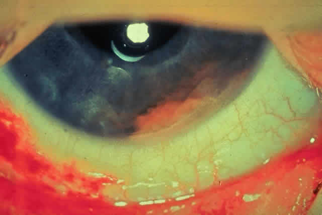
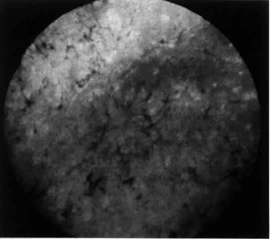
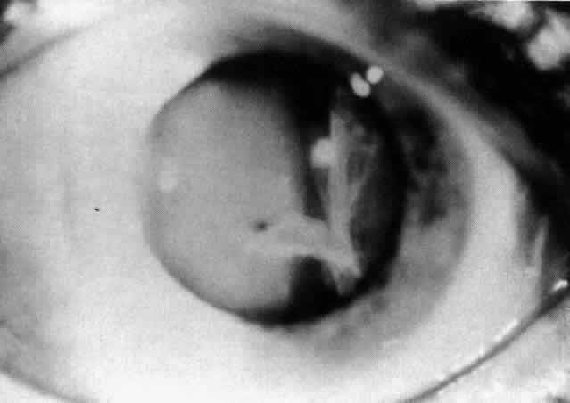
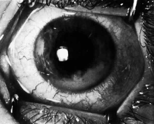
JRA uveitis is most frequently a chronic, nongranulomatous iridocyclitis that is bilateral in 71% of patients.14 Occasional African-American patients exhibit large granulomatous-appearing keratic precipitates. Since the ocular inflammation is usually asymptomatic and the affected eyes are frequently nonerythematous, it is not uncommon for complications to develop before the uveitis is detected.15 The complications include cataract, glaucoma, band keratopathy, posterior synechiae, and phthisis bulbi (Fig. 1; Color Plate 1A).12

Fig. 1. Patient with JRA demonstrating posterior synechia with scalloped pupil, band keratopathy and early cataract formation (see Color Plate 1A).

Color Plate 1. A. Patient with JRA demonstrating posterior synechia with scalloped pupil band keratopathy and early cataract formation. B. Fleshy, salmon-colored tumor resting on the inferior angle and attached to the iris in a patient with juvenile xanthogranuloma. C. Gray-white exudate present on the pars plana in a patient with early intermediate uveitis (pars planitis syndrome). D. The presence of leukocoria was the presenting sign in this young patient with toxocariasis. E. Cytomegalovirus retinitis in a patient with AIDS. F. Relatively subtle findings seen in the macula of a patient with subacute sclerosing panencephalitis. (Bloom JN: Uveitis in childhood. Ophthalmol Clin N Am 3:164, 1990)

The arthritis usually precedes the uveitis. If iridocyclitis does develop, it will occur in approximately half of the affected patients within 2 years of the onset of the arthritis.16 Pauciarticular individuals should be checked every 3 months. If they are ANA positive, the eye examinations are performed every 2 to 3 months. It has been recommended that JRA patients be examined for uveitis during the 7 years following the onset of uveitis.16 Polyarticular patients may be evaluated at 6-month intervals for the development of uveitis, while children with systemic-onset JRA are examined annually. There is no correlation between the degree of arthritis activity and the ocular inflammation. Although the arthritis may diminish and resolve with age, the uveitis often remains chronic and may persist into adulthood.17

Initial treatment of the iridocyclitis involves mydriatics and topical corticosteroids. Periocular corticosteroids, which may require a general anesthetic for administration in this pediatric population, and systemic corticosteroids are used for more severe ocular inflammation. Immunosuppressives are used for unresponsive vision-threatening cases of uveitis.18,19 These medications should only be used with the assistance of qualified pediatric consultants.

Ankylosing Spondylitis

Ten to 15% of patients with ankylosing spondylitis develop their arthritic disease in childhood.20 The onset of juvenile ankylosing spondylitis is usually at 8 to 10 years of age. Males are affected more commonly than females, as is true for the adult disorder form of the disease.20 Unlike the adult disorder, however, juvenile ankylosing spondylitis frequently presents as a peripheral arthropathy before physical or radiographic findings of sacroiliac joint involvement.21 Although both adult and juvenile ankylosing spondylitis manifest an approximately 95% association with HLA-B27,22,23 it is important to note that this histocompatibility antigen is not specific for ankylosing spondylitis and occurs in other forms of anterior uveitis,14 such as Reiter's syndrome and HLA-B27-associated anterior uveitis.

An acute, recurrent nongranulomatous iridocyclitis affects 10% to 20% of patients with juvenile ankylosing spondylitis. This anterior uveitis is typically described as unilateral because, although both eyes may be inflamed, it is unusual to note simultaneous involvement. The uveitis may precede or follow the arthritis, and the levels of activity of the two disorders are unrelated. Treatment of the ocular inflammation requires mydriatics and corticosteroids. Complications of the iridocyclitis include cataract, glaucoma, and posterior synechiae.

Reiter's Syndrome

Reiter's syndrome is infrequent in children, although it has been described in this patient age group.14,20 It is classically described as a triad of noninfectious urethritis, arthritis, and conjunctivitis.20 Males are more often affected than females. Although patients with Reiter's syndrome may have sacroiliitis and HLA-B27-positivity, the presence of urethritis, keratoderma blennorrhagica of the palms and soles, and ulcerations of the mouth and genitals differentiates this disorder from ankylosing spondylitis. Unlike Behçet's disease, the mucosal lesions are usually not painful.

A nongranulomatous anterior uveitis affects 3% to 12% of patients.22 This recurrent anterior uveitis is treated with topical and periocular corticosteroids.

Fuchs' Heterochromic Iridocyclitis

Although this disorder usually occurs in adults, it has been reported in the pediatric population.24 It is frequently asymptomatic with a chronic low-grade iridocyclitis, which is almost always unilateral. Hypochromia of the involved eye, as a result of iris atrophy, is typical but not invariable. The hypochromia may be difficult to detect in patients with blue irides. The keratic precipitates that accompany this anterior uveitis have a characteristic small, stellate appearance.25 Cataracts and open-angle glaucoma are the major complications of Fuchs' heterochromic iridocyclitis. Diagnosis is determined by the history and clinical manifestations, since there are no laboratory tests available for this disorder. Treatment with topical corticosteroids is usually ineffective. Mydriatics are often not required because posterior synechiae are uncommon.

Kawasaki Disease

Kawasaki disease is an acute multisystemic vasculitis characterized by fever, cervical lymphadenopathy, erythematous mucocutaneous lesions, and desquamation of the skin of the hands and feet.26–28 It is also referred to as mucocutaneous lymph node syndrome. This idiopathic disorder primarily affects infants and young children. It may be associated with arthritis and cardiac abnormalities, including coronary aneurysms, arrhythmias, and myocarditis. These heart complications produce sudden death in 1% to 2% of patients with Kawasaki disease.28

Bilateral bulbar conjunctival erythema has been reported in 96% of these children.26,29 Anterior uveitis is also common, occurring in 66% of individuals. If examined within 1 week of the onset of this disease, 83% of patients were reported to have an anterior uveitis.26 The ocular inflammation was mild and resolved without sequelae.26 No therapy is indicated for this uveitis.

Psoriatic Arthritis

Pauciarticular or polyarticular arthritis is an uncommon complication of psoriasis in childhood.20 The joint inflammation may precede or follow the development of the psoriasis. Patients with psoriatic arthritis may develop a bilateral, chronic anterior uveitis.14 Topical corticosteroids are the treatment for this ocular inflammation.

Acute Interstitial Nephritis

Acute interstitial nephritis is an uncommon renal disorder that may be the result of an immune reaction to antibiotics, nonsteroidal anti-inflammatory drugs, or infection.30,31 It is characterized by the nonspecific systemic complaints of low-grade fever, pallor, fatigue, and weight loss.30,31 Acute interstitial nephritis may be associated with an increased erythrocyte sedimentation rate, elevated serum creatinine value,31 proteinuria, glucosuria, microhematuria, leukocyturia, and excretion of casts.30,31 The diagnosis is established by renal biopsy. The prognosis in childhood is good.

A bilateral anterior uveitis may be associated with acute interstitial nephritis.30–33 The iridocyclitis may precede, follow, or occur concomitantly with this renal disorder. The ocular inflammation is treated with topical corticosteroids and responds well to therapy.33

Orbital Pseudotumor

Orbital pseudotumor is an idiopathic inflammatory lesion within the orbit that simulates a neoplasm.34 It is primarily a disorder of adults, although it may also occur in the first or second decade of life.34 This inflammatory disorder may present with multiple nonspecific signs and symptoms.34 Systemic complaints include headache, sore throat, lethargy, anorexia, weight loss, abdominal pain, and vomiting. Ocular findings include eyelid swelling, ptosis, pain, motility disorders, and diplopia. Proptosis occurs in 80% of patients.35

Unlike the adult form of the disease, uveitis may be associated with pediatric orbital pseudotumor.34 The ocular inflammation may involve the anterior, the posterior, or both segments of the eye. Patients with uveitis have an increased incidence of recurrence of orbital pseudotumor, as well as bilateral involvement.34

The possibility of an orbital pseudotumor should be considered in those pediatric patients with a persistent or recurrent uveitis and a previous negative diagnostic workup.36 These children may present without proptosis. Diagnostic evaluation includes ultrasonography, computed tomography, or magnetic resonance imaging. Treatment requires the use of systemic corticosteroids.

Lens-Induced Uveitis

Rupture of the lens capsule liberates lens material into the eye cavities. This results in one of two different types of uveitis that subside when the lens substance is removed. The uveitis is often associated with glaucoma. The phacotoxic reaction usually occurs in the presence of a hypermature cataract. The lens material acts as a chemical irritant, probably acting directly on the iris and ciliary body. Macrophages enter to engulf the liberated material. No polymorphonuclear cells are seen.

Endophthalmitis phacoanaphylactica results from an underlying sensitivity to lens protein that is probably amplified by bacteria or their toxins. Typically, a break in the lens capsule occurs in one eye as a result of surgery or injury. After inflammation has subsided in the first eye, the second eye develops a severe anterior granulomatous uveitis after surgery or trauma. Polymorphonuclear cells and macrophages are found in the aqueous iris and lens. Treatment involves removal of the lens, in addition to the general therapy for uveitis discussed in the section on treatment.

PROPOSED EVALUATION

Although it is traditional to present a flow sheet and table suggesting any etiologic evaluation in a patient with uveitis, Table 3 is offered with some hesitation. The “shotgun” approach to an etiologic evaluation should be abandoned in favor of evaluation on the basis of history and cost effectiveness. Table 3 is relevant only for the patient with severe disease in whom no localizing clue to etiology can be elicited.


TABLE 3. Diagnostic Evaluation of AnteriorUveitis in Children

  History
  Ocular examination
  Pediatric evaluation
  Complete blood cell count
  Serologic
  Antinuclear antibody
  Serum lysozyme
  Serum protein electrophoresis
  Fluorescent treponemal antibody absorption
  HLA typing
  Enzyme-linked immunosorbent assay, indirect fluorescent antibody for Lyme disease
  Skin tests
  Tuberculin
  Radiographic
  Chest
  Sacroiliac joint
  Gastrointestinal series
  Aqueous tap
  Fluorescein angiography
  Lumbar puncture


Routine blood studies should include a complete blood cell count to rule out the presence of leukemia as well as the fluorescent treponemal antibody absorption test for syphilis, which is most specific in cases of syphilitic ophthalmologic involvement. A number of serologic determinations can be performed to determine the etiology of anterior uveitis. These include determination of angiotensin converting enzyme levels, serum lysozyme elevation, and serum protein electrophoresis, antinuclear antibody, and HLA-B27 studies. The angiotensin converting enzyme is of great value in the presence of active sarcoid disease in adults, but results in children must be interpreted with caution (see Sarcoidosis). Serum lysozyme elevation and serum protein electrophoresis with an increased α2-globulin fraction are highly suggestive of sarcoid. Because of its association with JRA, antinuclear antibody determination is a most important serologic examination. Finally, while the presence of HLA-B27 is an important marker in patients with uveitis, it is not helpful for management and therefore is not obtained routinely except in a research setting. Until a clearer picture regarding the ocular manifestations of Lyme disease is available, immunologic testing for this spirochete (by enzyme-linked immunosorbent assay [ELISA] or indirect fluorescent antibody testing) is indicated in patients with idiopathic chronic uveitis.

Skin testing with the Kveim test has been used in some centers. Blind conjunctival biopsy has proved generally ineffective in attempts to diagnose sarcoid. When a significant conjunctival nodule is present, the yield is high. Biopsy of the lacrimal gland and surrounding conjunctiva has also proved helpful in establishing a diagnosis of sarcoid. The tuberculin skin test (intermediate-strength purified protein derivative), which is used to diagnose the rare case of tuberculous-induced uveitis, identifies patients in whom antituberculous therapy is indicated when systemic corticosteroids are used.

Radiographic studies include chest roentgenograms to rule out sarcoid and tuberculosis, a sacroiliac joint study to exclude rheumatoid spondylitis, and a gastrointestinal series in patients in whom ulcerative colitis or regional enteritis is suspected. Additional studies using fluorescein angiography, vitreous and aqueous aspiration, and lumbar puncture are only occasionally helpful.

TREATMENT

Over the past 35 years, corticosteroids have been the cornerstone of therapy for anterior uveitis. Of currently available local corticosteroids, prednisolone acetate 1% is the preferred solution because of its ability to penetrate the intact corneal epithelium. When this agent is used as frequently as every 30 minutes, most children do not need periocular and systemic corticosteroids.

Treatment requirements of the identified etiologic entities vary widely. Some, such as traumatic anterior uveitis and Fuchs' heterochromic iridocyclitis, require minimal amounts of local corticosteroids to control inflammation. On the other hand, acute fibrinous uveitis in patients with spondylitis may require maximal application of corticosteroids. With the onset of chronic anterior segment inflammation, careful monitoring is required to regulate drug dosage. Generally, the presence of flare without significant anterior chamber cells does not warrant treatment. We prefer to use chronic corticosteroid therapy to maintain the anterior segment with slightly less than a 1+ response present (scale = trace to 4+). Periocular corticosteroids have been widely employed for 2 decades, but their use in children younger than 14 years of age is somewhat limited because general anesthesia is usually needed for their delivery. Systemic corticosteroids are seldom required to treat anterior segment inflammatory disease.

Cycloplegic-mydriatic agents are important in the treatment of childhood uveitis because of the tendency for posterior synechiae to form. In the early phases of inflammation, atropine 1% may be used as often as two to four times daily. Some ophthalmologists are concerned that long-term use of mydriatic agents may result in a chronically dilated pupil. Most of the complications associated with the pupil are not related to dilatation, however, but to relative miosis and the formation of posterior synechiae. As soon as the inflammation has diminished, homatropine 5% four times daily may be substituted for atropine. In chronic anterior uveitis with minimal anterior chamber reaction, the pupil is dilated once daily. The parent is instructed to administer homatropine 5% one hour before the child's sleeping hour, to inspect the pupil carefully for change in size and regularity, and to report any change in the pupil to the ophthalmologist the next morning. Refraction and prescription of bifocals are important in these patients because of the potential long-term use of these cycloplegic agents. Chronic unilateral cycloplegia may produce amblyopia in children unless the refractive error is corrected.

Specific therapy for anterior uveitis in children is limited to the use of specific antiviral agents (trifluridine for herpes simplex) and antibacterial agents in infectious uveitis (penicillin for syphilis). Nonsteroidal anti-inflammatory agents such as indomethacin (Indocin), phenylbutazone (Butazolidin), piroxicam (Feldene), and aspirin are only occasionally effective in anterior segment inflammatory disease. Whenever high-dosage corticosteroids have failed, nonsteroidal anti-inflammatory agents have also failed.

COURSE AND COMPLICATIONS

With early diagnosis and prompt treatment, inflammation generally subsides within 2 to 6 weeks. The poor response reported in childhood anterior uveitis is directly related to the delay in diagnosis. It is this delay that makes complications of uveitis more common in children than those associated with adult-onset disease. keratopathy.Band keratopathy is the most consistent of all complications associated with chronic anterior chamber inflammation in children.Although hypercalcemia and phthisis bulbi also cause band keratopathy, most band formation is secondary to uveitis. Unless the band results in a cosmetic defect or reduces acuity, treatment is not indicated. When treatment is required, the band is easily removed. After the administration of general anesthesia, the cornea is de-epithelialized and Gelfoam, moistened with a 0.1 molar edentate disodium (sodium versenate) solution, is placed over it; 5- to 10-minute applications produce chelation of the band. If vision is present in the other eye, patching the affected eye after instilling a local antibiotic or sulfa medication for 24 hours aids in the re-epithelialization process. When there is little or no vision in the untreated eye, no patch is used. This procedure may be repeated as often as necessary and is almost uniformly successful.

The mechanism for the production of glaucoma is as varied in childhood uveitis as it is in the adult form of the disorder.Unfortunately, the prognosis for children with inflammatory glaucoma is far worse than for adults. Medical management, when successful, may require the long-term use of carbonic anhydrase inhibitors, since ß-adrenergic blocking agents (timolol, betaxolol) and epinephrine derivatives are not always successful. Medical therapy controls elevated intraocular pressure in 50% of patients who develop it.

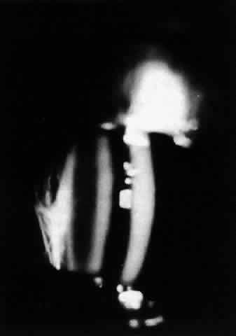
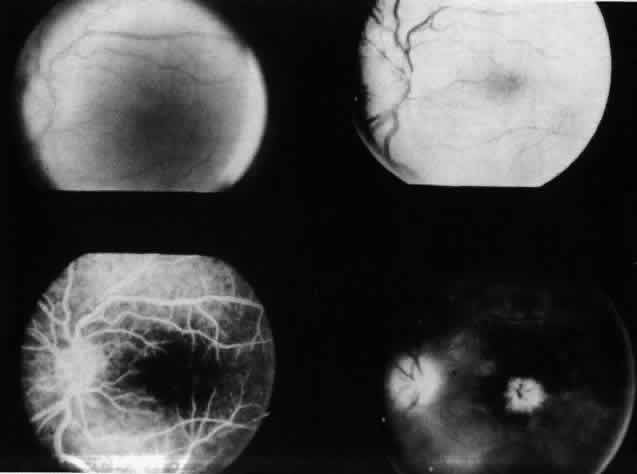
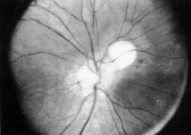
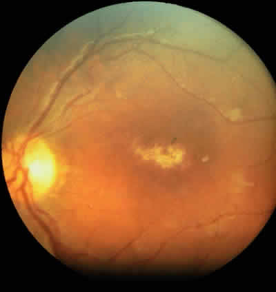
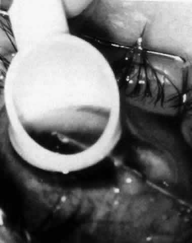
The variety of surgical procedures suggested by glaucoma specialists is adequate testimony to their lack of success. Filtering procedures have always been fraught with difficulty in all patients with inflammatory disease, but in younger patients the success rate is less than 50%. Trabeculectomy, peripheral iridectomy with thermal sclerostomy (advocated by Preziosa and Scheie), trabeculodialysis (advocated by Kanski; Fig. 2), and trabeculectomy with mitomycin have all failed to appreciably improve the prognosis of this complication.

Fig. 2. Intraoperative photograph of patient with inflammatory glaucoma undergoing trabeculodialysis. (Courtesy of Jack Kanski, MD)

Although the prognosis remains guarded in the management of cataract associated with inflammation, improved techniques offer significant hope for the young patient who presents with advanced cataract formation.Elimination of the posterior capsule prevents the later development of secondary membranes, thereby reducing the incidence of hypotony from ciliary body shutdown. A period of 6 months of relative inactivity of the inflammation is desirable before surgery. Periocular corticosteroids are administered at the time of surgery, and most of these eyes do not show marked additional inflammation. Orally administered postoperative corticosteroids are given only when indicated on a case-by-case basis. When extensive band keratopathy suggests that aphakic corneal contact lens fitting will prove difficult, the cornea should be chelated at the time of cataract surgery. Because of the character of the uveitis causing cataracts in children, intraocular lenses are seldom indicated. However, they may be used in carefully selected patients bulbi.Phthisis bulbi represents the end state of severe anterior uveitis in children. On occasion, however, these eyes respond well to a lensectomy-vitrectomy procedure even in the presence of a clear lens. The success of this operation probably depends on the removal of the cyclitic membrane, with restoration of aqueous formation and the elimination of hypotony.

PROGNOSIS

The prognosis for patients with anterior uveitis varies directly with the ability of the clinician to intervene and treat the disease in its early stages. The single largest group of patients who require careful monitoring are those with pauciarticular JRA.14,16 It is prudent to follow all patients who have a presumptive diagnosis of JRA in the previously described manner for 7 years after the diagnosis. For a few patients, early intervention will still fail to control many of the complications, and some of these patients may require immunosuppressive agents (e.g., cyclosporine). Although controlled evaluations of these agents in children are not available, results of preliminary studies are encouraging.

DIFFERENTIAL DIAGNOSIS

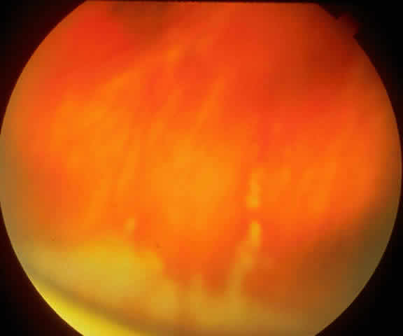
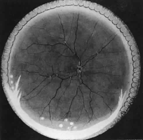
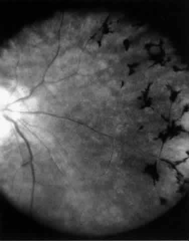
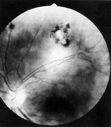
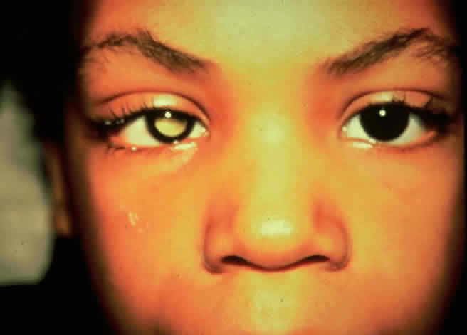
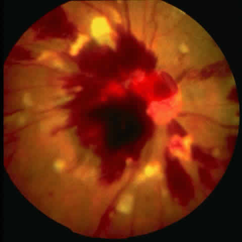
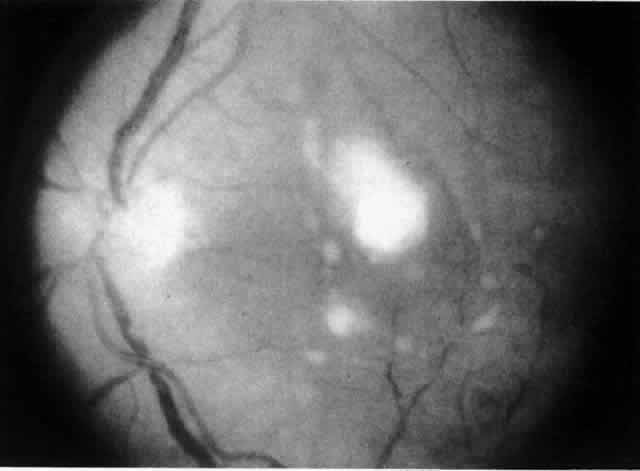
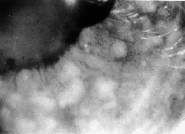
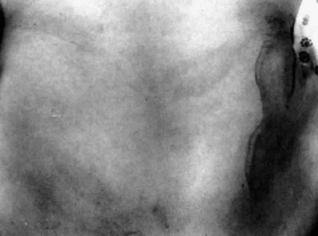
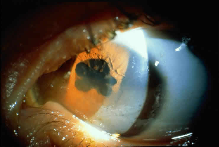
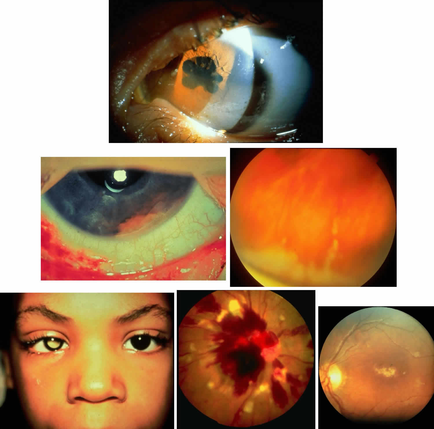
Table 4 lists the anterior segment masquerade syndromes. A pseudohypopyon in a patient with retinoblastoma (Fig. 3), although a diagnostic consideration, can easily be ruled out with a biomicroscopic or fundus examination. Juvenile xanthogranuloma characteristically presents as hyphema and a fleshy tumor on the iris (Fig. 4; Color Plate 1B).37 This rare condition commonly has associated skin lesions and is responsive to the use of local, periocular, or oral corticosteroids as well as to irradiation. Rarely, leukemia in childhood may present a significant anterior chamber reaction and hypopyon, although a general physical examination and associated laboratory signs will help identify this masquerade syndrome. Peripheral retinal detachment may have flare and cells in the anterior chamber, but careful ophthalmoscopy will reveal the presence of the detachment. An intraocular foreign body should be suspected in patients with unilateral uveitis who have a history of trauma and fail to respond to treatment.


TABLE 4. Masquerade Syndromes: Anterior Segment


Disease or DisorderAgeSigns of InflammationDiagnostic Studies
Retinoblastoma<15Flare, cells, pseudohypopyonAqueous tap for lactic dehydrogenase levels and cytology
Leukemia<15Flare, cells, heterochromiaBone marrow, peripheral blood smear, aqueous cytology
Intraocular foreign bodyAnyFlare, cellsRadiography, ultrasonography
Malignant melanomaAnyFlare, cells32P test, fluorescein angiography, ultrasonography
Juvenile xanthogranuloma<15Flare, cells, hyphemaExamination of skin, iris biopsy
Peripheral retinal detachmentAnyFlare, cellsCareful ophthalmoscopy


Fig. 3. Floccular cellular deposit present in anterior chamber of infant with retinoblastoma (pseudohypopyon). Courtesy of Robert Ellsworth, MD

Fig. 4. Fleshy salmon-colored tumor resting on the inferior angle and attached to the iris in a patient with juvenile xanthogranuloma (see Color Plate 1B).

Back to Top
INTERMEDIATE UVEITIS
Intermediate uveitis in children provides little problem in diagnosis.38 Experience, however, suggests that there is a great disparity between the ability to diagnose this entity and the clinician's ability to treat it. Emphasis will therefore be placed on the treatment of this disorder. Although intermediate segment inflammation can be associated with multiple sclerosis, toxocariasis, sarcoidosis, and Whipple's disease, the great majority of youngsters present with the “pars planitis syndrome” of unknown etiology.

SYMPTOMS

In its classic form, intermediate uveitis is largely asymptomatic until the disease is well advanced and vision is markedly reduced. Since the eyes are characteristically white, there are few clues to bring the condition to the parent's attention. Routine school or pediatric vision screening, the presence of strabismus associated with reduced acuity, or pain (in the more advanced forms) alert the parent to this disorder. Less characteristic but accounting for nearly one third of these patients are the classic symptoms of acute anterior uveitis with increased lacrimation, redness, and pain. When a patient appears with this symptom complex, the more subtle underlying intermediate component of what appears to be primarily an anterior uveal inflammation becomes more apparent as the anterior portion of the inflammation initially responds but then slips into a chronic phase. This expression of the disease is more common in children than in adults, most probably because children are unable to detect the early signs of minimal blurring of vision and the presence of vitreous floaters.

SIGNS

Except in eyes with associated acute anterior uveitis, the signs of this syndrome are relatively subtle. Early in the course of the disease, the presence of anterior vitreous cells is the only sign seen on routine examination (Fig. 5). Initially, these are fine and separate, but as the disorder progresses they aggregate and may acquire a fibrillar state in the anterior one third of the vitreous. The anterior segment may be quiet, although a trace anterior chamber flare and occasional cellular deposits may be present in the trabecular meshwork. There is usually posterior subcapsular cataract formation at the time of presentation. Scleral depression early in the course of the illness will reveal a gray-white exudate on the pars plana (Fig. 6; Color Plate 1C). Other ophthalmoscopic signs of the disease are peripheral retinal vasculitis with sheathing of both the venules and the arterioles. Peripapillary edema may be present at any time in its course (Fig. 7). When vision is reduced below a level of 20/30 (6/9),* ophthalmoscopically visible cystoid macular edema is usually seen (Fig. 8). Later in the course of the illness these cystoid changes may become loculated and may become associated with macular hemorrhage. An inconsistent finding in this disorder is peripheral retinal neovascularization and vitreous hemorrhage.

Fig. 5. Anterior vitreous cellular deposit with a fibrillar pattern in the retrolental space in a young patient with chronic intermediate uveitis (pars planitis syndrome).

Fig. 6. Gray-white exudate present on the pars plana in a patient with early intermediate uveitis (pars planitis syndrome; see Color Plate 1C).

Fig. 7. This represents an artist's rendering of an eye with chronic intermediate uveitis. Note the exudate present inferiorly at the vitreous base and the associated peripheral retinal vasculitis, with cystoid macular as well as peripapillary edema.

Fig. 8. Patient with cystoid macular edema and chronic anterior segment inflammation in association with intermediate uveitis as demonstrated with a fluorescein angiographic study.

TREATMENT

As a result of the experience of one of the authors (CLG) in managing 60 young patients with intermediate uveitis, a treatment algorithm has been devised (Fig. 9).39 Local corticosteroids are used only if the patient's vision is 20/40 (6/12) or better and significant anterior chamber reaction is present. Patients with minimal anterior chamber reaction and vision of 20/40 or better receive no treatment. If vision is less than 20/40 and subretinal exudation, optic nerve papillitis, or disturbing floaters are found, periocular corticosteroids are given weekly (up to six doses) until improvement occurs. In children younger than 14 years of age, general anesthesia is usually required. Children between the ages of 11 and 14 years may, however, become cooperative enough to permit periocular injection in the office without general anesthesia. If improvement does not continue and the signs triggering the intervention are not eliminated, reinjection should continue for six more weekly injections. If these fail to achieve the desired result, retinal cryopexy40,41 should be applied. If cryopexy also fails and the disease is bilateral, cyclosporine should be employed.42,43 Cyclosporine or other immunosuppressive agents are reserved for use in bilateral disease because of their potential long-term side effects. Vitrectomy for chronic cystoid macular edema is occasionally employed in those patients failing to respond to these aforementioned therapeutic measures.

Fig. 9. Treatment algorithm for intermediate uveitis.

COURSE AND COMPLICATIONS

When intervention is early and treatment is vigorous, most patients obtain normal vision. Again, difficulties in the management of this disorder are due to the late presentation of patients in the pediatric age group. Cystoid macular edema, if present for long periods, becomes irreversible. Cataract formation seen early in the course of the illness does little to reduce acuity, but with progression it may reduce vision to the point where cataract extraction is required. The presence of peripheral retinal neovascularization is especially threatening because recurrent vitreous hemorrhages may require ablation of the vessels and vitrectomy. Retinal detachment may also occur and provides a great clinical challenge because of the associated inflammatory changes. Secondary glaucoma is rarely a problem in patients with intermediate uveitis, although it may complicate the postoperative management of the cataract extraction.

DIFFERENTIAL DIAGNOSIS

Few conditions should confuse the clinician in the differential diagnosis of this disorder. Rarely, a patient with a peripheral lesion of toxoplasmosis may cause clinical confusion only if careful scleral depression is not carried out. “Mutton fat” keratic precipitates should also alert the clinician to the possibility of toxoplasmosis. Retinitis pigmentosa often presents with cells in the anterior one third of the vitreous (Fig. 10). The ophthalmoscopic changes in retinitis pigmentosa coupled with the electroretinographic findings should enable the ophthalmologist to differentiate retinitis pigmentosa from intermediate uveitis.

Fig. 10. This patient demonstrated retinitis pigmentosa with the presence of cells in the anterior third of the vitreous.

Back to Top
POSTERIOR UVEAL TRACT
The posterior segment of the uveal tract, especially the macular and paramacular areas of the retina and choroid, is frequently affected in childhood uveitis. Early diagnosis and prompt treatment obviously are important.

ETIOLOGY

The causes of posterior uveitis and panuveitis are given in Table 5.


TABLE 5. Etiology of Posterior Uveitis and Panuveitis in ChildrenPosterior Uveitis

  Toxoplasmosis (70%)
  Nematodiasis (10%–15%)
  Cytomegalovirus
  Tuberculosis
  Syphilis
  Rubella
  Subacute sclerosing panencephalitis
  Herpes simplex
  HIV
  Panuveitis
  Sarcoidosis
  Sympathetic ophthalmia
  Vogt-Koyanagi-Harada syndrome
  Behcet's disease
  Ocular Lyme borreliosis


Toxoplasmosis

Toxoplasmosis is the most common identifiable cause of pediatric uveitis.3,6 Congenital infection is the result of ingestion of Toxoplasma gondii cysts by a previously uninfected pregnant woman, followed by transplacental transmission of the organism to the fetus.44,45 These cysts may be found as tissue cysts in animal meats or as oocysts in cat feces or litter and in the soil. Infection during the first trimester is associated with neonatal convulsions, intracranial calcifications, and retinitis.46 Infants infected in the later trimesters often develop the retinitis only. Inactive atrophic scars, usually affecting the posterior pole, occur in approximately 80% of infected newborns. These lesions are bilateral in 85% of congenitally diagnosed cases of toxoplasmosis.47

It is postulated that ocular toxoplasmosis in older children and adults represents a reactivation of a subclinical congenital infection of the retina.48 Although active retinal lesions are almost always unilateral, the fellow eye may frequently be noted to have an inactive chorioretinal scar.48 It is also common to find active lesions adjacent to the healed, atrophic scars of a previous infection (Fig. 11). The necrotizing retinitis of toxoplasmosis is characterized by a fluffy white focus surrounded by retinal edema. The lesions are usually circular or oval and are of varying size. The posterior uveitis may be associated with a granulomatous or nongranulomatous iridocyclitis.

Fig. 11. Patient with an active focus of inflammation adjacent to a healed scar. The ELISA titer showed antibodies to toxoplasmosis.

The early symptoms of ocular toxoplasmosis include blurred vision and floaters. Unless the macula or optic nerve is affected, the active lesions spontaneously heal in several weeks with no significant visual sequelae except for a scotoma produced by the atrophic scar. The indications for treatment include lesions in the peripapillary, perifoveal, or maculopapillary bundle regions; loss of vision worse than 20/200 (6/60); or marked vitreous inflammation.49 Treatment consists of the combined use of pyrimethamine (Daraprim), folinic acid supplement (leucovorin), and sulfadiazine; clindamycin may be used in addition to these medications or in substitution for pyrimethamine.46,48,49 Careful monitoring of patients is required because pyrimethamine may produce thrombocytopenia and leukopenia. Folinic acid supplementation is concurrently administered to reduce the likelihood of this problem. Clindamycin may be associated with a pseudomembranous colitis.48 The use of sulfonamides may protect against this complication, which is treated with oral vancomycin if it does occur. Systemic corticosteroids are employed to decrease the inflammatory damage produced by the host's reaction to the Toxoplasma organism. This medication is not used without the antimicrobial coverage described earlier, however, since corticosteroids may otherwise suppress the immune response and worsen the infection.46 As a result, periocular injections of corticosteroids are not advisable.49 Topical corticosteroids may, however, be used to treat the anterior uveitis, since Toxoplasma gondii has never been detected in the anterior segment, except in an immunosuppressed patient.49,50

The use of live Toxoplasma organisms in the Sabin-Feldman methylene blue dye test has resulted in the replacement of this procedure by the indirect fluorescent antibody test, hemagglutination test, and ELISA.46,51 The ELISA is the most sensitive and specific of these tests.51 Any positive titer is significant in evaluating the diagnosis of toxoplasmosis, since there is no correlation between the height of a positive antibody titer level and either the clinical diagnosis or the degree of activity of the ocular disease.46

Toxocariasis

Toxocariasis is primarily a disease of children. It is acquired by the ingestion of soil containing the eggs of the canine intestinal roundworm Toxocara canis.52 After the eggs of the ascarid hatch in the intestines of the patient, the organism invades the blood vessels and migrates to various organs, especially the liver, brain, lungs, eyes, and skin. Since Toxocara does not develop beyond the larval state in the incidental human host, eggs are not found in the stool.

Visceral larva migrans is an acute, systemic disorder usually affecting children of approximately 2 years of age and characterized by fever, rash, coughing and wheezing, hepatosplenomegaly, and, at times, convulsions.52 Hematologic evaluation reveals eosinophilia. It is unusual for visceral larva migrans and ocular toxocariasis to affect the same person.53 The ocular form of the disease is typically found in patients approximately 7 years old, with a range of from 2 to 30 years.54 There are no associated systemic findings or eosinophilia. Ocular toxocariasis is a unilateral disorder that presents as strabismus, leukocoria (Fig. 12; Color Plate 1D), or decreased vision. Retinal damage is the result of the host's inflammatory response to the single infection nematode, which must usually be dead before the uveitis can develop. Although the posterior uveitis may be of severe intensity, it is not uncommon that little external evidence of inflammation is noted.

Fig. 12. The presence of leukocoria was the presenting sign in a child with toxocariasis (see Color Plate 1D).

Ocular toxocariasis may be present as a chronic endophthalmitis, peripheral chorioretinal granuloma, or posterior pole chorioretinal granuloma (Fig. 13).21 Toxocara endophthalmitis is characterized by a dense vitreous cellular reaction that may be associated with a peripheral granuloma or an exudate overlying the peripheral retina.21 It may be complicated by the development of a cataract, cyclitic membrane, or retinal detachment. The associated anterior uveitis is frequently mild.

Fig. 13. Ophthalmoscopic appearance of a solitary Toxocara lesion in the posterior pole.

A posterior pole Toxocara granuloma is usually located either in the macula or in the maculopapillary bundle.21 This round, elevated, yellow-white lesion elicits few cells in the overlying vitreous and no anterior uveitis. Its location and color may result in leukocoria.

A peripheral granuloma is often situated at or anterior to the equator. As is true for the posterior pole granuloma, minimal inflammation accompanies the peripheral granuloma.21 These lesions may, however, be associated with vitreous bands extending to the posterior fundus. Macular distortion or retinal detachment can result from the traction produced by this band. If the peripheral granuloma does not affect the posterior pole, there may be minimal or no visual effect.21

As discussed earlier, patients with ocular toxocariasis typically do not develop eosinophilia. Laboratory diagnosis is, however, aided by the high sensitivity and specificity (90%) of an ELISA titer for Toxocara.55 These titers may eventually decrease after infection with the organism, and therefore any positive titer, even less than 1:8, should be considered significant in the evaluation of a patient suspected of having this disease.56

Oral or periocular corticosteroids are used to treat Toxocara endophthalmitis. Corticosteroids are ineffective for posterior or peripheral granulomas, since there is minimal inflammation accompanying these lesions. Antihelminthics are not effective for ocular toxocariasis since it is the host's reaction to the dead nematode that initiates the retinal damage.21 Vitrectomy and scleral buckling procedures are used to treat retinal traction detachments.57

Toxocariasis is in the differential diagnosis of retinoblastoma.58 Although calcification has been reported in rare cases of ocular toxocariasis,52 this finding is frequently detected in retinoblastoma by ultrasonography or computed tomography. In addition, patients with retinoblastoma are frequently younger than persons with ocular toxocariasis. Unlike toxocariasis, retinoblastoma may be familial. Although a negative Toxocara ELISA result is evidence against this parasitic infection, a positive titer does not eliminate the possibility of retinoblastoma, since an increased titer may be present in a significant proportion of the general pediatric population in various regions of this country.59

Congenital Syphilis

Congenital syphilis is rare in the United States today for several reasons. The widespread use of penicillin and other antibiotic therapies for active syphilis, as well as serologic testing both before marriage and at the beginning of pregnancy, identifies cases before the fetus is infected.60

Infants with congenital syphilis have been born with active disease, as evidenced by fever, rash, pneumonitis, and hepatosplenomegaly. In some, active choroiditis may be seen; but in most the only evidence of choroiditis is the presence of segmental pigmentation in the periphery, which is seen later in life at routine examination or as a sequela to corneal inflammation. Corneal transplantation may be required for improved visual acuity. Cataract may begin earlier than would be expected. Glaucoma is a late sequela, both an insidious-onset, open-angle process and one of acute or subacute onset made of some narrow angle-closure events and, in some patients, a rapid decompensation of open angles. Decades later, recurrences of iritis and keratitis (interstitial keratitis) are puzzling but they respond to topical corticosteroids and cycloplegia.

Cytomegalovirus

With the increase in organ transplantation and the use of immune system-depressing agents in the treatment of neoplasm, cytomegalovirus has become a more important cause of posterior uveitis (Fig. 14; Color Plate 1E).61 Positive blood cultures and characteristic urinary inclusion bodies help establish this diagnosis.

Fig. 14. Cytomegalovirus retinitis in a patient with AIDS (see Color Plate 1E).

Cytomegalovirus retinitis may also be associated with the acquired immunodeficiency syndrome (AIDS). In the pediatric age group, the AIDS complex occurs in newborns of affected mothers, as well as in hemophiliacs who have received contaminated blood that has resulted in transmission of the human immunodeficiency virus. Ganciclovir and foscarnet are the drugs of choice for this disease.62

Herpes Simplex

Herpes simplex virus type 2 accounts for most herpetic infections in newborns. Acquired from the birth canal during delivery, its manifestations vary.63 Posterior involvement may disappear and leave few or no sequelae. Numerous areas of choroidal hemorrhage, choroidal and intraretinal exudates, and diffuse fine vitreous opacities have been seen, as well as extensive retinal edema and marked narrowing of the arterioles. In newborns with systemic herpes simplex virus infection, a severe necrotizing chorioretinitis has been observed that may need to be differentiated from the scars of toxoplasmic and cytomegalic retinitis.63 Acyclovir is used to treat herpes simplex virus.

Fungal Disease

Candidiasis and other fungal entities are rare causes of posterior uveitis in children (Fig. 15), although the spread of intravenous drug abuse may portend a future increase in these entities. Vitrectomy and systemic and intravitreal antifungal agents are used for treatment.

Fig. 15. Ophthalmoscopic appearance of a teenager with a history of intravenous drug abuse exhibiting classic candidiasis.

Viral Entities

Rubella retinitis is the most common ocular feature of the maternal rubella syndrome, being present in 25% to 50% of infants. Unilateral or bilateral pigment deposits, usually limited to the posterior pole, vary from fine, powdery, or granular to more discrete shapes. The retinopathy is benign and nonprogressive and does not interfere with vision unless neovascularization develops in the macula (Fig. 16).64

Fig. 16. Retinopathy associated with the maternal rubella syndrome in an infant.

Posterior uveitis without vitreous reaction of a mild, degenerative, speckled character is the most striking ocular manifestation of subacute sclerosing panencephalitis (SSPE),65 which is believed to be due to the measles virus.65,66 SSPE occurs most commonly in school-aged children and is marked by changes in behavior and intellectual deterioration. Most die within 1 to 2 years of onset. The disorder is characterized by myoclonic seizures followed by spastic paralysis and profound dementia. Cortical blindness, nystagmus, papilledema, and optic atrophy may also be observed (Fig. 17; Color Plate 1F).

Fig. 17. Relatively subtle findings seen in the macula of a patient with subacute sclerosing panencephalitis (SSPE) (see Color Plate 1F).

Back to Top
PANUVEITIS

ETIOLOGY

The following entities characteristically simultaneously affect both anterior and posterior segments.

Sarcoidosis

Sarcoidosis is a chronic, multisystemic disorder of unknown etiology characterized pathologically by the presence of noncaseating granulomas in affected organs. This disease is seldom diagnosed in the pediatric population, although it is suspected that its prevalence is underestimated because children, unlike adults, do not undergo the routine chest x-ray screenings that might detect asymptomatic persons.67

Pediatric sarcoidosis has two peaks of incidence, occurring between ages 8 and 15 and in children younger than 5 years old.68 Disease in older children resembles that in adults and often affects the lungs, lymph nodes, and eyes. Patients complain of fever, lethargy, malaise, and weight loss.67,69 Abnormal lung findings on chest roentgenography are common.67 Uveitis occurs in approximately one third of these children.24,67,70

Fewer patients younger than 5 years of age develop sarcoidosis.68 Disease in this group is significantly different from that in older children and adults in that it is characterized more commonly by arthritis, uveitis, and rash while pulmonary abnormalities affect only one third of these younger patients.70

Although pediatric sarcoidosis resembles the adult disorder in that black patients are more commonly affected than whites, there is no female predilection in the childhood disorder.67,69,70 As is true for adult sarcoidosis, the most common ocular problem is anterior uveitis,67,69,70 which occurs in approximately one third of the older children and three fourths of the younger group.69,70 The iridocyclitis, which may be either granulomatous or nongranulomatous (Fig. 18), is often not accompanied by complaints of pain or photophobia. Pediatric complications resemble those that occur in adults and include band keratopathy, glaucoma, cataract, and posterior synechiae secondary to the anterior uveitis. Other manifestations of pediatric sarcoidosis are conjunctival nodules, vitreitis, and chorioretinal granulomas. Unlike adults, lacrimal gland enlargement and retinal periphlebitis are not found in these pediatric patients.70

Fig. 18. Iris nodules with posterior synechiae formation in a young patient with chronic iridocyclitis associated with biopsy-proven sarcoidosis.

The diagnosis of sarcoidosis, in both adults and children, involves the combination of clinical, laboratory, histologic, and radiographic findings. A definitive diagnosis requires the identification of a noncaseating granuloma within a biopsy specimen.67,69 Enlarged peripheral lymph nodes, conjunctival nodules, rashes, liver, and mediastinum are potential biopsy sites.67

Children with sarcoidosis may also manifest an elevated erythrocyte sedimentation rate, neutropenia, hypergammaglobulinemia, hypercalciuria67 often in the absence of hypercalcemia,69 and a serum alkaline phosphatase value elevated beyond the usual pediatric range.69 Although children normally demonstrate a serum angiotensin converting enzyme level higher than that of adults, pediatric patients with sarcoidosis may have serum angiotensin converting enzyme concentrations greater than even these usual childhood values.71,72

As discussed earlier, chest roentgenograms are more commonly abnormal in the older group of children than in the younger patients. The most common radiographic finding is bilateral hilar adenopathy, with or without normal lung parenchyma.67,69 Pulmonary function testing may reveal restrictive lung changes.67

Ocular sarcoidosis may be treated with topical, periocular, or systemic corticosteroids. Topical mydriatics are also used for anterior uveitis. Patients with sarcoidosis often develop a severe flare-up of their iridocyclitis after cataract surgery. If such surgery is contemplated, the use of preoperative and postoperative systemic and intraoperative periocular corticosteroids is advisable.

Sympathetic Ophthalmia

Sympathetic ophthalmia is a bilateral granulomatous panuveitis that develops after a penetrating injury to one eye.73–75 The trauma may be either accidental or, in rare cases, surgical and is usually accompanied by uveal prolapse. The injured globe is the “inciting” or “exciting” eye and the fellow eye is categorized as “sympathizing.”75 A gradual onset of inflammation will develop within 3 months after the trauma in 70% of the cases.75 Ninety percent of patients with sympathetic ophthalmia will develop the disease within the first year after the injury.76 There have, however, been case reports of the onset of sympathetic ophthalmia as early as 5 days after globe penetration and as late as 12 years after the injury.75

The exciting eye frequently presents with persistent low-grade inflammation that fails to resolve after the trauma. Routine corticosteroid therapy of the post-traumatic uveitis may mask this sympathetic ophthalmia-related inflammation.76 The patient's complaints related to the sympathizing eye may initially include photophobia, as well as blurred vision secondary to a loss of accommodation. As the disease worsens, a bilateral granulomatous uveitis is noted. The iridocyclitis may be associated with mutton-fat keratic precipitates. Multiple signs of posterior segment inflammation develop, including cells and haze in the vitreous, choroidal thickening, and optic nerve edema.76 Small yellow-white infiltrates in the retinal periphery, at the level of the retinal pigment epithelium, are noted in approximately one third of patients.75,76 These lesions represent Dalen-Fuchs nodules, which are characteristic, but not pathognomonic, for sympathetic ophthalmia. There are no laboratory tests that assist in the diagnosis. Complications of this disorder include cataract, glaucoma, chorioretinal scarring, optic nerve atrophy, and phthisis bulbi, which may affect either or both the exciting and sympathizing eyes.76,77

Enucleation of the injured or exciting eye within 2 weeks of the onset of symptoms in the sympathizing eye improves the prognosis for the fellow, uninjured globe.75,77 It is not advisable, however, to surgically remove an exciting eye that retains useful vision, since this traumatized eye may ultimately retain better acuity than the sympathizing eye.74

The medical treatment of sympathetic ophthalmia, with or without enucleation, involves the use of corticosteroids delivered by topical, periocular, or systemic routes.74 If a patient is refractory to corticosteroid therapy, then immunosuppressives, such as azathioprine, methotrexate, chlorambucil, or cyclosporine may be necessary.74 Patients with sympathetic ophthalmia require close monitoring, since this is a recurrent disorder.

Sympathetic ophthalmia may mimic, in rare cases, Vogt-Koyanagi-Harada syndrome and present as the latter's characteristic extraocular abnormalities of vitiligo, alopecia, poliosis, and meningeal irritation. In addition, the ocular findings of these diseases may be similar, including the presence of Dalen-Fuchs nodules.76,78 The history of trauma in association with sympathetic ophthalmia and its absence with Vogt-Koyanagi-Harada syndrome is the primary distinguishing characteristic between these two inflammatory diseases.75

Vogt-Koyanagi-Harada Syndrome

The Vogt-Koyanagi-Harada syndrome is a multisystemic disorder of unknown etiology. Although it usually affects young and middle-aged adults there have been occasional reports in pediatric patients.14,38,79,80 Asian and heavily pigmented persons are more often affected than are whites.76 In the United States, patients of Native American extraction have a greater incidence of this disorder than does the general population. HLA-DR4, HLA-DRw53, and HLA-DQw3 antigens have been associated with an increased incidence of this syndrome.81

The extraocular findings of Vogt-Koyanagi-Harada syndrome81 include vitiligo, poliosis, alopecia, dysacousia (vertigo, tinnitus, and hearing loss), and meningeal irritation resulting in cerebrospinal fluid pleocytosis and headaches. The cerebrospinal fluid pleocytosis, if present, is detected during the first few weeks of this syndrome.79

The ocular abnormalities of the syndrome are frequently bilateral, although the onset may be unilateral.79 Pain and photophobia often accompany a granulomatous iridocyclitis. The posterior segment multiple exudative retinal detachments may be preceded by a multifocal choroiditis. These serous detachments are frequently located in the inferior retina. Pigmentary mottling and chorioretinal scarring develop as these initial lesions resolve.76 Other ocular findings include vitreitis, papillitis, and Dalen-Fuchs nodules similar to those observed in sympathetic ophthalmia.76

The diagnosis of Vogt-Koyanagi-Harada syndrome is based on the clinical findings, since there are no specific laboratory tests for this syndrome. The HLA association and cerebrospinal fluid pleocytosis discussed earlier may be of assistance in establishing the diagnosis, but the absence of these findings does not eliminate the possibility of this disorder. Vogt-Koyanagi-Harada syndrome is treated with corticosteroids, either topical, periocular, systemic, or a combination of these delivery modes. Immunosuppressives are used for patients refractory to this initial corticosteroid therapy.42 Surgery is not indicated for the serous retinal detachments, since these lesions will resolve spontaneously or after medical treatment.

Behçet's Disease

Behçet's disease may occur at any age82 and although most frequently a disorder of young adults, rare cases in childhood have been reported.14 It is a chronic multisystemic disorder classically described as a triad of iridocyclitis, oral ulcers, and genital ulcerations. The diagnosis of Behçet's disease is determined by the identification of various major and minor criteria within the spectrum of the patient's complaints.82 The major criteria include (1) recurrent painful mouth ulcers, (2) painful genital ulcerations, (3) ocular lesions, and (4) nonulcerative skin lesions. The minor criteria consist of (1) arthritis, (2) cardiovascular lesions, (3) epididymitis, (4) thrombophlebitis, (5) gastrointestinal lesions, and (6) central nervous system involvement.82,83 The pathologic basis for Behçet's disease is an occlusive, necrotizing vasculitis of unknown etiology.83 This disorder is most common in the Far East and the Mediterranean region. Patients with Behçet's disease demonstrate an increased incidence of positivity for HLA-B5 and its derivative HLA-Bw51.84

Ocular abnormalities are common in Behçet's disease and occur in 70% to 85% of patients.83 The most common finding is a recurrent bilateral iridocyclitis, which may be associated with a sterile hypopyon.83 The presence of a hypopyon is not pathognomonic for Behçet's disease, since this lesion occurs with other ocular inflammatory disorders. Posterior segment complications of this disease include vitreitis, retinal vasculitis, branch vein occlusion, areas of retinitis and retinal necrosis, cystoid macular edema, optic nerve edema, and optic nerve atrophy.83,85 The visual prognosis of patients with Behçet's disease is poor, despite intensive therapy.86 Corticosteroids are usually not effective for long-term treatment of these persons, who may require immunosuppressive medications.86–88

Lyme Disease

Lyme disease, originally described in 1975, is caused by the spirochete Borrelia burgdorferi and is transmitted by the tick Ixodes dammini. The full spectrum of ocular disorders associated with this complicated multisystemic disease has only recently begun to be appreciated.

The disorder has been divided into three clinical stages, each of which has specific ocular components.89 Conjunctivitis occurs in stage 1, which follows infections by the tick and its characteristic erythematous, spreading macular skin lesion (Fig. 19). It is associated with constitutional flulike symptoms, and there may be additional skin findings unassociated with the previous tick bite. Stage 2 involves significant cardiac and neurologic disease and has its primary ocular manifestation cranial nerve palsies, with involvement of the third, sixth, and seventh nerves. Meningitis and radiculoneuropathy are the other associated neurologic signs. Cardiac signs include acute myocarditis and the symptoms associated with acute rheumatic fever. In addition, keratitis has been described in this stage of the disease. Stage 3, characterized systemically by arthritis and chronic neurologic symptoms, is associated with migratory pain in muscles, joints, and tendons as well as the bursae. Neuropsychiatric disease, with the chronic fatigue syndrome and focal central nervous system disease, is also seen in this stage. It is in the third stage that the ocular manifestations involving the uveal tract probably occur with greatest regularity. Granulomatous anterior segment inflammatory disease, vitreitis (Fig. 20), diffuse choroiditis, and panophthalmitis, as well as the intermediate uveitis syndrome, have all been described in stage 3 or the latter part of stage 2 disease. In addition, ischemic optic neuropathy, retrobulbar neuritis, optic papillitis, and pseudotumor cerebri have been noted.

Fig. 19. Characteristic erythematous spreading macular skin lesion with bull's eye appearance in a patient after a tick bite.

Fig. 20. Severe vitreitis in a patient with presumed Lyme disease.

Laboratory diagnosis depends on an indirect immunofluorescent assay and the ELISA test. Results from both of these studies may be low or absent in stage 1, but in stages 2 and 3, antibodies are positive in 95% of patients with Lyme disease. At present the ELISA study is considered better than the indirect immunofluorescent assay in all stages (Table 6).


TABLE 6. Diagnostic Criteria for Lyme Disease


AreaCriteria
EndemicErythema migrans with exposure no more than 30 days before onset
 Involvement of one organ system and a positive antibody test
NonendemicErythema migrans with positive antibody test
 Erythema migrans with involvement of two organ systems


Treatment early in the course of the disease consists of oral antibiotics. Tetracycline, doxycycline, penicillin, erythromycin, and ceftriaxone have all been reported to be effective. Later, intravenous penicillin and ceftriaxone are required for remission. Early use of systemic corticosteroids makes treatment of the disorder with specific antibiotics far more difficult.

DIFFERENTIAL DIAGNOSIS

Many of the same masquerade symptoms that confuse the clinician in the diagnosis of anterior and intermediate uveitis may also contribute to confusion in the differential diagnosis of inflammatory disease in the posterior uveal tract (Table 7). Included in this group are retinitis pigmentosa, reticulum cell sarcoma, lymphoma, retinoblastoma, and malignant uveitis-like melanoma. Use of the electroretinogram and electro-oculogram, studies of the aqueous and vitreous, a complete physical examination including bone marrow studies, as well as the periodic employment of ultrasonography all sufficiently permit the ophthalmologist to rule out the above syndromes.


TABLE 7. Masquerade Syndromes: Posterior Segment


DiseaseAgeSigns of InflammationDiagnostic Studies
Retinitis pigmentosaAnyCell in vitreousElectroretinography, electro-oculography, visual fields
Lymphoma*15+Retinal hemorrhages, exudates, vitreous cellsNode biopsy, bone marrow, complete physical examination
Retinoblastoma*#5Vitreous cells, retinal exudatesUltrasonography, aqueous tap
Malignant melanoma15+Vitreous cellsP test, fluorescein angiography, ultrasonography
Multiple sclerosis15+PeriphlebitisNeurologic examination


This study was supported in part by grants from The Knights Templar Eye Foundation, Inc (JNB) and The National Children's Eye Care Foundation, Inc (JNB)

Back to Top
REFERENCES

1. Kimura SJ, Hogan MJ, Thygeson P: Uveitis in children. Arch Ophthalmol 51:80, 1954

2. Kimura SJ, Hogan MJ: Uveitis in children: Analysis of 274 cases. Trans Am Ophthalmol Soc 62:173, 1966

3. Perkins ES: Pattern of uveitis in children. Br J Ophthalmol 50:169, 1966

4. Witmer R, Korner G: Uveitis in children. Ophthalmologica 152:277, 1966

5. Kazdan JJ, McCulloch JC, Crawford JS: Uveitis in children. Can Med Assoc J 96:385, 1967

6. Makley TA Jr, Long J, Suie T et al: Uveitis in children: A follow-up study. J Pediatr Ophthalmol 6:136, 1969

7. Jutte A, Lempke L, Opitz J: Chronische Zyklitis im Kindesalter. Ophthalmologica 157:169, 1969

8. Bloom JN: Uveitis in childhood. Ophthalmol Clin North Am 3:163, 1990

9. Kanski JJ: Care of children with anterior uveitis. Trans Ophthalmol Soc UK 101:387, 1981

10. Key SM, Kimura SJ: Iridocyclitis associated with juvenile rheumatoid arthritis. Am J Ophthalmol 80:425, 1975

11. Kredich DW: Chronic arthritis in childhood. Med Clin North Am 70:305, 1986

12. Rosenberg AM: Uveitis associated with juvenile rheumatoid arthritis. Semin Arthritis Rheum 16:158, 1987

13. Cassidy JT, Levinson JE, Bass J et al: A study of classification criteria for a diagnosis of juvenile rheumatoid arthritis. Arthritis Rheum 29:274, 1986

14. Kanski JJ, Shun-Shin GA: Systemic uveitis syndromes in childhood: An analysis of 340 cases. Ophthalmology 91:1247, 1984

15. Wolf MD, Lichter PR, Ragsdale CG: Prognostic factors in the uveitis of juvenile rheumatoid arthritis. Ophthalmology 94:1242, 1987

16. Kanski JJ: Screening for uveitis in juvenile chronic arthritis. Br J Ophthalmol 73:225, 1989

17. Rosenberg AM, Oen KG: The relationship between ocular and articular disease in children with juvenile rheumatoid arthritis and associated uveitis. Arthritis Rheum 29:797, 1986

18. Mehra R, Moore TL, Catalano JD et al: Chlorambucil in the treatment of iridocyclitis in juvenile rheumatoid arthritis. J Rheumatol 8:141, 1981

19. Palestine AG: Medical therapy of uveitis. In: Focal Points 1989: Clinical Modules for Ophthalmologists, vol VII, No. 8. San Francisco, American Academy of Ophthalmology, 1989

20. Shaller JG: The seronegative spondyloarthropathies of childhood. Clin Orthop 143:76, 1979

21. Kanski JJ: Uveitis: A Colour Manual of Diagnosis and Treatment. London, Butterworths, 1987

22. Martenet A: Uveitis in connective tissue disorders. Trans Ophthalmol Soc UK 101:376, 1981

23. van der Linden SM, Rentsch HU, Gerber N et al: The association between ankylosing spondylitis, acute anterior uveitis and HLA-B27: The results of a Swiss family study. Br J Rheumatol 27(suppl II):39, 1988

24. Giles CL: Uveitis in childhood: I. Anterior. Ann Ophthalmol 21:13, 1989

25. Liesegang TJ: Clinical features and prognosis in Fuchs' uveitis syndrome. Arch Ophthalmol 100:1622, 1982

26. Burns JC, Joffe L, Sargent RA et al: Anterior uveitis associated with Kawasaki syndrome. Pediatr Infect Dis 4:258, 1985

27. Ohno S, Miyajima T, Higuchi M et al: Ocular manifestations of Kawasaki's disease (mucocutaneous lymph node syndrome). Am J Ophthalmol 93:713, 1982

28. Puglise JV, Rao NA, Weiss RA et al: Ocular features of Kawasaki's disease. Arch Ophthalmol 100:1101, 1982.

29. Morens DM, Anderson LJ, Hurwitz ES: National surveillance of Kawasaki disease. Pediatrics 65:21, 1980

30. Burghard R, Brandis M, Hoyer PF et al: Acute interstitial nephritis in childhood. Eur J Pediatr 142:103, 1984

31. Koskimies O, Holmberg C: Interstitial nephritis of acute onset. Arch Dis Child 60:752, 1985

32. Gafter U, Ben-Basat M, Zevin D et al: Anterior uveitis, a presenting symptom in acute interstitial nephritis. Nephron 42:249, 1986

33. Rosenbaum JT: Bilateral anterior uveitis and interstitial nephritis. Am J Ophthalmol 105:534, 1988

34. Mottow LS, Jakobiec FA: Idiopathic inflammatory orbital pseudotumor in childhood: I. Clinical characteristics. Arch Ophthalmol 96:1410, 1978

35. Mottow-Lippa L, Jakobiec FA, Smith M: Idiopathic inflammatory orbital pseudotumor in childhood: II. Results of diagnostic tests and biopsies. Ophthalmology 88:565, 1981

36. Bloom JN, Graviss ER, Byrne BJ: Orbital pseudotumor in the differential diagnosis of pediatric uveitis. J Pediatr Ophthalmol Strabismus 29:59, 1992

37. Cleasby GA: Nevoxanthoendothelioma (juvenile xanthogranuloma) of the iris: Diagnosis by biopsy and treatment with x-rays. Trans Am Acad Ophthalmol Otolaryngol 65:609, 1961

38. Giles CL: Uveitis in childhood: II. Intermediate. Ann Ophthalmol 21:20, 1989

39. Giles CL: Pediatric intermediate uveitis. J Pediatr Ophthalmol Strabismus 26:136, 1989

40. Aaberg TM, Cesarz TJ, Flickinger RR: Treatment of peripheral uveoretinitis by cryotherapy. Am J Ophthalmol 75:685, 1973

41. Devenyi RG, Mieler WF, Lambrou FH et al: Cryopexy of the vitreous base in the management of peripheral uveitis. Am J Ophthalmol 106:135, 1988

42. Nussenblatt RB, Palestine AG, Chan CC: Cyclosporin A therapy in the treatment of intraocular inflammatory disease resistant to systemic corticosteroids and cytotoxic agents. Am J Ophthalmol 96:275, 1983

43. de Vries J, Baarsma GS, Zaal DG et al: Cyclosporin A in the treatment of posterior uveitis. Trans Ophthalmol Soc UK 104:146, 1985

44. Daffos F, Forestier LF, Capella-Pavlovsky M: Prenatal management of 746 pregnancies at risk for congenital toxoplasmosis. N Engl J Med 318:271, 1988

45. McCabe IR, Remington JS: Toxoplasmosis: The time has come. N Engl J Med 318:313, 1988

46. Morgan CM, Campo RV: Ocular toxoplasmosis. Contemp Ophthal Forum 5:193, 1987

47. Schlaegel TF Jr: Toxoplasmosis. In Duane TD, Jaeger EA (eds): Clinical Ophthalmology. Philadelphia, JB Lippincott, 1986

48. Tessler HH: Diagnosis and treatment of ocular toxoplasmosis. In: Focal Points: Clinical Modules for Ophthalmologists, vol III, No. 2. San Francisco, Academy of Ophthalmology, 1985

49. Engstrom RE Jr, Holland GN, Nussenblatt RB et al: Current practices on the management of ocular toxoplasmosis. Am J Ophthalmol 111:601, 1991

50. Rehder JR, Burnier M Jr, Pavesio CE et al: Acute unilateral toxoplasmic iridocyclitis in an AIDS patient. Am J Ophthalmol 106:740, 1989

51. Rothova A, van Knapen F, Baarsma GS et al: Serology in ocular toxoplasmosis. Br J Ophthalmol 70:615, 1986

52. Barriga OO: A critical look at the importance, prevalence and control of toxocariasis and the possibilities of immunologic control. Vet Parasitol 29:195, 1988

53. Bass JL, Mehta KA, Glickman LT et al: Asymptomatic toxocariasis in children. Clin Pediatr 26:441, 1987

54. Brown DH: Ocular Toxocara canis: II. Clinical review. J Pediatr Ophthalmol 7:182, 1970

55. Pollard ZF, Jarrett WH, Hagler WS et al: ELISA for diagnosis of ocular toxocariasis. Ophthalmology 86:743, 1979

56. Pollard ZF: Long-term follow-up in patients with ocular toxocariasis as measured by ELISA titers. Ann Ophthalmol 19:167, 1987

57. Small KW, McCuen BW II, de Juan E Jr et al: Surgical management of retinal traction caused by toxocariasis. Am J Ophthalmol 108:10, 1989

58. Shields JA: Ocular toxocariasis: A review. Surv Ophthalmol 28:361, 1984

59. Worley G, Green JA, Frothingham TE et al: Toxocara canis infection: Clinical and epidemiological associations with seropositivity in kindergarten children. J Infect Dis 149:591, 1984

60. Wilhemus KR: Syphilis. In Insler MS (ed): AIDS and Other Sexually Transmitted Diseases and the Eye, pp 73–104. Orlando, Grune & Stratton, 1987

61. Friedman AH: The retinal lesions of the acquired immune deficiency syndrome. Trans Am Ophthalmol Soc 82:447, 1984

62. Holland GN, Sidikaro Y, Kreiger AE et al: Treatment of cytomegalovirus retinopathy with ganciclovir. Ophthalmology 94:815, 1987

63. Binder PS: Herpes simplex keratitis. Surv Ophthalmol 21(4):313, 1977

64. Collis WJ, Cohen DN: Rubella retinopathy: a progressive disorder. Arch Ophthalmol 84(1):33, 1970

65. Steele RW, Fuccillo DA, Hensen SA et al: Specific inhibitory factors of cellular immunity in children with subacute sclerosing panencephalitis. J Pediatrics 88:56, 1976

66. Otradovec J: Chorioretinitis centralis bei Leuco-encephalitis subacuta sclerotisans. Ophthalmologica 146:65, 1963

67. Pattishall EN, Strope GL, Spinola SM et al: Childhood sarcoidosis. J Pediatr 108:169, 1986

68. Hetherington S: Sarcoidosis in young children. Am J Dis Child 136:13, 1982

69. Clark SK: Sarcoidosis in children. Pediatr Dermatol 4:291, 1987

70. Hoover DL, Khan JA, Giangiacomo J: Pediatric ocular sarcoidosis. Surv Ophthalmol 30:215, 1986

71. Rodriguez GE, Shin BC, Abernathy RS et al: Serum angiotensin-converting enzyme activity in normal children and in those with sarcoidosis. J Pediatr 99:68, 1981

72. Weinreb RN, Tessler H: Laboratory diagnosis of ophthalmic sarcoidosis. Surv Ophthalmol 28:653, 1984

73. Gass JDM: Sympathetic ophthalmia following vitrectomy. Am J Ophthalmol 93:552, 1982

74. Jennings T, Tessler HH: Twenty cases of sympathetic ophthalmia. Br J Ophthalmol 73:140, 1989

75. Lubin JR, Albert DM, Weinstein M: Sixty-five years of sympathetic ophthalmia. Ophthalmology 87:109, 1980

76. Chan CC: Relationship between sympathetic ophthalmia, phacoanaphylactic endophthalmitis, and Vogt-Koyanagi-Harada disease. Ophthalmology 95:619, 1988

77. Reynard M, Riffenburgh RS, Maes EF: Effect of corticosteroid treatment and enucleation on the visual prognosis of sympathetic ophthalmia. Am J Ophthalmol 96:290, 1983

78. Rao NA, Marak GE: Sympathetic ophthalmia simulating Vogt-Koyanagi-Harada's disease: A clinicopathologic study of four cases. Jpn J Ophthalmol 27:506, 1983

79. Ohno S, Minakawa R, Matsuda H: Clinical studies of Vogt-Koyanagi-Harada disease. Jpn J Ophthalmol 32:334, 1988

80. Weber SW, Kazdan JJ: The Vogt-Koyanagi-Harada syndrome in children. J Pediatr Ophthalmol 14:96, 1977

81. Davis JL, Mittal KK, Freidlin V et al: HLA associations and ancestry in Vogt-Koyanagi-Harada disease and sympathetic ophthalmia. Ophthalmology 97:1137, 1990

82. Arbesfeld SJ, Kurban AK: Behçet's disease: New perspectives on an enigmatic syndrome. J Am Acad Dermatol 19:767, 1988

83. Michelson JB, Chisari FV: Behçet's disease. Surv Ophthalmol 26:190, 1982

84. Ohno S, Ohguchi M, Hirose S et al: Close association of HLA-Bw51 with Behçet's disease. Arch Ophthalmol 100:1455, 1982

85. Nussenblatt RB, Palestine AG: Uveitis, Fundamentals and Clinical Practice. Chicago, Year Book Medical Publishers, 1989

86. BenEzra D, Cohen E: Treatment and visual prognosis in Behçet's disease. Br J Ophthalmol 70:589, 1986

87. Masuda K, Urayama A, Kogure M et al: Double-masked trial of cyclosporin versus colchicine and long-term open study of cyclosporin in Behçet's disease. Lancet 1:1093, 1989

88. Nussenblatt RB, Palestine AG, Chan CC et al: Effectiveness of cyclosporin therapy for Behçet's disease. Arthritis Rheum 28:671, 1985

89. Bertuch AW, Rocco E, Schwartz EG: Eye findings in Lyme disease. Conn Med 51:151, 1987

Back to Top