Chapter 51
Sympathetic Ophthalmia and Vogt-Koyanagi-Harada Syndrome
CHI-CHAO CHAN, SCOTT M. WHITCUP, and ROBERT B. NUSSENBLATT
Main Menu   Table Of Contents

Search

SYMPATHETIC OPHTHALMIA
VOGT-KOYANAGI-HARADA SYNDROME
REFERENCES

SYMPATHETIC OPHTHALMIA
Sympathetic ophthalmia (SO) is defined as a bilateral granulomatous panuveitis that occurs after the uvea of one eye is subjected to a penetrating injury because of either accidental trauma or surgery. An alternative definition can be found in the work of Duke-Elder and Perkins:1

“Sympathetic ophthalmitis is a specific bilateral inflammation of the entire uveal tract of unknown etiology, characterized clinically by an insidious onset and a progressive course with exacerbation and pathologically by a nodular or diffuse infiltration of the uveal tract with lymphocytes and epithelioid cells. It almost invariably follows a perforating wound involving uveal tissue.”

This disease was known to Hippocrates and was fully described and named by MacKenzie in 1840.2 Schirman, as cited in Duke-Elder and Perkins,1 presented clinical data and supportive rabbit experiments in 1892 and 1905; however, it was Fuchs3 who described the classic histopathology as a distinct ocular disease in a treatise published in 1905. Capitalizing on the theme of SO, Sir Frederick Treves was even able to develop a totally different medical subject into an entertaining horror story in his famous book, The Elephant Man and Other Reminiscences, in 1923.4

PREVALENCE

The prevalence of SO is difficult to measure because it has always been a relatively rare disease; as a result of improvements in modern surgical and medical treatment, it has become even more uncommon. Still, on average, the older literature generally has found the incidence after a perforating injury to be approximately 2%.1 In the American Civil War (1861–1865), 16% of all ocular injuries led to the development of SO. In the Franco-Prussian War (1870–1871), the prevalence of SO after ocular injuries was 55.5% among the Germans and 50% among the French.5 In contrast, only rare cases of SO were reported in World Wars I and II,3,5,6 and none were reported in the Korean, Vietnam, and Persian Gulf conflicts. In surveys unrelated to warfare, Von Holland7 and Allen and Ho8 found SO in 0.7% of 838 and in 0.3% of 348 cases of ocular trauma, respectively.

After trauma, surgical wound is the second most important cause of SO. Allen and Ho8 found one case (0.015%) of SO in a survey of 6,613 cases of ocular surgery (5,325 cataract extraction and 1,288 glaucoma procedures). In 105 cases of SO, Lubin and associates9 found that traumatic perforating wounds accounted for 53.5% of cases, surgical wounds for 40.4%, perforating corneal ulcers for 6%, and malignant melanoma for 4%. Nevertheless, postsurgical SO has been a rare event.

With the development of vitreous surgery and exploratory oculotomy in ocular trauma, however, SO is becoming more prevalent. This may be partly explained by increased awareness and better diagnosis. In 1975, Michaels and Ryan10 reported that SO is an extremely rare complication after vitrectomy. Since then, however, more isolated cases of SO after a number of different intraocular surgeries, including pars plana vitrectomy, have been described.11–16 Other ocular surgeries that may induce SO include iridectomy,9,17,18 iridencleisis,9,19 evisceration,20 retinal detachment repair,21–25 keratectomy,1,9,19 laser photocoagulation,16,26 laser cyclotherapy,27–29 and local irradiation.30,31 Recently, Kilmartin reported a minimum estimated incidence of 0.03/100,000 in the United Kingdom and the Republic of Ireland. The main current risk is surgery, particularly retinal surgery.32

Men have a higher prevalence of trauma-induced SO than women, probably because men have higher overall injury rates.9,33 In postsurgical cases, however, the rates are equal between the sexes.33,34 Trauma-induced disease is more common among children, whereas postsurgical disease is more prevalent among older patients because of their increasing need for ophthalmic surgery.33,35

CLINICAL MANIFESTATIONS

The interval between ocular injury and the onset of SO is important. It has been reported to be as short as 5 days9,36 or as long as 66 years.37,38 In general, 65% of SO cases occur 2 weeks to 2 months after injury, and 90% occur before 1 year.1,9,39

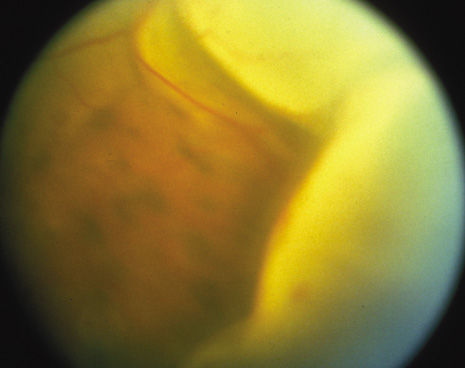
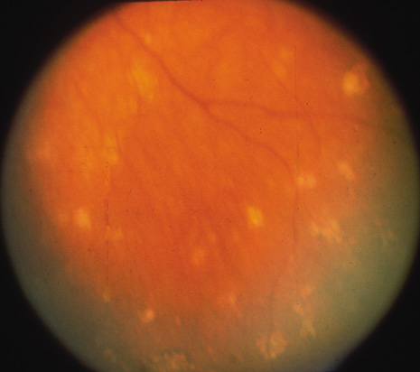
The onset of the disease is insidious. The injured eye becomes painful and photophobic, and visual acuity is diminished. The uninjured (sympathetic) eye then follows a similar course. On ocular examination, the injured eye shows persistent low-grade uveitis despite the healing wound. Characteristic bilateral panuveitis can be recognized by “mutton-fat” keratic precipitates, ciliary flush, aqueous cell and flare, posterior synechiae, vitreous cells and haze, choroidal thickening and infiltration, and optic nerve head edema. The presence of small, yellowish-white inflammatory infiltrates, 60 to 700 μm in diameter, at the level of the retinal pigment epithelium (Dalen-Fuchs nodules), but more commonly located at the midperiphery, is pathognomonic of SO (Fig. 1). In severe cases, rubeosis iridis, cataracts, pupillary membrane formation, and exudative retinal detachment can occur. Chorioretinal scarring may eventually form, and the eyes may become phthisic.1,40–42

Fig. 1. Multiple subretinal and choroidal infiltrates seen in the midperiphery of a patient with sympathetic ophthalmia.

In some cases of SO, the bilateral uveitis may be so mild or transient that the diagnosis is missed.1,40,43 Sympathetic ophthalmia can present as a mild, nongranulomatous uveitis that is easily misdiagnosed as idiopathic uveitis. This atypical clinical presentation may result from early enucleation of the injured eye before the classic histopathology of SO has appeared. Alternatively, the pathology may be modified by immunosuppressive treatment.43,44 In addition, progressive subretinal fibrosis with multifocal granulomatous chorioretinitis45 can occur in SO.46 This rare entity is characterized by rapid vision loss with progressive subretinal fibrosis involving the macula.45 This form of the disease progresses rapidly and often is unresponsive to systemic immunosuppressive therapy.

Fluorescein angiography of patients with SO often shows multiple areas of hyperfluorescence and leakage at the level of the retinal pigment epithelium (Dalen-Fuchs nodules) and the choroid (choroidal granuloma).39–41,47,48 If there is a serous retinal detachment, pooling of dye in the late frames of the angiogram can be observed.11,40,48–50 Although early hypofluorescence followed by late hyperfluorescence has been reported, this may indicate obliteration of the choriocapillaris and the presence of numerous Dalen-Fuchs nodules, as well as choroidal granulomas to obscure choroidal fluorescence in the early phase.11,41,50,51 Indocyanine green angiography (ICG) can reveal hypofluorescent patterns that persist throughout angiography or fade in the late phase that represent active or cicatricial lesions in the choroids.29,52,53

There may be a genetic component to disease susceptibility. With racially matched controls, HLA-DR4 and HLA-DRw53 were found to be highly associated with SO in Japanese patients,54 whereas the occurrence of HLA-DR4 and DQw3 was reported significantly elevated in American patients with SO.55 Using HLA serological and polymerase chain reaction amplification (PCR)–based DNA typing in 16 Japanese patients and 50 controls, Shindo and associates56 reported HLA-DRB1*04, DQA1*03, and DQB1 04 were significantly associated with SO. Kilmartin and colleagues57 also confirmed this genotype association (HLA-DRB1*04 and DQA1*03) in Caucasian patients with SO.

The differential diagnosis of SO includes Vogt-Koyanagi-Harada syndrome, phacoanaphylactic uveitis, sarcoidosis, chronic idiopathic uveitis, and other granulomatous uveitis induced by mycobacteria or fungi.1,41,42 A history of ocular injury and a lack of systemic involvement or proven ocular infection, however, are helpful indicators in diagnosing SO. Vitiligo, poliosis, alopecia, dysacusis, and meningeal irritation, findings more commonly reported in Vogt-Koyanagi-Harada syndrome, may be noted rarely in SO.1,40,58 Blodi59 and Lubin and colleagues9 reported SO in 23% of 170 and in 46% of 105 cases of lens-induced endophthalmitis, respectively; in the latter study, of 31 cases of lens-induced endophthalmitis occurring after 1950, there was only one case of SO. In reviewing 100 cases of SO from the Armed Force Institute of Pathology (AFIP) file, Croxatto and associates60 found 14 cases (22% of the 46 eyes enucleated before 1950, in contrast to 7% of the 54 eyes enucleated after 1950) associated with phacoanaphylactic endophthalmitis; the occurrence of SO was unrelated to the severity of the choroidal inflammation.

The more severe the inflammation, the poorer the prognosis of SO. The sooner the diagnosis and more intensive the therapy, the better the outlook.19,34,61,62 In their study of 105 cases, Lubin and associates9 found that 93.3% of the patients with mild inflammation had 20/70 (6/21)* vision or better; and 100% of patients with severely inflamed eyes had 20/70 vision or worse. In a study with long-term follow-up data (mean follow-up of 23 years), Makley and Azar61 demonstrated ocular complication in 70% of patients. In this study, only 65% of the 17 patients treated with corticosteroids attained a visual acuity of 20/60 (6/18) or better. In a study at Moorfield hospital, a visual acuity of between 20/20 and 20/60 was achieved in 14 of 18 patients (77%).62 Chan and colleagues34 reported on 32 SO patients from 1982 to 1992: 16 patients had a final visual acuity of 20/40 or better, and 10 had a visual acuity worse than 20/200. The remaining six patients had visual acuity ranging from 20/40 to 20/200. In this series, prompt and aggressive application of anti-inflammatory therapy was the key to achieving better visual outcome.

HISTOPATHOLOGY

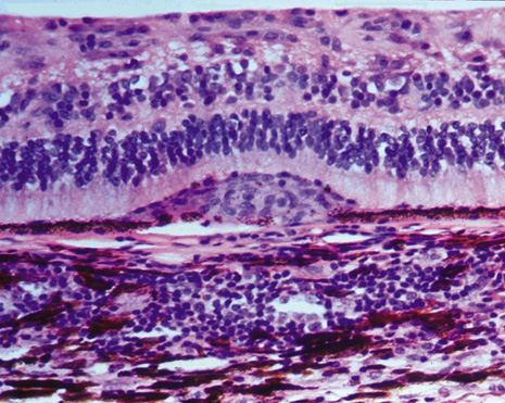
The classic histopathologic features of SO were first described by Fuchs in 1905,3 confirmed by Easom and Zimmerman,63 and summarized by Green.64 Both the injured and the sympathetic eyes show similar histopathologic features. The main feature of SO is a diffuse, non-necrotizing, granulomatous inflammation in the uvea. The choroid is markedly thickened and infiltrated by lymphocytes and nests of macrophages, epithelioid cells, and giant cells. Other classic histopathologic findings include a relative lack of retinal involvement, sparing of the choriocapillaris, and the formation of Dalen-Fuchs nodules, consisting mainly of epithelioid cells located between Bruch's membrane and the retinal pigment epithelium (Fig. 2). Fuchs3 found that approximately 25% of his classic cases of SO had Dalen-Fuchs nodules.

Fig. 2. Microphotograph of Dalen-Fuchs nodule composed of mainly epithelioid histiocytes (macrophages) and a few degenerative pigment epithelial cells between the retinal pigment epithelial cell layer and Bruch's membrane.

Lubin and associates9 reported that more than one nodule was present in 33 of 93 cases (35.5%). Kuo and co-workers65 observed Dalen-Fuchs nodules in all 50 Chinese patients with severe SO. Nine of 29 patients (31%) had Dalen-Fuchs nodules in an AFIP series.66 Breaks in Bruch's membrane at the site of Dalen-Fuchs nodules also have been identified.50,67

Many other histopathologic features of SO have been reported in the literature. Retinal involvement (e.g., retinal perivasculitis, retinitis, retinal gliosis) has been documented in 30% of cases of SO by Winter,19 in 42.2% by Lubin and associates,9 and in 55% by Croxatto and colleagues.60 Other features include choriocapillaris obliteration, reported in 40% of cases by Croxatto and colleagues60 and in 52% by Kuo and co-workers;65 chorioretinal scarring was reported in 7% of cases by Croxatto and colleagues60 and in 25% by Makley and Azar.61 Lubin and associates9 found optic atrophy in 54.4% of SO cases. Nongranulomatous inflammation and focal inflammation also can be observed in SO. In the 29 AFIP cases of SO, 11 did not have granulomatous inflammation and 13 presented only with focal choroidal inflammation.66 Eosinophils and plasma cells were more commonly seen in the non–corticosteroid-treated and more severely inflamed cases.

Marak and co-workers68 observed histologic variations related to race in SO. They found thickened choroid, an increase in the number and size of choroidal granulomas, and eosinophilia more commonly among African-American patients than among white American patients. Kuo and associates65 did not find any significant histopathologic differences between Chinese patients and white American patients. The finding of Marak and co-workers may reflect the fact that the cases in the African-American group were more severe at the time of diagnosis.

As stated, there is a rare form of SO characterized by a progressive subretinal fibrosis with multifocal granulomatous chorioretinitis leading to rapid vision loss.45 Histopathologic findings include multifocal granulomatous inflammation involving the outer retina and choroid with giant cells distributed along the Bruch's membrane.46 In this variant serum antibodies against photoreceptor and retinal pigmented epithelium can be detected.

Immunohistopathologic studies of SO show that choroidal infiltrates are predominantly composed of T lymphocytes.16,67,69–72 We have demonstrated a greater number of CD4+ T cells (T helper/inducer cells) in the early stages of the disease and CD8+ T cells (T suppressor/cytotoxic cells) in the late stages of the disease.67 In contrast, B cells are found in less than 5%16,69,70,72 to 15%16,67 of choroidal infiltrates; however, higher percentages of B cells were found in four of the 29 AFIP eyes at the end stage of the disease.66 Formation of pseudogerminal center with aggregate of B cells surrounding by T cells is found in the variant of SO with progressive subretinal fibrosis and blindness associated with multifocal granulomatous chorioretinitis.73

The main cellular component of both choroidal granuloma and Dalen-Fuchs nodules are bone marrow–derived monocytic (histocytic) cells. The granulomas of SO have immunohistochemical staining profiles that are similar to the granulomas of sarcoidosis.74 In the late stage of SO, however, degenerated retinal pigment epithelium can be an important component of Dalen-Fuchs nodules.75 Despite the accumulation of T lymphocytes in eyes with SO, there is no reported difference in the lymphocytic subpopulations in the peripheral blood of patients with SO, with the exception of one atypical case reported by Kaplan and co-workers.72 Expressions of cell adhesion molecules and major histocompatibility (MHC) class II antigens on the ocular resident cells have been observed in eyes with SO and may be important in the pathogenesis of the disease.16,76,77

PATHOGENESIS

The etiology of SO is still an enigma. Two hypotheses dominate the older literature: (a) autoimmunity against uveal melanin, uveal melanocytes, retinal pigment epithelium, or retinal antigens; or (b) a viral or bacterial infection.1 Later studies have emphasized immune system involvement in patients with ocular inflammation.41 In 1971, investigators reported that lymphocytes from patients with SO might respond to heterogeneous or homogeneous retinal and uveal tissues.78–80 Using lymphocyte transformation and a leukocyte migration inhibition assay, Rahi and associates43 reported that cells from five of six patients with SO showed positive reactions to uveoretinal tissues, whereas seven of ten cases of posttraumatic nongranulomatous uveitis showed negative responses to uveoretinal antigen. These observations suggested that cell-mediated hypersensitivity could be an important pathogenic mechanism. The immunohistopathologic finding of the predominantly T-lymphocytic infiltration in ocular tissue supports a cell-mediated immunologic response (delayed hypersensitivity) to ocular antigens in both typical and atypical cases of SO.16,70–72,81,82

Although the immune nature of SO appears to be established, the inciting antigen is still in question.40,41,43,44,69 Retinal extracts are highly antigenic and easily produce retinouveitis in experimental animals. Four main retinal antigens are rhodopsin,83 retinal soluble antigen,84,85 interphotoreceptor retinoid binding protein,86 and recoverin.87 Using a single injection of purified rhodopsin with adjuvant in monkeys, Wong and co-workers79 observed choroidal granulomatous inflammation, loss of outer segment photoreceptor, perivascular retinitis, and optic disc edema. Extensive investigations on experimental autoimmune uveoretinitis models have been reported since the isolation of retinal soluble antigen in 1977 by Wacker and colleagues,84 who found it to be located on the membrane of the outer segment of the photoreceptor. Like retinal soluble antigen, interphotoreceptor retinoid binding protein (located in the retinal interphotoreceptor space) and recoverin (a retinal calcium-binding protein) are highly uveitogenic and can induce T-cell–mediated autoimmune uveoretinitis.86,87 Similar clinical and histopathologic pictures of SO can be produced in experimental models by alternating the immunizing dose, route of administration, adjuvants, and animal species.88,89 These features include Dalen-Fuchs nodules, nongranulomatous and granulomatous uveitis, retinal vasculitis, retinal detachment, corneal keratic precipitates, iridocyclitis, and vitreitis.

In 1992, Broekhuyse and co-workers90 described an insoluble uveal melanin preparation that can produce an inflammation limited to the uvea in immunized animals. Chan and associates91 reported that spontaneous recurrence occurred in this experimental model; however, Bora and colleagues92,93 emphasized the anterior uveal involvement with immunization of melanin protein. Such a role of a uveal antigen is more consistent with clinical data because SO usually follows uveal tissue trauma. Recently Bora's group reported that type I collagen is the autoantigen in their experimental autoimmune anterior uveitis model.94

The role of an ocular penetrating wound in the development of SO has been proposed.95,96 Barker and Billingham proposed that absence of lymphatics within the eye lets intraocular antigens circulate to the blood and spleen and may induce blocking antibodies or suppressor cells; when the antigens go to the regional lymph nodes, a cell-mediated immune response can be induced. Thus, in SO cases, penetrating trauma may result in exposure of uveoretinal antigens to the conjunctival lymphatics, as confirmed by subconjunctival injection of retinal soluble antigen in rabbits.88 Exposure to bacteria (e.g., Propionibacterium acne), viruses, and other infectious agents through the wound may serve as an adjuvant to induce the disease.41,97

TREATMENT

Enucleation

The classic and only known prevention against SO is enucleation of the injured eye before the other eye develops disease.1,40 However, because this is an extremely rare disease and techniques for the surgical repair of injured eyes have greatly improved, one should always make every attempt to save the injured eye if it has useful vision. Once SO develops, enucleation of the injured eye usually cannot improve the symptoms and final visual acuity of the sympathizing eye.19,40 Lubin and associates9 did suggest, however, that enucleation within 2 weeks after symptoms of SO occurred may improve the visual prognosis. Kuo and co-workers65 reported significantly fewer recurrences of inflammatory disease in patients who underwent early enucleation but a lack of improvement in ultimate visual acuity. In a retrospective clinicopathologic study of 30 cases of SO, Reynard and colleagues98 found that early enucleation of the existing eye was associated with a benign clinical course: visual acuity better than 20/50 (6/15) and fewer and milder relapses than eyes that underwent late enucleation (p = 0.008). Even when corticosteroid-treated patients were excluded in the study, the association between early enucleation and a benign clinical course remained significant (p = 0.018). There still is controversial whether enucleation is helpful to prevent development of SO in the uninjured eye.32,99

Sympathetic ophthalmia occasionally develops after enucleation of the injured eye. Of the 18 cases of SO from the Moorfield Eye Hospital, there was one case of SO that occurred in a patient whose injured eye was enucleated before the onset of disease;62 similarly, two of the 29 AFIP cases occurred after enucleation of the traumatized eye.66

Pharmaceutical Agents

CORTICOSTEROIDS.

Intensive systemic and topical corticosteroid treatment can improve visual outcome and decrease relapse of SO.1,9,34,40,61,65,98 Adequate high-dose corticosteroids should be given for the first few days until the disease is controlled. Makley and Azar61 found that 9 of 14 treated patients attained 20/60 vision or better, Lubin and associates9 noted that 13 of 18 treated patients achieved 20/50 vision or better, and Reynard and co-workers98 reported that 18 of 22 treated patients had 20/50 vision or better. It is known, however, that prophylactic corticosteroids do not prevent the development of SO. This is based on the repeated observation in the 1950s that SO developed in patients who were on high-dose steroids.1 Recently intravitreal steroids have been successfully used in the management of SO.10

Cytotoxic Agents

Despite the advantages of corticosteroids, their side effects should be taken into consideration. Other immunosuppressive therapies such as azathioprine, chlorambucil, or cyclophosphamide may be used when patients are unresponsive to corticosteroids or are having severe corticosteroid-related side effects.35,101 Corticosteroids also may be used in combination with other immunosuppressive agents. When these cytotoxic agents are used, it is essential to monitor the status of the patient's bone marrow; it is also advisable to coordinate the patient's care with an internist.

CYCLOSPORINE.

Because eyes with SO usually are infiltrated with numerous activated T cells, cyclosporine, a potent inhibitor of T-cell function, can be a very effective therapeutic agent in the treatment of SO. Nussenblatt and colleagues34,97,102 have used cyclosporine as a corticosteroid-sparing agent in patients with SO, and the results have been encouraging. However, because cyclosporine can cause hepatic and renal toxicities, patients need to be monitored closely. The recommended dosages for a combination of cyclosporine and steroids are cyclosporin A, 3 to 5 mg/kg per day; and prednisone, 15 to 20 mg/day. An optimal therapy for SO depends on improved understanding of the disease mechanism

Biologics

Recently several biologics agents are available as therapeutic target for various autoimmune diseases such as Crohn's disease, Behçet's syndrome, arthritis, psoriasis, and uveitis.103–105 A number of these agents are available and effective for the treatment of uveitis.106,107 Nussenblatt and his associates108,109 have reported the success of humanized anti–interleukin-2 receptor alpha antibody in the treatment of noninfectious uveitis. Several investigators have reported the success of anti–tumor necrosis factor-alpha in the treatment of uveitis associated with Behçet's syndrome and other autoimmune uveitis.110,111 Although these biologics are relatively expensive, they could be considered as an alternative therapeutic agents for sympathetic ophthalmia.

Back to Top
VOGT-KOYANAGI-HARADA SYNDROME
In 1906, Vogt described a case of anterior uveitis associated with poliosis and dysacusis.112 In 1929, Koyanagi discussed the details of six cases in Japan, all with symptoms similar to the case described by Vogt, but also including alopecia and vitiligo.113 In 1926, Harada reported on a second condition that included posterior uveitis and exudative retinal detachment associated with a cerebrospinal fluid pleocytosis and meningeal signs.114 Because of the similarities and overlapping features of both clinical conditions, many investigators considered these diseases to be related, hence the name Vogt-Koyanagi-Harada syndrome (uveoencephalitis).115–118

PREVALENCE

The worldwide prevalence of Vogt-Koyanagi-Harada syndrome is not easy to assess because this syndrome occurs more frequently among Asians.119 In Japan, approximately 8% of uveitic patients have Vogt-Koyanagi-Harada syndrome,117 with an incidence of 800 new cases per year.120 Ohno and co-workers121 reported that 1% of 5,500 uveitic patients were diagnosed with Vogt-Koyanagi-Harada syndrome, of whom 41% were Asian, 29% were white, 16% were Hispanic, and 14% were African American. Snyder and Tessler122 found that 3.7% of 455 patients seen in their uveitis clinic had Vogt-Koyanagi-Harada syndrome, 75% of whom were either of Native American or Hispanic descent; none were Asian. In the National Eye Institute series of 75 patients, 52% of the patients with this syndrome were African American, 23% white, 9% Hispanic, and 9% Asian.123 Of the 65 patients with Vogt-Koyanagi-Harada syndrome seen in Doheny Eye Institute, 51 (78%) were Hispanic, seven (10%) Asian, four (6%) African American, two (3%) white, and one Native American.124

Overall, no significant sex differences have been found in this syndrome.117,118,122 In North America, however, women seem to be affected more frequently than men.121,125,126 This syndrome typically affects adults between the ages of 20 and 50 years, although a case in a 6-year-old child has been described.127

CLINICAL MANIFESTATIONS

The progression of Vogt-Koyanagi-Harada syndrome used to be divided into three stages117,118,121 but is now divided into four:

  Stage 1: The prodromal stage. This occurs a few days before the ocular symptoms and is characterized by headaches, deep orbital pain, vitiligo, nausea, slight fever, and occasional photophobia and lacrimation. Cerebrospinal fluid may show pleocytosis.
  Stage 2: The ophthalmic or uveitis stage. This is characterized by bilateral uveitis that may affect one eye first with sudden blurring vision, photophobia, impairment of ocular movement, dysacusis, and meningism. This stage can last weeks to months. On ocular examination, panuveitis can be recognized by mutton fat keratic precipitates, aqueous cells and flare, iris nodules, synechiae, vitreous haze and cells, optic disc swelling, retinal edema, hemorrhages, exudate and nonrhegmatogenous detachment (Fig. 3), choroidal infiltrates, and infiltrates at the retinal pigment epithelial level. Approximately 15% of patients may note skin hyperesthesia.
  Stage 3: The convalescent (recovery) or chronic stage. This stage may last from weeks to months, or it may continue chronically. It is characterized by subsiding uveitis with possible visual and neurologic improvement but continuous dysacusis (especially to high-frequency sound), poliosis, vitiligo (Fig. 4), and alopecia. Ocular examination reveals depigmentation of the perilimbus (Sugiura sign)117 and a pale fundus (sunset-glow fundus) (Fig. 5). In general, the retina has reattached with a mottled appearance and with white spots interspersed with a pattern of mottled pigmentation. The periphery and equator show focal depigmentation. The sunset glow fundus is often associated with previous ocular inflammation.128 Sometimes strands of chorioretinal scarring with pigmentation or depigmentation can be observed. Occasionally, subretinal neovascular membrane and subretinal fibrosis develop.
  Stage 4: The recurrent stage. This stage has been added recently to the classification of Vogt-Koyanagi-Harada syndrome.124,125,129 It usually involves complications such as mutton fat keratic precipitates, iris Koeppe's nodules, synechiae, cataract, glaucoma, subretinal neovascularization, and subretinal fibrosis.124,129

Fig. 3. Large, bullous, exudative retinal detachments seen in a patient with Vogt-Koyanagi-Harada syndrome.

Fig. 4. Depigmentation of the skin (vitiligo) and whitening of the eyelashes (poliosis) seen in a patient with Vogt-Koyanagi-Harada syndrome. (Courtesy of Dr. David Cogan.)

Fig. 5. Depigmentation of the peripheral retina (sunset-glow fundus) seen in a patient with Vogt-Koyanagi-Harada syndrome.

Since 1999, participants of the International Workshop and Symposium on Vogt-Koyanagi-Harada syndrome have revised the criteria for the disease.119 The diagnosis requires absence of penetrating ocular trauma or surgery preceding the initial onset of uveitis, no other ocular disease, bilateral uveitis, neurologic and auditory findings, and integumentary finding of alopecia, poliosis, and/or vitiligo. Often the clinical course of Vogt-Koyanagi-Harada syndrome is quite varied. Some patients may have a limited period of severe ocular inflammation, followed by rapid depigmentation and no further recurrences. Others, however, may continue to have ongoing, chronic disease.130,131

Fluorescein angiography in patients with Vogt-Koyanagi-Harada syndrome shows multifocal hyperfluorescence at the level of the retinal pigment epithelium and the optic nerve head in the early phase (Fig. 6). In the late phase, either pooling of dye behind the detached retina or a combination of chorioretinal scarring and abnormal retinal pigment epithelium is observed.122,125,132–135 Fluorescein angiography may confirm retinal vasculitis with vascular sheathing, subretinal neovascularization with neovascular membrane network, and retinochoroidal anastomosis.39 Indocyanine green angiogram illustrates choriocapillary perfusion delay and perivascular leakage in the early stage, hypofluorescent dark dots in the intermediate phase, and choroidal diffuse leakage and hyperfluorescence in the late stage.136 Optic disc edema and retinal detachment also can be identified by indocyanine green angiogram. Maruyama and associates137 reported tomographic features of serous retinal detachment in Vogt-Koyanagi syndrome in 42 eyes of 21 patients: 69% had true retinal detachment and 40% had intraretinal edema. Standardized echography and magnetic resonance imaging are useful adjuncts in the diagnosis of Vogt-Koyanagi-Harada syndrome to confirm the presence of a thickened choroid and retinal detachment.138,139

Fig. 6. Multiple areas of hyperfluorescence seen in a fluorescein angiogram of a patient with Vogt-Koyanagi-Harada syndrome.

Similar to SO, there appears to be a genetic susceptibility to Vogt-Koyanagi-Harada syndrome. Ohno140 studied HLA haplotype typing in 185 Japanese patients with the syndrome. All patients were typed positively for HLA-DRw53, and the relative risk for this antigen was determined to be 74.5. The number of subjects who typed positively for HLA-DR4 and HLA-DQwa was significantly higher among Japanese patients with Vogt-Koyanagi-Harada syndrome than among normal Japanese controls.140 A close association between HLA-DR4 and HLA-DRw53 positivity and Vogt-Koyanagi-Harada syndrome also was reported in American and Chinese patients.55,141,142 Hispanic patients living in southern California were found to have a significantly higher positivity for HLA-DR4 and HLA-DR1.143 Using two combined testing methods (PCR with single-strand conformation polymorphism, and PCR with restriction fragment-length polymorphism), Islam and associates144 examined 57 patients with Vogt-Koyanagi-Harada syndrome in Japan and concluded that the combination of the HLA-DQA1*0301 gene as the primary and HLA-DR4 as an additive factor would lead to the development of Vogt-Koyanagi-Harada syndrome.

The differential diagnosis of Vogt-Koyanagi-Harada syndrome includes other causes of posterior uveitis and panuveitis, such as SO, acute posterior multifocal placoid pigment epitheliopathy, uveal effusion syndrome, geographic choroidopathy, posterior scleritis, sarcoidosis, and primary intraocular B-cell lymphoma.118,125,131 However, the systemic presentations of meningeal signs, auditory disturbances, and integumentary changes usually are associated with Vogt-Koyanagi-Harada syndrome.

The prognosis of untreated Vogt-Koyanagi-Harada syndrome is poor, especially for chronic recurrent cases.117,118,129,131 Sugiura117 found anterior and posterior synechiae, secondary cataract, and glaucoma in 35% of patients; however, with proper corticosteroid treatment, 70% of the eyes had a final visual acuity of better than 20/50, 10% had a final visual acuity of between 20/50 and 20/200, and only 20% had worse than 20/200 vision. Forster and associates145 reported that glaucoma developed in 16 of 42 (38%) of their patients with Vogt-Koyanagi-Harada syndrome, of whom 11 required surgical intervention. Moorthy and co-workers146 reported that 26 of 65 (40%) patients required surgery for cataracts that had developed. The surgical outcome in these patients was satisfactory. Reed and colleagues126 reported the association between prognosis and complications that 51% of their 101 patients developed complications including cataracts (42%), glaucoma (27%), choroidal neovascularization (11%), and subretinal fibrosis (6%). In a study of 75 patients with Vogt-Koyanagi-Harada syndrome, Lertsumitkul and associates123 also demonstrated that poor prognosis was associated with complications, particularly subretinal fibrosis. Sheu and co-workers found the association between early severe retinal detachment and poor prognosis in 31 patients.147

HISTOPATHOLOGY

There are only a few histopathologic reports on eyes with Vogt-Koyanagi-Harada syndrome. In general, these cases resemble the histopathologic features of SO, with granulomatous inflammation in the uveal tract and formation of Dalen-Fuchs nodules.58,64,125,148–150 Unlike SO, Vogt-Koyanagi-Harada syndrome frequently involves obliteration of choriocapillaris, focal active chorioretinitis, old chorioretinal scarring, choroidal neovascularization, migration of pigment into the retina, and marked retinal pigment epithelial involvement.64,125,148,149,151,152 Close contact between lymphocytes and choroidal melanocytes has been demonstrated by electron microscopic study.129,153

The data on immunopathology of eyes with Vogt-Koyanagi-Harada syndrome are extremely limited.154–156 An increase of T cells, especially T helper/inducer cells, was observed in cerebrospinal fluid compared with that observed in blood.157–160 A decrease in the percentage of T cells in peripheral blood was found compared with a control population. Aziga and associates161 studied limbus biopsies from four patients with Vogt-Koyanagi-Harada syndrome and noted T-cell infiltration, with predominant T helper/inducer cells in the early stage (within 20 days) and T suppressor/cytotoxic cells in the late stage (60 days) of the disease. Norose and co-workers162 observed dominance of activated T cells as well as elevated interleukin-6 levels in the aqueous humor of 11 patients with the disease. Gocho and colleagues160 found autoreactive T cells against tyrosinase or tyrosinase-related protein in the peripheral blood of patients with Vogt-Koyanagi-Harada syndrome.

We have reported an enucleated eye with a long-standing history of Vogt-Koyanagi-Harada syndrome and found mild to moderate T-lymphocytic infiltration, with loci of B-cell aggregation and scattering macrophages in the uvea and retina.154 Major histocompatibility class II expression was present on the ocular resident cells.77,154 Sakamoto and associates156 performed an autopsy study of the eyes of two patients who had had Vogt-Koyanagi-Harada syndrome for 2.5 and 7 years, respectively. Diffuse choroidal infiltration of mainly activated T helper/inducer cells was observed. B cells were scattered, having a tendency to cluster in small aggregates. Kahn and associates155 reported on an enucleated eye with active Vogt-Koyanagi-Harada syndrome, demonstrating a predominance of T cells and MHC class II–positive macrophages in the dense uveal infiltration. Nondentritic, CD1-positive cells were located adjacent to choroidal melanocytes. No microorganisms were identified. More retinal cells were found apoptosis in Vogt-Koyanagi-Harada syndrome as compared to SO.163

PATHOGENESIS

The etiology of Vogt-Koyanagi-Harada syndrome is unknown; a viral origin has long been speculated in the literature but definitive data are lacking.117,118 The manner of onset of stage 1 of the disease and the fact that disease onset is more prevalent in the spring and fall suggest that an infectious agent is responsible.164 Isolation of an untyped virus was reported in a cerebrospinal fluid sample from a patient with Vogt-Koyanagi-Harada syndrome,165 but numerous other studies have reported negative virologic findings.125,131

Antigen-specific immune response is believed to be the leading mechanism of Vogt-Koyanagi-Harada syndrome. Hammer80 used the leukocyte migration test to demonstrate cellular hypersensitivity to uveal pigment in patients with Vogt-Koyanagi-Harada syndrome. Immunopathologic studies have found predominantly T-lymphocytic infiltration in the eye,154–156,166 cerebrospinal fluid,157,159 and skin. Ohno140 reported an increase of interferon-γ in the sera and cerebrospinal fluid of patients with Vogt-Koyanagi-Harada syndrome and suggested that the interferon-γ was originating from the T cells. In Vogt-Koyanagi-Harada syndrome, humoral immunity may play certain roles that are not as evident in SO. This may be confirmed by the accumulation of plasmacytic and B-cell infiltration in the eye and the formation of exudative retinal detachment. Antibodies against retinal elements were detected in serum of some patients with Vogt-Koyanagi-Harada syndrome.167

Pathologic findings of the close contact between lymphocytes and melanocytes;153 the lymphocyte-mediated cytotoxicity on the melanoma cells;162,168 and the involvement of skin, ear, and meninges suggest the possibility that Vogt-Koyanagi-Harada syndrome is an autoimmune reaction against the melanocytes caused by an unknown triggering agent. Norose and co-workers162 showed the cytotoxic effect of peripheral blood lymphocytes and cerebrospinal fluid lymphocytes obtained from patients with Vogt-Koyanagi-Harada syndrome on the B36 melanoma cell line. McClellan and associates168 detected antibodies to melanoma cell lines in a cytotoxic assay of a patient with Vogt-Koyanagi-Harada syndrome and identified interleukin-2–dependent T cells with specificity for normal melanocytes and for melanoma cells in cytotoxic and proliferation assays. Suzuki and colleagues169 found serum melanin protein antibodies in patients with Vogt-Koyanagi-Harada syndrome and discovered that the titers of these antibodies were associated with disease activity. Yamaki and associates170 reported that exposure of lymphocytes from patients with Vogt-Koyanagi-Harada syndrome to a 30-mer peptide derived from the tyrosinase family proteins led to significant proliferation of the lymphocytes. Immunization of these peptides into pigmented rats induced ocular and extraocular changes that highly resembled human Vogt-Koyanagi-Harada syndrome.170 These authors concluded that Vogt-Koyanagi-Harada syndrome is an autoimmune disease against the tyrosinase family protein.171 This hypothesis is strengthened by the establishment of 28 T-cell clones directed against tyrosinase and another 34 directed against tyrosinase-related protein 1.160

TREATMENT

Currently, the most valuable therapeutic medication for Vogt-Koyanagi-Harada syndrome is systemic corticosteroids.130,131 As in SO, an early and aggressive high-dose steroid regimen at the beginning of the disease, followed by gradual tapering of the dosage, is recommended for successful treatment.172 The early administration of systemic steroids may prevent the progression of the disease, shorten the duration, and decrease complications and other systemic involvement. Ohno140 reported that in 78 eyes treated with systemic steroids, 85% achieved better than 20/40 vision. Rubsamen and Gass172 reported that in 44 eyes treated with steroids, 29 (66%) had a final vision of better than 20/30. They also documented recurrence in nine of 21 (43%) patients within the first 3 months, usually in association with a rapid tapering of steroid dosage. Application of pulse steroids and intravitreal triamcinolone was able to rapidly improve serous retinal detachment associated with acute Vogt-Koyanagi-Harada syndrome.173,174 Other immunosuppressive medications, such as chlorambucil, cyclosporine, and FK506, have had positive effects in patients in whom steroid therapy failed.175–177 Before one chooses any of these medications, however, their side effects must be considered and carefully monitored. Recent usage of various novel biologics such as anti–tumor necrosis alpha or anti–interleukin-2 receptor antibodies may be considered alternative agents for autoimmune uveitis, including chronic recurrent Vogt-Koyanagi-Harada syndrome.106,107

In summary, both SO and Vogt-Koyanagi-Harada syndrome are bilateral granulomatous panuveitis. The former is associated with ocular injury and the latter with systemic signs of poliosis, vitiligo, alopecia, and auditory and neurologic manifestations. The diseases probably involve autoimmune mechanisms influenced by genetic factors. When they are treated promptly and aggressively with corticosteroids or other immunosuppressive medications, their complications can be minimized and their prognoses can be optimal.

Back to Top
REFERENCES

1. Duke-Elder S, Perkins ES: Sympathetic ophthalmitis. In Diseases of the Uveal Tract. St. Louis: Mosby, 1966:558

2. MacKenzie W: A Practical Treatise on the Diseases of the Eye. London: Longmans, 1840

3. Fuchs E: Über sympathisierende Entzündung zuerst Bemerkungeen über seröse traumatische Iritis. Albrecht Von Graefes Arch Ophthalmol 61:365, 1905

4. Ravin JG: Sir Frederick Treves and sympathetic ophthalmia. Arch Ophthalmol 122:99, 2004

5. Fuchs E: Uber Ophthalmia sympathica. Graefes Arch Clin Exp Ophthalmol 70:465, 1909

6. Albert DM, Diaz-Rohena R: A historical review of sympathetic ophthalmia and its epidemiology. Surv Ophthalmol 34:1, 1989

7. Von Holland G: Über indikation und zeitpunkt der entfernung eines verletzten auges. Klin Monatsbl Augenheilkd 145:732, 1964

8. Allen JC, Ho JK: Sympathetic ophthalmia. A disappearing disease. JAMA 209:1090, 1969

9. Lubin JR, Albert DM, Weinstein M: Sixty-five years of sympathetic ophthalmia. A clinicopathologic review of cases (1913–1978). Ophthalmology 87:109, 1980

10. Michels RG, Ryan SJ: Results and complications of 100 consecutive cases of pars plana vitrectomy. Am J Ophthalmol 80:24, 1975

11. Lewis ML, Gass DM, Spencer WH: Sympathetic uveitis after trauma and vitrectomy. Arch Ophthalmol 96:263, 1978

12. Smith RS, Webb R, van Heuven WAJ: Sympathetic ophthalmia as a complication of pars plana vitrectomy. Perspectives in Ophthalmol 2:117, 1978

13. Croxatto JO, Galentine P, Cupples HP, et al: Sympathetic ophthalmia after pars plana vitrectomy-lensectomy for endogenous bacterial endophthalmitis. Am J Ophthalmol 91:342, 1981

14. Gass JD: Sympathetic ophthalmia following vitrectomy. Am J Ophthalmol 93:552, 1982

15. Tamai M, Obara J, Mizuno K, Koisumi A: Sympathetic ophthalmia induced by vitrectomy not by trauma. Jpn J Ophthalmol 28:75, 1984

16. Chan C-C, Nussenblatt RB, Fujikawa LS, et al: Sympathetic Ophthalmia. Immunopathological findings. Ophthalmology 93:690, 1986

17. Burns DMJ, Ainley RH: Sympathetic ophthalmia after glaucoma surgery. Trans Ophthalmol Soc UK 86:757, 1966

18. Daxecker F, Kieselbach G: Sympathische ophthalmie nach eingriffen wegen eines hämorrhagischen glaukoms. Klin Monatsbl Augenheilkd 188:601, 1986

19. Winter FC: Sympathetic uveitis. A clinical and pathologic study of the visual field. Am J Ophthalmol 39:340, 1955

20. Green WR, Maumenee AE, Sanders TE, Smith ME: Sympathetic ophthalmia following evisceration. Trans Am Acad Ophthalmol Otolaryngol 76:625, 1972

21. Jain IS, Gangwar DN, Kaul RL, Jain GC: Sympathetic ophthalmitis simulating Vogt-Koyanagi-Harada disease after retinal detachment surgery. Annals Ophthalmol 11:1121, 1979

22. Laroche L, Pavlakis C, Saraux H, Orcel L: Ocular findings following intravitreal silicone injection. Arch Ophthalmol 101:1422, 1983

23. Maisel JM, Vorwerk PA: Sympathetic uveitis after giant tear repair. Retina 9:122, 1989

24. Pusin SM, Green WR, Tasman W, Griffith WR, Rodrigues M: Simultaneous bacterial endophthalmitis and sympathetic uveitis after retinal detachment surgery. Am J Ophthalmol 81:57, 1976

25. Wang WJ: Clinical and histopathological report of sympathetic ophthalmia after retinal detachment surgery. Br J Ophthalmol 67:150, 1983

26. Lam S, Tessler HH, Lam BL, Wilensky JT: High incidence of sympathetic ophthalmia after contact and noncontact Neodymium:YAG cyclotherapy. Ophthalmol 99:1818, 1992

27. Brancato R, Carassa RG, Bettin P, Fiori M, Trabucchi G: Contact transscleral cyclophotocoagulation with diode laser in refractory glaucoma. Eur J Ophthalmol 5:32, 1995

28. Bechrakis NE, Müller-Stolzenburg NW, Helbig H, Foerster MH: Sympathetic ophthalmia following laser cyclocoagulation. Arch Ophthalmol 112:80, 1994

29. Kumar N, Chang A, Beaumont P: Sympathetic ophthalmia following ciliary body laser cyclophotocoagulation for rubeotic glaucoma. Clin Experiment Ophthalmol 32:196, 2004

30. Fries PD, Char DH, Crawford JB, Waterhouse W: Sympathetic ophthalmia complicating helium ion irradiation of a choroidal melanoma. Arch Ophthalmol 105:1561, 1987

31. Margo CE, Pautler SE: Granulomatous uveitis after treatment of a choroidal melanoma with proton-beam irradiation. Retina 10:140, 1990

32. Kilmartin DJ, Dick AD, Forrester JV: Prospective surveillance of sympathetic ophthalmia in the UK and Republic of Ireland. Br J Ophthalmol 84:259, 2000

33. Rao NA, Cook GR: Sympathetic uveitis. Contemp Ophthalmol 1:1, 1980

34. Chan CC, Roberge RG, Whitcup SM, Nussenblatt RB: Thirty-two cases of sympathetic ophthalmia. A retrospective study at the National Eye Institute, Bethesda, Md., from 1982 to 1992. Arch Ophthalmol 113:597, 1995

35. Power WJ, Foster CS: Update on sympathetic ophthalmia. Int Ophthalmol Clin 35:127, 1995

36. Thies O: Gedanken über den Ausbruch der sympathetischen ophthalmie. Klin Monatsbl Augenheilkd 112:185, 1947

37. McClellan KA, Billson FA, Marijan F: Delayed onset sympathetic ophthalmia. Med J Australia 147:451, 1987

38. Zaharia MA, Lamarche J, Laurin M: Sympathetic uveitis 66 years after injury. Can J Ophthalmol 19:240, 1984

39. Goto H, Rao NA: Sympathetic ophthalmia and Vogt-Koyanagi-Harada syndrome. Int Ophthalmol Clin 30:280, 1990

40. Marak GE, Jr: Recent advances in sympathetic ophthalmia. Surv Ophthalmol 24:141, 1979

41. Chan CC, Roberge FG: Chapter 57 Sympathetic ophthalmia. In Ocular Infection and Immunity. St. Louis: Mosby, 1996:723

42. Chan CC, Mochizuki M: Sympathetic ophthalmia: An autoimmune ocular inflammatory disease. Springer Semin Immunopathol 21:125, 1999

43. Rahi A, Morgan G, Levy I, Dinning W: Immunological investigations in post-traumatic granulomatous and non-granulomatous uveitis. Br J Ophthalmol 62:722, 1978

44. Lubin JR, Albert DM: Sympathetic ophthalmia. Ample room for controversy. Surv Ophthalmol 24:137, 1979

45. Gass JD, Margo CE, Levy MH: Progressive subretinal fibrosis and blindness in patients with multifocal granulomatous chorioretinitis. Am J Ophthalmol 122:76, 1996

46. Wang RC, Zamir E, Dugel PU, et al: Progressive subretinal fibrosis and blindness associated with multifocal granulomatous chorioretinitis: A variant of sympathetic ophthalmia. Ophthalmology 109:1527, 2002

47. Kayazama F: A case of sympathetic uveitis. Ann Ophthalmol 12:1106, 1980

48. Spitznas M: Fluoreszenangiographie der sympathischen ophthalmie. Klin Monatsbl Augenheilkd 169:195, 1976

49. Castier P, Six A, Prin L: Ophtalmie sympathique à forme postérieure. Bull Soc Ophtalmol Fr LXXXV:47, 1985

50. Sharp D, Bell RA, Patterson E, Pinkerton RMH: Sympathetic ophthalmia. Histopathologic and fluorescein angiographic correlation. Arch Ophthalmol 102:232, 1984

51. Allinson RW, Le TD, Kramer TR, Snyder RW: Fluorescein angiographic appearance of Dalen-Fuchs nodules in sympathetic ophthalmia. Ann Ophthalmol 25:152, 1993

52. Bernasconi O, Auer C, Zografos L, Herbort CP: Indocyanine green angiographic findings in sympathetic ophthalmia. Graefes Arch Clin Exp Ophthalmol 236:635, 1998

53. Saatci AO, Pasa E, Soylev MF, et al: Sympathetic ophthalmia and indocyanine green angiography. Arch Ophthalmol 122:1568, 2004

54. Ohno S: Immunogenetic studies on ocular diseases. In The XXVth International Congress of Ophthalmology. Rome: Kugler & Ghedini, 1986:144

55. Davis JL, Mittal KK, Freidlin V, et al: HLA association and ancestry in Vogt-Koyanagi-Harada disease and sympathetic ophthalmia. Ophthalmology 97:1137, 1990

56. Shindo Y, Ohno S, Yamamoto T, Nakamura S, Inoko H: Complete association of the HLA-DRB1*04 and -DQB1*04 alleles with Vogt-Koyanagi-Harada's disease. Hum Immunol 39:169, 1994

57. Kilmartin DJ, Wilson D, Liversidge J, et al: Immunogenetics and clinical phenotype of sympathetic ophthalmia in British and Irish patients. Br J Ophthalmol 85:281, 2001

58. Rao NA, Marak GE: Sympathetic ophthalmia simulating Vogt-Koyanagi-Harada's disease: A clinico-pathologic study of four cases. Jpn J Ophthalmol 27:506, 1983

59. Blodi FC: Sympathetic ophthalmia as an allergic phenomenon. With a study of its association with phacoanaphylactic uveitis and a report on the pathological findings in sympathizing eyes. Trans Am Acad Ophthalmol Otolaryngol 63:642, 1959

60. Croxatto JO, Rao NA, McLean IW, Marak GE: Atypical histopathologic features in sympathetic ophthalmia. A study of a hundred cases. Int Ophthalmol 4:129, 1981

61. Makley TA, Jr, Azar A: Sympathetic ophthalmia. Arch Ophthalmol 96:257, 1978

62. Hakin KN, Pearson RV, Lightman SL: Sympathetic ophthalmia: Visual results with modern immunosuppressive therapy. Eye 6:453, 1992

63. Easom HA, Zimmerman LE: Sympathetic ophthalmia and bilateral phacoanaphylaxis. Arch Ophthalmol 72:9, 1964

64. Green WR: The uveal tract. In Ophthalmic Pathology. Philadelphia: W.B. Saunders 1996:1913

65. Kuo PK, Lubin JR, Ni C, Wang KM, Albert DM: Sympathetic ophthalmia: A comparison of the histopathological features from a Chinese and American series. Int Ophthalmol Clin 22:125, 1982

66. Shah DN, Piacentini MA, Burnier MN Jr, et al: Inflammatory cellular kinetics in sympathetic ophthalmia. A study of traumatized (exciting) eyes. Ocul Immunol Inflamm 1:255, 1993

67. Chan C-C, BenEzra D, Rodrigues MM, et al: Immunohistochemistry and electron microscopy of choroidal infiltrates and Dalen-Fuchs nodules in sympathetic ophthalmia. Ophthalmology 92:580, 1985

68. Marak GE, Jr, Font RL, Zimmerman LE: Histologic variations related to race in sympathetic ophthalmia. Am J Ophthalmol 78:935, 1974

69. Jakobiec FA, Marboe CC, Knowles D, II, et al: Human sympathetic ophthalmia. An analysis of the inflammatory infiltrate by hybridoma-monoclonal antibodies, immunochemistry, and correlative electron microscopy. Ophthalmology 90:76, 1983

70. Müller-Hermelink HK, Kraus-Mackiw E, Daus W: Early stage of human sympathetic ophthalmia. Histologic and immunopathologic findings. Arch Ophthalmol 102:1353, 1984

71. Rao NA, Xu S, Font RL: Sympathetic ophthalmia. An immunohistochemical study of epithelioid and giant cells. Ophthalmology 92:1660, 1985

72. Kaplan HJ, Waldrep JC, Chan WC, Nicholson JKA, Wright JD: Human sympathetic ophthalmia. Immunologic analysis of the vitreous and uvea. Arch Ophthalmol 104:240, 1986

73. Lim WK, Chee SP, Sng I, Nussenblatt RB, Chan CC: Immunopathology of progressive subretinal fibrosis: a variant of sympathetic ophthalmia. Am J Ophthalmol 138:475, 2004

74. Chan C-C, BenEzra D, Hsu S-M, Palestine AG, Nussenblatt RB: Granulomas in sympathetic ophthalmia and sarcoidosis. Immunohistochemical study. Arch Ophthalmol 103:198, 1985

75. Font RL, Fine BS, Messmer E, Rowsey JF: Light and electron microscopic study of Dalén-Fuchs nodules in sympathetic ophthalmia. Ophthalmology 90:66, 1983

76. Kuppner MC, Liversidge J, McKillop-Smith S, Lumsden L, Forrester JV: Adhesion molecule expression in acute and fibrotic sympathetic ophthalmia. Curr Eye Res 12:923, 1993

77. Whitcup SM, Chan CC, Li Q, Nussenblatt RB: Expression of cell adhesion molecules in posterior uveitis. Arch Ophthalmol 110:662, 1992

78. Marak GE, Jr, Font RL, Johnson MC, Alepa FP: Lymphocyte-stimulating activity of ocular tissues in sympathetic ophthalmia. Invest Ophthalmol 10:770, 1971

79. Wong VG, Anderson R, O'Brien PJ: Sympathetic ophthalmia and lymphocyte transformation. Am J Ophthalmol 72:960, 1971

80. Hammer H: Lymphocyte transformation test in sympathetic ophthalmia and Vogt-Koyanagi-Harada syndrome. Br J Ophthalmol 72:960, 1971

81. Jakobiek FA, Lefkowitch J, Knowles DM, II: B- and T-lymphocytes in ocular disease. Ophthalmology 91:635, 1984

82. Kraus-Mackiw E: Sympathetic ophthalmia, a genuine autoimmune disease. Curr Eye Res 9:1, 1990

83. Schalken JJ, Winkens HJ, Van Vugt AHM, De Grip WJ, Broekhuyse RM: Rhodopsin-induced experimental autoimmune uveoretinitis in monkeys. Br J Ophthalmol 73:168, 1989

84. Wacker WB, Donoso LA, Kalsow CM, Yankeelow JA, Jr, Organisciak DT: Experimental allergic uveitis. Isolation, characterization and localization of a soluble uveitopathogenic antigen from bovine retina. J. Immunol 119:1949, 1977

85. de Kozak Y, Sakai J, Thillaye B, Faure JP: S-antigen-induced experimental autoimmune uveoretinitis in rats. Curr Eye Res 1:327, 1981

86. Gery I, Wiggert B, Redmond TM, et al: Uveoretinitis and pinealitis induced by immunization with interphotoreceptor retinoid-binding protein. Invest Ophthalmol Vis Sci 27:1296, 1986

87. Gery I, Chanaud NPI, Anglade E: Recoverin is highly uveitogenic in Lewis rats. Invest Ophthalmol Vis Sci 35:3342, 1994

88. Rao NA, Robin J, Hartmann D, Sweeney JA, Marak GE Jr: The role of penetrating wound in the development of sympathetic ophthalmia. Arch Ophthalmol 101:102, 1983

89. Roberge FG, de Kozak Y, Utsumi T, Faure JP, Nussenblatt RB: Immune response to intraocular injection of retinal S-antigen in adjuvant. Graefe's Arch Clin Exp Ophthalmol 227:67, 1989

90. Broekhuyse RM, Kuhlmann ED, Winkens HJ: Experimental autoimmune anterior uveitis (EAAU). II. Dose-dependent induction and adoptive transfer using a melanin-bound antigen of the retinal pigment epithelium. Exp Eye Res 55:401, 1992

91. Chan CC, Hikita N, Dastgheib K, et al: Experimental melanin-protein-induced uveitis in the Lewis rat. Immunopathologic processes. Ophthalmology 101:1275, 1994

92. Bora NS, Kim MC, Kabeer NH, et al: Experimental autoimmune anterior uveitis. Induction with melanin-associated antigen from the iris and ciliary body. Invest Ophthalmol Vis Sci 36:1056, 1995

93. Bora NS, Woon MD, Tandhasetti MT, Cirrito TP, Kaplan HJ: Induction of experimental autoimmune anterior uveitis by a self-antigen: Melanin complex without adjuvant. Invest Ophthalmol Vis Sci 38:2171, 1997

94. Bora NS, Sohn JH, Kang SG, et al: Type I collagen is the autoantigen in experimental autoimmune anterior uveitis. J Immunol 172:7086, 2004

95. Cousins SW, McCabe MM, Danielpour D, Streilein JW: Identification of transforming growth factor-beta as an immunosuppressive factor in aqueous humor. Invest Ophthalmol Vis Sci 32:2201, 1991

96. Barker CF, Billingham RE: Immunological privileged sites. Adv Immunol 25:1, 1977

97. Nussenblatt RB, Whitcup SM: Sympathetic ophthalmia. In Uveitis. Fundamentals and Clinical Practice. Philadelphia: Mosby, 2004:311

98. Reynard M, Riffenburgh RS, Maes EF: Effect of corticosteroid treatment and enucleation on the visual prognosis of sympathetic ophthalmia. Am J Ophthalmol 96:290, 1983

99. Hollander DA, Jeng BH, Stewart JM: Penetrating ocular injuries in previously injured blind eyes: Should we consider primary enucleation? Br J Ophthalmol 88:438, 2004

100. Jonas JB: Intravitreal triamcinolone acetonide for treatment of sympathetic ophthalmia. Am J Ophthalmol 137:367, 2004

101. Yang CS, Liu JH: Chlorambucil therapy in sympathetic ophthalmia. Am J Ophthalmol 119:482, 1995

102. Kilmartin DJ, Forrester JV, Dick AD: Cyclosporine-induced resolution of choroidal neovascularization associated with sympathetic ophthalmia. Arch Ophthalmol 116:249, 1998

103. Reimold AM: TNF-alpha as therapeutic target: New drugs, more applications. Curr Drug Targets Inflamm Allergy 1:377, 2002

104. Calamia KT: Current and future use of anti-TNF agents in the treatment of autoimmune, inflammatory disorders. Adv Exp Med Biol 528:545, 2003

105. Skurkovich B, Skurkovich S: Anti-interferon-gamma antibodies in the treatment of autoimmune diseases. Curr Opin Mol Ther 5:52, 2003

106. Lustig MJ, Cunningham ET Jr: Use of immunosuppressive agents in uveitis. Curr Opin Ophthalmol 14:399, 2003

107. Dunn JP: Review of immunosuppressive drug therapy in uveitis. Curr Opin Ophthalmol 15:293, 2004

108. Nussenblatt RB, Fortin E, Schiffman R, et al: Treatment of noninfectious intermediate and posterior uveitis with the humanized anti-Tac mAb: A phase I/II clinical trial. Proc Natl Acad Sci USA 96:7462, 1999

109. Nussenblatt RB, Thompson DJ, Li Z, et al: Humanized anti-interleukin-2 (IL-2) receptor alpha therapy: Long-term results in uveitis patients and preliminary safety and activity data for establishing parameters for subcutaneous administration. J Autoimmun 21:283, 2003

110. Markomichelakis NN, Theodossiadis PG, Pantelia E, et al: Infliximab for chronic cystoid macular edema associated with uveitis. Am J Ophthalmol 138:648–50, 2004

111. Ohno S, Nakamura S, Hori S, et al: Efficacy, safety, and pharmacokinetics of multiple administration of infliximab in Behçet's disease with refractory uveoretinitis. J Rheumatol 31:1362, 2004

112. Vogt A: Fruhzeitiges Ergraunen der Zilien und Bemerkungen uber den sogenannten plotzlichen Eintritt dieser Veranderung. Klin Monatsbl Augenheilkd 44:228, 1906

113. Koyanagi Y: Dysakusis, Alopecia und Poliosis bei schwerer Uveitis nicht traumatischen Ursprungs. Klin Monatsbl Augenheilkd 82:194, 1929

114. Harada Y: Beitrag zur klinischen Kenntnis von michteitrifer Choroiditis (choroiditis diffusa acta). Acta Soc Ophthalmol Jpn 30:356, 1926

115. Bruno MG, Jr:S.D M. Harada's disease. Am J Ophthalmol 32:513, 1949

116. Babel J: Syndrome de Vogt-Koyanagi (Uveite bilaterale, poliosis, alopecie, vitiligo et dysacousie). Schweiz Med Wochenschr Nr 44:1136, 1932

117. Sugiura S: Vogt-Koyanagi-Harada disease. Jpn J Ophthalmol 22:9, 1978

118. Duke-Elder S, Perkins ES: Vogt-Koyanagi-Harada syndrome. In Diseases of the Uveal Tract. St. Louis: Mosby, 1966:373

119. Read RW, Holland GN, Rao NA, et al: Revised diagnostic criteria for Vogt-Koyanagi-Harada disease: Report of an international committee on nomenclature. Am J Ophthalmol 131:647, 2001

120. Murakami S, Inaba Y, Mochizuki M, Nakajima A, Urayama A: A nationwide survey on the occurrence of Vogt-Koyanagi-Harada disease in Japan. Jpn J Ophthalmol 38:208, 1994

121. Ohno S, Char DH, Kimura SJ, O'Connor GR: Vogt-Koyanagi-Harada syndrome. Am J Ophthalmol 83:735, 1977

122. Snyder DA, Tessler HH: Vogt-Koyanagi-Harada syndrome. Am J Ophthalmol 90:69, 1980

123. Lertsumitkul S, Whitcup SM, Chan CC, Nussenblatt RB: Subretinal fibrosis in Vogt-Koyanagi Harada syndrome. Ophthalmology 108:1371, 2001

124. Moorthy RS, Inomata H, Rao NA: Vogt-Koyanagi-Harada syndrome. Surv Ophthalmol 39:265, 1995

125. Rao NA, Inomata H, Moorthy RS: Vogt-Koyanagi-Harada syndrome. In Ocular Infection and Inflammation. St. Louis: Mosby, 1996:734

126. Read RW, Rechodouni A, Butani N, et al: Complications and prognostic factors in Vogt-Koyanagi-Harada disease. Am J Ophthalmol 131:599, 2001

127. Lal SK, Nath LS: An atypical case of Vogt-Koyanagi-Harada syndrome. J Indian Med Assoc 84:382, 1986

128. Keino H, Goto H, Usui M: Sunset glow fundus in Vogt-Koyanagi-Harada disease with or without chronic ocular inflammation. Graefes Arch Clin Exp Ophthalmol 240:878, 2002

129. Inomata H, Kato M: Vogt-Koyanagi-Harada disease. In Handbook of Clinical Neurology. Amsterdam: Elsevier, 1989:611

130. Read RW: Vogt-Koyanagi-Harada disease. Ophthalmol Clin North Am 15:333, 2002

131. Nussenblatt RB, Whitcup SM: Vogt-Koyanagi-Harada syndrome. In Uveitis. Fundamentals and Clinical Practice. Philadelphia: Mosby, 2004:324

132. Carlson MR, Kerman EM: Hemorrhagic macular detachment in the Vogt-Koyanagi-Harada syndrome. Am J Ophthalmol 84:1977, 1977

133. Kanter PJ, Goldberg MF: Bilateral uveitis with exudative retinal detachment. Arch Ophthalmol 91:13, 1974

134. Ober RR, Smith RE, Ryan SJ: Subretinal neovascularization in Vogt-Koyanagi-Harada syndrome. Int Ophthalmol 6:225, 1983

135. Shimizu K: Harada's, Behçet's, Vogt-Koyanagi syndrome: Are they clinical entities? Trans Acad Ophthalmol Otolaryngol 77:281, 1973

136. Bouchenaki N, Herbort CP: The contribution of indocyanine green angiography to the appraisal and management of Vogt-Koyanagi-Harada disease. Ophthalmology 108:54, 2001

137. Maruyama Y, Kishi S: Tomographic features of serous retinal detachment in Vogt-Koyanagi-Harada syndrome. Ophthalmic Surg Lasers Imaging 35:239, 2004

138. Forster DJ, Cano MR, Green RL, Rao NA: Echographic features of the Vogt-Koyanagi-Harada syndrome. Arch Ophthalmol 108:1421, 1990

139. Ibanez HE, Grand MG, Meredith TA, Wippold FJn: Magnetic resonance imaging findings in Vogt-Koyanagi-Harada syndrome. Retina 14:164, 1994

140. Ohno S: Vogt-Koyanagi-Harada's disease. In Uveitis Update. Amsterdam: Elsevier 1984:401

141. Zhang XY, Wang X-M, Hu T-S: Profiling human leukocyte antigens in Vogt-Koyanagi-Harada syndrome. Am J Ophthalmol 113:567, 1992

142. Zhao M, Jiang Y, Abrahams W: Association of HLA antigens with Vogt-Koyanagi-Harada syndrome in a Han Chinese population. Arch Ophthalmol 109:368, 1991

143. Weisz JM, Holland GN, Roer LN, et al: Association between Vogt-Koyanagi-Harada syndrome and HLA-DR1 and -DR4 in Hispanic patients living in southern California. Ophthalmology 102:1012, 1995

144. Islam SM, Numaga J, Fujino Y, et al: HLA class II genes in Vogt-Koyanagi-Harada disease. Invest Ophthalmol Vis Sci 35:3890, 1994

145. Forster DJ, Rao NA, Hill RA, Nguyen QH, Baerveldt G: Incidence and management of glaucoma in Vogt-Koyanagi-Harada syndrome. Ophthalmology 100:613, 1993

146. Moorthy RS, Rajeev B, Smith RE, Rao NA: Incidence and management of cataracts in Vogt-Koyanagi-Harada syndrome. Am J Ophthalmol 118:197, 1994

147. Sheu SJ, Kou HK, Chen JF: Significant prognostic factors for Vogt-Koyanagi-Harada disease in the early stage. Kaohsiung J Med Sci 20:97, 2004

148. Perry HD, Font RL: Clinlcal and histopathologic observations in severe Vogt-Koyanagi-Harada syndrome. Am J Ophthalmol 83:242, 1977

149. Lubin JR, Ni C, Albert DM: A clinicopathological study of the Vogt-Koyanagi-Harada syndrome. Int Ophthalmol Clin 22:141, 1982

150. Ikui H, Kiroishi M, Furuyoshi Y: Histologic investigations of idiopathic uveitis (Vogt-Koyanagi-Harada syndrome): Report of two cases. Acta Soc Ophthalmol Jpn 56:1089, 1952

151. Inomata H, Minei M, Taniguchi Y, Nishimura F: Choroidal neovascularization in long-standing case of Vogt-Koyanagi-Harada disease. Jpn J Ophthalmol 27:9, 1983

152. Inomata H, Rao NA: Depigmented atrophic lesions in sunset glow fundi of Vogt-Koyanagi-Harada disease. Am J Ophthalmol 131:607, 2001

153. Sugiura S, Matsuda H: Ultrastructural changes of the melanocyte in Vogt-Koyanagi-Harada syndrome and sympathetic ophthalmia. Jpn J Ophthalmol 15:69, 1971

154. Chan CC, PAlestine AG, Kuwabara T, Nussenblatt RB: Immunopathological study of Vogt-Koyanagi-Harada syndrome: Report of a case. Am J Ophthalmol 105:607, 1988

155. Kahn M, Pepose JS, Green WR, Miller J, Foos RY: Immunocytologic findings in a case of Vogt-Koyanagi-Harada syndrome. Ophthalmology 100:1191, 1993

156. Sakamoto T, Murata T, Inomata H: Class II major histocompatability complex on melanocytes of Vogt-Koyanagi-Harada disease. Arch Ophthalmol 109:1270, 1991

157. Aziga H, Ohno S, Higuchi M, Matsuda H: Immunological studies on lymphocytes in the cerebrospinal fluid of patients with Vogt-Koyanagi-Harada disease and sympathetic ophthalmia. Acta Soc Ophtahlmol Jpn 92:225, 1988

158. Okubo K, Kurimoto S, Okubo K, Matsumoto K, Yokoyama MM: Surface markers of peripheral blood lymphocytes in Vogt-Koyanagi-Harada disease. J Clin Lab Immunol 17:49, 1985

159. Shoji H, Kuwasaki N, Nakashima K, et al: CSF findings and distribution of T and B lymphocytes in Vogt-Koyanagi-Harada disease. Kurume Med J 31:203, 1984

160. Gocho K, Kondo I, Yamaki K: Identification of autoreactive T cells in Vogt-Koyanagi-Harada disease. Invest Ophthalmol Vis Sci 42:2004, 2001

161. Aziga H, Ohno S, Higuchi M, Matsuda H: Immunohistochemical study on corneal limbus and vitiligo in Vogt-Koyanagi-Harada disease. Clin Immunol (Jpn) 20:279, 1988

162. Norose K, Yano A, Aosai F, Segawa K: Immunologic analysis of cerebrospinal fluid lymphocytes in Vogt-Koyanagi-Harada disease. Invest Ophthalmol Vis Sci 31:1210, 1990

163. Chan CC, Matteson DM, Li Q, Whitcup SM, Nussenblatt RB: Apoptosis in patients with posterior uveitis. Arch Ophthalmol 115:1559, 1997

164. Morris WR, Schlaegel JTF: Virus like inclusion bodies in subretinal fluid in uveo-encephalitis. Am J Ophthalmol 58:940, 1969

165. Erbakan S: Harada's disease: The first case reported in Turkey. Am J Ophthalmol 53:368, 1962

166. Inomata H, Sakamoto T: Immunohistochemical studies of Vogt-Koyanagi-Harada disease with sunset sky fundus. Curr Eye Res 9:35, 1990

167. Chan C-C, Palestine AG, Nussenblatt RB, Roberge FG, BenEzra D: Anti-retinal autoantibodies in Vogt-Koyanagi-Harada syndrome, Behçet's disease, and sympathetic ophthalmia. Ophthalmology 92:1025, 1985

168. McClellan KA, MaDonald M, Hersey P, Billson FA: Vogt-Koyanagi-Harada syndrome: Isolation of cloned T cells with specificity for melanocytes and melanoma cells. Aust NZ J Ophthalmol 17:347, 1989

169. Suzuki S, Chan CC, Ando Y, Nussenblatt rB: Humoral immune response to melanin protein in Vogt-Koyanagi-Harada syndrome. Ophthalmology 102(Suppl), 1995

170. Yamaki K, Gocho K, Hayakawa K, Kondo I, Sakuragi S: Tyrosinase family proteins are antigens specific to Vogt-Koyanagi-Harada disease. J Immunol 165:7323, 2000

171. Yamaki K, Gocho K, Sakuragi S: Pathogenesis of Vogt-Koyanagi-Harada disease. Int Ophthalmol Clin 42:13, 2002

172. Rubsamen PE, Gass JDM: Vogt-Koyanagi-Harada syndrome. Clinical course, therapy, and long-term visual outcome. Arch Ophthalmol 109:682, 1991

173. Yamanaka E, Ohguro N, Yamamoto S, et al: Evaluation of pulse corticosteroid therapy for Vogt-Koyanagi-Harada disease assessed by optical coherence tomography. Am J Ophthalmol 134:454, 2002

174. Andrade RE, Muccioli C, Farah ME, Nussenblatt RB, Belfort R Jr: Intravitreal triamcinolone in the treatment of serous retinal detachment in Vogt-Koyanagi-Harada syndrome. Am J Ophthalmol 137:572, 2004

175. Nussenblatt RB, Palestine AG, Chan CC: Cyclosporine therapy in the treatment of intraocular disease resistant to systemic corticosteroids or cytotoxic agents. Am J Ophthalmol 96:275, 1983

176. Hemady R, Tauber J, Foster CS: Immunosuppressive drugs in immune and inflammatory ocular disease. Surv Ophthalmol 35:369, 1991

177. Mochizuki M, Masuda K, Sakane T, et al: A clinical trial of FK506 in refractory uveitis. Am J Ophthalmol 115:763, 1993

Back to Top