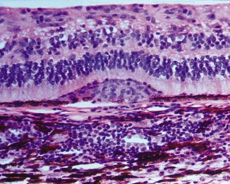
Fig. 2.
Microphotograph of Dalen-Fuchs nodule composed of mainly epithelioid histiocytes (macrophages) and a few degenerative pigment epithelial cells between the retinal pigment epithelial cell layer and Bruch's membrane.